Amylin’s role in nociception: study in amylin KO
mice
Sara João Lourenço Paulo
Integrated Master in Bioengineering, Molecular Biotechnology
July, 2015
Supervisor
Dr. Catarina Alexandra Soares Potes, IBMC
Co-supervisor
3
Acknowledgements
I would like to express my sincere gratitude to my supervisor Dr. Catarina Potes, for all the knowledge conveyed, for all the patience, support, availability and friendship. It was an honor to work with such a complete researcher that helped me to grow both professionally and as a person. I will never forget her sympathy, willingness to help and encouragement. I am indeed deeply grateful to her.
Dr. Fani Neto, my co-supervisor, for all support, kindness, availability, encouragement and willingness to help, despite her full professional agenda. Thank you for providing such a nice and healthy work environment.
My colleagues and friends within the research group, it was an honor to work with so pleasant people, always willing to help and so full of joy.
To the Animal Housing of Faculty of Medicine of the University of Porto and animal care takers, for the breeding of the colony used in these experiments and for performing and collecting the ear biopsies necessary for mice genotyping.
To LAIM the Laboratory of Support to Research in Molecular Medicine (LAIMM) at FMUP for performing the DNA extraction and PCR, necessary to genotype the mice.
To whole the Department of Experimental Biology of the Faculty of Medicine of the University of Porto, a sincere appreciation for the warm welcome and constant collaboration.
To my parents, for their unconditional support, for always believing in me and for their wise advice. I wouldn’t make it without them. Their absolute love gives me courage and confidence to achieve my goals. I hope they can feel proud of the person I become.
To my sister, for all love, support and encouragement. For all moments we lived together, for making me grow and for never leaving my side. Words fall short in expressing my deep gratitude and admiration.
To my brother-in-law, who supported me all these years. Thank you for your patience and friendship.
My best friend Sara Branco, for all support and sincere friendship which I will never forget. Thank you for being present when I most needed you.
To my friend and colleague Sofia Ferreira, for all support during these extraordinary years. We made it together.
5
Resumo
A dor é definida pela Associação Internacional do Estudo da Dor (IASP) como "uma experiência sensorial e emocional desagradável associada a um dano tecidual real ou potencial ou descrita em termos de tal dano", que pode ter um papel protetor (dor nociceptiva e inflamatória), ou pode ser mal-adaptativa (dor crónica).
A amilina, um membro da família de peptídeos relacionados com o gene da calcitonina, também conhecida como polipeptídeo amilóide, é um péptido com 37 aminoácidos segregado pelas células β-pancreáticas. Esta hormona é segregada simultaneamente com a insulina, em resposta à ingestão de nutrientes. As ações da amilina melhor estudadas a concentrações plasmáticas fisiológicos são na inibição da ingestão de alimentos e do esvaziamento gástrico. Em consequência disso, esta hormona está a ser usada em co-terapia com insulina em pacientes diabéticos nos EUA e é objeto de estudos clínicos de terapias para a redução do peso. O papel da amilina na nocicepção tem sido estudado recentemente, no entanto, as investigações nesta área não são suficientes e os resultados obtidos são muitas vezes contraditórios. Inicialmente foram descritos locais de ligação de amilina em áreas do cérebro envolvidas na nocicepção e a sua presença foi também detetada em gânglios raquidianos dorsais do rato (DRG), sugerindo um papel sensorial deste neuropeptídeo. Após estas descobertas interessantes, alguns grupos focaram-se no estudo do papel da amilina na nocicepção e sugeriram que este péptido pode estar envolvido na fase inicial de inflamação e que pode ter um papel excitatório em condições fisiológicas. Por conseguinte, Gebre Medhin e os seus colegas [2] demonstraram que a depleção genética da amilina produziu um fenótipo em que os ratinhos eram mais tolerantes à estimulação nóxica. Por outro lado, resultados recentes obtidos pelo nosso grupo de investigação mostraram que a administração de amilina em animais com dor inflamatória crónica produziu um efeito analgésico. Além disso, o nosso grupo também observou que a administração de amilina no teste de formalina em rato modulou o comportamento de dor manifestado durante a interfase e a fase de dor sustentada. Os nossos resultados indicam que o efeito da amilina na dor parece variar de acordo com a natureza do estímulo nóxico (aguda/crónica, a origem inflamatória/origem de uma lesão do nervo) e com a via de administração (sistémica/central). Estes resultados sugerem um papel importante de amilina na nocicepção.
O estudo apresentado nesta tese de mestrado teve como objetivo esclarecer o efeito da amilina no sistema nociceptivo por comparação de respostas comportamentais e neuro-químicas entre ratinhos sem o gene da amilina (ratinhos KO) e ratinhos de estirpe selvagem (ratinhos WT). Ambos os grupos de animais foram sujeitos a testes de dor aguda e a três diferentes modelos de dor: dor visceral, dor inflamatória crónica e dor neuropática.
Os testes de dor aguda realizados em ratinhos naïve sugerem que os ratinhos KO são geralmente mais sensíveis aos estímulos mecânicos do que os ratinhos WT. Foram observadas algumas diferenças nos resultados obtidos pelos testes térmicos de dor aguda, uma vez que no teste de Hargreaves não houve diferenças significativas entre os dois grupos de animais, indicando que a falta da amilina em animais KO não causa alterações na sensibilidade ao calor, ao contrário do teste de Cold plate em que os animais KO toleraram melhor o frio nóxico do que os animais WT.
A indução de dor inflamatória utilizando o Adjuvante Completo de Freund produziu uma resposta imunitária forte, que resultou numa reação artrítica localizada, resultando no desenvolvimento de uma inflamação grave na pata, de alodínia mecânica e hiperalgesia térmica. Uma vez que as patas ipsilaterais de ratinhos WT apresentaram diferenças significativas em comparação com as patas não inflamadas contralaterais, ao contrário do observado para as patas ipsilaterais de ratinhos KO, pudemos concluir que os ratinhos WT são mais sensíveis aos estímulos nóxicos aplicados.
A indução da dor neuropática foi realizada usando um modelo de lesão parcial do nervo ciático onde dois dos seus três ramos são ligados e axotomizados, denominado SNI (do inglês spared nerve injury) e resultou em alodínia e hiperalgesia intensas na região lateral da pata posterior, que recebe inervação do ramo do nervo ciático que foi deixado intacto, a qual se encontra hipersensível. Os resultados observados em testes de dor aguda sugerem que os ratinhos KO foram menos sensíveis aos diferentes estímulos aplicados, mostrando menos sinais de dor neuropática. Estes resultados são suportados pela quantificação de c-Fos, um gene de expressão imediata, que é expresso pelos neurónios rapidamente em resposta a estímulos. Assim, ratinhos WT expressaram mais c-Fos do que ratinhos KO, sugerindo que mais neurónios nociceptivos específicos da medula espinhal foram ativados em ratinhos WT.
A indução da dor visceral foi conseguida através de uma injeção intraperitoneal de ácido acético. As respostas nociceptivas foram medidas através da contagem do número de contrações em intervalos de 5 minutos, durante vinte minutos. Foi observado um efeito temporal e os ratinhos KO contraíram mais vezes o abdómen do que os ratinhos WT no período entre os 5 e os 15 minutos apos injeção, sugerindo um papel anti-nociceptivo da amilina na dor visceral. Estes resultados são suportados pela quantificação de neurónios positivos para o c-Fos na medula espinhal, uma vez que se observou que animais KO expressaram significativamente mais c-Fos do que animais WT.
Para avaliar alterações na densidade neuronal em gânglios raquidianos, foi realizada a coloração por hematoxilina e eosina, sendo o número total de neurónios quantificado. Contudo, não houve diferenças significativas entre os dois grupos
7 animais. Por outro lado, a medição das áreas neuronais mostrou que os ratinhos KO têm uma tendência para ter mais neurónios de pequeno tamanho (<600 µm2) e têm significativamente menos neurónios de grande área (>1200 µm2), em comparação com ratinho WT.
Para avaliar alterações nas populações de nociceptores entre ratinhos WT e KO, os níveis de expressão de CGRP, um marcador bem conhecido de nociceptores do tipo C péptidergicos, foram quantificados. Não foram detetadas diferenças significativas na percentagem de neurónios que expressam CGRP em DRGs da região L4 e L5.
Resumindo, o papel da amilina na nocicepção parece variar dependendo do modelo de dor em causa. Além disso, a falta da amilina em ratinhos KO parece envolver alterações no sistema nociceptivo em populações neuronais especificas dos gânglios raquidianos.
Palavras-chave: amilina, nocicepção, dor visceral, dor inflamatória, dor crónica, alodínia, hiperalgesia, medula espinhal, c-Fos, DRG, CGRP.
9
Abstract:
Pain is defined by the International Association for the Study of Pain (IASP) as "an unpleasant sensory and emotional experience associated with actual or potential tissue damage, or described in terms of such damage", which may have a protective role (nociceptive and inflammatory pain), or can be maladaptive (chronic pain).
Amylin, a member of the calcitonin gene-related peptide family, also known as islet amyloid polypeptide, is a 37 amino-acid-long secretory product of pancreatic β-cells. This peptide hormone is secreted simultaneously with insulin, in response to nutrient ingestion. The best studied actions of amylin at physiological plasma concentrations are on inhibition of food intake and gastric emptying. Thus, this hormone is being used in co-therapy with insulin in diabetic patients in the US, and is employed in clinical studies for weight reduction. Lately, amylin’s role in nociception has been studied, however the investigations in this area are still not sufficient and the results obtained are often contradictory. At first, amylin’s binding sites have been described in brain areas involved in nociception and its presence has also been detected in rat’s dorsal root ganglions (DRG), suggesting a sensorial role for this neuropeptide. After these interesting findings, some groups focused on amylin’s role in nociception and suggested that this peptide could be involved in the initial phase of inflammation and that it could have an excitatory role under physiological conditions. Accordingly, Gebre Medhin and his colleagues [2] have shown that amylin genetic depletion produced a phenotype in which mice are more tolerant to noxious stimulation. On the other hand, recent results obtained by our research group show that chronic subcutaneous amylin administration in animals with chronic inflammatory pain has analgesic effects. Additionally, our group has also observed that amylin administration in the rat formalin test modulates the pain behavior manifested at the interphase and in the sustained pain phase. Our data indicates that amylin’s effect on pain seems to fluctuate according to the nature of the noxious stimulus (acute/chronic, inflammatory origin/ origin in a nerve lesion) and to the route of administration (systemic/spinal). Overall, these results suggest an important role of amylin in nociception.
The study presented in this master thesis aimed at clarifying the effect of amylin in the nociceptive system by comparing the behavioral and neurochemical responses of mice with a general ablation of the amylin gene (amylin knock-out, KO, mice) with their wild-type (WT) littermates. Both mice genotypes were subjected to acute pain and to three different models of ongoing pain: visceral pain, chronic inflammatory pain and neuropathic pain.
The acute pain tests performed on naïve mice suggested that amylin KO mice are generally more sensitive to mechanical stimuli than WT mice. Some dissimilarity in
the results were noted when analyzing both thermal acute pain tests, since the Hargreaves test showed no significant differences between both animals groups, indicating no changes in noxious heat sensitivity due to amylin’s lack, contrarily to the cold plate test, which suggested that KO animals tolerate better the noxious cold than WT animals.
The induction of inflammatory pain using complete Freund’s adjuvant (CFA) elicited a strong immune response that resulted in a localized arthritic reaction, since mice developed a severe paw inflammation, mechanical allodynia and thermal hyperalgesia. As WT ipsilateral hind paws presented significant differences to non-inflamed hind paws, contrarily to what was observed in KO ipsilateral hind paws, we could conclude that WT-CFA inflamed mice were generally more responsive to the noxious stimuli applied.
Induction of neuropathic pain was performed using the spared nerve injury (SNI) model and resulted in long-lasting and intense allodynia and hyperalgesia in the lateral surface of the hind paw receiving innervation from the spared hypersensitive branch of the injured sciatic nerve. The behavior observed in the neuropathic animals in response to acute pain tests suggested that KO mice were less sensitive to the different applied stimuli, showing less signs of neuropathic pain. These results were supported by the quantification of c-Fos immunolabeling, an immediate early gene that is rapidly expressed by neurons in response to stimulation. Thus, WT mice showed more c-Fos positive neurons than KO mice suggesting that more spinal cord nociceptive-responsive neurons were activated in WT SNI-mice.
Induction of visceral pain was done by an intraperitoneal injection of acetic acid. The nociceptive response was measured by counting the number of writhes in 5 minute time-periods for 20 minutes. A time effect was noted and KO animals had in total more writhes than WT animals between 5 and 15 minutes post injection, suggesting an anti-nociceptive role of amylin in visceral pain. These results were supported by quantification of the number of c-Fos positive spinal cord neurons after acetic acid injection, since amylin KO animals expressed significantly more c-Fos than WT animals.
To assess changes on the neuronal density in dorsal root ganglia (DRG), a hematoxylin and eosin staining was performed and the total number of neurons was quantified, but no significant variations were detected when comparing WT and KO animals. However, the measurement of the cell body area showed that KO animals tended to have more small-sized neurons (<600 µm2) and had significantly lesser amounts of large-sized neurons (>1200 µm2), comparing to WT animals.
To assess changes in nociceptors populations between KO and WT mice, we also quantified the expression of calcitonin gene-related peptide (CGRP), a known
11 marker of peptidergic C nociceptors in DRG. We observed no significant differences in the percentage of neurons which expressed CGRP in L4 and L5 DRGs.
Overall, amylin’s role in nociception seems to vary depending on the model of pain. Moreover, the lack of amylin in KO mice seems to involve alterations in the nociceptive system in specific populations in the DRG.
Key-words: amylin, nociception, visceral pain, inflammatory pain, chronic pain, allodynia, hyperalgesia, spinal cord, c-Fos, DRG, CGRP.
13
Index
List of figures ... 15 Nomenclature ... 17 Introduction ... 19 Nociception ... 19Animal Pain Models... 21
Inflammatory Pain Models ... 21
Pathological Pain Models: Neuropathic Pain ... 22
Nociceptors ... 23
Responses to stimulus ... 25
Neuronal markers... 27
Amylin ... 28
Effects of amylin ... 29
Calcitonin and Calcitonin Gene Related Peptide (CGRP) ... 29
Amylin’s receptor ... 30
... 31
Amylin and Pain ... 31
Objectives ... 35
Methods and Materials ... 37
Animals and Habituation ... 37
Visceral Pain: Writhing test induced by intraperitoneal acetic acid injection ... 39
Induction of Chronic Inflammatory Pain: Complete Freund’s Adjuvant Model ... 39
Induction of Chronic Neuropathic Pain: spared nerve injury (SNI) model ... 40
Acute pain behavioral tests ... 40
Von frey ... 41
Tail Pressure ... 41
Cold plate ... 42
Hargreaves ... 43
Acetone ... 44
Results and Discussion ... 51
Acute pain tests ... 51
Acute tests performed on naïve animals ... 51
Immunohistochemistry results ... 66
Amylin immunolabeling ... 66
Quantification of CGRP expression ... 66
Conclusions and future perspectives ... 69
15
List of figures
Figure 1 - Pain classification: Nociceptive pain, Inflammatory Pain and Pathological pain. ... 21
Figure 2 - Schematic drawing of nociceptor's structure ... 23
Figure 3 - Illustration of a lumbar 4 (L4) cross section of the mouse spinal cord. ... 26
Figure 4 Spinal cord anatomy ... 27
Figure 5 - Amino acid sequence of human amilyn ... 29
Figure 6 - Representation of the amylin receptor structure ... 31
Figure 7 - Agarose gel ... 38
Figure 8 - Contraction of the abdominal musculature and extension of the hind limbs. ... 39
Figure 9 - A) Von Frey test. B) Calibrated von Frey filaments. ... 41
Figure 10 - Randall Selitto apparatus. ... 42
Figure 11 - Cold plate test. ... 43
Figure 12- Hargreaves test. ... 43
Figure 13 - Acetone test. ... 44
Figure 14 - Illustration of the spinal cord region considered for quantification of c-Fos ... 48
Figure 15 - von Frey test in the plantar surface of KO and WT naïve mice hind paws. ... 51
Figure 16 - Tail Pressure test in KO and WT naïve mice ... 52
Figure 17 - Hargreaves test in WT and KO naive mice ... 52
Figure 18 - Grubb's test result for the data regarding the cold plate test in KO and WT naïve mice. ... 53
Figure 19 - Cold plate test in KO and WT naïve mice ... 53
Figure 20 - Evolution of inflammation in WT-CFA and KO-CFA animals. ... 54
Figure 21 - Mechanical allodynia evolution in WT and KO mice after CFA injection. ... 55
Figure 22 - Thermal hyperalgesia and allodynia evolution for KO and WT animals after CFA injection. ... 57
Figure 23 - Mechanical allodynia progression for WT and KO mice after SNI surgery ... 59
Figure 24 - Assessment of cold allodynia by the acetone test, after SNI surgery in WT and KO mice. ... 60
Figure 25 - Thermal hyperalgesia and allodynia evolution in SNI pain model animals. ... 61
Figure 26 - Cold Plate tests on KO and WT mice, 14 days after SNI pain model induction. ... 61
Figure 27 - c-Fos quantification on the ipsilateral side of WT and KO animals’ spinal cords, after 14 days of SNI surgery. ... 62
Figure 28 - Writhing test in WT e KO mice after acetic acid injection. ... 63
Figure 29 - c-Fos quantification on WT and KO animals’ spinal cords, after acetic acid injection ... 64
Figure 30 - Neuronal density and cell body area in L4 and L5 DRGs from WT and KO naïve mice. ... 65
Figure 31 - Fluorescence microphotographs of Immunohistochemistry reaction amylin (green) in lumbar 4 DRG of WT naïve mice (A) and KO naïve mice (B). ... 66
Figure 32 - Results from CGRP immunohistochemistry in lumbar 4 and 5 DRGs of WT and KO mice. ... 67
17
Nomenclature
BDNF Brain derived neurotrophic factor
CFA Complete Freund’s Adjuvant
CGRP Calcitonin gene related peptide
CMH C mechano-heat nociceptors
CNS Central nervous system
CTR Calcitonin receptor
DAB 3,3-diaminobenzidine
DRG Dorsal root ganglia
GABA γ-aminobutyric acid
H&E Hematoxylin and eosin
IAPP islet amyloid polypeptide
IASP International Association for the Study of Pain
KO Knock-out
L3-L6 Lumbar 3 until Lumbar 6
L4 Lumbar 4
L5 Lumbar 5
NGF Nerve growth factor
NHS Normal horse serum
NS Nociceptive specific
NSAID Non steroidal inflammatory drugs
NSS Normal swine serum
NT-3 Neurotrophin 3
PBS Phosphate-buffered saline
PBST Phosphate-buffered saline with triton-X 100
PCR Polymerase chain reaction
PNS Peripheral nervous system
PWL Paw withdrawal latency
RAMP Receptor activity modifying protein
sCT Salmon calcitonin
SNI Spared nerve injury
SNL Spinal nerve ligation
SP Substance P
TRPM8 Transient receptor potential melastatin 8
TRPV1 Transient receptor potential vanilloid-1
T5 Thoracic 5
T13 Thoracic 13
T5-L2 Thoracic 5 until Lumbar 2
WDR Wide dynamic range
19
Introduction
Nociception
The International Association for the Study of Pain (IASP) defined pain as ‘’an unpleasant sensory and emotional experience associated with actual or potential tissue damage or described in terms of such damage’’ [3]. Pain can be classified as being nociceptive or neuropathic. The first can occur as a result of the noxious stimulation of nociceptors localized on skin, viscera and other organs. When pain is resultant from a dysfunction or lesion in the nervous system, it is classified as neuropathic pain [4]. However, this definition of pain was shown not to be appropriate for animals, as it becomes hard to analyze if an emotional experience occurred or not. Therefore, Zimmermann defined pain as ‘’an aversive sensorial experience derived from a concrete or potential lesion that causes progressive motor and vegetative reactions, results in behaviors of evasion and can modify specific behaviors of species, including social behavior’’ (1986) [5].
Although the pathways of pain perception are not fully described or understood, pain is a process of utmost importance since it is a symptom of many diseases, being considered as a major cause of demand for health professionals among the population in general, as it is very debilitating for the patient. The sensation of pain is present in diseases and in medical procedures and its ineffective treatment can lead to elevated socio-economic costs [4].
Besides the non-steroidal anti-inflammatory drugs (NSAIDs), usually prescribed to alleviate pain of inflammatory origin, the remaining therapies currently used to treat pain are mainly based on the use of narcotic analgesics like morphine, and sedative agents which include barbiturates and benzodiazepines. These agents promote analgesia, i.e., absence or reduction of pain in response to a noxious stimulus. However, these analgesics also produce undesirable side effects, such as depressant actions on respiration [6] and circulation, urinary retention [7], pruritus [7], constipation as well as possible somnolence or drowsiness [8]. Moreover, narcotic analgesics may induce tolerance in patients, as well as dependence.
It is therefore important to invest in the discovery of new more effective therapies that show no significant adverse effects and do not induce tolerance in patients, to combat pain.
From a neurobiological perspective, pain can be classified in three major different types: nociceptive pain, inflammatory pain and, finally, pathological pain, as summarized in Figure 1 [9].
The first one, nociceptive pain, is an early-warning physiological protective system, essential to detect and minimize contact with damaging or noxious stimuli
which can lesion the tissues [9]. This is the pain we feel when touching something too hot, cold, or sharp. This kind of pain is concerned with the sensing of noxious stimuli. It is associated to the stimulus detection and minimizes the contact with damaging stimuli, and so it has a protective role. This protective role demands instantaneous attention and action, which occurs by the withdrawal reflex that it activates, the intrinsic unpleasantness of the sensation caused, and the emotional anguish it involves [9]. Nociceptive pain is, therefore, essential for maintaining the body integrity [9].
Inflammatory pain has also an adaptive and protective role. This pain assists the healing of the injured body part, since it creates a situation that discourages physical contact and movement. The process of inflammatory pain is caused by the activation of the immune system by tissue injury or infection [9]. Pain hypersensitivity, or tenderness, reduces further risk of damage and promotes recovery. In a surgical wound or in an inflamed joint, for example, stimuli that are normally innocuous now elicit pain (allodynia). In this condition, an increased perception of noxious stimuli in the affected area (primary hyperalgesia) or in the adjacent region (secondary hyperalgesia) also occurs [10].
At last, there is pathological pain which is not protective, but maladaptive, and results from abnormal functioning of the nervous system. This pain is not a symptom of disorder but a disease state of the nervous system. It occurs when excessively intense or prolonged stimuli induce tissue damage that results in extended discomfort and abnormal sensitivity. This may arise spontaneously and is characterized by a low threshold to noxious stimuli, causing an exaggerated response to these stimuli, and leading to the presence of primary and secondary hyperalgesia, as well as allodynia. It is caused by tissue damage-associated inflammation (inflammatory pain), or by central or peripheral nerve injury (neuropathic pain) [11].
21 Figure 1 - Pain classification: Nociceptive pain, Inflammatory Pain and Pathological pain. From [9].
Animal Pain Models
Since pain plays an important role on actual society, and its ineffective treatment can lead to elevated socio-economic costs, it has been object of intense research. The major purpose of these studies has been to promote knowledge that will be important to treat acute and chronic pain. To study pain’s underlying pathological mechanisms, animal models which mimic different human clinical conditions have been developed throughout the last decades.
Inflammatory Pain Models
In order to understand the mechanisms of persistent pain, animal models of inflammatory pain that simulate human clinical pain conditions have been developed. The majority relies on the injection of inflammatory agents into the rat or mouse hind paw [12]. The main inflammatory agents currently used are carrageenan [13], formalin [14], complete Freund’s adjuvant (CFA) and capsaicin [15], although there are more agents available.
Complete Freund’s adjuvant injection is a model widely used which consists in the injection of a mycobacteria in a fatty excipient [16] in the animal’s hind paw, which will trigger a strong immune response. This method is known for causing pannus formation, cartilage erosion, inflammation and hyperplasia, the same symptoms associated with rheumatoid arthritis, an inflammatory autoimmune disorder [17]. After CFA injection, the cutaneous inflammation appears within two hours and the hyperalgesia and edema are present for at least one or two weeks [17]. It is widely considered as a reliable and useful model to study inflammation and nociception in both rat and mice, which produces a consistent, localized arthritic reaction with associated inflammation-induced nociceptive behavioral changes [18].
Pathological Pain Models: Neuropathic Pain
Neuropathic pain results from trauma in the central (CNS) or peripheral nervous systems (PNS), causing allodynia, hyperalgesia and spontaneous pain. There are several animal models that mimic human peripheral neuropathic pain, which often combine both intact and injured fibers [19]. These models include the spinal nerve ligation (SNL) [20] and the spared nerve injury (SNI) models [19]. The last one consists on the injury of two of the three branches of the sciatic nerve (the tibial and the peroneal nerves) while the third (sural nerve) is left intact. This procedure will induce very rapidly hypersensitivity in the skin area adjacent to the denervation [20].
The SNI has been described as an intense, reproducible and simple model that produces long-lasting and intense allodynia and hyperalgesia for about six months as well as behavioral changes that can be measured by stimulating the non-injured sural nerve skin territory [20]. Acute sensory and pain behavioral tests like the von Frey and Cold Plate, respectively, are conducted to measure hypersensitivity to mechanical and thermal stimuli and the intensity of pain induced in the animals by the axotomy of the tibial and peroneal nerves. Given the similarities to human neuropathic pain and the advantages shown by the SNI model, it is highly used for the study of the mechanisms involved in the development of neuropathic pain and to test the efficacy of new treatments.
Acute Visceral Pain Models
Visceral pain is the most usual form of pain in the clinical setting and one of the most relevant reasons for patients to seek medical supervision [21]. It results from pain in the internal organs and is described as being vague, poorly defined and more similar to a discomfort than a real sense of pain. Visceral pain is usually perceived indistinctly in the same site (lower sternal region of epigastrium) no matter what organ is involved [22].
In order to induce this type of pain in rats or mice, it is usual to perform an intraperitoneal injection of a noxious agent [22]. This method is called ‘’the writhing
23
test’’. After the injection of the chemical irritant, the animal will respond with
characteristic contractions of the abdominal muscles accompanied by a hind limb extensor motion. The most frequently used agents for the writhing test have been phenylquinone and acetic acid [23].
Intraperitoneal injection of acetic acid is a well-known noxious chemical visceral stimulus in animals that produces the already mentioned abdominal contractions or writhes, and also affects the gastrointestinal ileus by inhibiting gastric emptying and small intestine transit, which is associated with visceral pain [24].
Nociceptors
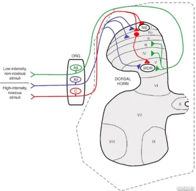
The PNS neurons responsible for the detection and transmission of noxious stimuli are known as nociceptors [25]. The nociceptor has four major functional components: the peripheral terminal that transduces external stimuli and initiates action potentials, the axon that conducts action potentials, the cell body that controls the identity and integrity of the neuron and is localized in sensory ganglia, and the central terminal which forms the presynaptic element of the first synapse in the sensory pathway in the CNS (Figure 2) [26]. Stimuli arising from the face and head will be sensed by nociceptors with their cell bodies located in trigeminal sensory ganglia while stimuli in the remaining parts of the body will be conducted to cell bodies located in dorsal root ganglia (DRG).
Figure 2 - Schematic drawing of nociceptor's structure. It is represented the location of the cell body of a nociceptor
responsible for detecting stimuli in all parts of the body except the face and head. From: [25]
Each nociceptor structure has an essential role in the perception and transduction of the stimulus. The cell body is necessary to maintain the other regions of the cell and is located in the ganglia of the spinal cord dorsal roots, as already mentioned. The axon detects peripheral stimuli, transduces their energy into an electrical signal and conducts the action potential to the synaptic terminal where the information is transmitted to the primary sensory area. Thus, stimuli arising from trigeminal ganglia will be conveyed into the trigeminal nucleus while stimuli from DRG, arising from any part of the body except the face and head, will convey into the dorsal horn of the spinal cord. These stimuli can be thermal, mechanical or chemical from
different parts of the organism. The release of transmitters at the synaptic terminal is subject to modulation by agents released by other neurons and possibly by glial cells [25].
Nociceptors can be broadly divided into two classes: one class has small-diameter cell bodies and slowly conducting, unmyelinated axons (C fibers), whereas the other class has medium-diameter cell bodies and faster conducting, lightly myelinated axons (Aδ fibers) [27]. There are also Aβ fibers which transmit non-nociceptive information [4]. These different fibers are divided into three big groups, in result of their different conduction velocities, fiber diameter and myelination, as it is shown in Table 1:
Table 1 - Classification of cutaneous sensitive fibers
Fiber Myelination Conduction velocity Fiber Diameter Aβ Thick 30-100 m/s >10 µm Aδ Thin 12-30 m/s 2-6 µm C Absent 0,5-2 m/s 0,4-1,2 µm
Nociceptors can also be further divided into three groups according to their neurochemistry: peptidergic C nociceptors, non peptidergic C nociceptors and Aδ nociceptors. All of them have glutamate, the most abundant excitatory neuropeptide. However, the peptidergic C nociceptors also express substance P (SP) and the calcitonin gene related peptide (CGRP). These CGRP-containing neurons are activated by chemical, thermal, and high-threshold mechanical stimuli, and innervate essentially all peripheral tissues [28]. They also send primary afferent input to nociceptive and viscerosensitive neurons in the dorsal horn, trigeminal nucleus caudalis, or nucleus of the solitary tract that project to the brainstem, amygdala, hypothalamus, and thalamic nuclei, which in turn transmit these inputs to the somatosensory and insular cortexes [28]. This group of nociceptors relies on neuronal growth factor (NGF) for developing and survival.
The second group of nociceptors (non peptidergic C nociceptors) does not have peptides and depends on brain derived neurotrophic factor (BDNF) for their development and survival. This later type of nociceptors can be identified for the presence of specific isolectins, purinergic receptors or other specific enzymes. The final group, Aδ nociceptors, relies on neurotrophin-3 (NT-3) and BDNF and is easily recognized by the presence of specific neurofilaments [4].
25 Responses to stimulus
As mentioned above, Aβ fibers are responsible for sensing innocuous stimulus, like vibration and pressure. Under physiological conditions, C and Aδ fibers conduct nociceptive information [4]. When a noxious stimulus is applied, Aδ fibers are responsible for the immediate acute pain, exhibiting a high conduction velocity of the stimulus. On the other hand, C fibers are responsible for a more diffuse delayed pain sensation, as these fibers exhibit a lower conduction velocity and diameter [4].
Aδ fibers can be divided into type I and type II Aδ fibers [4]. Type I fibers respond mostly to mechanical stimulus, but can also respond to chemical and thermal stimulus above 53oC [29]. Type II Aδ fibers are insensible to mechanical stimuli, but respond to lower thermal stimuli than Aδ fibers type I.
C fibers can be divided according to the noxious stimulus that activates them. Many of them are polymodal, so they respond to all different stimuli (mechanical, thermal and chemical) [27]. However, some C-nociceptors are only sensitive to thermal or mechanical stimulus, or to both [4].
The C fibers which are simultaneously sensitive to mechanical and thermal stimuli are known as C mechano-heat nociceptors (CMH) and are activated by a range of pressures from 30 to 17 mN in human and mice and by temperatures around 39-51oC [30]. Moreover, there are other types of C nociceptors that are insensitive to noxious mechanical and heat stimuli, known as silent nociceptors. These nociceptors are present in skin, viscera and joints. In case of inflammation, where there is release of histamine or other substances, these C silent nociceptors are capable of being activated by noxious stimuli, exhibiting a decrease in their activation threshold [31]. Central projections of nociceptors
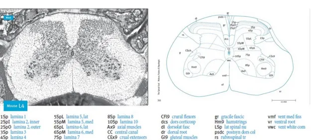
The spinal cord consists of a gray inner zone covered by white matter. This gray zone is divided in ten laminae, which are anatomically and electrophysiological distinct [32]. The lamina I (or marginal zone) is the more superficial region of the dorsal horn, which extends to lamina VI. The ventral horn comprises laminae VII-IX and the center canal is surrounded by lamina X (Figure 3).
Figure 3 - Illustration of a lumbar 4 (L4) cross section of the mouse spinal cord. Left: Nissl staining of a L4 cross section of the mouse spinal cord. Right: respective atlas schematic drawing from [33].
The central projections of the different types of nociceptor are distributed differentially in the spinal cord, occupying different areas and, therefore, different laminae [32]. Aβ nociceptors, responsible for transmitting innocuous information, terminate in laminae III and IV. They also terminate in lamina V, as well as Aδ and C fibers, where convergent non-noxious and noxious inputs are received [32]. Most peptidergic C fibers terminate in lamina I and in the most dorsal part of lamina II. On the other hand, the non-peptidergic afferents terminate in the mid-region of lamina II. Electrophysiological analyses demonstrate that spinal cord neurons within lamina I are generally responsive to noxious stimulation (via Aδ and C fibers) (Figure 4).
Spinal cord neurons can be divided in three major groups, considering their functional features. They can be nociceptive specific (NS) therefore responding to noxious stimuli only; non-nociceptive, which respond equally to both innocuous and noxious stimuli, and wide dynamic range (WDR) neurons, which respond to both noxious and innocuous stimuli according to their intensities, so that the response is proportional to the intensity of the stimulus [32].
27 Figure 4 Spinal cord anatomy. NS: nociceptive specific neurons; WDR: Wild-dynamic range neurons. Adapted from: [34]
Neurons in the spinal cord can also be classified as being inhibitory or excitatory, or according to the terminus of the axon relatively to the cell body as interneurons, propriospinal or projection neurons. Interneurons communicate with nearby neurons, propriospinal neurons connect with other regions of the spinal cord, in particular the contralateral side, and projection neurons, which are mainly present in laminas I, V and X, have long axons and transmit the information from the spinal cord to supraspinal regions such as the thalamus and parabrachial area. The thalamus activates the pain matrix while the parabrachial area is involved in descending and affective modulation [35], facilitating or inhibiting the transmission of painful information from the spinal cord.
During nociceptive transmission, the output of the spinal cord is dependent on various spinal mechanisms that can increase or decrease the activity of dorsal horn neurons. These mechanisms comprise local excitatory and inhibitory interneurons [34]. The inhibitory neurons release γ-aminobutyric acid (GABA) and/or glycine, while the preferred neurotransmitter for the excitatory neurons is glutamate.
Neuronal markers
The study of the expression of some neuronal markers can be very useful to determine the amount of noxious information that is being transmitted to the CNS by primary afferent neurons.
Quantification of c-Fos expression has been widely used as a marker of neuronal excitation [36] and is easily detected by an immunohistochemistry reaction.
The immediate early gene c-Fos is quickly expressed by spinal cord neurons following noxious stimulation of the body tissues and its laminar distribution is connected to the nature of the sensory stimulus [37]. Its transcriptional activation takes place within minutes after noxious stimulation and the levels of this proteins reach its peak about two hours after stimulation [38] and returns to baseline values after eight to twenty-four hours after the initial stimulation [39]. It is widely accepted that analysis of c-Fos expression may help clarify the central neural activity occurring during the development of persistent pain or prolonged inflammatory pain [40]. Indeed, in the spinal cord, c-Fos expression is considered specific of neurons activated by noxious stimulus while its supraspinal expression may occur independently of a painful stimulus [35].
Another cellular marker related to the nociceptive system is the calcitonin gene related peptide (CGRP). CGRP is a 37–amino acid peptide that is a member of the calcitonin family and is widely expressed in the PNS and CNS, frequently coexisting and interacting with other neurotransmitters [28]. CGRP is known for its vasodilator properties and for participating in many central and peripheral pain mechanisms. It is upregulated in peripheral nerve injury or tissue inflammation conditions and produces sensitization of dorsal horn and trigeminal neurons. CGRP is also known for eliciting behavioral pain sensitization [28]. Thus, CGRP is considered a classic marker of nociceptive DRG neurons [41], namely, C-peptidergic nociceptors as mentioned above, which respond to stimuli that evoke sensations of pain and itch. As for c-Fos, its expression is easily detected by an immunohistochemistry reaction.
Amylin
Amylin, also known as islet amyloid polypeptide, is a 37 amino-acid-long beta-cell secretory product and the main protein component of the pancreatic islet amyloid found in human subjects with type 2 diabetes (Figure 3). Amylin is a product of a gene located on chromosome 12. It is transcribed as an 89-aminoacid prepolypeptide, is cleaved to form the mature peptide in the β-cells of the pancreas, where it is stored along with insulin and C-peptide in the same granules. Amylin is a normal product of β-cells and is co-released with insulin in a molar ratio of 1 to 100 in healthy non-diabetic subjects in response to nutrient stimuli (carbohydrate-and protein-containing meals) [42] although this rate is not always constant.
29 Figure 5 - Amino acid sequence of human amilyn. [43]
Effects of amylin
Some of amylin’s physiological functions include decrease of appetite [44], inhibition of gastric emptying, gastric acid and of secretion of digestive hormones [45], therefore amylin controls nutrient appearance in plasma, and plays an important role in weight control.
Amylin presents a regulatory effect on glucose homeostasis, presenting an important role on the control of insulin secretion, as it was reported that amylin inhibits insulin secretion [46]. While the human amylin peptide readily forms aggregates, as mentioned above, rat amylin does not. Therefore, co-therapy treatments in type 2 diabetes mellitus patients use a soluble and stable rat amylin analogue (pramlintide) to control glycaemia in these patients [47]. Additionally to these roles of amylin there are reports of other functions, many of which totally unrelated to metabolism control, such as induction of kidney’s epithelial cells proliferation [48], development of proximal tubules [49], differentiation of osteoclasts and osteoblasts, protection of the gastric mucosa [50], blood pressure regulation where it functions as a vasodilator [51], modulation of memory [52] and of motor activity [53].
Calcitonin and Calcitonin Gene Related Peptide (CGRP)
It is known that some actions of amylin are identical to the known metabolic actions of CGRP and calcitonin. In fact, these peptides belong to the same peptide family and possess related structures.
Amylin’s structure is 50% identical to that of CGRPs, 37 amino acid peptides which are widespread neurotransmitters with many potent biological actions [51]. CGRP is formed from the precalcitonin gene on chromosome 11 [54] and as result of transcription, two different peptides are produced by alternative splicing: procalcitonin (in thyroid tissue) and proCGRP (in neural tissue).
Calcitonin is a 32 amino acid peptide which is produced by thyroid C-cells. Calcitonin is synthesized at first as a 132 amino acid precursor molecule and then is processed by proteolytic cleavage and by amidation of its carboxy terminal proline residue before secretion [55].
The gene which encodes calcitonin is also responsible for encoding one of the CGRPs, the CGRP, which is largely expressed in neural tissue, in both PNS and CNS. α-CGRP is known to be a potent vasodilator [55]. Cell specific alternate processing of the calcitonin/α-CGRP transcripts is the mechanism that regulates the formation of either calcitonin or α-CGRP in different cell types.
Another molecule described as being very similar to calcitonin and α-CGRP is β-CGRP. This is a product of another gene which is expressed by enteric neurons and it differs from α-CGRP by just 3 amino acids in humans [55].
The peptides just described are extensively distributed in various peripheral tissues as well as in the PNS and CNS and induce multiple biological effects. Effects such as vasodilatation (CGRP) and inhibition of bone resorption (calcitonin) are shared, though with much less potency, by amylin [56].
Amylin’s receptor
Earlier reports suggested that amylin acted via CGRP receptors in order to achieve its biological effects. Later it was discovered that amylin receptors can be reconstituted in cellular systems by co-expressing the calcitonin receptor (CTR) with receptor activity modifying proteins (RAMPs). These receptors exhibit high affinity for salmon calcitonin, which is recognized as a very potent agonist of amylin receptors, while, importantly, CGRP shows much less affinity for these receptors [56].
The specific amylin receptor is a heterodimeric complex that consists on the association of CTR with one of the three RAMPs, RAMP1, RAMP2 or RAMP3 (Figure 4) [57], having high affinity for amylin [58]. It is known that the CTR belongs to the family of the G protein-coupled receptors which have 7 transmembrane domains. Analysis of CTR transcripts from the porcine kidney epithelial cell line LLC-PK1 has exhibited two
splice variants, CTR1a and CTR1b, being this last transcript longer due to the presence of an additional 48 base pairs coding sequence [59]. These receptors induce the accumulation of cyclic AMP, leading to activation of the subordinate signaling cascade. As previously stated, RAMPs play an important role in this receptor’s structure, offering a mechanism for the ligand specificity variation and regulation of receptor function. These proteins have the ability to enhance the affinity of CTR to amylin, building up the amylin receptor. When associated with a calcitonin like receptor, RAMPs build up the CGRP receptors.
Six different isoforms of amylin receptors were reported: AMY1(a)
(CTRa/RAMP1), AMY2(a) (CTRa/RAMP2), AMY3(a) (CTRa/RAMP3), AMY1(b)
(CTRb/RAMP1), AMY2(b) (CTRb/RAMP2) and AMY3(b) (CTRb/RAMP3), being the most
common ones AMY1(a) and AMY3(a). CTR(b) displays greater capacity to generate
RAMP2 AMY receptors than CTR(a) [56]. It was also reported that different RAMPs promote different pharmacologic properties and different affinities for the ligands. For
31 Figure 6 - Representation of the amylin receptor structure. Amy: amylin; RAMP: Receptor activity modifying proteins; CTR: Calcitonin receptor. Source: [1]
example, RAMP3 provides a greater affinity for salmon’s calcitonin and amylin while RAMP1 increases CTR affinity for mammal’s CGRP and calcitonin [60].
Amylin and Pain
Some studies have suggested a role of amylin in nociception; however, the research in this area has not been sufficient and the results obtained so far are often contradictory. Amylin’s binding sites were described in the CNS for the first time by Sexton and his colleagues in 1994 [61]. Many of those brain areas are involved in nociception such as the nucleus of solitary tract, parabrachial nucleus, hypothalamus, the periaquedutal grey, the locus coeruleus and the dorsal raphe, and it is known that amylin is transported across the blood-brain barrier to these areas [62]. In fact, intracerebroventricular administration of amylin elicited anti-nociceptive effects in thermal acute pain, as it increased the time latency in the hot plate test response in rats [63]. Moreover, Mulder detected the presence of amylin messenger RNA and of the amylin peptide in the rat’s DRG neurons, specifically in C peptidergic nociceptors, suggesting a sensorial role for this neuropeptide [64]. They also identified amylin expressing fibers in the superficial laminae of the spinal cord [64]. In fact, amylin’s expression pattern in rat sensory neurons mimics that of CGRP and substance P following noxious stimulation, occurring in small to medium sized nerve cell bodies known to receive sensory input. Taking all these findings together, some research groups proposed to study amylin’s role in the nervous system, more specifically in nociception.
Mulder and his colleagues continued their research in this area and studied the expression of amylin in rat DRGs after induction of inflammatory pain by intradermic injection of CFA [65]. Their results showed an increase in amylin expression at early time points, suggesting that this peptide could be involved in the initial phase of inflammation. Other important finding of this group focused on a neuropathic model in rats, which caused a decrease of amylin expression in rat DRGs as well of fiber density in the dorsal horn of the spinal cord in the ipsilateral side to the affected limb. This led
the Mulder group to consider an excitatory role for amylin under physiological conditions [66]. Accordingly, Gebre Medhin and his colleagues have confirmed that amylin is a constituent of sensory neurons in mice and its genetic ablation produced a phenotype in which mice are more tolerant to noxious stimulation. However, in the paw inflammation model induced by CFA, there were no significant differences in the ankle diameter between knock-out (KO) mice and wild-type (WT) mice [2].
On the other hand, Huang et al demonstrated that amylin, possibly acting on AMY1(a/b) or AMY3(a/b) receptors in the spinal cord, induced antinociceptive effects as
assessed by the acetic acid writhing model of visceral pain. Some studies also show anti-nociceptive properties for salmon calcitonin (sCT), a strong amylin receptor agonist [67]. Intra-nasal sCT relieved bone pain in patients suffering from malignant tumors [68, 69] and improved pain symptoms in patients with knee osteoarthritis [70]. When centrally administered, sCT also produced strong antinociceptive effects in the tonic pain phase of the formalin test in mice [71]. It is important to note that calcitonin’s expression has not been identified in the adult CNS, unlike amylin and CGRP [72]. This fact shows the relevance of amylin in pain, suggesting that salmon calcitonin may act through amylin’s receptors, rather than via CTR alone, to attenuate pain.
Despite these findings on antinociceptive effects resulting from amylin-receptor activation, Bouali and his colleagues were not able to observe significant alterations in the nociceptive response when they immersed the rat’s tail in a 49oC water bath after central administration of amylin [73].
Interestingly, recent results obtained by our research group show that amylin administration in the rat formalin test modulates the pain behavior manifested at the interphase, when auto-analgesia in response to the noxious stimulus takes place, and in the sustained pain phase, when inflammatory processes triggered by the chemical insult are observed. This effect was dependent on the amylin dose, time of injection and route of administration [74]. It was also shown by our group that amylin’s effect on pain seems to fluctuate according to the noxious stimulus nature (acute/chronic, inflammatory origin/ origin in a nerve lesion). Indeed, while chronic amylin infusion was shown to aggravate allodynia to cold stimuli in animals with neuropathic pain [75], in animals with chronic inflammatory pain induced by intra-articular CFA injection, amylin promoted analgesia [76]. These results propose an important role for amylin in nociception.
It is important to note that studies in this area are not abundant. Although some results demonstrated to be contradictory, it is settled that amylin has a role in nociception and its study can bring major progresses in pain understanding and treatment. Additionally, it is very important to determine whether overlapping or related functions of amylin and CGRP are mediated via amylin receptors or via CGRP
33 receptors, and to determine which are the effects specifically related to amylin’s peptide and receptor.
35
Objectives
Amylin has been related to nociceptive mechanisms and several studies highlight amylin’s role in the nervous system. However, these studies are often contradictory and limited, not looking into all the behaviors that are measurable in certain pain conditions. Other studies assessed amylin’s role by intracerebroventricular administration, but these neglected the fact that amylin might have a direct peripheral effect or act at the spinal cord, especially since amylin is produced by DRG neurons. Furthermore, there are intriguing results linking amylin lack in KO mice to defective nociception [2]. The only study evaluating nociception in amylin KO mice used few pain models, did not test the effect of restoring amylin in these animals and did not investigate plastic changes in the nociceptive system of these animals. In consequence, in this project we intended to assess whether nociception and the nociceptive system were altered in animals lacking the amylin gene (KO mice).
More specifically, in this project we intended:
1) To investigate changes in the nociceptive behavior in response to noxious stimuli in amylin KO mice when comparing to their WT littermates. To achieve this goal we induced four different types of pain conditions, namely, animals were subjected to acute noxious stimuli, to visceral pain, to chronic inflammatory and neuropathic pain. The sensitivity to visceral pain as well as to standard acute mechanical and thermal stimuli was tested in naïve animals to evaluate whether nociceptive behaviors were altered in animals due to amylin´s lack. Animals ongoing inflammatory or neuropathic pain were tested with appropriate acute pain tests in order to evaluate the pain behavior changes between the two mice genotypes when submitted to the same painful condition.
2) To assess putative changes in the nociceptive system in neuronal populations of the DRG of amylin KO mice when comparing to their WT littermates. Thus, the number of neurons per area was determined as well as the neuronal density of CGRP positive neurons, as a first step towards the investigation of the type of sensory fibers that could be altered in KO mice.
3) To study amylin’s effect on the nociceptive-responsive spinal cord neurons. Thus, we analyzed the c-Fos protein expression by immunohistochemistry in the dorsal horn of amylin KO and WT mice. This represented an indicator of the neuronal activity following noxious stimulation.
37
Methods and Materials
Animals and Habituation
For all experiments, adult amylin KO (IAPP -/-) and WT (IAPP +/+) littermate mice with a C57BL/6 background, bred at the Faculty of Medicine of Porto animal house, were used. The founder IAPPheterozygous F1 breeding pairs were provided by Prof. Thomas Lutz from the University of Zurich, Switzerland, in 2012. All experiments were performed with males, except the writhing test where females were used. Ear biopsies were used to tag the animal and the tissue was used for genotyping by polymerase chain reaction (PCR) analysis. DNA extraction and PCR were performed by the Laboratory of Support to Research in Molecular Medicine (LAIMM) at FMUP and are described below.
The 59 C57BL/6 mice used for the experiments were kept under controlled conditions (temperature and humidity of 22 ± 2°C and 55 ± 5%, respectively, and a light cycle of 12h light / 12 hours darkness).
The acclimatization to the conventional animal house area, where tests were performed, was of at least 7 days, during which only animal caretakers had contact with the animals. Later, the process of habituation began, with simple manipulations of the animal, such as picking them up and placing them in the test cages. Animals were habituated to the elevated cage with a grid floor used for von Frey and acetone tests, to the Hargreaves apparatus cages, as well as to the cold plate test and writhing test chambers. Moreover, animals were habituated to freely enter into the metal tube restrainer used for the tail pressure test, and to the noise of the Randall Selitto equipment used for this test. This preliminary step reduces stress, which is important to minimize confusion with stress-induced analgesia, and allows the animals to be accustomed to the noise, investigator, handling and manipulation. So, in the 4-5 days leading up to the first day of testing and just before the execution of the tests thereof, the animals were maintained for about 30 minutes in the testing room without being disturbed, and were then transferred to the test chamber where they remained for further 30 minutes without any noxious stimulation, in order to acclimatize to the conditions of the room and reduce stress and exploratory behaviors. All behavioral tests were performed during the light period for all experimental groups, in random order, in order to eliminate any change caused by the circadian cycle.
The physical condition of the animals was monitored throughout the experiment. Special attention was given to the presence of stress signs, illness or poor physical condition, such as loss or gain of excessive weight, dehydration, aggressive social behavior, low mobility, bleeding and poor wound healing, infection of the sutures and opening of the stitches in the post-surgery period.
The experimental procedures were performed according to ethical standards for the study of experimental pain in conscious animals [77], the Directive 2010/63/EU of the European Parliament and of the European Council [Strasbourg, 22 September 2010] and the rules of the regulations of local authorities [Decree-Law 129/92, Ordinance 1005/92] on the use of animals for scientific purposes.
Mice genotyping protocol
For the DNA extraction, a fresh solution of 100mM NaOH was prepared and added to the microtubes containing the mice tissue. Samples were placed in the thermocycler at 99oC for about 1 hour. After cooling down, 100uL of 1M Tris HCl (pH 8) was added. After centrifugation, a 1 µL aliquot of the extracted DNA was used for the polymerase chain reaction (PCR). To each PCR tube was further added the following solutions from the Citomed BM-10002 KIT: 0.1 µL Taq polymerase, 2 µL of Buffer and
2µL of MgSO4 (2 mM,). Finally, 1 µL of each primer (at 10 µM) was added, namely,
primer 206: 5’-CTTGGGTGGAGAGGCTATTC-3’, primer 207:
5’-CACAGCTGCGCAAGGAAC-3’, primer 208: 5’-GTAGCAACCCTCAGATGGAC-3’ and primer 209: 5’-GAGGACTGGACCAAGGTTGT-3’, all produced by Stab Vida.
The samples were placed in a thermocycler, which conducted 35 cycles of reaction. Each cycle included 5 minutes at 95oC, followed by 30 second at 95oC, 62oC and 72oC, and finally 10 minutes at 72oC. At last, the samples were run in an agarose gel for electrophoresis. The amplified target sequences included a mutated allele with 200 base pairs (knock-out mice) and a wild-type allele with 100 base pairs (wild-type mice). When both products were found in the gel, the sample belonged to a heterozygous mouse, as we can see in the following gel-example (Figure 7).
Figure 7 - Agarose gel. Het- heterozygote mice; WT- wild-type mice (100 base pairs); KO- knock-out mice (200 base pairs).
39
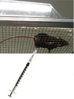
Visceral Pain: Writhing test induced by intraperitoneal acetic acid injection
The acetic acid-induced visceral pain model is widely used in experimental research to produce a writhing reaction, characterized by contraction of the abdominal musculature and extension of the hind limbs (Figure 8), associated with visceral pain [24]. Animals were placed in the test chamber, a clear 20 × 26 × 12 cm plastic cage (figure 8), for 10 to 15 minutes to acclimatize. Following the intraperitoneal injection of 1% acetic acid (10ml/kg of body weight; Sigma-Aldrich, [24]), animals were placed back into the test chamber and were video-recorded for twenty minutes. Behavior was analyzed later with the program Etholog 2.25 [78]. The number of writhes was monitored for twenty minutes and recorded in five minutes intervals [24]. The total number of writhes between 5 and 15 min after injection [51, 79] and the latency time to the occurrence of the first writhe were also assessed.
Figure 8 - Contraction of the abdominal musculature and extension of the hind limbs.
Induction of Chronic Inflammatory Pain: Complete Freund’s Adjuvant Model
The animals were anesthetized with volatile anesthesia (isoflurane), at first in a glass chamber at 4% (to induce) and then directly on the nose at 1.5% (to maintain). Then, animals received an intraplantar injection of 20 µL of Complete Freund’s adjuvant (CFA) into the left hind paw. The injection was performed slowly to avoid the reflux of CFA. The CFA solution consists of water in oil emulsion containing killed and dried Mycobacterium butyricum (Difco Laboratories [80]; see composition in annex).The inflammation reaction was monitored daily using a scale that takes into account the animal’s behavior and local inflammation signs. The inflammatory scale considered has the minimum value of zero, where the animal shows no signs of inflammation, and the maximum value of four, where the animal demonstrates severe inflammation and persistent flexion of the paw [81].
Mice were transferred to the behavioral testing room at least one hour before testing to acclimatize. The von Frey and Hargreaves behavioral tests (see below) were performed at three hours, one day and two days after injection. Both hind paws were tested.
Induction of Chronic Neuropathic Pain: spared nerve injury (SNI) model
The spared nerve injury surgery in mice was performed in order to induce a painful neuropathy, as described by Richner et al. [82] which is based on the procedure defined by Decostered and Woolf [19] in rats.
Briefly, the animals were anesthetized using the combination of Domitor (1 mg/kg, medetomidine hydrochloride) with Imalgene (75 mg/kg, Ketamine hydrochloride) diluted in distilled water and administered intraperitoneally. The left thigh was shaved and disinfected with Betadine and 70% ethanol. A 1 cm incision was made in the skin in the longitudinal direction proximal to the knee, subsequently detaching the skin from the underlying connective tissue layer covering the muscle. The muscle layer was separated with the help of blunt scissors, right next to the clearly visible blood vessel, close to the thigh bone (femur). At this point, a stereo microscope was required so the sciatic nerve and its different branches (tibial, peroneal and sural) could be visualized. A silk suture (6-0, No. 18020-60, Fine Science Tools) was applied around the tibial and peroneal branches, and a tight surgical knot was made, leaving the sural branch intact. Both nerves (tibial and peroneal) were held with a sterile tweezer bellow the suture and a 1 mm portion was cut with a small scissor. The muscle layer and the skin were sutured using absorbable 4-0 suture (simple knots in the muscle layer and U-eversion knots in the skin; C1048213, Safil, Braun) and lidocaine was applied to minimize local discomfort. After surgery, the wound was disinfected, the animals were rehydrated with a subcutaneous injection of 0.5 mL of saline solution (0.9 % NaCl) and finally, the anesthesia was reverted with a subcutaneous injection of Antisedan (atipamezol, 1 mg/Kg). In the days after surgery, easy access to water and food were provided. The von Frey, Acetone and the Hargreaves behavioral tests (by this order, see details of the tests below) were performed one day before surgery and on day one, three, seven and fourteen after surgery. The Cold Plate test was performed just on day 14 of SNI (see details below) after the other three tests.
Acute pain behavioral tests
These tests were applied to naïve mice and some were also used to assess the development of allodynia and hyperalgesia in chronic pain animals, as mentioned above. Acute pain tests were performed in naïve animals to detect differences in acute
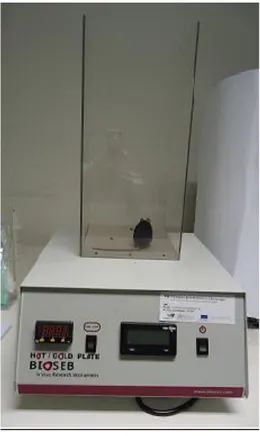
41 sensitivities between both genotypes. The tail pressure and cold plate tests were performed in these animals to assess mechanical and cold hyperalgesia, respectively.
To assess acute mechanical sensitivity and heat hyperalgesia in naïve animals, the von Frey and Hargreaves tests were performed, respectively, at the baseline time point of the CFA animals (before CFA injection), since at baseline all the animals were naïve.
Von frey
Mechanical allodynia was assessed both in naïve and in chronic pain animals using a set of calibrated von Frey filaments (Figure 9B; Touch test sensory evaluator kit, Stoelting). Starting from the thinnest hair, each filament was applied on the plantar surface of the hind paw (Figure 9A) (for naïve and CFA-mice) or on the lateral area of the hind paw (innervated by the spared sural nerve for the SNI-mice) five consecutive times for thirty seconds until a response was observed. A response was considered positive if the mouse licked or removed the paw from the platform in response to the application of the filament. Both paws were tested. The threshold was considered as being the lowest filament force that provoked a paw withdrawal response (adjusted from [83]).
Figure 9 - A) Von Frey test. B) Calibrated von Frey filaments.
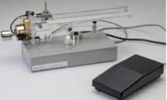
Tail Pressure
In naïve animals, mechanical hyperalgesia was assessed by only using the tail pressure test [84]. The animals were introduced in the metal restrainer, leaving the tail out and loose in order to place it on a small platform under the conic tip of the Randall Selitto apparatus (Figure 10) (Ugo Basile, Biological Research, Comerio). Pressing the apparatus pedal, an increasing force was applied to the animals’ distal tail portion (about 1-2 cm from tail tip), until the first pain response was observed. A response was considered positive if the mouse struggled, squeaked, or tried to remove the tail from

![Figure 5 - Amino acid sequence of human amilyn. [43]](https://thumb-eu.123doks.com/thumbv2/123dok_br/19174595.942626/29.892.200.494.121.299/figure-amino-acid-sequence-of-human-amilyn.webp)