PHENOTYPIC AND FUNCTIONAL ASPECTS OF
CD8+ T CELLS IN ATOPY
from populations to cells
Olga Maria Marques Lourenço
Supervisors: Professor Luís Taborda-Barata, M.D., Ph.D.
Dissertação com vista à obtenção do grau de Doutor em Biomedicina, apresentada à Faculdade de Ciências da Saúde da Universidade da Beira Interior.
Acknowledgements
First and foremost, I would like to thank my supervisor, Professor Luís Taborda-Barata for his invaluable support during my postgraduate studies, guidance, continuous help and friendship. It has been a privilege.I would like to thank my co-supervisor Professor Fernando A. Arosa, for his availability and helpful criticisms and recommendations.
I would like to thank Professor Mafalda Fonseca, for her support, advice and friendship. Moreover, I would like to express my gratitude to all present and former colleagues in the Immunology Group, as well as to everyone involved in the Health Sciences Research Centre of the University of Beira Interior.
I would like to thank all the study volunteers without whom this project would have been impossible.
I am also grateful to my parents for their caring and fruitful cooperation during my academic formation and postgraduate studies.
I thank my sister for her encouragement, for giving time unselfishly and her helpful suggestions in reviewing the English version of the manuscript.
Finally, very special thank you to my husband, who supported me during the days of intensive work, for sharing my dreams, even those implying his personal sacrifice.
This research was partially supported in the form of a fellowship (BD16448/2004) by the Portuguese Foundation for Science and Technology and this is gratefully acknowledged.
“To give less than your best is to sacrifice the gift” Steve Prefontaine
Contents
Index of Figures ...vii
Index of Tables...viii
Resumo Alargado...ix
Abstract ...xvii
Abbreviations...xix
List of Publications ...xxi
Related to this Thesis ...xxi
Unrelated to this Thesis ...xxi
Thesis Overview...xxii
1. General Introduction...1
1.1. Atopy and Allergic Diseases ...2
1.2. CD8+ T Cells...4
Memory and Naïve CD8+ T cells...5
Tc1 and Tc2 cells...6
CD8+CD28+ and CD8+CD28− T cells ...7
Suppressor CD8+ T cells...10
1.3. CD8+ T Cells in Allergy...14
Animal models of allergy...14
Human allergic disease...16
HYPOTHESES...19
AIMS OF THE THESIS...20
2. Background on the Methods...21
2.1. Collection of Biological Material ...22
Peripheral Blood...22
Induced Sputum ...22
2.2. Cell Isolation ...23
Immunomagnetic cell sorting ...23
2.3. T Cell Phenotype...25
Flow Cytometry...25
2.4. Cytokine Synthesis...26
Cytometric Bead Array ...26
2.5. T Cell Proliferation...26
Tritiated Thymidine ([3H]-thymidine) Incorporation...26
CFSE Fluorescence Loss...27
3.1. Demographic, Laboratory and Clinical Characterisation of Adult Asthmatic Patients ...29
3.1.1. Abstract ...29
3.1.2. Introduction...30
3.1.3. Materials and Methods ...31
3.1.4. Results ...31
3.1.5. Discussion...37
3.2. Asthma is more frequently associated with Non-allergic than Allergic Rhinitis in Portuguese Patients ...40
3.2.1. Abstract ...40
3.2.2. Introduction...41
3.2.3. Materials and Methods ...42
3.2.4. Results ...43
3.2.5. Discussion...47
3.3. Functional Characterisation of CD8+CD28+ and CD28− T Cells in Atopic Individuals sensitised to Dermatophagoides pteronyssinus...53
3.3.1. Abstract ...53
3.3.2. Introduction...54
3.3.3. Materials and methods ...55
3.3.4. Results ...58
3.3.5. Discussion...61
3.4. T Cells in Sputum of Asthmatic Patients are Activated Independently of Disease Severity or Control ...64
3.4.1. Abstract ...64
3.4.2. Introduction...65
3.4.3. Materials and Methods ...66
3.4.4. Results ...68 3.4.5. Discussion...74 4. General Discussion...79 4.1. Demographic aspects...80 4.2. CD8+ T cells in allergy...85 5. Appendix...89
5.1. Isolation of Leucocytes from Depletion Filters ...90
Introduction ...90
Materials and Methods...91
Results and Discussion ...91
5.2. Generation of Allergen-specific T cell lines...94
Introduction ...94
Materials and Methods...94
Results...95
Discussion and Conclusions ...97
Index of Figures
FIGURE 1–SUPPRESSOR T CELLS SCHEMATIC REPRESENTATION. ...11
FIGURE 2–RELATIONSHIP BETWEEN SEVERITY OF ASTHMA AND AGE IN ALLERGIC ASTHMATIC (PANEL A) AND NON-ALLERGIC ASTHMATIC (PANEL B) PATIENTS...32
FIGURE 3–PREVALENCE OF EXERCISE-INDUCED AND COUGH-VARIANT ASTHMA IN ALLERGIC AND NON -ALLERGIC ASTHMATIC PATIENTS...33
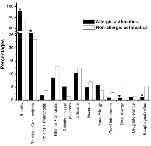
FIGURE 4–MAIN CO-MORBIDITIES ASSOCIATED WITH BRONCHIAL ASTHMA...34
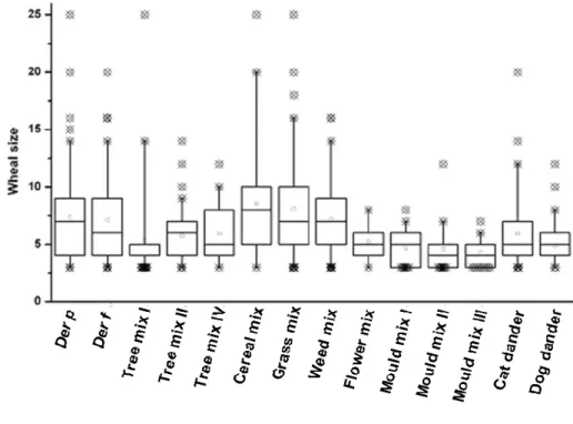
FIGURE 5–CUTANEOUS REACTIVITY TO AEROALLERGENS IN ALLERGIC ASTHMATIC PATIENTS...35
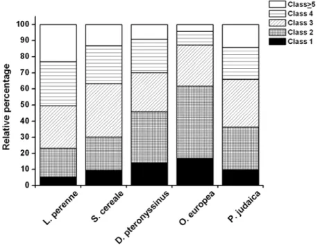
FIGURE 6–DEGREE OF SENSITISATION TO AEROALLERGENS IN ALLERGIC ASTHMATIC PATIENTS. ...36
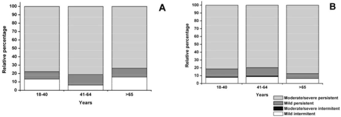
FIGURE 7–PROFILES OF DISEASE SEVERITY IN DIFFERENT AGE GROUPS OF NON-ALLERGIC RHINITIS (PANEL A) AND ALLERGIC RHINITIS (PANEL B) PATIENTS...44
FIGURE 8–MAIN CO-MORBIDITIES ASSOCIATED WITH RHINITIS...44
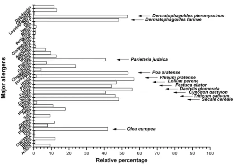
FIGURE 9–MAJOR AEROALLERGENS IN ALLERGIC RHINITIS. ...46
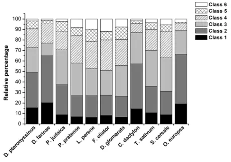
FIGURE 10–DEGREE OF SENSITISATION TO AEROALLERGENS IN ALLERGIC RHINITIS PATIENTS. ...47
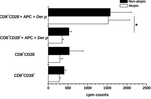
FIGURE 11–CD8+CD28−T CELLS PROLIFERATE MORE THAN CD8+CD28+T CELLS WHEN STIMULATED WITH DER P. ...58
FIGURE 12–CD8+CD28−T CELLS PROLIFERATE MORE THAN CD8+CD28+T CELLS WHEN STIMULATED WITH DER P BUT LESS WITH A POLYCLONAL STIMULUS. ...59
FIGURE 13–CYTOKINE SYNTHESIS IS SIMILAR FOR BOTH ISOLATED POPULATIONS. ...59
FIGURE 14–CD8+CD28− CO-CULTURES WITH PBMC PROLIFERATE MORE THAN CD8+CD28+ CO -CULTURES. ...60
FIGURE 15–CYTOKINE SYNTHESIS IS SIMILAR IN ALL THE CO-CULTURES STIMULATED WITH DER P EXTRACT. ...61
FIGURE 16–CD25(PANELS A,D AND G, N=25),CD69(PANELS B,E AND H, N=28), AND CD28(PANELS C, F AND I, N=23) SURFACE EXPRESSION IN PERIPHERAL BLOOD AND INDUCED SPUTUM T CELLS FROM PATIENTS WITH INTERMITTENT AND MODERATE PERSISTENT ASTHMA...71
FIGURE 17–COMPARISON OF SURFACE EXPRESSION OF CD25(PANEL A, N=25),CD69(PANEL B, N=28)) AND CD28(PANEL C, N=23) BETWEEN CD4+ AND CD8+T CELLS FROM PERIPHERAL BLOOD AND INDUCED SPUTUM OF PATIENTS WITH INTERMITTENT AND MODERATE PERSISTENT ASTHMA. ...71
FIGURE 18–EXPRESSION OF CD25 ON CD8+T CELLS FROM PERIPHERAL BLOOD OF PATIENTS WITH INTERMITTENT (N=18) AND MODERATE PERSISTENT (N=8) ASTHMA. ...73
FIGURE 19–FLOW CYTOMETRY DOT PLOTS OF FILTER MONONUCLEAR CELLS. ...92
FIGURE 20–OVERNIGHT ADHERENCE TO PLASTIC INCREASES PURITY OF LEUCOCYTE DEPLETION FILTERS’ FRACTIONS...92
Index of Tables
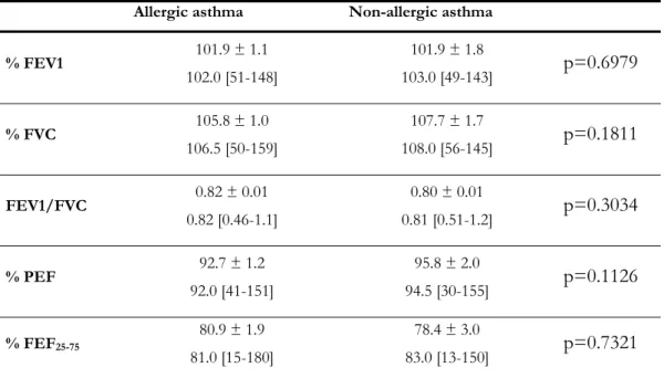
TABLE 1–LUNG FUNCTION TESTING VALUES IN ALLERGIC AND NON-ALLERGIC ASTHMATIC PATIENTS...33 TABLE 2–SENSITISATION PROFILE IN THE ALLERGIC ASTHMATIC PATIENTS...35 TABLE 3–CLINICAL CHARACTERISTICS OF STUDY SUBJECTS. ...69 TABLE 4–THE EFFECT OF DITHIOTHREITOL (DTT) ON THE PERCENTAGE OF SPUTUM LYMPHOCYTES
EXPRESSING CD28,CD25 AND CD69(N=6)...69 TABLE 5–EXPRESSION OF CD25,CD69 AND CD28 IN SPUTUM TOTAL LYMPHOCYTES,CD4+ AND CD8+T
CELLS FROM PATIENTS WITH INTERMITTENT (N=11) AND MODERATE PERSISTENT (N=6) CONTROLLED ASTHMA...73 TABLE 6–EXPRESSION OF CD25,CD69 AND CD28 IN SPUTUM TOTAL LYMPHOCYTES,CD4+ AND CD8+T
CELLS FROM PATIENTS WITH CONTROLLED (N=6) AND UNCONTROLLED (N=6) MODERATE PERSISTENT ASTHMA...74
Resumo Alargado
DOENÇA ALÉRGICAA prevalência das doenças alérgicas tem vindo a aumentar nas últimas décadas, especialmente nos países industrializados. Estas doenças surgem em indivíduos geneticamente predispostos, sob influência de factores ambientais. A “hipótese higiénica” procurou explicar este aumento de prevalência, relacionando-o com uma diminuição do contacto com microrganismos e do número de infecções, características do modo de vida ocidental. Inicialmente, pensou-se que a redução do contacto com microrganismos impedia o desvio imune de respostas do tipo T auxiliar 2 (“T helper 2” ou Th2) para respostas do tipo Th1, que ocorre geralmente com a idade (de acordo com o paradigma Th1/Th2). A inflamação, que ocorre particularmente ao nível das vias aéreas na asma e na rinite seria resultado de uma perda de equilíbrio entre respostas do tipo Th1, importantes nos mecanismos de defesa intracelular e respostas do tipo Th2, que caracterizam a inflamação alérgica. No entanto, estudos epidemiológicos posteriores puseram esta interpretação em causa. Foi então sugerido que, por diminuição da actividade reguladora/supressora dos linfócitos T, ocorre não um desvio imune, mas sim um desenvolvimento de respostas inadequadas face a estímulos antigénicos, resultando quer em doenças alérgicas quer em doenças auto-imunes.
A atopia consiste numa predisposição genética para produzir anticorpos IgE contra antigénios ambientais comuns (alergénios) e correlaciona-se com o desenvolvimento de uma ou mais doenças alérgicas. Estas caracterizam-se pela infiltração ao nível do órgão-alvo de células inflamatórias activadas, das quais se destacam os eosinófilos, os mastócitos e os linfócitos T.
De facto, as células T, especialmente as células T CD4+ do tipo Th2, são consideradas as grandes orquestradoras da resposta alérgica. As suas funções são mediadas principalmente pela secreção de citocinas do tipo 2 (interleucina (IL)-4, IL-5, IL-9 e IL-13). Posteriormente, foi mostrado que também células T CD8+ eram capazes de sintetizar estas citocinas (padrão “T cytotoxic 2” ou Tc2). No entanto, o papel desempenhado pelas células T CD8+ na doença alérgica ainda não está bem esclarecido. Diversos estudos,
nomeadamente em modelos animais, referem resultados contraditórios, atribuindo às células T CD8+ quer funções reguladoras, quer funções promotoras da inflamação.
A existência de subpopulações de células T CD8+ com características fenotípicas e funcionais distintas poderá ser um dos motivos para esta disparidade de resultados.
Estudos efectuados em indivíduos atópicos sugeriram que as células T CD8+ poderiam estar envolvidas no desenvolvimento de asma alérgica. Células T CD8+ estavam presentes nos infiltrados celulares no órgão-alvo, sendo que esta presença se correlacionava com asma severa e morte por asma brônquica. No entanto, estudos noutras patologias relacionaram a existência de células T do tipo CD8+ com funções reguladoras. A evidência é mais relevante em casos de transplantes de órgão sólido, onde a presença de células T supressoras do tipo CD8+CD28− se correlaciona com aumento de tolerância ao transplante e menor necessidade de medicação imunossupressora.
Tendo em atenção estes e outros estudos prévios, procurámos, neste estudo, clarificar o papel das células T CD8+ na atopia, nomeadamente no que diz respeito ao seu fenótipo e função na asma e rinite alérgicas. Definimos cinco objectivos: avaliar a proliferação alergénio-específica e a síntese de citocinas por células T CD8+; verificar se as células T CD8+ isoladas do sangue periférico têm propriedades supressoras face a respostas alergénio-específicas; verificar se as células T CD8+ estão presentes ao nível do órgão-alvo e se se encontram activadas; observar se existe correlação entre a expressão dos marcadores de activação expressos por células T CD8+ e a severidade da asma ou o seu controlo e por último, pretendemos caracterizar demograficamente a nossa população de estudo.
De modo a contextualizar os resultados a nível celular, decidimos iniciar o nosso estudo pela caracterização epidemiológica da população alvo (doentes com rinite alérgica e doentes com asma brônquica alérgica). Apesar do número de publicações sobre prevalência, diagnóstico e tratamento das doenças alérgicas ter aumentado nos últimos anos, dados relativos aos perfis de sensibilização continuam escassos. Dados relativos a prevalência em crianças foram obtidos maioritariamente através do estudo ISAAC (“International Study on
Asthma and Allergy in Children”) nas suas diversas fases. Neste estudo, os dados relativos a
Portugal mostram um aumento da prevalência na rinoconjuntivite alérgica e na asma brônquica em ambos os grupos etários de crianças (6-7 anos e 13-14 anos) entre as fases I e III, separadas no tempo por sete anos.
Relativamente aos dados na população adulta, Portugal participou no estudo ECHRS (“European Community Respiratory Health Survey”), que avaliou, através de questionário,
adultos entre os 20 e os 44 anos. Dados relativos a este estudo estimam uma prevalência de asma de cerca de 5% e de rinite de 17% para a população avaliada.
Apesar da região da Cova da Beira ter dos mais altos níveis polínicos a nível nacional (como descrito no Mapa Polínico de Portugal (1998-1999), neste momento não está incluída na Rede Portuguesa de Aerobiologia, nem existe nenhuma estação de monitorização de pólenes na região. Os dados sobre doença alérgica, especificamente relativos à região da Cova da Beira são muito escassos, revelando esforços mais ou menos individuais para clarificar a situação em termos de prevalência, sensibilizações e severidade de patologia. Pareceu-nos, pois, essencial caracterizar a doença alérgica na Cova da Beira de um modo mais sistematizado, como complemento importante dos nossos estudos de Imunologia básica da Alergia.
Recolhemos dados relativos aos doentes da consulta de Imunoalergologia do Hospital da Cova da Beira, centrando-nos especificamente em doentes com asma brônquica e rinite alérgica. Para o estudo da asma brônquica foram incluídos doentes admitidos à Consulta de Imunoalergologia entre 2003 e 2006 (1078 doentes), enquanto o estudo da rinite incluiu doentes admitidos à Consulta de Imunoalergologia entre 2003 e 2007 (1092 doentes). Para a análise posterior foram excluídos doentes com menos de 18 anos, doentes não residentes no concelho e todos aqueles com resultados laboratoriais discordantes em termos de avaliação de alergia.
A asma brônquica é uma doença inflamatória crónica, caracterizada por obstrução intermitente, reversível espontaneamente ou mediante tratamento e por hiper-reactividade brônquica. A rinite é também uma doença com características inflamatórias, caracterizada clinicamente por rinorreia, obstrução nasal e crises esternutatórias. Apesar dos avanços no conhecimento da sua fisiopatologia e terapêutica, a asma e a rinite mantém-se como duas das doenças crónicas mais frequentes em crianças e adultos.
O diagnóstico da asma e da rinite foi feito com base numa história clínica positiva e no exame objectivo (incluindo rinoscopia anterior), suportado por testes específicos de alergia (testes cutâneos por picada e determinação de IgE específica sérica), bem como por provas funcionais ventilatórias, no caso da asma brônquica. De acordo com a presença ou não de processos mediados pela IgE, os doentes foram divididos em duas grandes categorias: alérgicos e não alérgicos. Para a classificação de aspectos ligados à severidade da doença e ao seu controlo foram aplicadas “guidelines” internacionais (“Global Initiative for Asthma” para a asma e “Allergic Rhinitis and its Impact on Asthma” para a rinite).
Foram também avaliadas as principais co-morbilidades da asma e da rinite, nomeadamente no que diz respeito ao conceito actual de “one airway one disease” que considera asma e rinite manifestações de uma mesma entidade. Especificamente, doentes com rinite foram avaliados quanto à presença de asma pela história clínica, exame objectivo do tórax e avaliação de obstrução e, do mesmo modo, doentes com asma foram avaliados quanto à presença de rinite pela história clínica e rinoscopia anterior.
Trinta por cento dos doentes asmáticos e vinte e oito por cento dos doentes com rinite eram não alérgicos. Globalmente, os doentes não alérgicos eram mais velhos, predominantemente do sexo feminino e com uma maior prevalência de alergia/intolerância medicamentosa. Como descrito atrás e de acordo com as “guidelines” do projecto ARIA (“Allergic Rhninitis and its Impact on Asthma”), verificámos a associação de asma e rinite. Na população em estudo, a rinite estava mais frequentemente associada com a asma não alérgica do que com a asma alérgica. Pelo contrário, a asma brônquica estava preferentemente associada com rinite alérgica. No primeiro caso, estes resultados podem dever-se ao facto da rinite alérgica geralmente preceder o desenvolvimento de asma alérgica (marcha atópica). De facto, a imunoterapia específica parece atrasar ou mesmo impedir a progressão da rinite alérgica para asma. Pelo contrário, não existe esta progressão temporal quando se fala de doença não alérgica, em que geralmente existem desencadeantes comuns. Os dois tipos de asma e de rinite (alérgica e não alérgica) não puderam ser distinguidos em termos de severidade da doença, nem em termos de sintomatologia clínica. Este facto era inesperado, uma vez que estudos anteriores mostravam que asma e rinite não alérgicas eram mais severas do que quando se deviam a alergia. Contudo, não podemos descurar que esta conclusão possa ter tido origem num viés na análise: geralmente a severidade da doença aumenta com a idade e os doentes com patologia não alérgica são mais velhos, pelo que, em estudos de amostras não emparelhadas quanto à idade, a patologia não alérgica vai parecer mais severa.
Os alergénios mais frequentes na região são os pólenes de gramíneas (Poaceae), sejam elas cultivadas ou não, os ácaros e os pólenes de oliveira. A monosensibilização não é comum e, nos casos em que existe, refere-se unicamente a alergia a ácaros. Um dado interessante prende-se com o facto de os pólenes das gramíneas não serem apenas responsáveis pela maioria das sensibilizações, mas serem também os responsáveis pelos níveis mais elevados de IgE específica. No entanto, não foi possível estabelecer uma correlação entre os níveis de IgE específica e a severidade da asma ou da rinite.
Estudos semelhantes levados a cabo noutras regiões mostraram uma associação significativa entre os níveis de poluição atmosférica e o aumento da sintomatologia ligada à asma e à rinite. A prevalência de atopia e doença alérgica era maior nas zonas urbanas do que nas zonas rurais, devido quer a um aumento da poluição atmosférica nas zonas urbanas, quer a um efeito protector do ambiente rural. Nas áreas urbanas, os grãos de pólen podem ficar revestidos com resíduos de combustível e com produtos da combustão, sendo que esta ligação a partículas de exaustão de diesel pode alterar os epítopos alergénicos e mesmo aumentar a sua alergenicidade. Convém salientar que irritantes não específicos como a poluição atmosférica são capazes de agravar ou mesmo induzir sintomas em doentes asmáticos.
No nosso estudo, asmáticos residentes em ambiente rural ou urbano apresentavam perfis de sensibilização distintos, tendo, no entanto severidades da doença semelhantes. Doentes com rinite alérgica apresentavam perfis de sensibilização semelhantes. Estes resultados podem ser explicados pelo tamanho relativo dos grãos de pólen que condiciona a sua passagem pelas vias aéreas. Geralmente, grãos de pólen intactos são incapazes de penetrar as regiões mais distais das vias aéreas, sendo a maioria dos sintomas localizados ao nível dos olhos, nariz e faringe. No entanto, alergénios derivados dos grãos de pólen podem depositar-se em partículas sub-micrónicas e desencadear asma, tal como ocorre durante as trovoadas.
Os nossos estudos epidemiológicos forneceram uma base de trabalho extensa e pormenorizada da nossa população, permitindo delinear melhor os estudos celulares subsequentes. Por outro lado, estes estudos vieram reforçar a necessidade de tratar em conjunto doenças das vias aéreas superiores e inferiores usando uma estratégia simultaneamente mais eficaz e mais segura. No entanto, estes estudos apresentam uma importante limitação, visto analisarem amostras provenientes de uma consulta da especialidade. De um modo geral, a maioria dos doentes seguidos em consultas da especialidade são aqueles em que a severidade da doença é maior. Outro ponto a considerar prende-se com a classificação da severidade da doença. Apesar de esta ter sido efectuada na primeira consulta, alguns dos doentes já se encontravam sob medicação, o que poderia enviesar a classificação.
De qualquer forma, trata-se, na nossa opinião, de um estudo muito interessante uma vez que inclui parâmetros clínicos, contrastando com a maioria dos estudos anteriores que apenas se baseavam em questionários.
CÉLULAS T CD8+ NA ALERGIA
A asma e a rinite alérgicas caracterizam-se pela presença de uma inflamação crónica mediada por múltiplas proteínas inflamatórias, incluindo citocinas, quimiocinas e enzimas. Em ambas as patologias ocorrem exacerbações quando a intensidade da inflamação aumenta. Diversos estudos sugeriram que as células T desempenham um papel fundamental nesta inflamação. Através da síntese e secreção de citocinas do tipo 2, as células T promovem a diferenciação de células Th2 e a síntese de IgE pelas células B (IL-4 e IL-13); promovem a diferenciação, a activação e a sobrevivência dos eosinófilos (IL-5) e aumentam a diferenciação de mastócitos e a produção de muco (IL-9). Tal como foi referido, a grande maioria dos estudos avaliou fenotípica e funcionalmente as células T CD4+, sendo escassos os resultados relativos às células T CD8+.
Uma vez que a atopia se caracteriza por elevados níveis de IgE específica sérica, muito provavelmente alterações no órgão-alvo vão reflectir-se ao nível do sangue periférico. O estudo das células T CD8+ do sangue periférico de doentes alérgicos constituiu o nosso primeiro projecto ao nível da imunologia básica da doença alérgica. Uma vez que a expressão da molécula co-receptora CD28 permite definir duas subpopulações distintas ao nível das células T CD8+, procurámos, ao longo do estudo, aprofundar a sua caracterização fenotípica e funcional.
As células T foram isoladas a partir de sangue periférico de doentes com asma e/ou rinite por centrifugação em gradiente de densidade. Em seguida, as células T CD8+CD28+ e CD28− foram isoladas por separação imunomagnética num sistema semi-automático (MACS®) e usadas, posteriormente, para os estudos funcionais. Para avaliar a proliferação e síntese de citocinas em resposta a aeroalergénios usámos extracto purificado de
Dermatophagoides pteronyssinus. Para avaliar a capacidade supressora das células T CD8+ face
às respostas alergénio-específicas desenvolvemos um sistema de co-culturas com células mononucleares do sangue periférico tratadas com mitomicina. A pureza das fracções usadas foi avaliada por citometria de fluxo.
Observámos que a atopia não estava relacionada com alterações nas percentagens ou fenótipo das células T CD8+; no entanto, células T CD8+CD28+ eram distintas em termos fenotípicos de células T CD8+CD28−. Esta alteração fenotípica era acompanhada
por uma resposta proliferativa distinta relativamente a mitogénios e alergénios, se bem que semelhante do ponto de vista da secreção de citocinas.
Ambas as subpopulações apresentavam uma capacidade proliferativa semelhante em doentes atópicos e não atópicos e não apresentavam características supressoras.
Estes resultados devem, no entanto, ser analisados com cuidado, uma vez que as culturas in
vitro não conseguem imitar o estímulo repetido e prolongado que ocorre in vivo. Além disso,
as células T do sangue periférico não partilham o mesmo ambiente das células T dos órgãos-alvo, não estando sujeitas ao mesmo tipo de interacções e contacto com alergénio. Para clarificar se o que ocorre ao nível do sangue periférico reflecte o que se passa no órgão-alvo, procurámos estudar células T CD8+ provenientes deste. Procurando uma abordagem menos invasiva do que os lavados broncoalveolares ou as biópsias, realizámos o estudo em células T provenientes da expectoração induzida de doentes asmáticos alérgicos. Foram incluídos no estudo asmáticos alérgicos com asma intermitente ou moderada persistente com diversos graus de controlo.
A expectoração induzida foi obtida por inalação de uma solução hipertónica salina (NaCl a 5%). A expectoração foi seguidamente homogeneizada e incubada com ditiotreitol, sendo posteriormente filtrada através de um filtro celular de 0.40μm de diâmetro de poro. A viabilidade da suspensão celular obtida foi avaliada pelo método de exclusão de Azul Tripano em câmara de Neubauer.
Neste estudo mostramos claramente que, quer as células T CD4+, quer as células T CD8+ apresentam um fenótipo activado na expectoração induzida, o que sugere que ambas as subpopulações celulares participam no processo inflamatório ao nível do órgão-alvo. Uma vez que esta inflamação se relaciona com a severidade da doença, procurámos avaliar a correlação existente entre a activação celular e o grau de severidade da asma.
Apesar da escolha criteriosa de voluntários, emparelhados quanto a sexo, idade e controlo da doença, não observámos correlação entre a percentagem relativa de expressão de marcadores de activação (CD25 e CD69) e o grau de severidade da doença.
Atendendo a que o controlo é um dos objectivos mais determinantes do tratamento da asma, avaliámos também a correlação existente entre os nossos marcadores de activação em estudo e o controlo da doença. Optámos aqui por incluir apenas asmáticos alérgicos com asma moderada persistente, sob tratamento mais ou menos equivalente. Tal como se verificou com a severidade, também não se observou correlação entre a percentagem relativa dos marcadores de activação e o controlo da doença.
Para clarificar o efeito dos corticosteróides inalados na expressão dos marcadores de activação, comparámos a percentagem relativa dos marcadoress de activação entre asmáticos com asma intermitente controlados e não controlados. Não observámos correlação entre a percentagem relativa dos marcadores e o grau de controlo dos doentes, o que parece indicar um efeito mínimo dos corticosteróides inalados em termos de avaliação de controlo da asma.
Em resumo, nesta Tese clarificámos as características demográficas da população de asmáticos e doentes com rinite da consulta de especialidade do Hospital Pêro da Covilhã e caracterizámos as células T, especialmente da população T CD8+ nestes indivíduos em termos fenotípicos e funcionais. Finalmente, procurámos novas estratégias para isolar células T do sangue periférico e diferenciar linhas celulares T alergénio-específicas.
Relativamente às nossas hipóteses iniciais, verificámos que as células T CD8+ isoladas a partir do sangue periférico não apresentavam características supressoras das respostas alergénio-específicas. Em segundo lugar, as células T CD8+ da expectoração induzida de doentes asmáticos expressavam marcadores de activação, sendo a percentagem relativa de expressão maior do que a encontrada ao nível do sangue periférico. No entanto, embora apresentando um fenótipo de células activadas, a percentagem relativa dos marcadores de activação não se correlacionou com a severidade da doença nem com o seu grau de controlo.
Abstract
The prevalence of allergic diseases has been increasing during the last decades. It is believed that the development of allergic diseases in susceptible individuals is dependent on T cells, especially type 2 CD4+ T cells. CD8+ T cells may also be involved in the pathophysiology of allergic diseases, albeit studies in animal models of asthma have found confounding results.With our study we attempted to clarify the phenotypic and functional properties of CD8+ T cells in allergic disease settings. The specific aims were to assess allergen-specific proliferation and cytokine synthesis of CD8+ T cells and evaluate their suppressor function on antigen-specific responses. In addition, it was also our goal to assess the presence of CD8+ T cells at the target organ and their activation status, correlating our findings with disease severity and control. In order to contextualize our cellular findings, we initially characterised our study population from an epidemiological point of view.
We enrolled asthma and rhinitis patients attending the Allergy Clinic of the Cova da Beira Hospital. Asthma and rhinitis were diagnosed by a positive clinical history and specific diagnostic tests, and severity was assessed by current guidelines. We also identified the main co-morbidities in both disease settings and evaluated asthma symptoms in rhinitis patients and rhinitis symptoms in asthma patients. In the allergic groups we investigated the sensitisation profiles to aeroallergens and concluded that major allergens included grass and cereal pollen, mites and olive tree pollen. Sensitisation profiles and severity were also compared between rural and urban-based allergic patients. In this regard, we found contrasting results between asthmatic patients and patients with rhinitis.
In the second part of our study we clarified the role of CD8+ T cells in allergic inflammation through basic cellular systems. We assessed proliferation capacity and cytokine synthesis in response to Dermatophagoides pteronyssinus extract, and devised a co-culture system in order to evaluate the suppressor function of CD8+ T cells. CD8+CD28+ and CD28− T cells isolated from the peripheral blood had distinct phenotypes and proliferated at different levels to common stimuli, although sharing similar cytokine production patterns. A potential suppressor activity was not found in the co-culture systems, either for CD8+CD28+ or CD28− T cells.
We then studied CD8+ T cells at the target-organ, assessing activation phenotype in cells from the induced sputum of asthmatic patients. We showed that CD8+ T cells are
activated at the target-organ, although we were unable to demonstrate a relationship between activation of this T cell subset and severity or control of asthma.
Abbreviations
ACT Asthma Control Test
AHR Airway hyperresponsiveness
AICD Activation induced cell death APC Antigen presenting cell
-APC Allophycocyanin conjugated
AR Allergic rhinitis
ARIA Allergic Rhinitis and its Impact on Asthma
BAL Bronchoalveolar lavage
BSA Bovine serum albumin
BW Bronchial washing
CBA Cytometric bead array
CD Cluster of differentiation
CFSE Carboxylfluorescein diacetate succinimidyl ester COPD Chronic obstructive pulmonary disease
cpm Cintilations per minute
CRA Cockroach antigen
CTL Cytotoxic T lymphocyte
CTLA-4 Cytotoxic T lymphocyte-associated antigen 4
DC Dendritic cell
DC2 Plasmacytoid dendritic cell Der p Dermatophagoides pteronyssinus DTT Dithiothreitol
ECRHS European Community Respiratory Health Survey ELISA Enzyme linked imuno-sorbent assay
FACS Fluorescence-activated cell sorting
FCS Fetal calf serum
FcεRI High affinity IgE receptor FcεRII Low affinity IgE receptor (CD23) FEV1 Forced expiratory volume in one second
-FITC Fluoresceine isothiocyanate conjugated FOXP3 Forkhead box p3
GINA Global Initiative for Asthma
GITR Gluocorticoid-induced TNF receptor
HCMV Human cytomegalovirus
HCV Hepatitis C virus
HIV Human immunodeficiency virus IFN Interferon
Ig Immunoglobulin IL Interleukin
ILT Immunoglobulin-like transcript ISAAC International Study of Asthma and Allergies in Childhood
mAb Monoclonal antibody
MHC Major histocompatibility complex
NAR Non-allergic rhinitis
NFкB Nuclear factor kappa-B
NK Natural killer
OVA Ovalbumin PBL Peripheral blood lymphocytes PBMC Peripheral blood mononuclear cells
PBS Phosphate buffered saline
-PE Phycoerythrin conjugated
PEF Peak expiratory flow
-PerCP Peridinin chlorophyll protein conjugated PHA Phytohaemagglutinin PMA Phorbol myristate acetate sem Standard error of the mean
Tc T cytotoxic cells TCC T cell clones TCL T cell lines
TCM Central memory CD8+ T cell
TCR T cell receptor
TEFF Effector memory CD8+ T cell
Th T helper cells
TLR Toll-like receptor
TNF Tumour necrosis factor
Treg T regulatory (cells)
List of Publications
Related to this Thesis
Functional and phenotypic characterization of CD8+CD28+ and CD28− T cells in atopic individuals sensitized to Dermatophagoides pteronyssinus.
Lourenço O, Fonseca AM, Arosa FA, Taborda-Barata L Allergol. et Immunopathol. 2006; 34 (6): 234-41
Demographic, laboratory and clinical characterisation of adult Portuguese asthmatic patients.
Lourenço O, Fonseca AM, Taborda-Barata L Allergol. et Immunopathol. 2007; 35 (5): 177-83
Non-allergic rhinitis is more frequently associated with asthma than allergic rhinitis in adult Portuguese patients.
Lourenço O, Fonseca AM, Taborda-Barata L
Rhinology (Accepted for publication)
T cells in Sputum of asthmatic patients are activated independently of disease severity or control.
Lourenço O, Fonseca AM, Taborda-Barata L
Submitted to Respiration
Unrelated to this Thesis
Respostas celulares T aos alergénios do látex.Santos S, Tavares P, Lourenço O, Fonseca AM, Taborda-Barata L Rev. Port. Imunoalergologia 2007; 15 (1): 7-17.
Thesis Overview
This Thesis is divided into five chapters.The first chapter consists of a brief literature review running from atopy and allergic diseases to specific issues regarding CD8+ T cells in allergy settings.
The second chapter includes an overview of the methods used.
Subsequently, the third chapter is based on original research papers published or submitted during the Ph.D. course, and directly related to this Thesis.
The fourth chapter comprises the general discussion and conclusion remarks.
Finally, the fifth chapter refers to unpublished work developed during the course and directly related to this Thesis.
1.1. Atopy and Allergic Diseases
Atopy is an individual predisposition for excessive immunoglobulin (Ig)-E production against harmless environmental allergens. It is a genetically and environmentally determined condition 1, 2, which may lead to allergic diseases such as asthma, allergic rhinitis,
conjunctivitis, and atopic dermatitis 3. It is estimated that as many as 300 million people of
all ages, and all ethnic backgrounds, suffer from asthma and the burden of this disease to governments, health care systems, families, and patients is increasing worldwide 4.
The prevalence of allergic diseases has been increasing during the last decades, especially in developed countries. The “hygiene hypothesis” attempts to explain this increase on the basis of reduced exposure to infectious agents, especially during early life. Early studies suggested that a skewing of the T cell response towards type 2 CD4+ T cells could explain the subsequent development of allergy 5. Inflammation, particularly in the airways in
asthma and rhinitis would represent a loss of balance between T-helper (Th)-1 cells, critical for intracellular defence mechanisms, and Th2 cells, which mediate allergic inflammation. However, several observations argue against this. Firstly, epidemiological studies showed that both Th2-related allergies and Th1-related autoimmune diseases could co-exist in the same populations 6, 7. Secondly, high parasite burdens, which are associated with strong Th2
responses, seem to protect against allergic disease 8, 9. Thirdly, in asthma, IFN-γ levels are
enhanced, suggesting that Th1 cytokines contribute to the pathogenesis of the disease, instead of protecting from it 10, 11.
The first interpretation of the “hygiene hypothesis” states that a reduced microbial burden impairs the shift of allergen-specific responses from the Th2 to the Th1 phenotype that usually takes place with age (thereby failing to undergo immune deviation). The more recent interpretation states that the lower microbial burden reduces the activity of regulatory T cells leading to allergy and autoimmunity (reduced immune suppression). (For review on the “hygiene hypothesis” see 12, 13, 14).
It is generally believed that the development of allergic disease in susceptible individuals first requires repeated and persistent exposure to allergens, leading to aberrant activation and recruitment of T cells with subsequent differentiation of increased numbers of allergen-specific Th2 cells. These T cells provide interleukin (IL)-4 and contact-mediated
signals that promote differentiation of B cells and class-switching to the production of IgE. The produced IgE binds to high affinity IgE receptors (FcεRI) on the surface of circulating basophils and on mast cells in various tissues. Following such sensitisation, subsequent exposure to the specific allergen initiates a secondary immune response. The allergen binds to cell-associated IgE and cross-links the IgE molecules and the FcεRI receptors to which they are bound. Cross-linking of the IgE-FcεRI complexes on basophils and mast cells signals these cells to release preformed and newly generated mediators, including biogenic amines such as histamine, lipid mediators such as leukotrienes, and cytokines (early phase response) 15. Biogenic amines and lipid mediators create the signs and symptoms of allergic
disease that typically begin within one hour of allergen exposure. These include rapid vascular leakage of plasma fluid and protein, and vasodilatation. Six to twenty-four hours later, a second round of symptoms (late phase reaction) may develop, mainly driven by the release of cytokines and chemokines. Specifically, in terms of target-organs, in the nose, this late reaction is characterised by an inflammatory infiltrate including activated eosinophils, basophils, neutrophils, and lymphocytes; resulting in major congestion, itching and rhinorrhea 2. Furthermore, in the lung, repeated episodes of this late phase reaction can
lead to tissue damage and remodelling.
Antigen presenting cells (APC) including monocytes, macrophages, B cells, dendritic and Langerhans cells are important in IgE-mediated allergic responses. Both the high affinity, FcεRI, and the low affinity FcεRII (CD23) IgE receptors are expressed on these APC and are important in facilitating allergen presentation to allergen-specific T cells. Triggering of the CD23 receptor by IgE on B cells not only enhances antigen presentation, but also increases synthesis of IgE.
The priming of allergen-specific CD4+ T cells and their differentiation towards a Th2 phenotype are key events for the development of allergic diseases. Th2 cells orchestrate atopic inflammation through the secretion of a subset of cytokines, mainly IL-4, IL-5, IL-9 and IL-13. Similarly, CD8+ T cells from atopic patients have the capacity to produce type-2 cytokines (T cytotoxic type 2 or Tc2 phenotype) 16.
1.2. CD8+ T Cells
CD8+ T cells are major histocompatibility complex (MHC) class I-restricted. As MHC class I molecules contain mainly peptides derived from the cytosol, this is an effective mechanism for killing cells infected with viruses or other intracellular pathogens. Antigen recognition by naïve CD8+ T cells triggers a programme of proliferation and differentiation that leads to the production of effector lymphocytes (cytotoxic T lymphocytes or CTL) directly able to lyse antigen-bearing cells. The lytic mechanism primarily involves release of cytoplasmic granules loaded with perforin (a pore-forming protein) and granzyme B (a serine protease) at the contact site between the CTL and the target cell 17, resulting in specific killing without bystander cell damage. The nonsecretory
mechanism is initiated by receptor-mediated triggering of apoptosis-inducing target cell surface molecules (Fas-FasL)17.
However, CD8+ T cells have also been associated with suppression of immune responses, and their ability to produce various cytokines, such as IFN-γ and TNF-α, suggests an additional function.
Generally, the peptides bound in the grooves of the MHC class I molecules are derived from proteins synthesised within the cell that bears the class I molecules (self-proteins and viral proteins); in contrast, allergic responses are thought to occur when CD4+ T cells react to exogenous antigen presented on MHC class II molecules. It was believed that CD8+ T cells were unable to respond to allergens. However, both macrophages and dendritic cells (DC) have been reported to cross-present exogenous antigen to CD8+ T cells in some circumstances, by at least two different mechanisms (the phagosome-to-cytosol pathway and the vacuolar pathway)(for review see 18).
Altough most allergens are proteins, some are lipids or carbohydrates. Peptides are not the only target of T cell responses, as T cells can recognize lipid antigens through a CD1-dependent pathway. In fact, lipids are important in pollen grain structure and function, and may be relevant in enhancing grain capture by DC, which is likely to be required for their subsequent recruitment of pollen-specific T cells in sensitised subjects 19.
So, a potential pathogenic role can be envisioned for CD1-restricted T cells, as lipid components of allergens can be presented by epithelial DC to T cells 20. T cell clones
function for IgE production 19. CD1-restricted T cells can be TCR-αβ, -γδ, or NKT cells,
but the majority of lipid-specific clones decribed to date are CD4+ 19, 21.
Memory and Naïve CD8+ T cells
Immune responses to viral infections involve the proliferation and differentiation of CD8+ T cells into an effector population that controls viral replication. The expansion of the effector cells is accompanied by an increased sensitivity to apoptosis, which regulates proliferation and maintains lymphocyte homeostasis. A small proportion of activated CD8+ T cells survive, forming the memory populations. Memory CD8+ T cell subsets have been broadly divided into central memory CD8+ T cells (TCM) and effector memory
CD8+ T cells (TEFF) distinguishable by phenotype and function. TCM reside in lymphoid
organs, do not express immediate lytic functions, are CD62hiCCR7hi and can be generated
following culture with IL-15 after a short stimulation with allergen 22. T
EFF are found in
non-lymphoid tissues, express lytic activity, are CD62loCCR7lo and are generated in culture
with IL-2 22. T
EFF can play a critical role in the development of airway hyperresponsiveness
(AHR) and allergic inflammation by producing IL-13 in the lung.
When activated under optimal conditions, these cells will emerge as functionally competent CTL that are armed with various effector mechanisms to eliminate abnormal cells. There are two mechanisms of cytotoxicity: a membranolytic one, depending on the formation of pores in the target cell membrane, and mediated by perforin and granzymes; and a nonsecretory one, receptor-mediated (Fas-FasL) 17. Helper CD4+ T cells may not be
required to initiate CTL activity but are critical for preserving antigen-specific CD8+ T cell responsiveness during chronic infection. Since some pathogens are able to directly stimulate APC to properly present antigen to CD8+ T cells, helper cells may not be
required to activate APC during the early stages of infection. During persistent infection, where antigen may be minimal and thus unable to effectively condition professional APC, CD4+ T cells play a critical role in sustaining CD8+ T cell immunity by activating the APC to properly stimulate CD8+ T cells 23, 24.
In humans, the CD45RA and CD45R0 surface antigens have been used to separate T cells into naïve and memory pools. However, discrimination based only on CD45 isoform expression is insufficient since, with time, some CD45R0 T cells may revert to CD45RA 25.
with concomitant phenotypic changes and acquisition of functional properties (IFN-γ and TNF-α synthesis, and cytotoxic function) that are difficult to distinguish from antigen-primed T cells 26.
Tc1 and Tc2 cells
The current model of naïve T cell activation postulates that full activation requires two signals. The first signal is antigen displayed by the APC in the form of peptides bound to histocompatibility molecules; the recognition of antigen by the T cell receptor (TCR) provides specificity to the response. The second signal, called the costimulatory signal, is provided by molecules on the APC that engage particular costimulatory receptors on T cells 27. It can dictate the fate of the response and provides a mechanism for regulating the
appropriateness of a given response. A third signal or polarization directs T cell differentiation into various effector types, such as Tc1 and Tc2; it can be derived from cytokines or from inflammatory signals supplied by Toll-like receptor (TLR) ligands 28, 22.
CD8+ T cells can differentiate into two effector phenotypes, Tc1 and Tc2, secreting different cytokine patterns. Tc1 cells are defined as CD8+ T cells that secrete IFN-γ but not IL-4 or IL-5, and Tc2 cells are CD8+ cells that secrete IL-4, IL-5, and low amounts of IFN-γ 29, 30. The synthesis of IL-2 by mouse Tc1 cells is subjected to further regulation, and
IL-6 and IL-10 are preferentially, but not exclusively, synthesized by Tc2 cells 29. Overall,
CD8+ T cells are able to produce the same range of type 1 and type 2 cytokines as CD4+ T cells 31.
Naïve CD8+ T cells show a strong preference for differentiating into Tc1 cells. Tc2 differentiation requires substantial amounts of IL-4 and is very much dependent upon the APC 29, whereas IFN-γ and IL-12 encourage differentiation to Tc1 cells 30.
Human Tc1 CD8+ T cell clones express little CD28, CD30 or CD40L, while Tc2 cells express significant amounts of these ligands, which may facilitate their interaction with other immune cells 32.
Both subsets are cytotoxic via the perforin and the Fas pathways, and both are able to kill resting and activated B cells 29, 30. Furthermore, both subsets induce inflammation with
The CTL response to acute infection can generally be divided into four phases. During the effector phase, naïve CTL precursors are primed, undergo dramatic expansion, acquire effector function, travel to sites of infection, and mediate pathogen clearance by killing infected cells and secreting effector cytokines. During the contraction phase, most effector CTL die, leaving behind 5-10% of the original burst size as long-lived memory cells. During the memory maintenance phase, memory CTLs are maintained at stable levels throughout the mouse lifespan and for many years in humans. The rapid recall response of memory CTL following re-exposure to the pathogen provides enhanced protection to the host.
It is unlikely that CD8+ T cells provide help for B cells, since, via the regular antigen-processing pathways, foreign antigens would be presented on MHC class II in the B cells leading to help by CD4+ T cells. In contrast, if the B cells were infected, the antigen would be presented in MHC class I leading to killing of these cells by CD8+ T cells 29.
CD8+CD28+ and CD8+CD28− T cells
CD8+ T cells can be subdivided into two subsets with different biological properties according to the surface expression of CD28.
At birth, the great majority of T cells express CD28 (less than 1% of CD28− T cells in cord blood 33). Aging is characterised by a progressive exhaustion of naïve T cells involving
both CD4 and CD8 subsets 34 which is accompanied by a progressive expansion of CD28−
T cells, that is particularly evident in CD8+ T lymphocytes 35, 36. The CD28+ T cell
population is predominant in young healthy individuals and expands during primary viral infection, whereas CD28− T cells are more common in elderly healthy individuals 37 and
increase dramatically in human immunodeficiency virus (HIV) patients 38, 25. In these
patients, specific cytotoxic activity is mainly mediated by CD8+CD28− T cells, while inhibition of HIV replication is controlled by CD8+CD28+ T cells 39.
CD8+CD28− T cells have been characterised by oligoclonal expansions 40, 41, impaired
proliferative responses, but preserved cytotoxicity and reduced telomeres 42. Some authors
suggested that the CD28− subset has a longer replicative history (more rounds of cell division), and consistent with the loss of CD28 might have reached a state of replicative senescence 42. The average telomere length shortens between 50 to 100 base pairs with each
round of replication, and thus, telomere lengths can be used to assess the replicative history of cell populations.
It has been proposed that CD8+CD28−T cells derive from CD8+CD28+precursors 43,
because the CD28 negative phenotype can be induced with in vitro stimulation of cord blood lymphocytes which typically contain only CD8+CD28+ T positive cells 44. BrdU
labelling showed that CD8+CD28− T cells derive from CD8+CD28+ precursors in vitro 45.
IL-4 was able to prevent, but not reverse, the switch in CD28 expression in one study 43,
but in another study, treatment of CD8+ T cells with IL-4 decreased the levels of both CD28 surface expression and message and increased CD8 expression 46.
Signalling through CD28 is the major co-stimulatory signal for activation of naïve T cells. Its interaction with CD80/CD86 expressed on B cells, macrophages, and dendritic cells stimulates IL-2 mRNA production 47, and stabilises IL-2 mRNA, thereby enabling T
lymphocytes to proliferate in response to antigen. It also inhibits T cell receptor-induced apoptosis during primary T cell responses 48, 49. Once established, the proliferative response
appears to be less dependent on CD28 co-stimulation, since exogenous growth factors such as IL-2 can restore mitotic progression. Normally, most CD8+ T cells do not produce enough IL-2 to support their own expansion 43.
Freshly-isolated peripheral blood CD8+CD28− T cells are enriched for large and granular lymphocytes as assessed by flow cytometry 33, 50, and have a limited Vβ repertoire 51. This
oligoclonality is common in healthy subjects as well as in pathological conditions 40. Loss of
CD28 expression is also associated with a phenotype of antigen-experienced CD8+ T cells: CD57+CD11b+CD27−CD49+ with variable expression of CD45RO/RA 33, 37, 52.
Co-expression of chronic activation antigens such as CD38 and HLA-DR can occur, particularly in HIV-infected patients 53, but acute activation antigens, such as, CD25, CD69
and CD71 are not usually expressed 33, 37, 52,54.
When phenotypically compared with their CD28 positive counterparts, CD8+CD28− T cells have a higher expression of CD11b, CD29, CD57, and CD94 and lower expression of CD25 55. Affymetrix gene chip analysis of CD8+CD28− and CD28+ T cells from five
different human T cell lines (TCL) showed differences in the level of expression of genes that encode cell surface molecules, signal transduction molecules, chemokines, cytokines, apoptosis-related proteins, cell growth regulators and metabolic enzymes 56. Natural killer
(NK)-related receptors (CD16, CD56, CD57, CD85j, CD94, CD161, CD244, EB6, gl183, NKG2A, NKB1) were preferentially expressed on the CD28 negative subset, both in
healthy donors and also in melanoma patients 57. It is possible that the specific
up-regulation of some inhibitory NK cell-related receptors on CD8+CD28− T cells can block their cytotoxic capacity, accounting for the inability of these cells to kill their target cells 58.
In our previous work, we evaluated the expression of several phenotypic markers on CD3+CD8+CD28+ and CD28− subpopulations in whole blood from ten atopic and ten non-atopic volunteers. Although the phenotypic expression was similar in both atopic and non-atopic volunteers, overall the phenotypic expression in CD8+CD28+ T cells was different from that in CD8+CD28− T cells, as previously described by other authors 33, 37, 52.
The functional response of the CD8+CD28− T cells is distinct from that of the CD8+CD28+ T cells. After stimulation of T cells with immobilised anti-CD3, freshly isolated peripheral blood CD8+CD28− T cells proliferated poorly, compared to the vigorous response of the CD8+CD28+ T cells 33. Increased anti-CD3 can compensate for
the loss of co-stimulatory signal caused by reduced CD28 levels but fails to significantly increase the responsiveness of the CD28− population 46. This hyporesponsiveness could
not be overcome by addition of exogenous IL-2, excluding the lack of IL-2 as the cause for this anergic response 33, 46. Proliferation of these cells was impaired, even when activated
with mitogens that bypass TCR signalling 55. Similarly, T cell clones also demonstrated
modest growth in response to stimulation with anti-CD3 plus IL-2, whereas CD8+CD28+ clones proliferated vigorously 33. Both CD8+CD28+ and CD28− T cell lines were
observed to expand when cultured in the presence of IL-2, IL-7 or IL-15, and survived without significant expansion in the presence of IL-4, suggesting that proliferation might be important in inducing the loss of CD28 59. The reduction in proliferation might be due
to increased RNA and protein levels of the cyclin dependent kinase inhibitor p16, as described by Scheuring et al. in CD8+CD28− T cells from aged adults 55.
Overall, CD8+CD28− T cells are a heterogeneous subset that contains both memory and effector cells 60. The most established view considers CD8+CD28− T cells as terminally
differentiated or end stage/senescent cytotoxic T cells 45, 52, although these cells were first
described as suppressors of B and T cell function. Interestingly enough, in a number of situations, ranging from chronic inflammatory conditions 61 and infectious diseases 53, 62 to
aging 52, 63, immunodeficiency 64, iron overload 65 and heavy alcohol intake 66, an increase in
known whether this increase in CD8+CD28−T cells is a cause for the pathology or an immunological response to it 50, but this association with disease suggests that they are an
effector population.
Suppressor CD8+ T cells
A central finding from experimental models and human studies shows that allergic diseases are due to an aberrant immune response mediated through Th2 cells, characterised by a specific cytokine pattern. To avoid chronic cell activation and inflammation against non-pathogenic antigens, and control harmful T cell responses, the immune system has evolved a variety of regulatory mechanisms mediated by distinct T cell subsets. The regulation mediated by these suppressor subsets is superimposed on intrinsic regulatory mechanisms (such as deletion and anergy). The cellular basis of suppression was initially proposed in the early 1970s by Gershon and Kondo, who demonstrated that spleen cells from mice tolerant to sheep red blood cells could transfer the unresponsive state to naïve syngeneic mice 67. However, for many years, their existence was questioned until animal
model studies of multiple sclerosis demonstrated a role for suppressor cells in protection against disease recurrence and exacerbation (for review see 68).
Although, in the beginning, suppressor activity was restricted to the CD8+ T cell subset, nowadays the most extensively characterised population of regulatory/suppressor cells is that of natural CD4+CD25+ T regulatory (Treg) cells. These cells suppress immune responses via cell-to-cell interactions and/or the production of IL-10 and TGF-β.
Three major subsets of CD8+ T suppressor (Ts) cells have been identified. The first type acts in an antigen-dependent manner via the transfer of inhibitory signals to APC by direct cell-to-cell contact 69, 70, 71. The second type does not require antigen recognition and acts via
cytokine secretion 72. The third type is antigen-specific but acts through IL-10 secretion 73.
Several studies have shown that Ts cells can act in inflammation models in chronic infections, organ transplantation, and autoimmunity.
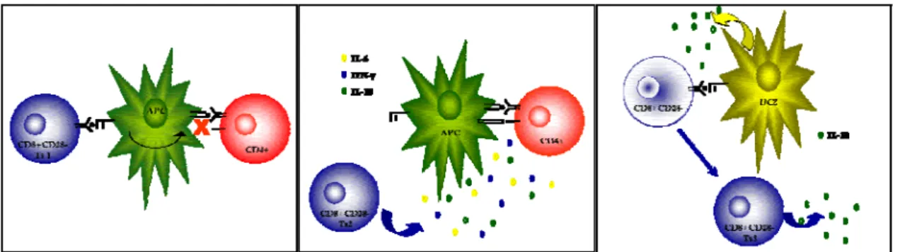
Figure 1 – Schematic representation of the mode of action of different suppressor T cells. From left to right: type1, type 2, and type 3 CD8+ T suppressor cells.
(Adapted from Filaci and Suciu-Foca, 2002)
Type 1 Ts are represented by CD8+CD28−T cells which inhibit allo-antigen, xeno-antigen and nominal antigen-specific CD4+ T cell responses. Type 1 CD8+CD28− Ts cells can be generated in vitro by multiple rounds of stimulation of peripheral blood mononuclear cells (PBMC) with allogeneic 69, xenogeneic 70 or autologous (antigen-pulsed) APC 71. These Ts
cell lines express specific molecular markers related to natural CD4+CD25+ regulatory T cells, namely FOXP3 56, 74, have a limited Vβ usage 51, 70, and are MHC-class-I-restricted 69.
Unprimed CD8+CD28− T cells from fresh peripheral blood, which have no regulatory activity, do not express FOXP3, GITR, OX40, CD25, CD62L or 4-1BB 56. Inhibition of
Th cell proliferation is not caused by killing either the APC or the Th cell, nor is the suppressor effect mediated by the production of cytokines. Instead, it requires direct interactions between the suppressor cell and the APC used for priming 69, 70, 75. The
consequence of this interaction is the inhibition of the CD40-mediated up-regulation of other costimulatory molecules (CD80 and CD86) 69, 76 and the up-regulation of expression
of the immunoglobulin-like transcript (ILT)3 and ILT4 on the surface of the APC 77, 78. The
suppressed APC are rendered unable to induce and sustain the full programme of CD4+ Th cell activation due to the inhibition of the NFкB activation and transcription of costimulatory molecules in the APC 76, 78. Of note is the fact that Ts have a low
proliferating rate at first but reach a sizable population upon repeated stimulation with antigen 79. Type 1 Ts can also be generated in vitro by allo-stimulation of PBMC from the
peripheral blood of baboons or ACI rats. These Ts suppressed the proliferative response of CD4+ T cells from the same T cell line to allogeneic APC in a dose-dependent manner 80.
Non-antigen-specific Ts, or Type 2 Ts, as well as Type 1 Ts are phenotypically characterised by the lack of expression of the CD28 costimulatory molecule. Type 2 Ts can be generated in vitro from purified circulating CD8+CD28− lymphocytes incubated for 1
week with IL-2 and IL-10 72. These Ts inhibit the proliferative response of T cells
stimulated with specific antigens, anti-CD3 monoclonal antibodies or mitogens in a non-antigen specific way. They also seem to suppress the lysis mediated by cytotoxic cells
72. The suppressor effect is mediated by the secretion of soluble factors, namely IFN-γ,
IL-6 and IL-10, without direct interaction with APC 72, 81. They have a phenotype
(CD45RA+CD27−CCR7−) and TCR repertoire suggesting that they are pre-activated and expanded clones 72 (for review see 82). Type 2 Ts seem to be involved in the pathogenic
mechanisms of diseases, since failure to generate non-antigen-specific Ts is associated with relapse in patients with multiple sclerosis and systemic lupus erythematosus 81, 83, and in
HIV- or hepatitis C virus (HCV)-infected patients 84. These findings may reflect two
opposite conditions: the physical elimination of CD8+ Ts cell precursors or the compartmentalization of these cells in tissues. In fact, in autoimmune thyroiditis and cancer, type 2 Ts were found to infiltrate affected organs leading to different outcomes: a protective function in thyroiditis and a pathogenic role in cancer 84, 85.
A third Ts subpopulation was also identified. These cells are generated by stimulating purified, naïve CD8+ T lymphocytes with plasmacytoid dendritic cells (DC2). Their generation depends on antigen presentation and secretion of IL-10 by DC2. DC2 primed CD8+ T cells develop anergy to further stimulation with the same antigen and start secreting IL-10 themselves. These DC2 cells induce a population of CD8+ regulatory T cells that are anergic, non-cytolitic and capable of inhibiting primary T cell responses through the production of IL-10. Their inhibitory activity affects naïve but not pre-activated CD8+ T cells and is directly mediated by IL-10. IL-10 producing CD8+ T suppressor cells are directly induced via antigen presentation by DC2 and require antigen-specific re-stimulation to deliver their immunosuppression through the production of IL-10 73.
CD8+ T suppressor cells may be involved in the pathogenic processes of chronic inflammatory diseases with immunological involvement, either due to an impairment of their function (autoimmune diseases) or due to an unregulated function (tumours). The presence of Type 1 suppressor T cells has been associated with the absence of acute rejection in transplanted patients 86, 87 and is responsible for immunosuppression in animal
models of transplantation 88. The failure to generate Type 2 Ts has been associated with
it is the presence of these cells that allows the disease to go unchecked 82, 84. However, a
great deal of uncertainty remains about differentiation factors, antigen specificity, and mechanisms of action of Ts cells.
1.3. CD8+ T Cells in Allergy
Animal models of allergy
CD8+ T cells which are pivotal in tumour cell killing and protection during viral infection through secretion of IFN-γ and cytolytic factors have been considered to be much less important or even negative regulators of the development of allergic inflammation.
A number of animal studies reported conflicting effects of CD8+ T cells in allergic airway disease.
In a high IgE-responder rat model of asthma (Brown Norway rat), the depletion of CD8+ T cells with the monoclonal antibody (mAb) OX8 (a mouse anti-rat anti-CD8α monoclonal cytotoxic antibody) enhanced allergen-induced airway hyperresponsiveness (AHR) and eosinophil numbers in bronchoalveolar lavage (BAL) fluid. There was also a reduction in gene expression of Th1 cytokines in the lungs (IL-2 and IFN-γ), while the levels of IL-4 and IL-5 remained unchanged 89.
Using the same approach in a low IgE-responder rat (Sprague-Dawley rat), Olivenstein et
al. observed an increased magnitude of the late airway response to inhaled allergen. This
was accompanied by a significant increase in antigen-specific IgE and airway inflammation characterised by an influx of total leucocytes, macrophages, neutrophils and lymphocytes into the airway lumen 90.
In an attempt to clarify the role of CD8+ T cells in the regulation of IgE and CD4+ T cell responses to ovalbumin (OVA), Holmes and co-workers not only depleted but also reconstituted sensitised rats with purified CD8+ T cells. Depletion of CD8+ T cells in vivo was time-dependent: depletion, 7 days after immunisation, failed to enhance IgE production, while depletion on days 12-18 greatly enhanced it. Reconstitution of CD8-depleted rats on day 12 with purified CD8+ T cells completely inhibited the IgE response. This effect was antigen-specific since CD8+ T cells from OVA-primed animals had little effect on the IgE response of bovine serum albumin immunised rats 91.
The mechanism of this IgE regulation by CD8+ T cells was further investigated in wild-type and in IL-12 and IFN-γ knock-out mice. Contrary to common knowledge, the authors concluded that IFN-γ was required for inhibition, but had an origin other than the
CD8+ T cell (indirect suppression). The immunoregulatory potential of the CD8+ T cells was dependent on their ability to stimulate IL-12 production by the APC, leading to the activation of Th1 cells. In turn, the generation of OVA-specific Th1 cells inhibited Th2-dependent IgE class switching via production of IFN-γ 92. Surprisingly, both Tc1 and
Tc2 cells promoted Th1 cell development and inhibited the generation of Th2 cells 92.
Finally, Yang et al. examined the cellular basis for the poor IgE production in SJL/J mice upon OVA immunisation. The authors found that increased IFN-γ production by CD8+ T cells was responsible for their failure to make IgE 93.
However, other studies suggested that CD8+ T cells are important contributors and might be necessary for AHR and eosinophilic airway inflammation.
In fact, Euthymic Hooded Lister rats depleted of CD8+ cells by repeated injection of OX8 mAb did not mount a greater IgE response when immunised with antigen alone, or antigen plus ricin, suggesting that CD8+ in this setting might have a “helper” function 94.
Moreover, CD8−/− mice develop significantly lower AHR and airway inflammation compared with wild type mice after sensitisation and challenge with OVA. Reconstitution of the CD8−/− mice with magnetically isolated, OVA-primed CD8+ T cells fully restored the development of AHR and lung inflammation to levels comparable to those seen in wild type mice 95, 96. In another murine OVA model of asthma, if CD8+ T cells were removed
prior to sensitisation, a significant decrease in AHR was observed. This was correlated with a decrease in the levels of IL-5 and eosinophil numbers in the lungs of these mice 97.
Schaller et al. investigated CD8+ T cell responses in a murine cockroach antigen (CRA) model of asthma. Their results showed that depletion of CD8+ T cells after allergen sensitisation to CRA significantly reduced airway hyperreactivity, airway eosinophilia and pulmonary type 2 cytokine levels. CD8+ T cells from CRA-sensitised mice produced type 2 cytokines IL-4, IL-5, and IL-13 upon antigen challenge, and the transfer of these cells into naïve mice caused airway hyperreactivity when exposed to CRA. This transferred airway response was dependent both on IL-4 and IL-13 98. The author’s data further suggests that
both CD4+ and CD8+ T cells can mediate an airway response, and that when both cells are transferred into naïve mice from CRA-sensitised mice, the airway response is much greater than when one population alone is transferred 98.
Direct comparison between these studies is difficult because of the different protocols used: different species (rat vs. mice) and strain models, differences in recipient status at the
time of reconstitution (naïve/sensitised), different routes of sensitisation, different adjuvants; all these factors can play a role in the final effect. In addition, different subpopulations of CD8+ T cells do exist and can be at different functional stages of differentiation, some of which suppress and others enhance Th2-responses. It is noteworthy that it is being recognised that the cell and cytokine requirements during the initiation/sensitisation phase differ considerably from those that might be necessary during the challenge/maintenance/progression phase 99, 100. CD8+ T cells could play an important
inhibitory role early on during the sensitisation phase on the allergen-induced response, but once the systemic sensitisation is established, CD8+ T cells may play a bystander or pro-inflammatory role 101.
Human allergic disease
As we have seen, CD8+ T cells are important immunoregulatory cells in some animal models of allergic disease, but proinflammatory in others. Dichotomous findings with respect to CD8+ cells led to attempts to further characterise CD8+ cells in human allergic immune responses.
According to the “Th2 hypothesis for asthma” asthmatic subjects have a relative deficiency in IFN-γ production or an imbalance towards type 2 cytokine production 102. Because Th1
cells antagonise Th2 cell functions, it was proposed that immune deviation towards a Th1 pattern could protect against asthma. It was then suggested that CD8+ T cells might play a protective role in the pathogenesis of allergic inflammation via their ability to produce IFN-γ. This IFN-γ could inhibit the development of Th2 cells directly and inhibit IgE synthesis by B cells.
Several authors investigated the secretion of IFN-γ by CD8+ T cells from patients with allergic asthma and healthy controls.
One study showed that PMA (phorbol myristate acetate) and calcium-ionophore-induced IFN-γ secretion by isolated CD8+ T cells in patients with asthma was lower than that of healthy controls, but was significantly increased after stimulation with anti-CD3 antibodies
103. Using a bronchial explant system without any stimulation, the mRNA expression for