Peripheral neuropathy in patients in
haemodialysis treatment
Adriana Ondina Pestana Santos
Thesis dissertation for MSc Degree in
Medicine
(Integrated cycle of studies)
Scientific Orientation: Maria Assunção Vaz Patto, M.D., PhD
Scientific Co-orientation: Rui Alves Filipe, M.D.
II I’m beginning to know myself. I don’t exist. I’m the space between what I’d like to be and the others made of me. Or half that space, because there’s life there too… So that’s what I finally am… Turn off the light, close the door, stop shuffling your slippers out there in the hall. Just let me at ease and all by myself in my room. It’s a cheap world.
III To the people that make me strive
to something better: My family My friends ‘My’ patients
IV
Acknowledgments
I would like to express my deepest gratitude to my mentor Maria Assunção Vaz Patto, M.D., PhD for all the support and shared knowledge.
I would also like to thank my other mentor Rui Alves Filipe, M.D. for having shared with me a labor of years and for the advices.
I would like to thank Nuno Pinto, BSc for being always free to help in patient’s examination and for his friendship.
I deeply thank to Jorge Gama, PhD for the precious help with the statistical analysis. I must thank Raquel Chorão, MD for helping with patients.
I also want to thank my family for the constant support. Without you, all, I would not be here.
Márcia, there are no words to express the role you had throughout this journey. Thanks for always being near.
V
Abstract
Background: Chronic kidney disease is a worldwide public health problem. Its prevalence is
15% in developed countries. End-stage kidney disease is known to be associated with peripheral neuropathy, which is classically a distal symmetrical length-dependent, sensorimotor polyneuropathy. Diagnosis of peripheral neuropathy is complex. For its early detection and appropriate intervention, it is required careful clinical assessment with history and physical examination including neurological examination, laboratory testing and electrodiagnostic studies or nerve biopsy, if the diagnosis remains unclear. Objectives: To evaluate the electrophysiological changes in a subgroup of patients with end-stage kidney disease treated with haemodialysis and correlate them with the clinical course of the disease.
Methods: Twenty seven patients with end-stage kidney disease in haemodialysis maintenance
treatment from the dialysis unit of Amato Lusitano Hospital’s were submitted to electrophysiological evaluation from October 2011 to January 2012 in the Faculty of health Sciences of the University of Beira Interior. As inclusion criteria we considered the duration of haemodialysis treatment and the ability to do the exam. All patients with any disease that might give rise to peripheral neuropathy, except diabetes mellitus were excluded. Results: Peripheral neuropathy was observed in 92.6% of patients. We did not find any correlation with neurologic symptoms neither with haemodialysis duration, p=0.051. Diabetes did not increase the risk of peripheral neuropathy. Diabetic patients when compared with non-diabetic patients had 6.7 times the risk of having sensorimotor neuropathy. Diabetic patients alone had 3.094 times more risk to have sensorimotor neuropathy. Conclusions: Peripheral neuropathy seems to be a silent partner in the multifactorial pathology of this group of patients. The absence of clinical findings may delay the diagnosis of peripheral neuropathy. Thereafter a multidisciplinary approach for prevention, diagnosis and treatment of this type of complications is crucial.
VI
Keywords
Peripheral neuropathy; end-stage kidney disease; haemodialysis; nerve conduction studies; Diabetes Mellitus; Uraemia
VII
Resumo Alargado
Introdução: A doença renal crónica é um problema de saúde pública mundial. A sua prevalência é 15% nos países desenvolvidos. A idade, a diabetes mellitus e a hipertensão arterial são indicadores independentes para esta doença. Em Portugal as principais etiologias de doença renal crónica nos doentes em hemodiálise são diabetes mellitus (33.6%), causa indeterminada (20.7%) e hipertensão arterial (15.%). A doença renal crónica apresenta complicações neurológicas na maioria dos doentes. No estadio terminal, está geralmente associada a neuropatia periférica. Esta neuropatia é, classicamente, uma polineuropatia sensitivo-motora, distal, simétrica. Clinicamente, os doentes com polineuropatia poderão ser assimtomáticos. No entanto, os principais sintomas são aumento do limiar vibratório, perda dos reflexos tendinosos profundos, parestesias, hiperestesias ou hipoestesias, cãimbras, pernas inquietas (restless legs), fraqueza e atrofia muscular. O diagnóstico de neuropatia periférica é complexo. Para a sua deteção precoce e intervenção adequada, é necessária uma avaliação clínica minuciosa com anamnese e exame objectivo detalhados, incluindo exame neurológico, testes laboratoriais e, se o diagnóstico permanece incerto, estudos electrodiagnósticos e biópsia do nervo. Objetivo: Avaliar as características electrofisiológicas num subgrupo de doentes com doença renal crónica terminal em tratamento hemodialítico e relacioná-las com a etiologia e o tempo em hemodiálise. Métodos: Estudo transversal em doentes com doença renal crónica terminal em hemodiálise na Unidade de Diálise do Hospital Amato Lusitano, em Castelo Branco. Os critérios de inclusão e exclusão considerados foram, no primeiro, o tempo de hemodiálise – 6 a 60 meses. Todos os doentes incapazes de serem submetidos a estudo de condução nervosa por patologia associada ou com patologias que podem causar polineuropatia, como sarcoidose, lupus eritimatoso sistémico, história de radio-quimioterapia, doença dos plasmócitos ou doença primária neurológica primária foram excluídos. De acordo com os critérios usados 27 dos 78 doentes em hemodiálise na referida unidade de diálise foram submetidos a uma avaliação electrofisiológica entre Outubro de 2011 e Janeiro de 2012 na Faculdade de Ciências da Saúde da Universidade da Beira Interior. A nossa amostra continha 9 mulheres e 18 homens, na faixa etária dos 39 aos 87 anos. Todos os doentes eram submetidos a hemodiálise três vezes por semana, durante 3.75 a 4.5 horas por
VIII
sessão, usando-se uma membrana biocompatível de baixo fluxo (Polyfluxo Gambro 17L). Todos os doentes estudados tinham outras patologias associadas e eram polimedicados. Resultados: De acordo com a etiologia da insuficiência renal terminal classificámos os doentes em diabéticos (n=13) e não diabéticos (n=14). A média da duração da hemodiálise nos doentes diabéticos é significativamente inferior à média da duração da hemodiálise nos doentes não-diabéticos (p=0.002). Noventa e dois vígula seis por cento dos doentes estudados têm neuropatia periférica. No entanto, esta parece não ter correlação com os sintomas ou duração de hemodiálise. Na nossa amostra constatou-se que a diabetes não aumenta o risco de neuropatia periférica e que não há significância estatística do efeito da duração da hemodiálise na neuropatia periférica, p=0.051. Conclusões: Este estudo permite-nos concluir que, neste grupo, a diabetes não aumenta o risco de neuropatia periférica. Apesar de não termos encontrado significância estatística quando analizamos o efeito da duração do tratamento hemodialítico na neuropatia periférica, acreditamos que este poderá ser um factor que tem influência na neuropatia, pois, apesar do tamanho da amostra ser pequeno, p=0.051. A neuropatia periférica aparenta ser uma doença silenciosa na patologia multifactorial deste grupo de doentes. Em suma, uma abordagem multidisciplinar é preponderante na prevenção, diagnóstico e tratamento destas complicações.
Palavras-chave
Neuropatia periférica; insuficiência renal crónica terminal; hemodiálise; estudos de condução nervosa; Diabetes Mellitus; uremia.
IX
Index
Dedication ... III Acknowledgments ... IV Abstract... V keywords ... VIResumo Alargado ... VII
Palavras-chave ... VIII
List of tables ... XI
List of acronyms ... XII
1. Introdution ... 1
2. Aims ... 4
2.1. Specific Aims ... 4
3. Methods ... 5
3.1. Study design and sample selection ... 5
3.2. Clinical evaluation and electrophysiology ... 6
3.3. Statistical analysis ... 9
3.4. Conflict of interest disclosure ... 9
4. Results ... 10
4.1. Clinical characteristics of the patients ... 10
4.2. Clinical findings ... 11
4.3. Neuropathy characterization ... 13
4.4. Correlation of aetiology, haemodialysis duration and neuropathy characteristics ... 14
X
6. Study limitations ... 19
7. Conclusions and Future Perspectives ... 21
References ... 22
Appendixes ... 26
Appendix 1 - Informed consent ... 26
XI
List of Tables
Table 1 - Clinical data of patients on haemodialysis treatment ... 5
Table 2 - Haemodialysis therapy ... 7
Table 3 - Outpatient therapy ... 8
Table 4 - Patient characteristics ... 10
Table 5 - Clinical manifestations and electrophysiological findings ... 12
Table 6 - Correlation between clinical manifestations and electrophysiological findings ... 13
Table 7 - Relationship between aetiology and peripheral neuropathy ... 13
Table 8 - Peripheral neuropathy characteristics ... 14
Table 9 - Correlation between aetiology and peripheral neuropathy characteristics ... 15
Table 10 - Mean of haemodialysis duration in the groups represented by each peripheral neuropathy variables ... 16
XII
List of Acronyms
CKD Chronic Kidney Disease DM Diabetes Mellitus EPO Erythropoietin
GFR Glomerular Filtration Rate HIV Human Immunodeficiency Virus
K/DOQI Kidney Disease Outcomes Quality Iniciative LL Lower Limb
LSD Least Significant Difference
MGUS Monoclonal Gammopathy of Undetermined Significance MM Multiple Myeloma
NA Not-applicable
NSS Neuropathy Symptom Score OR Odds Ratio
PKDAD Polycystic Kidney Disease Autossomic Dominant PNP Peripheral Neuropathy
POEMS Polyneuropathy, Organomegaly, Endocrinopathy, Monoclonal gammopathy, Skin changes
SD Standard Deviation UL Upper Limb
Φ Phi
1
1. Introduction
Chronic kidney disease (CKD) is a worldwide public health problem. Its prevalence is 15% in developed countries1, 2, 3, 4 and it causes neurological complications in the majority of patients5, 6. CKD can occur at any age of life. Age is an independent major predictor of CKD as well as diabetes mellitus (DM) and hypertension7.
There are several aetiologies for CKD. It can occur due to either a primary kidney disease or as a complication of a multisystemic disorder8. DM is the most common cause in developed nations8, whereas inflammatory kidney disease, namely glomerulonephritis and interstitial nephritis remains the most common causes in developing countries9. DM along with hypertension - the second most common cause – and glomerulonephritis accounts for about 75% of all adult cases10. In young adults a common aetiology of CKD is genetic kidney disease10.
In Portugal the main aetiologies for CKD in the patients under haemodialysis treatment are DM (33.6%), undetermined (20.7%) and arterial hypertension (15.5%)11.
The prevalence of CKD symptoms depends on the glomerular filtration rate (GFR)7. When the GFR is less than 15 mL/min/1.73 m2 – the end-stage kidney disease or stage 5 of CKD according to the Kidney Disease Outcomes Quality Iniciative (K/DOQI) – symptoms of uraemia are almost always present10.
End-stage kidney disease is known to be associated with peripheral neuropathy12, 13, 14, 15. Uremic neuropathy in end-stage kidney disease is classically a distal symmetrical length-dependent, sensorimotor polyneuropathy12, 16, 17 which is more common in lower limbs than in upper extremities12. According to the diagnostic criteria used, its prevalence rate varies from 60 to 100% in people that undergo hemodialysis12, 18, with an unexplained male predominance12, 16, 17.
Despite the huge effort developed in this area, the pathophysiology of uremic neuropathy has not been established yet. Nevertheless, there are two main postulated hypotheses. First, with the ‘Middle Molecule Hypothesis’ it was postulated that uremic neuropathy occurred as consequence of retention of neurotoxic molecules in the middle
2 molecular range of 300-12000 Da, given that such molecules were slowly dialyzable by haemodialysis membrane19.However, there is little evidence that such molecules are actually neurotoxic20. Second, recent nerve excitability studies, undertaken over the course of a dialysis session, demonstrated that patients with uremic neuropathy had motor and sensory axonal changes before dialysis suggesting that hyperkalaemia could be a contributing factor to the development of neuropathy21, 22, 23.
Research on the subjects is contradictory. Recent investigation reports demonstrated that improvement of uremic neuropathy with dialysis is uncommon12. Some authors suggested that dialysis retards the progression of neuropathy in most patients, while others suggested that in patients on dialysis there is a gradual worsening of the neuropathy12.
Diabetes is a common cause of CKD. Among the diabetic complications, diabetic peripheral neuropathy is the most common complication24. The incidence of diabetic peripheral neuropathy varies from 10 to 50%25, 26. Diabetic neuropathy is also length-dependent and of greater severity than other neuropathies with different aetiologies8, 27. In diabetic patients, males with type 2 DM may develop earlier diabetic neuropathy than females28. The pathophysiology of diabetic neuropathy has not been established but it seems to be related with metabolic disturbances, such as hyperglycaemia, dyslipidaemia, oxidative and nitrosative stress and growth factor deficiencies, microvascular insufficiency and autoimmune damage to nerve fibres29.
Clinically, patients with either uremic neuropathy or diabetic neuropathy can be asymptomatic. When symptomatic they can present with increased vibratory thresholds, loss of deep tendon reflexes, paraesthesias, hyperesthesia or hypoesthesia, cramps, restless legs, muscle weakness and atrophy8, 13, 24.
Diagnosis of peripheral neuropathy is complex. For its early detection and appropriate intervention, it is required careful clinical assessment with history and physical examination including neurological examination, laboratory testing and electrodiagnostic studies or nerve biopsy, if the diagnosis remains unclear30, 31. Electrodiagnostic tests, namely electromyography and nerve conduction studies are reliable and sensitive methods to access peripheral nerve function26. They can support the clinical diagnosis and provide information
3 about the type of fibres involved – motor, sensory or both -, the pathophysiology – axonal loss versus demyelination - and the pattern of involvement – symmetric or asymmetric30, 31.
4
2. Aim
The aim of our study was to evaluate the electrophysiological characteristics of a subgroup of patients in chronic haemodialysis treatment and to correlate them with the aetiology and with the number of years of haemodialysis treatment.
2.1.
Specific Aims
SA 1: To evaluate the correlation of aetiology and peripheral nervous system
neuropathy.
SA 2: To evaluate the effect of haemodialysis duration in peripheral nervous system
5
3. Methods
3.1.
Study design and sample selection
This was a cross-sectional study composed by 27 out of 78 patients in haemodialysis treatment in the dialysis unit at Amato Lusitano Hospital’s, in Castelo Branco. In our sample there were 9 women and 18 men (age range 39 to 87 years), with end-stage kidney disease receiving chronic maintenance haemodialysis treatment three times weekly, for 3.75 to 4.5 hours per session, using a biocompatible low-flux membrane (Polyfluxo Gambro 17L). All patients had been on chronic haemodialysis treatment for six to sixty months [Table 1].
†Deceased.
Abbreviations: PKDAD, polycystic kidney disease autossomic dominant; F, female; M, male.
Table 1. Clinical data of patients on haemodialysis treatment
Case No. Age Gender No. of months of haemodialysis Diagnosis
1 2 3 4† 5 6 7 8 9 10 11 12 13 14 15 16 17 18 19 20 21 22 23 24 25 26 27 68 69 80 81 76 79 67 51 82 75 73 74 81 65 78 76 67 71 77 77 70 82 73 56 87 39 56 M F M M F M M F M M M F F M F M M F M M M F M M M F M 21 54 8 25 7 48 16 15 60 9 34 17 28 41 30 25 19 46 10 9 36 19 28 58 48 50 40 Diabetes Mellitus Undetermined Diabetes Mellitus Diabetes Mellitus Diabetes Mellitus Undetermined Diabetes Mellitus Undetermined Nephroangisclerosis Diabetes Mellitus Nephroangisclerosis Nephroangisclerosis Undetermined Diabetes Mellitus Diabetes Mellitus Chronic pyelonephritis Diabetes Mellitus Diabetes Mellitus Diabetes Mellitus Diabetes Mellitus Undetermined Undetermined Diabetes Mellitus PKDAD Undetermined IgA glomerulonephritis Undetermined
6 This study was approved by the Ethics Committee of Sousa Martins Hospital’s, in Guarda and throughout the study medical confidentiality was kept.
In our study we included all the patients from the dialysis unit of Amato Lusitano Hospital’s with 6 to 60 months of haemodialysis treatment and could be submitted to electrophysiological examination. As exclusion criteria we considered any disease which might give rise to a peripheral neuropathy, except diabetes mellitus. Thus none of the studied subjects had clinical, laboratory or histopathological evidence of plasma-cell dyscrasias – monoclonal gammopathy of undetermined significance (MGUS), multiple myeloma (MM), Waldenström’s macroglobulinemia, Castleman’s disease, POEMS (polyneuropathy, organomegaly, endocrinopathy, monoclonal gammopathy, skin changes) syndrome , light-chain amyloidosis -, sarcoidosis, systemic lupus erythematous, neoplasms pressing on nerves, previous history of radio or chemotherapy, HIV infection or primary neurologic disease.
According to the inclusion and exclusion criteria used our sample was composed by 14 non diabetic patients and 13 diabetic patients. The main aetiologies in the former group were IgA glomerulonephritis in 1 patient (case 26), chronic pyelonephritis in 1 patient (case 16), nephroangiosclerosis in 3 patients (cases 9, 11 and 12); 8 patients had undetermined aetiology (cases 2, 6,8, 13, 21, 22, 25 and 27) [Table 1].
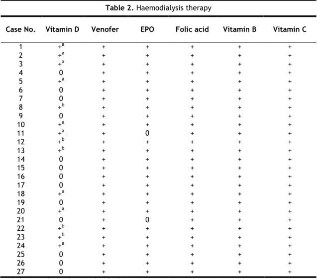
Besides therapy patients took during haemodialysis [Table 2], all patients took chronically at least four drugs. The outpatient therapy is summarized in table 3.
During haemodialysis treatment, all patients took folic acid and vitamins B and C. The main outpatient drugs that the majority of patients were taking belong to the following therapeutic groups: platelet aggregation inhibitor, serum lipid reducing agents, proton pump inhibitor, anxiolytics and ions exchange resins.
3.2.
Clinical evaluation and electrophysiology
All the twenty seven patients were studied in the Faculty of Health Sciences of the University of Beira Interior between October 12, 2011 and January 3, 2012. Before undergoing any procedure, patients were explained the purpose of the study and signed a written informed consent based on Helsinki’s Declaration - see appendix 1.
7 During the initial clinical assessment each patients was questioned specifically for symptoms of peripheral neuropathy. Neuropathic symptoms were graded using subsets IB, IIA and IIB of the Neuropathy Symptom Score (NSS) – see appendix 2. A full neurological examination was performed, as well.
The electrophysiological assessment of the studied sample was done with nerve conduction studies. Motor nerve conduction velocity was performed in the cubital nerve in the right arm, median nerve in the left arm, tibial nerve in the right leg and peroneal nerve in the left leg. Sensory nerve conduction was examined in the cubital and median nerve, respectively in the right and left arm. Sural sensory and peroneal nerves conduction were also studied, respectively in the right and left legs.
Table 2. Haemodialysis therapy
Case No. Vitamin D Venofer EPO Folic acid Vitamin B Vitamin C
1 2 3 4 5 6 7 8 9 10 11 12 13 14 15 16 17 18 19 20 21 22 23 24 25 26 27 +a +a +a 0 +a 0 0 +b 0 +a +a +b +b 0 0 0 0 +a 0 +a 0 +b +b +a 0 0 0 + + + + + + + + + + + + + + + + + + + + + + + + + + + + + + + + + + + + + 0 + + + + + + + + + 0 + + + + + + + + + + + + + + + + + + + + + + + + + + + + + + + + + + + + + + + + + + + + + + + + + + + + + + + + + + + + + + + + + + + + + + + + + + + + + + + + + + + + + + +
0, does not take the drug; +, takes the drug. a zemplar; b rocalterol.
8
Table 3. Outpatient therapy
Therapeutic Group 1 2 3 4 5 6 7 8 9 10 11 12 13 14 15 16 17 18 19 20 21 22 23 24 25 26 27
Antiarrhythmic – Class III + + +
β-blockers + + + + + + + + +
Calcium channel blockers + + + + +
ACE inhibitor +
Angiotensin II antagonist + + + +
Loop diuretics + + + + + + + +
Platelet aggregation inhibitor + + + + + + + + + + + + + + + + + + + +
Vasodilators + + + + +
Serum lipid reducing agents + + + + + + + + + + + + + + +
Proton pump inhibitor + + + + + + + + + + + + + + + + + + + + + + + +
H2 receptor antagonists + + Insulin + + + + + + + + + Anxiolytics + + + + + + + + + + + + + + + + + Anti-depressant + + Analgesics + + + Anti-androgens + Thyroid hormones + Anti-thyroid preparations + Antigout preparations + + Anti-emetics + + Laxatives + + + + + Anti-fungal +
Ion exchange resins + + + + + + + + + + + + + + + + + + +
Vitamins and minerals + + + + + + + +
+, takes the drug.
9
3.3.
Statistical analysis
Aetiology of end-stage kidney disease was the primary outcome variable. Collected data were analysed in terms of absolute and relative frequencies of each variable studied by descriptive statistics. Data are presented as the mean ± standard deviation (SD). We analysed the correlation between the variables of the study through Fisher’s Exact Test. We also used the association measures Φ (Phi) and odds ratio (OR). To measure the factors effects we used partial Eta square (η2). Parametric tests were used (t test or ANOVA) after verifying the normality and variances homogeneity assumptions with Shapiro-Wilk test and Levene’s test, respectively. For all statistical analyses, a p-value (p) less than 0.05 was considered statistically significant. Analyses were done using IBM SPSS Statistics 19®.
3.4.
Conflict of interest disclosure
10
4. Results
4.1.
Clinical characteristics of the patients
In our sample of 27 patients with end-stage kidney disease the female to male ratio was 9:18 (33.3% and 66.7%, respectively). The age range was between 39 and 87 years, with a mean age of 71.48 years ± 10.74 (SD) and a median of 74 years. The median duration of haemodialysis treatment was 28 months and the mean was 29.67 months ± 16.53 (SD)
The enrolled patients in the study were 13 (48.1%) diabetic and the remaining 14 (51.9%) patients were non-diabetic. The female to male ratio in the diabetic patients group was 3:10, with a mean age of 76.46 years ± 5.36 (SD). In this group, the mean duration of haemodialysis was 20.69 months ± 12.82 (SD). On the other hand, in the non-diabetic patient’s female to male ratio was 6:8. The mean age was 69.64 years ± 14.01 (SD) and the mean duration of haemodialysis was 38 months ± 15.46 (SD) [Table 4]. The mean duration of haemodialysis treatment was shorter in patients with diabetes when compared to non-diabetic patients (20.69 and 38 months, respectively), and this difference was statistically significant (T-test with p=0.002). The mean ages of diabetic and non-diabetic patients are not considered significantly different (T-test with p=0.357) [Table 4] and there is no association between gender and aetiology (Fisher’s exact test with p=0.420).
a T-test=0.357.
b T-test=0.004. η2=0.334.
Data are expressed as mean ± standard error of the mean.
(continued)
Table 4. Patient characteristics
Aetiology ANOVA
Diabetic Non-diabetic p-value
No. of patients 13 14 NA
Gender (F:M) 3:10 6:8 NA
Age (yr) 76.46 ± 5.36 69.64 ± 14.01 0.374a
11
Table 4. Patient characteristics (continuation)
Haemodialysis session duration (hours) ANOVA
≤4.25 >4.24 p-value
No. of patients 21 6 NA
Haemodialysis duration (mo) 25.67 ± 15.367c 43.67 ± 13.155c 0.023d
c LSD=0.034 d η2=0.213. p<0.05.
Data are expressed as mean ± standard error of the mean. Abbreviations: NA, non-applicable.
There is also a statistical significance between the haemodialysis duration and the time of each session of haemodialysis treatment. Thus, patients with more time of haemodialysis treatment also have longer sessions of haemodialysis, LSD with p=0.034.
4.2.
Clinical findings
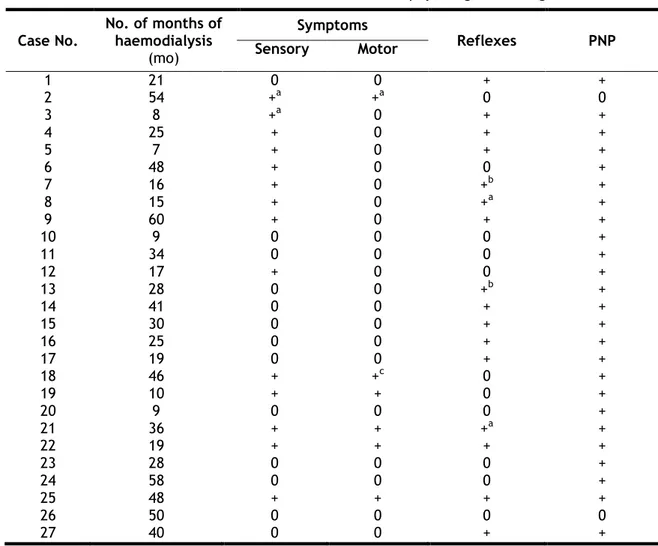
Clinical manifestations and electrophysiological findings have been summarized in table 5.
In our sample of 27 patients, sensory involvement was present in 14 patients. In those, the symptoms were bilateral, except in cases 2 and 3 which had symptoms in the right lower limb. Motor involvement was present in 6 patients, being in case 18 due to stroke sequelae. All the patients with motor symptoms also had sensory symptoms, but not all the patients with sensory symptoms had motor symptoms. Reduction or loss of reflexes was present in 16 patients, usually in the lower limbs. On examination, cases 7 and 13 manifested bilateral hyperreflexia due to other subjacent clinical conditions, specifically stoke and vascular dementia, respectively.
12
0, absence; +, presence.
a asymmetric; b hyperreflexia; c stroke sequelae. Abbreviations: PNP, peripheral neuropathy.
Thirteen, from the 25 patients with peripheral neuropathy, had sensory symptoms. However, when we correlated these two variables (symptoms and electrophysiological results), we did not obtain statistical significance on Fisher’s exact test, p=1 [Table 6]. It was also not possible to establish statistical significance between sensory symptoms and haemodialysis duration, (T-test with p=0.886). When we correlated the group of patients with sensorimotor symptoms with the electrophysiological results, no statistical significance was achieved, p= 0.402 [Table 6]. We obtain the same result when we compared sensorimotor symptoms and haemodialysis duration, (T-test with p=0.376). In summary, the presence of peripheral neuropathy in this group of patients seems to have no correlation neither with the clinical symptoms or signs nor with haemodialysis duration.
Table 5. Clinical manifestations and electrophysiological findings Case No. No. of months of haemodialysis (mo) Symptoms Reflexes PNP Sensory Motor 1 2 3 4 5 6 7 8 9 10 11 12 13 14 15 16 17 18 19 20 21 22 23 24 25 26 27 21 54 8 25 7 48 16 15 60 9 34 17 28 41 30 25 19 46 10 9 36 19 28 58 48 50 40 0 +a +a + + + + + + 0 0 + 0 0 0 0 0 + + 0 + + 0 0 + 0 0 0 +a 0 0 0 0 0 0 0 0 0 0 0 0 0 0 0 +c + 0 + + 0 0 + 0 0 + 0 + + + 0 +b +a + 0 0 0 +b + + + + 0 0 0 +a + 0 0 + 0 + + 0 + + + + + + + + + + + + + + + + + + + + + + + 0 +
13
Table 6. Correlation between clinical manifestations and electrophysiological findings
Sensory symptoms Exact Test Fisher’s Motor symptoms Exact Test Fisher’s
Absence Presence p-value Absence Presence p-value
PNP Absence 1 1 1 1 1 0.402 Presence 12 13 20 5
p<0.05.
Abbreviations: PNP, peripheral neuropathy
4.3.
Neuropathy characterization
All the thirteen diabetic patients studied had evidence of peripheral neuropathy on nerve conduction study. On the non-diabetic group (n=14) of patients there were two subjects with no evidence of peripheral neuropathy on nerve conduction studies; the remaining 12 patients had peripheral neuropathy [Table 7]. This is, in our sample 25 out of 27 patients had peripheral neuropathy. The ratio between not having to having peripheral neuropathy was 2:25 (7.4% and 92.6%, respectively). When comparing both with Fisher’s exact test we did not obtain any statistical significance [Table 7]. In this group of patients, diabetes does not increase the risk of peripheral neuropathy. Moreover, there was no significant difference between diabetic versus non diabetic patients in the distribution of neuropathy characteristics.
p<0.05.
Abbreviations: PNP, peripheral neuropathy.
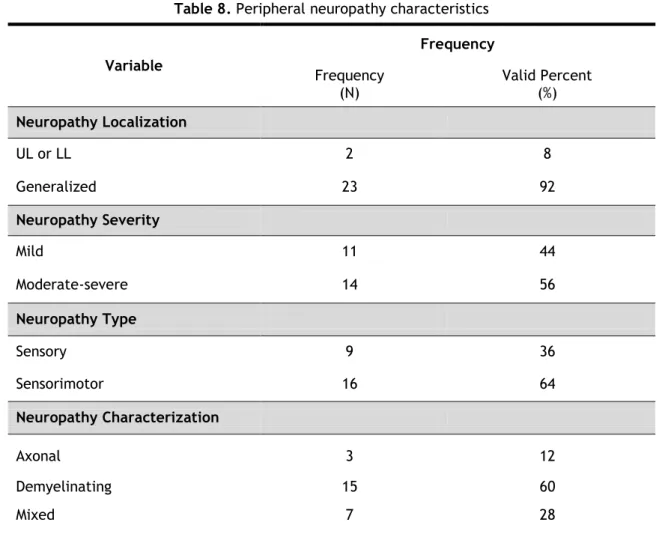
As stated above 25 out of 27 patients had findings of peripheral neuropathy on nerve conduction study. Table 8 summarizes the neuropathy characteristics of the patients. Ninety two percent of patients with peripheral neuropathy presented with the generalized form. There were 56% of patients with moderate-severe neuropathy, while the remaining 44% had
Table 7. Relationship between aetiology and peripheral neuropathy
Aetiology Fisher’s Exact Test
Diabetic Non-diabetic p-value
PNP
Absence 0 2
0.48 Presence 13 12
14 mild neuropathy. Almost two-thirds of the patients presented with a sensorimotor neuropathy, while 36% presented with a sensory neuropathy. Sixty percent of patients presented with demyelinating neuropathy, 12% with axonal neuropathy and 28% with mixed neuropathy.
Abbreviations: UL: upper limb; LL: lower limb.
4.4.
Correlation of aetiology, haemodialysis duration and
neuropathy characteristics
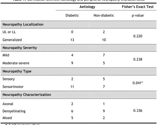
Tables 9 and 10 show the correlation of aetiology, haemodialysis duration and neuropathy characteristics.
In this group of patients we found a significant correlation between aetiology and neuropathy type (p=0.041 on Fisher’s exact test). The odds ratio for sensorimotor neuropathy
Table 8. Peripheral neuropathy characteristics
Variable Frequency Frequency (N) Valid Percent (%) Neuropathy Localization UL or LL 2 8 Generalized 23 92 Neuropathy Severity Mild 11 44 Moderate-severe 14 56 Neuropathy Type Sensory 9 36 Sensorimotor 16 64 Neuropathy Characterization Axonal 3 12 Demyelinating 15 60 Mixed 7 28
15 when compared with sensory neuropathy was 7.7 times more. The Φ was 0.447 meaning that dimension of effect is moderate. Diabetic patients when compared with non-diabetic patients had 6.7 times the risk of having sensorimotor neuropathy. Diabetic patients alone had 3.094 times more risk to have sensorimotor neuropathy than sensory neuropathy.
Table 9. Correlation between aetiology and peripheral neuropathy characteristics
Aetiology Fisher’s Exact Test
Diabetic Non-diabetic p-value
Neuropathy Localization UL or LL 0 2 0.220 Generalized 13 10 Neuropathy Severity Mild 4 7 0.238 Moderate-severe 9 5 Neuropathy Type Sensory 2 5 0.041* Sensorimotor 11 7 Neuropathy Characterization Axonal 2 1 0.336 Demyelinating 6 9 Mixed 5 2 Φ=0.447 (moderate effect). OR=7.7. p<0.05.
Abbreviations: OR, odds ratio; UL, upper limb; LL, lower limb.
When we analysed the effect of haemodialysis duration in peripheral neuropathy, we obtained no statistical significance, with a p=0.051, but suggesting a trend to significance.
16
Table 10. Mean of haemodialysis duration in the groups represented by each peripheral
neuropathy variables
Haemodialysis duration (mo) t-test ANOVA
Mean p-value p-value
Neuropathy Localization UL or LL 21.50 ± 9.192 0.564 NA Generalized 28.43 ± 16.295 Neuropathy Severity Mild 27.45 ± 13.945 0.908 NA Moderate-severe 28.21 ± 17.686 Neuropathy Type Sensory 28 ± 15.508 0.978 NA Sensorimotor 27.81 ± 16.514 Neuropathy Characterization Axonal 21.691 NA 0.923 Demyelinating Mixed p<0.05.
Data are expressed as mean ± standard error of the mean. Abbreviations: UL, upper limb; LL, lower limb, NA, Non-appicable.
17
5. Discussion
Almost all patients with severe chronic kidney disease have neurological complications8, explicitly peripheral neuropathy. The aim of our study was to evaluate the electrophysiological characteristics of a subgroup of patients in chronic haemodialysis treatment and to correlate them with the aetiology and with the number of years of haemodialysis treatment.
In our sample men were present twice as often as women (66.7% and 33.3%, respectively) probably representing the same distribution as seen in CKD patients. According to scientific data there are differences in the development of diabetic neuropathy between genders. It is postulated that men with type 2 DM may develop peripheral neuropathy earlier than women28. Diabetic subgroup of patients was composed by more than three-quarters (77%) of men and all patients have peripheral neuropathy. In the non-diabetic subgroup of patients up to two-thirds of men also had peripheral neuropathy. However in the studied sample gender was not significantly different in the two groups (p=0.420) and the age showed no difference, as well. On the other hand, there is a significant correlation between aetiology and haemodialysis treatment duration – we found that diabetic patients are in haemodialysis for less time.
In our study, the electrophysiological findings confirmed the results of previous studies12, 32. Even in patients without clinical evidence of peripheral neuropathy, many studies through nerve conduction studies have disclosed evidence of high prevalence of subclinical peripheral neuropathy33. In spite of small sample’s size, the rate of peripheral neuropathy in the present study was 92.4% in keeping with previous studies which have demonstrated similarly high rates of neuropathy. However we did not find a clinical expression of this peripheral neuropathy even in subjects with a definite pathology. Peripheral neuropathy seems to be a silent partner in the multifactorial pathology of this group of patients.
Peripheral neuropathy in end-stage kidney disease is usually a length-dependent, distal sensorimotor polyneuropathy21. According to the literature this length-dependent neuropathy is more severe in diabetic than in non-diabetic patients with CKD8. In our study, the majority of patients (92%) had generalized peripheral neuropathy, specifically, all
18 diabetic patients presented generalized peripheral neuropathy and only 2 out of 14 non-diabetic patients had peripheral neuropathy localized to the limbs, either upper or lower limbs. While these results are in accordance with scientific data8 we did not obtain statistical significance, probably due to the small number of patients in our sample. The prevalence of patients with sensorimotor neuropathy (64%) was higher when compared with patients with sensory neuropathy (36%). Diabetic patients when compared with non-diabetic patients had 6.7 times the risk of having sensorimotor neuropathy. Diabetic patients alone have 3.094 times more risk to have sensorimotor neuropathy. This result supports the results achieved by other previous studies which conclude that sensorimotor neuropathy is the main type of neuropathy in diabetic patients24, 26.
In this group of patients peripheral neuropathy did not have any correlation neither with the clinical symptoms or signs nor with haemodialysis duration. Diabetes did not increase the odds of having peripheral neuropathy, and there was no significant difference between diabetic versus non-diabetic patients in the distribution of neuropathy characteristics. However, we cannot exclude the possibility that the small sample of patients may have affected these results. A subsequent study with a larger number of patients may achieve other conclusions. Despite not finding any correlation between the presence of neuropathy and the number of years of dialysis, with a p=0.051, we believe that with a bigger sample we would have statistical significance.
We conclude that in our group of patients neuropathy is a serious complication of end-stage kidney disease. Diabetes may be an adjuvant factor for sensorimotor involvement, but other factors like uraemia or the dialitic process may be of importance as well. Moreover, diabetes does not seem to increase the risk to develop peripheral neuropathy in these patients.
In some of these patients peripheral neuropathy presented with no signs or symptoms. The absence of clinical findings may delay the diagnosis of peripheral neuropathy. Thereafter a multidisciplinary approach for prevention, diagnosis and treatment of these types of complications is crucial.
19
6. Study Limitations
The main limitation of the present study relates primarily to the size of the sample. This limitation arises from the fact that only few patients under chronic haemodialysis maintenance treatment in the dialysis unit at Amato Lusitano Hospital’s fulfilled the study criteria. A study carried out in a longer period of time, namely a cohort study, would allow the inclusion of more people and so, it would perhaps make statistically significant some trends shown in this study. Furthermore, with a larger sample we would be able to set additional aims in order to better understand the studied problem. Despite meeting the study criteria, debilitated patients could not be submitted to nerve conduction study because as described above the study took place in a different city and the journey could worsen their clinical condition or they were not even able to do it. In addition, one patient had to be excluded from the study due to inability to tolerate the exam. In short, all these factors contributed for the small size of the sample and, thus, it conditioned the results obtained.
Other important issue was economic sustainability of the project. Eventually, with other economic resources, we could have thought to investigate the nerve excitability abnormalities before dialysis and nerve excitability changes following dialysis and correlate its implications with neuropathy clinical course.
The electrophysiological diagnosis in most patients was established by the study. Thus, we did not know which where the electrophysiological characteristics of the neuropathy before patients undergone haemodialysis treatment and which was the neuropathy clinical course until the electrophysiological diagnosis. Because of these, we were unable to correlate which is the haemodialysis relevance in neurological abnormalities manifested by this group of patients with end-stage kidney disease. We could not establish if haemodialysis treatment improved or worsened the peripheral neuropathy in this subset of patients. As stated, in our sample we have patients with many years of haemodialysis that have clinical manifestations of the disease, but we were unable to know if symptoms were better or worse before haemodialysis treatment because there are not previous electrophysiological recordings from all the patients.
20 Despite all limitations the study was doubtless extremely important to characterize this clinical problem in the studied sample. It was also relevant in emphasizing the huge importance of having a multidisciplinary team working all together, especially neurologists and nephrologists in the care of these patients.
21
7. Conclusions and Future Perspectives
The present study emphasizes the high prevalence of peripheral neuropathy in a group of patients with end-stage kidney disease under haemodialysis maintenance treatment.
Despite the short period of time the study was conducted and, consequently, small sample’s size, the obtained results allow us to highlight the huge importance of having neurologists and nephrologists as well as other specialists working all together to better diagnose and manage neurological complications of end-stage kidney disease in these patients. Its importance is increasing because CKD has become worldwide a public health issue.
Since some patients with CKD have subclinical peripheral neuropathy and neurological complications impair their quality of life, early diagnosis of this condition is essential. The gold standard exam for diagnosis confirmation is nerve conduction studies. Thus, before undergoing dialysis, it would be recommended to submit all patients with CKD to nerve conduction studies. It is, however, equally important to frame clinically which is the CKD aetiology and patient’s clinical condition, as well. Nerve conduction studies would also be recommended to evaluate the role of dialysis in such patients.
Hereafter the studied subjects should be clinical and electrophysiological reassessed to evaluate the evolution of their condition. It would also be important to compare both results and to rule out the importance of dialysis in the management of such patients.
In the future it would be judicious to study the Portuguese population or at least a significant sample with end-stage kidney disease through a cohort study to characterize its neurological complications – namely peripheral neuropathy. Moreover, according to these conclusions, it would also be important to establish a protocol or guidelines that could provide orientation on the management of these patients.
22
References
1. Chadban SJ, Briganti EM, Kerr PG, Dunstan DW, Welborn TA, Zimmet PZ, et al. Prevalence of kidney damage in Australian adults: The AusDiab kidney study. Journal of The American Society of Nephrology. 2003;14(7 Suppl 2):S131–S138.
2. Fox CS, Larson MG, Vasan RS, Guo C-Y, Parise H, Levy D, et al. Cross-sectional association of kidney function with valvular and annular calcification: the Framingham heart study. Journal of The American Society of Nephrology. 2006;17(2):521–7.
3. Nitsch D, Felber Dietrich D, Von Eckardstein A, Gaspoz J-M, Downs SH, Leuenberger P, et al. Prevalence of renal impairment and its association with cardiovascular risk factors in a general population: results of the Swiss SAPALDIA study. Nephrology Dialysis Transplantation. 2006;21(4):935–44.
4. Ninomiya T, Kiyohara Y, Kubo M, Tanizaki Y, Doi Y, Okubo K, et al. Chronic kidney disease and cardiovascular disease in a general Japanese population: the Hisayama Study. Kidney International Supplement. 2003;68(1):228–36.
5. Murray AM. Cognitive impairment in the aging dialysis and chronic kidney disease populations: an occult burden. Advances in Chronic Kidney Disease. 2008;15(2):123–32.
6. Krishnan AV, Pussell BA, Kiernan MC. Neuromuscular disease in the dialysis patient: an update for the nephrologist. Seminars in Dialysis. 2009;22(3):267–78.
7. Arora P, Verrelli M. Chronic Renal Failure [Internet]. In Medscape, Batuman V, WebMD LLC, New York, Updated: Nov 23, 2010. Available from: http://emedicine.medscape.com/article/238798.
8. Krishnan AV, Kiernan MC. Neurological complications of chronic kidney disease. Nature reviews Neurology. 2009;5(10):542–51.
23 9. Barsoum RS. Chronic kidney disease in the developing world. The New England Journal
of Medicine. 2006;354(10):997–9.
10. Krause RS. Renal Failure, Chronic and Dialysis Complications [Internet]. In Medscape, Schraga ED, WebMD LLC, New York, Updated: Jun 28, 2010. Available from: http://emedicine.medscape.com/article/1918879.
11. Macário F, Filipe R, Carvalho MJ, Galvão A, Lopes JA, Amoedo M, et al. Diálise Domiciliária. SPNews - Sociedade Portuguesa de Neurologia. 2011;VII(24):1–20.
12. Krishnan AV, Kiernan MC. Uremic neuropathy: clinical features and new pathophysiological insights. Muscle and Nerve. 2007;35(3):273–90.
13. Brouns R, De Deyn PP. Neurological complications in renal failure: a review. Clinical Neurology and Neurosurgery. 2004;107(1):1–16.
14. Asbury AK. Recovery from uremic neuropathy. The New England Journal of Medicine. 1971;284(21):1211–2 [Abstract].
15. Nielsen VK. The peripheral nerve function in chronic renal failure. Acta Medica Scandinavica. 1971;190(1‐6):105–11 [Abstract].
16. Pan Y. Uremic neuropathy [Internet]. In Medscape, Ramachandran TS, WebMD LLC, New York, Updated: Aug 3, 2011. Available from: http://emedicine.medscape.com/article/1175425.
17. Burn DJ, Bates D. Neurology and the kidney. Journal of Neurology Neurosurgery and Psychiatry. 1998;65:810–21.
18. Van den Neucker K, Vanderstraeten G, Vanholder AR. Peripheral motor and sensory nerve conduction studies in haemodialysis patients. A study of 54 patients. Electromyography and Clinical Neurophysiology. 1998;38(8):467–74.
24 19. Babb AL, Ahmad S, Bergström J, Scribner BH. The middle molecule hypothesis in
perspective. American Journal of Kidney Diseases. 1981;1(1):46–50.
20. Vanholder R, De Smet R, Hsu C, Vogeleere P, Ringoir S. Uremic toxicity: the middle molecule hypothesis revisited. Seminars in Nephrology. 1994;14(3):205–18.
21. Krishnan AV, Phoon RKS, Pussell BA, Charlesworth JA, Bostock H, Kiernan MC. Altered motor nerve excitability in end-stage kidney disease. Brain: A Journal of Neurology. 2005;128(9):2164–74.
22. Krishnan AV, Phoon RKS, Pussell BA, Charlesworth JA, Kiernan MC. Sensory nerve excitability and neuropathy in end stage kidney disease. Journal of Neurology, Neurosurgery and Psychiatry. 2006;77(4):548–51.
23. Krishnan AV, Phoon RKS, Pussell BA, Charlesworth JA, Bostock H, Kiernan MC. Neuropathy, axonal Na+/K+ pump function and activity-dependent excitability changes in end-stage kidney disease. Clinical Neurophysiology. 2006;117(5):992–9.
24. Ko SH, Cha BY. Diabetic peripheral neuropathy in type 2 Diabetes Mellitus in Korea. Diabetes and Metabolism Journal. 2012;36(1):6–12.
25. Krishnan AV, Lin CS-Y, Kiernan MC. Activity-dependent excitability changes suggest Na+/K+ pump dysfunction in diabetic neuropathy. Brain: A Journal of Neurology. 2008;131(5):1209–16.
26. Karagoz E, Tanridag T, Karlikaya G, Midi I, Elmaci NT. The Electrophysiology of Diabetic Neuropathy. The Internet Journal of Neurology. 2005;5(1).
27. Mitz M, Di Benedetto M, Klingbeil GE, Melvin JL, Piering W. Neuropathy in end-stage renal disease secondary to primary renal disease and diabetes. Archives of Physical Medicine and Rehabilitation. 1984;65(5):235–8.
28. Aaberg ML, Burch DM, Hud ZR, Zacharias MP. Gender differences in the onset of diabetic neuropathy. Journal of Diabetes and its Complications. 2008;22(2):83–7.
25 29. Vinik A. The approach to the management of the patient with neuropathic pain. The
Journal of Clinical Endocrinology and Metabolism. 2010;95(11):4802–11.
30. Poncelet AN. An algorithm for the evaluation of peripheral neuropathy. American Family Physician. 1998;57(4):755–64.
31. Azhary H, Farooq MU, Bhanushali M, Majid A, Kassab MY. Peripheral neuropathy: differential diagnosis and management. American Family Physician. 2010;81(7):887–92.
32. Bazzi C, Pagani C, Sorgato G, Albonico G, Fellin G, D’Amico G. Uremic polyneuropathy: a clinical and electrophysiological study in 135 short- and long-term hemodialyzed patients. Clinical Nephrology. 1991;35(4):176–81 [Abstract].
33. Ramírez BV, Gómez PAB. Uraemic neuropathy : A review. International Journal of Genetics and Molecular Biology. 2012;3(11):155–60.
26
Appendixes
Appendix 1 – Informed consent
Adriana Ondina Pestana Santos, estudante de medicina da Faculdade de Ciências da Saúde da Universidade da Beira Interior, a realizar um trabalho de investigação no
âmbito da Tese de Mestrado, subordinada ao tema ”Neuropatia Periférica em Doentes em
Hemodiálise”, vem solicitar a sua colaboração neste estudo. Informo que a sua participação é
voluntária, podendo desistir a qualquer momento sem que por isso venha a ser prejudicado nos cuidados de saúde prestados; informo ainda que todos os dados recolhidos serão confidenciais.
Consentimento Informado
Ao assinar esta página está a confirmar o seguinte:
Entregou esta informação
Explicou o propósito deste trabalho
Explicou e respondeu a todas as questões e dúvidas apresentadas pelo doente.
____________________________________ Nome do Investigador (Legível)
____________________________________ ______________ (Assinatura do Investigador) (Data)
Consentimento Informado
Ao assinar esta página está a confirmar o seguinte:
O Sr. (a) leu e compreendeu todas as informações desta informação, e teve tempo para as ponderar;
Todas as suas questões foram respondidas satisfatoriamente;
Se não percebeu qualquer das palavras, solicitou ao investigador que lhe fosse explicado, tendo este explicado todas as dúvidas;
O Sr. (a) recebeu uma cópia desta informação, para a manter consigo.
________________________________ ________________________________ Nome do Doente (Legível) Representante Legal
_________________________________________ ______________ (Assinatura do Doente ou Representante Legal) (Data)
27
Appendix 2 – Neuropathy Symptom Score
Neuropathy Symptom Score
Left Right
Symptoms of muscle weakness
Bulbar Extraocular _______ _______ Facial _______ _______ Tongue _______ _______ Throat _______ _______ Limbs
Shoulder girdle and upper arm _______ _______ Hand _______ _______ Glutei and thigh _______ _______ Legs _______ _______
Sensory disturbances
Negative symptoms
Dificulty identifying objects in month _______ _______ Dificulty identifying objects in hands _______ _______ Unsteadness walking _______ _______
Positive Symptoms
Numbness, asleep feeling, like Novocain, prickling at any site _______ _______ Pain – burning, deep aching, tenderness at any location _______ _______
Autonomic symptoms
Postural fainting _______ _______ Impotence in male _______ _______ Loss of urinary control _______ _______ Night diarrhea _______ _______
SUM _______


