Outubro de 2011
Escola de Engenharia
Ana Isabel Leal
Asymmetric PDLLA Membranes Containing
Bioglass® for Guided Tissue Regeneration
UMinho|20
11
Ana Isabel Leal
Asymme
tric PDLL
A Membranes Cont
aining Biog
lass® for Guided Tissue R
Dissertação de Mestrado
Mestrado Integrado em Engenharia Biomédica
Ramo de Biomateriais, Reabilitação e Biomecânica
Trabalho realizado sob a orientação do
Professor Doutor João Filipe Colardelle da Luz Mano
Universidade do Minho
Escola de Engenharia
Ana Isabel Leal
Asymmetric PDLLA Membranes Containing
Bioglass® for Guided Tissue Regeneration
É AUTORIZADA A REPRODUÇÃO PARCIAL DESTA TESE, APENAS PARA EFEITOS DE INVESTIGAÇÃO, MEDIANTE DECLARAÇÃO ESCRITA DO INTERESSADO, QUE A TAL SE COMPROMETE.
Acknowledgments
Ironicamente, a última página a ser escrita será, de facto, a primeira passiva de ser lida nesta tese. Ao longo desta jornada, tive o privilégio de conhecer e privar com pessoas que, com todo o seu conhecimento, experiência ou simples companhia e apoio, contribuíram para a realização deste trabalho. É a todos eles que tenho o prazer de demonstrar a minha gratidão.
Começarei por agradecer ao Professor João F. Mano, meu orientador neste projecto. As suas perspicazes ideias, orientação e tenacidade permitiram-me acreditar e ir construindo este trabalho. Sobretudo, estou-lhe muito grata pelo planeamento e grande incentivo à integração no programa ERASMUS, contributo imenso para a minha formação e crescimento neste período.
Irmano aqui um agradecimento a toda a equipa do grupo 3B’s – Biomateriais, Biodegradáveis e
Biomiméticos, especialmente à Sofia Caridade pela partilha, ajuda e constante disponibilidade, pela preocupação. Aqui agradeço também à Joana, pela amizade e companhia nas comuns lides laboratoriais e em todo este capítulo quase incessante! Agradeço ainda ao Doutor Vítor Correlo pela ajuda nos ensaios mecânicos e ao Álvaro, por sempre se mostrar disponível. À Ana Rita Duarte em especial, porque o sorriso simpático com que cruzámos no corredor é reflexo da sua enorme entrega ao que verdadeiramente é investigar, dela retirei esse ensinamento e fomentei o meu gosto.
Working in the Department of Periodontology and Biomaterials at Radboud University Medical Centre, during those super-busy five months was an honor for me. I would like to thank Professor Frank Walboomers for all the guidance, support, encouragement and warm welcoming in his laboratory even being in the quite frozen Netherlands. To Jinling and Na I would like to deeply express my gratitude for the extremely correct example of Chinese hard work and mainly for the untiring availability and help they have shown, all the time, even in the extraordinary! Some more people have left their own mark during this experience. They are Daniel, Matilde (the owner of my precious Dutch buddy, my bike), Ruggero, my group: Ljupcho (the excellent technical support in PCR), Manuela, Edwin and all the researchers and staff, who kindly welcomed me in their department. Thank you all very much!
ERASMUS was a truly unforgettable experience for me! It would be insane naming all the people I have met and which contributed for this success, but in an incomparable manner, Aga, Joachim, Ilja, Seyda and Marta, I thank you so much for all we have shared! Hoogeveldt, especially the Kitchen 116 encloses such great moments we have spent together! As they already have done, after my coming back to Portugal, I know these people will cross my life again, uncountable times!
Tenho gravado na memória um sem fim de grandes momentos, ao longo dos últimos 5 anos! Aos meus amigos e colegas de curso, agradeço pela energizante força que é ―Engenharia…Biomédica!". E aqui encaixo um obrigada à Clarinha, pelas simpáticas e muito úteis dicas para esta tese!
Os que se seguem são agradecimentos especiais. Aos meus amigos de longa data, especialmente a D, pela osmose de sucessos/fracassos mesmo à distância e já em presença, pelas longas conversas de C&C, de verdadeiras amigas, onde várias vezes também se incluem C, F, e, telepaticamente, Z, J, R e R. Às minhas blackberries, que antes, durante e após um 5D, me fizeram sempre sentir ―em casa‖. A dois caracteres que descrevem momentos de partilha das mesmas emoções, encaradas por amigas diferentes até à mais ínfima ponta de cabelo, mas complementares, W e 7. A menor que 3, 4 e 8, algarismos bem significativos de um ―círculo fechado‖ que me proporcionou grandes momentos, excepcionais fugas ao marasmo da falta de inspiração! Uma Seta por vezes se lançou neste domínio, ―cerejando‖ o topo do bolo. E, dentro ou fora da jaula, houve um omnipresente Leão que soube, sempre habilmente, tomar conta do seu Reino. Nobres indivíduos, velhos e novos contactos que me (re)inspiraram, melodicamente embalando o meu rasgo de horizontes. Todas estas pessoas, muitas vezes sem se aperceberem, contribuíram em grande escala para o meu característico right mood.
Um grande sentido de gratidão para com a minha família, pai e irmão, avó, primos e tios (de primeiro e segundo grau). Agradeço em especial a alguns dos meus tios que são como uns segundos pais para mim, e, de uma forma peculiar, àqueles que em corpo ou espírito me ensinaram tanto! À minha mãe dirijo o mais extenso agradecimento. Obrigada por todos os valores transmitidos, o sentido de responsabilidade, os conselhos e todo o esforço desde sempre empregue. Em suma, por tornar tudo isto possível! Este é o porto seguro onde posso atracar, quer nos bons, quer nos maus momentos. Finalizando, remeto-me a um mundo encantado na minha vida, alicerçado em extrema amizade, amor e companheirismo. Obrigada Luís, pelo carinho e compreensão, por estares comigo, sempre.
Abstract
In the treatment of periodontal defects, composite and asymmetric membranes might be applied to protect the injured area and simultaneously stimulate distinct tissue regeneration. This work describes the development and characterization of poly(D,L-lactic acid)/Bioglass® (PDLLA/BG) membranes with asymmetric bioactivity, prepared by an adjusted solvent casting method that promoted a non-uniform distribution of the inorganic component along the membrane thickness. We hypothesized that an improvement on structural and osteoconductive properties of the composite membranes would occur by the addition of BG, comparing to the pure PDLLA ones. To test this hypothesis a wide range of assays was performed.
In vitro asymmetric bioactive behavior was proved. SEM micrographs revealed the smoothness of pure PDLLA membranes surface contrasting to the homogeneous asperities distribution of BG on the composite membranes bottom side surface, in which was exhibited an apatite layer upon immersion in simulated body fluid. The detection of BG presence was complemented by FT-IR spectra analysis. Owing to the BG microparticles hydrophilicity, an enhancement on swelling ratio would be expected by their incorporation on the membranes. Such result was no significantly visible, which may have been influenced by the weight loss induced through BG dissolution in PBS, and which percentage was consequently statistically higher for PDLLA/BG membranes. Such process is consistent with the abovementioned event of the formation of an apatite layer. The mechanical properties of the membranes were not significantly compromised with the introduction of BG. Revealing that this formulation maintains the necessary integrity for the membranes function.
Human bone marrow stromal cells (hBMSC) and human periodontal ligament cells (hPDL) were seeded in osteogenic medium on the membranes surface, such as the ideally cell culture choice for the assessment of biological performance, respectively concerning to alveolar bone and periodontal ligament tissues. SEM observation, DNA content and metabolic activity quantification revealed an improved cell adhesion and proliferation for the PDLLA/BG membranes. A significant enhancement on cell differentiation was further detected by the measurement of APL activity, as well as a promoted mineralization, an extended extracellular matrix (ECM) and calcium nodule formation, suggesting the positive effect of the BG microparticles added. These last results were confirmed by both Ca content measurement and Von Kossa staining assays. Accordingly, from this formulation is expected a higher and even better regeneration of the abovementioned tissues. The results indicate that the proposed asymmetric PDLLA/BG membranes could have potential to be used in guided tissue regeneration therapies or in orthopaedic applications, with improved outcomes.
Resumo
No tratamento de defeitos periodontais, a utilização de membranas compósitas de design
assimétrico deve ser aplicada de forma a proteger a área afectada e, simultaneamente, estimular a regeneração de tecidos distintos. Este estudo descreve o desenvolvimento e caracterização de membranas de poli(D,L-ácido láctico) (PDLLA) e biovidro (BG, do comercial Bioglass®) com bioactividade assimétrica, através de um método ajustado de evaporação de solvente que permitiu uma distribuição não-uniforme da componente inorgânica ao longa da espessura da membrana. Hipotetizou-se que um melhoramento das propriedades estruturais e osteoconductivas das membranas compósitas ocorreria graças à adição do BG, comparativamente às de PDLLA puro. Para testar esta hipótese, um alargado leque de testes foi aplicado.
O carácter bioactivo assimétrico foi comprovado in vitro. Micrografias SEM revelaram a suavidade da superfície das membranas de PDLLA puro, contrastante com a homogénea distribuição de asperidades do BG à superfície da face inferior das membranas compósitas, na qual foi exibida uma camada de apatite, após imersão em SBF. A detecção da presença de BG foi complementada por análise dos espectros FT-IR. Graças à hidrofilicidade do BG, seria de esperar um aumento da razão de dilatação pela sua incorporação nas membranas. Tal não foi visível significativamente, podendo ter sofrido influência da perda de peso que a dissolução do BG em PBS provoca e cuja percentagem, por conseguinte, se revelou estatisticamente superior para as membranas de PDLLA/BG. Este processo é consistente com o evento de formação da camada de apatite, acima mencionado. As propriedades mecânicas das membranas não foram significativamente comprometidas com a introdução do BG. Revelando esta ser uma formulação que mantém a integridade exigida à função da membrana.
Células humanas do estroma da medula óssea (hBMSC) e células humanas do ligamento periodontal (hPDL) foram cultivadas em meio osteogénico na superfície das membranas, como sendo a cultura celular ideal para a avaliação da performance biológica, no que respeita a tecidos como osso alveolar e ligamento periodontal, respectivamente. A observação SEM, bem como a quantificação do conteúdo em DNA e da actividade metabólica acusaram adesão e proliferação celular superiores nas membranas de PDLLA/BG. Um melhoramento significativo da diferenciação celular foi ulteriormente detectado por mensuração da actividade ALP, assim como uma promovida mineralização e extensa formação de matriz extra-celular e nódulos cálcicos, sugerindo o efeito positivo da adição do BG,
confirmado inclusive por medição do conteúdo em Cálcio e procedimento Von Kossa. De acordo com
estes resultados, desta formulação espera-se uma maior e melhor regeneração dos tecidos visados. Os resultados indicam que as membranas assimétricas de PDLLA/BG propostas podem ter potencial êxito se utilizadas em terapias de regeneração guiada de tecidos ou em aplicações ortopédicas.
Table of Contents
Acknowledgments ... iii
Abstract ... v
Resumo ... vii
List of Abbreviations ...xi
List of Figures ... xiii
List of Tables ... xv
CHAPTER I. GENERAL INTRODUCTION ... 1
1. Motivation and Outline ... 3
2. Periodontal Defects ... 4
2.1 Current Therapies and Outcomes ... 5
3. Periodontal Defects and Guided Tissue Regeneration... 8
4. Membranes for Guided Tissue Regeneration ... 9
4.1 Properties and Characteristics ... 10
4.2 Types of Membranes ... 12
4.3 Materials Employed and Current Marketed Membranes ... 12
5. Biocomposite Materials ... 14
5.1 Biodegradable Polymers, the Poly(α-hydroxy-esters) ... 14
5.1.1 Poly (D,L-Lactic Acid) ... 16
5.2 Bioactive Ceramic phases ... 17
5.2.1 Bioglass® ... 18
5.3 Bioactive Composites Processing Techniques ... 20
5.3.1 The Solvent Casting Method ... 20
6. Asymmetric Biocomposite Membranes ... 21
7. References ... 22
CHAPTER II. MATERIALS &METHODS ... 29
1. Materials ... 31
2. Methods ... 31
2.1 Preparation of PDLLA and Bioglass® Membranes ... 31
2.2 Bioactivity Tests ... 31
2.3 Physico–chemical Characterization ... 32
2.3.1 Scanning Electron Microscopy (SEM) ... 32
2.3.2 Fourier Transform Infrared Spectroscopy (FTIR)... 32
2.3.3 Swelling Properties ... 32
2.3.4 Degradation Test ... 33
2.4 Mechanical Characterization ... 33
2.4.1 Mechanical properties ... 33
2.4.2 Nanoindentation Tests ... 33
2.5 Cell culture studies using hBMSC and hPDL... 34
2.5.1 Cell Seeding ... 35
2.6 Cell Adhesion, Proliferation and Metabolic Activity ... 36
2.6.1 DNA content ... 36
2.6.2 Alamar Blue® staining ... 37
2.7.1 Alkaline Phosphatase Activity Measurements (ALP)... 37
2.7.2 Von Kossa Staining ... 38
2.7.3 Ca Content ... 39
2.8 Statistical Analysis ... 39
3. References ... 39
CHAPTER III. ASYMMETRIC PDLLAMEMBRANESCONTAINING BIOGLASS® FOR GUIDED TISSUE REGENERATION ... 41
Abstract ... 43
1. Introduction ... 44
2. Materials and methods ... 45
2.1 Materials ... 45
2.2 Preparation of PDLLA and PDLLA/Bioglass® Membranes ... 46
2.3 Bioactivity Tests ... 46
2.4 Physico–chemical Characterization ... 46
2.4.1 Scanning Electron Microscopy (SEM) ... 46
2.4.2 Fourier Transform Infrared Spectroscopy (FTIR) ... 46
2.4.3 Swelling Properties ... 47
2.4.4 Degradation Test ... 47
2.5 Mechanical Characterization ... 48
2.5.1 Mechanical properties ... 48
2.5.2 Nanoindentation Tests ... 48
2.6 Cell culture studies using hBMSC and hPDL... 48
2.6.1 Cell seeding ... 49
2.7 Cell Adhesion, Proliferation and Metabolic Activity ... 49
2.7.1 DNA content ... 49
2.7.2 Alamar Blue® staining ... 50
2.7.3 Scanning Electron Microscopy (SEM) observation ... 50
2.8 Cell Differentiation and Mineralization ... 50
2.8.1 Alkaline Phosphatase Activity Measurements (ALP) ... 50
2.8.2 Von Kossa Staining ... 51
2.8.3 Ca Content ... 51
2.9 Statistical Analysis ... 51
3. Results and Discussion ... 51
3.1 Membranes characterization ... 51
3.1.1 Bioactivity, morphology and microstructure ... 51
3.1.2 Material stability properties ... 53
3.1.3 Mechanical properties ... 53
3.2 In vitro hBMSC and hPDL cells culture ... 56
3.2.1 Cell Adhesion, Proliferation and Metabolic Activity ... 56
3.2.2 Cell Differentiation and Mineralization ... 58
4. Conclusions ... 60
5. References ... 60
CHAPTER IV. GENERAL CONCLUSIONS &FUTURE RESEARCH ... 63
1. General Conclusions and Future Research ... 65
List of Abbreviations
3D – Three-dimensional
A
ALP – Alkaline Phosphatase
B
BF – Bottom Face
bFGF – Basic Fibroblast Growth factor BG – Bioglass®
D
DNA – Dhesoxy Ribonuclease
E
ECM – Extracellular Matrix
ePTFE - Expanded Poly(Tetraluorethylene)
F
FBS – Fetal Bovine Serum
FDA – Food and Drugs Administration FT-IR – Fourier Transform Infra-Red Spectroscopy
G
GBR – Guided Bone Regeneration GTR – Guided Tissue Regeneration
H
HA - Hydroxyapatite
HCA – Hydroxy-carbonate apatite hBMSC – Human Bone Marrow Stromal Cells
hPDL – Human Periodontal Ligament Cells
M
MEM – Minimal Essential Medium
O
OCPC - -cresolphthalein complexone
P
PBS – Phosphate Buffered Saline PCL – Poly(ε – caprolactone) PCR – Polymerase Chain Reaction PDL – Periodontal Ligament PDLA – Poly(D-lactic) Acid PDLLA – Poly(D,L-lactic) Acid PGA – Polyclycolic Acid PLA – Poly(lactic) Acid
PLGA – Poly(lactic-coglycolic) Acid PLLA – Poly(L-lactic) Acid
RT – Room Temperature
RT-PCR – Real Time Polymerase Chain Reaction
S
SBF – Simulated Body Fluid SD – Standard Deviation
SEM – Scanning Electron Microscopy
T
TIPS – Thermally Induced Phase Separation TMC – Trimethylene-Carbonate
TMS – Tetramethylsilane
U
UF – Upper Face
V
VEGF – Vascular Endothelial Growth Factor
W
List of Figures
Chapter I. General Introduction
Figure 1.1 – Tooth anatomy and different distinguished structures. (Adapted from [9])
Figure 1.2 – Periodontal disease. (A) A case of advanced periodontitis, evidencing very swollen gums, loose teeth, staining and heavy plaque and calculus deposits on all the teeth. (B) Tartar deposits on the inside aspect of the lower incisor teeth in a patient with periodontitis. (Adapted from [14])
Figure 1.3 – Progressive action of one of the guided tissue regeneration marketed membranes. (A) Adhesion of PDL fibroblasts to the smooth upper face, promoting soft tissue healing. (B) Osteoblasts adhesion to the rough porous bottom face, allowing increased mineralization. (C) Complete vascularization and porous interconnectivity, at 2 weeks. (Adapted from [37; 38])
Figure 1.4 – General chemical structure of poly(lactic acid). (Adapted from [112])
Figure 1.5 – SEM micrographs illustrating the typical ―cauliflower‖ morphology of hydroxyapatite formed on the surface of a 45S5 Bioglass® based foam after immersion in SBF for 28 days. (Adapted from [151])
Chapter III. Asymmetric PDLLA Membranes Containing Bioglass® for Guided Tissue Regeneration
Figure 3.1 – In vitro bioactivity tests of the developed membranes: (A) SEM micrographs of the surfaces
of the PDLLA (A1) and PDLLA/BG membranes, from upper and bottom face (B1 and C1), before and after
2 days of immersion in SBF (A2, B2, C2). The scale bar represents 50 μm. (B) Schematic design of the
PDLLA/BG membrane, evidencing the distinct faces. (C) FT-IR spectra of both sides of the composite membrane: bottom face rich in BG (red line) and upper face exposing basically PDLLA (orange line).
Figure 3.2 – Weight loss percentage of PDLLA and PDLLA/BG membranes, in PBS solution, at 37°C up to 120 days. Values are reported as mean ± SD (n=3). Statistically significant differences in the weight loss values of the PDLLA/BG membranes when compared to those of the pure PDLLA are indicated by (*) for p < 0.1, (**) for p < 0.01 and (***) for p < 0.001.
Figure 3.3 – Representative stress versus strain curves obtained for PDLLA and PDLLA/BG membranes in dry (solid lines) and wet (dashed lines) conditions.
the upper face (UF) and the bottom face (BF) of the membrane and (B) hardness values for both sides of the membranes. There were no statistically significant differences (p > 0.05) observed between the parameters of the analyzed materials.
Figure 3.5 – Results obtained from DNA and Alamar Blue assays at 1, 3, 7, 14 and 28 days of culture: (A) DNA content, in ng/mL, of hBMSC and hPDL cells seeded onto each membrane type and controls (plastic cell culture in polystyrene), (B) metabolic activity ratio between the samples with and without BG (solid lines) for both cell types, where the border line (dashed orange line) is y=1. Values are reported as mean ± SD (n=3). (*) shows significant differences for p < 0.1, (**) for p < 0.01 and (***) for p < 0.001.
Figure 3.6 – SEM micrographs of PDLLA and PDLLA/BG membranes after cultured with hPDL and hBMSC cells. The subscripts indicate the incubation time.
Figure 3.7 – Alkaline phosphatase activity, normalized by the DNA content, of hBMSC and hPDL cells on PDLLA and PDLLA/BG membranes and controls(plastic cell culture in polystyrene) at 1, 3, 7, 14 and 28 days of culture. Values are reported as mean ± SD (n=3). (*) shows significant differences for p < 0.1, (**) for p < 0.01 and (***) for p < 0.001.
Figure 3.8 – Evaluation of the mineralization of hBMSC and hPDL cells on PDLLA and PDLLA/BG membranes and controls (plastic cell culture in polystyrene), after 21 and 28 days of culture: (A) Calcium content quantification and comparison between 21 and 28 days of incubation. (B) Calcium nodules formation, verified by Von Kossa staining at day 28 of incubation, on PDLLA/BG membrane cultured with hPDL cells. (C) Ratio of mineralization between seeded and bare membranes. Values are reported as mean ± SD (n=3). (*) shows significant differences for p < 0.1, (**) for p < 0.01 and (***) for p < 0.001.
List of Tables
Chapter I. General Introduction
Table 1.1 – Factors that negatively influence guided tissue regeneration outcomes. (Adapted from [8; 16])
Table 1.2 – Membrane requirements for guided tissue regeneration and its correspondent description (information collected and adapted from [41-52]).
Table 1.3 – Synthetic biodegradable marketed membranes, with respective constituent polymer, dimensions in mm, biodegradation time in weeks, price in Euros per unit and produced company.
Table 1.4 – Physical properties of synthetic, biocompatible and biodegradable polymers from saturated aliphatic polyesters family used as scaffold materials. (Adapted from [75])
Table 1.5 – Mechanical properties of dense and highly porous 45S5 Bioglass®, hydroxyapatite, A/W glass-ceramic, and human cortical bone. (Adapted from [75])
Chapter II. Materials & Methods
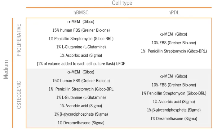
Table 2.1 – Proliferative and osteogenic medium composition.
Table 2.2 – Standard curve values for PicoGreen dsDNA Quantification, proliferation assay.
Table 2.3 – Needed solutions and respectively preparation instructions.
Table 2.4 – Standard curve values for ALP assay.
Chapter III. Asymmetric PDLLA Membranes Containing Bioglass® for Guided Tissue Regeneration
Table 3.1 – Young modulus, maximum strain and maximum stress of the PDLLA and PDLLA/BG membranes obtained in dry and wet conditions. Values are reported as mean ± SD. Statistically significant differences in the mechanical properties of the PDLLA/BG membranes when compared to those of the pure PDLLA are indicated by (*) for p < 0.1 and (**) for p < 0.01.
CHAPTER I. General Introduction
CHAPTER I.
G
ENERAL
I
NTRODUCTION
CHAPTER I. General Introduction
1| Motivation and Outline
Periodontal defect exists when tooth-supporting tissues, including the alveolar bone, periodontal ligament and cementum destruction occurs, as a consequence of periodontitis. [1] Mechanical removing of the damaged structures is the first procedure applied [2], however it is usually followed by a surgical intervention. [3] These conventional therapies reveal to be efficient in halting the periodontal disease progression; even so they involve some drawbacks that restrict their efficiency, being the major limitation related to the promotion of tissue regeneration. It is crucial to repopulate the defect with viable specific cells which are able to promote the regeneration of periodontal ligament and alveolar bone, as well as do not allow the growth of undesirable tissues, which cells have a higher migration rate [4; 5]. Considering that, regenerative procedures appeared using physical barrier membranes to create a segregated space and reach the aforementioned cell manipulation, which is often referred to as guided tissue regeneration (GTR).
Periodontal defects can be a complex problem to treat and represents the main cause of tooth loss in adults. [1] Accordingly, although originally GTR membranes were used specifically for periodontal regeneration, they were afterwards introduced in the implants surgery field, to regenerate bone tissue and thus allowing the dental implants application and conferring the needed stability for the implants [6; 7]. This evolution has to be congruent with the emergent raise of implantology cases, nowadays, factor that more impulses the development of new membranes systems. In this case, the major challenge is the distinct tissue regeneration [8]. Combine bioactive ceramic or glasses with polymers, conferring an asymmetric bioactivity, seems to be a really promising option.
Poly(D,L-lactic acid), PDLLA, is a biodegradable and biocompatible polymer that could be adequate to be used as the matrix in the production of membranes. The polymer may be formulated with inorganic particles and processed into membranes using different techniques. Bioglass® is an approved osteoconductive biomaterial used in orthopedic and dental applications. Therefore, the aim of this work was to prepare a PDLLA membrane containing Bioglass® microparticles, hypothesizing that these microparticles could enhance structural and biological performance of PDLLA membranes, regarding to periodontal regeneration. Moreover, due to the distinct biological environment
experimented in vivo by the two sides of an implanted membrane, it seems reasonable to produce
membranes with distinct properties in each face. To test this hypothesis, asymmetric PDLLA/Bioglass® membranes were prepared, in a single step, by an adjusted solvent casting method.
This concrete chapter presents an overview of the guided tissue regeneration field, specifically applied to periodontal defects regeneration, as well as an additional analysis to the performance of poly(D,L-lactic acid) and Bioglass® in this area.
2| Periodontal Defects
Teeth anatomy involves a variety of different components, which can be classified as hard or soft tissues. We can identify three structural different layers in the tooth: enamel, dentine and pulp chamber. Enamel is a crystalline structure, extremely
hard and highly mineralized, thus it is the outer layer and its function is to cover and protect the crown of the tooth. Dentine constitutes the core structure and pulp chamber composes the center of the tooth. Containing this center area, there are blood vessels and nerves, which connects the jaws vascular and the nervous supply through tooth apices. The tooth root is attached to the adjacent alveolar bone by the periodontal ligament. [9]
Periodontal ligament is the investing and supporting connective tissue structure that anchors a tooth within its alveolus. It is composed by collagen fibers that connect the cementum of the tooth to both gingival and alveolar bone and to the cementum of surrounding teeth. [10]
The etiology of periodontal diseases is related to the dental biofilm, of which evolution results in a progressive loss of dental insertion. [11] The presence of many bacteria in the supra and sub-gingival plaque was considered the major etiologic factor, and the accumulation of many microorganisms on it promotes the starting of periodontal destruction and consequent progression of the disease. [11; 12] Later on, the same author [13] revealed that inclusively some metabolic properties of dental biofilm assure the resistance of microorganisms to the natural body defenses. Logically, besides the bacterial intervention, aspects as genetic influences, immune host response and environmental factors are also included in the contribution for bacteria accumulation on gingival plaque and consequently in the progress of periodontitis.
Periodontitis is a disease, which destroys the tooth-supporting tissues, including the alveolar bone, periodontal ligament (PDL) and cementum, creating defects in the oral cavity. This is the major cause of tooth loss in adults. [1] The treatment of periodontal defects can be a complex process.
Figure 1.1 – Tooth anatomy and different distinguished structures. (Adapted from [9])
CHAPTER I. General Introduction
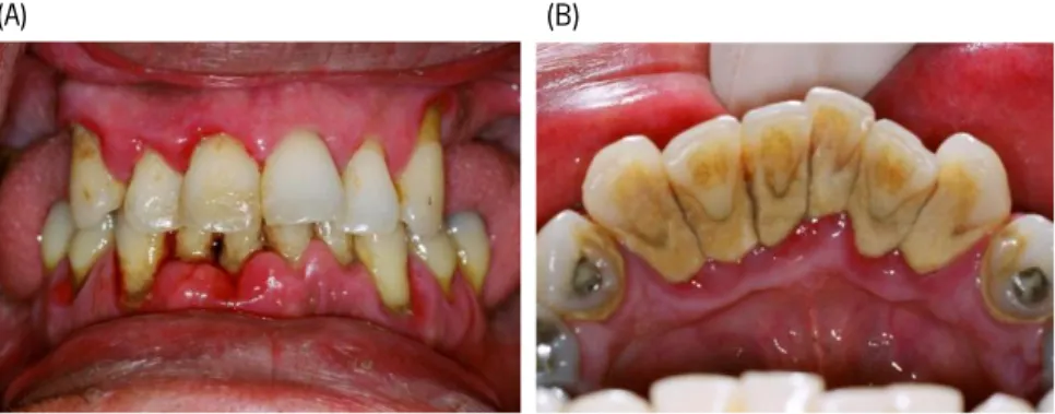
Figure 1.2 – Periodontal disease. (A) A case of advanced periodontitis, evidencing very swollen gums, loose teeth, staining and heavy plaque and calculus deposits on all the teeth. (B) Tartar deposits on the inside aspect of the lower incisor teeth in a patient with periodontitis. (Adapted from [14])
2.1 Current Therapies and Outcomes
Periodontal therapy mainly pretends to achieve reduction or even elimination of the tissue inflammation induced by the dental biofilm and correction of the anatomic defects, reestablishing the dental insertion integrity like before the disease. [15]
The mechanical regular removing of dental biofilm (as well as calculus, infected cementum, granulated tissue and crown portion of epithelium) is the first procedure applied. [2] After an evaluation of the initial phase outcomes, if the inflammation signs persist, the surgical treatment emerges as the best option. [3] Therefore, conventional periodontal therapy includes not just nonsurgical (debridement of root surfaces or root canals) but also surgical approaches (periodontal flap procedures, recessive surgeries). These last mentioned ones provide a better access for the cleaning of root surfaces and apical lesions, as well as for restoring the surrounding bone/root apex. [16]
Traditional recessive surgeries reveal to be efficient even for advanced disease stages, halting the periodontal disease progression, which is a good outcome once that allows and makes easy the removing of subgingival deposits, restituting the morphology of sustaining and recovering periodontal tissues. On the other hand, soft tissue recession, leading to poor esthetics in the anterior dentition, and residual pockets usually inaccessible to adequate cleaning, which negatively affect lon-term prognosis of the treated tooth, constitute the major drawbacks of this technique. [8] Nonetheless, the principal limitation of conventional therapies concerns to the promotion of periodontal regeneration. [17] Regeneration is though the most desirable but also the most difficult outcome for any therapy. [16]
Considering the overcoming of these limitations and difficult challenges, new approaches such as regenerative techniques have been proposed. The main goal is the inhibition of undesirable tissues growth, essentially epithelial tissue which migration and growth rate is higher (approximately 10 times faster) than conjunctive and bone tissues, dominating the initial healing phase. [18] According to that, it
is crucial to colonize the defect with viable specific cells which are able to promote the periodontal regeneration. [7; 18] This concept is named compartmentalization and was introduced by Melcher in 1976. [7] Thus, regenerative procedures used physical barrier mechanisms to reach the aforementioned cell manipulation, which is often referred to as guided tissue regeneration (GTR).
GTR is specifically indicated for narrow intrabony and class II mandibular defects treatment, because the close proximity, between the defect and periodontal mesenchymal cell sources, allow their adequate migration, repopulation and differentiation into the defect. Based on the extensively proved concept that the ability to recreate the original periodontal attachment belongs only to the fibroblasts from the periodontal ligament or undifferentiated mesenchymal cells, GTR therapy was applied with success in the regeneration of periodontal defects. [8] Regarding to the furcation lesions, just for the treatment of class II mandibular furcations GTR can be effectively used, actually allowing the passage of class II into a class I mandibular furcation, easier to maintain overtime. However, on class II maxillary furcations it has a limited clinical effect (namely reduction in the horizontal furcation depth) doubtful clinical significant. [19] In respect to class III furcations, GTR efficacy is unpredictable. [19; 20] In the treatment of intrabony defects, apart from the high variability in clinical outcomes, GTR results show greater probing depth reductions and higher gain in attachment levels compared to conventional flap procedures. [21]
Behind the successful GTR strategy to regenerate periodontal defects, many factors can influence the clinical responses to GTR. For example, the majority of patient-related factors such as smoking and residual periodontal disease could be controlled trough behavioral and therapeutic interventions [8], respectively. Several patient and local factors, as well as factors related to the surgical treatment that should be evaluated are listed on Table 1.1 and contribute for the increasing of the predictability and success of outcoming results of GTR.
Table 1.1 – Factors that negatively influence guided tissue regeneration outcomes. (Adapted from [8; 16])
FACTORS
INFLUENCE ON THE OUTCOMES
PA
TI
EN
T
Smoking habitDecreased vascular flow, altered neutrophil function, and impaired fibroblast function, increased prevalence of periodontal pathogens, decreased IgG production and lymphocyte proliferation.
Poor plaque control Residual periodontal disease
Increased residual periodontal pockets and percentage of sites with bleeding. Higher risk of membrane contamination.
CHAPTER I. General Introduction Table 1.1 (continuation) – Factors that negatively influence guided tissue regeneration outcomes. (Adapted from [8; 16])
FACTORS
INFLUENCE ON THE OUTCOMES
PA
TI
EN
T
Systemic compromised patient Occlusal trauma
Poor oral hygiene/compliance
Mechanical trauma (aggressive tooth brushing)
Inflamed gingival tissues Immunosuppression Diabetes
Stress
These ones were also cited as negative factors to the clinical outcomes of GTR, however the evidences are not enough.
LOCA
L
Local anatomy and morphology strongly affect the predictability of GTR.
Case selection Increased residual periodontal pockets and percentage of sites with bleeding represent a higher risk of membrane contamination.
Gingival thickness (if less than 1 mm) Increased prevalence and severity of flap dehiscence over GTR membranes. Calculus
Overhanging restorations Favored plaque accumulation. Shallow infrabony defects
Wide defect angle > 45 degrees Horizontal bone loss
< 3-wall defects
Deep furcation involvement
Many studies have related significantly better results for GTR strategy than control groups (without GTR membranes) conjugated with the highest success rate for 3-wall defects with a deep infrabony component of ≥ 4 mm and defect angle < 45 degrees.
SURG
ERY
Excessive flap tension Early mechanical disruption Contamination during surgery Lengthy/traumatic surgery Inadequate wound closure Poorly designed incisions Membrane exposure
Basically, to achieve predictable tissue regeneration, 4 summarized factors are critical (the so-called PASS [22]): primary wound closure; angiogenesis as a blood supply and source of undifferentiated mesenchymal cells; space maintenance; and, finally, stability of the wound.
After the GTR procedures some postoperative care and maintenance are also required. Systemic antibiotics and nonsteroidal analgesics may be prescribed, either to reduce the risk of infection or to control pain. [8] Even the usual mechanical tooth cleaning should be avoided. If a non-resorbable membrane is used, it is needed a second surgical intervention, and, ultimately, if the postsurgical problems extend (e.g. membrane exposure as referred in Table 1.1) membrane has to be removed earlier. Membrane exposure frequency can vary between 50% and 100% and represents the major complication associated to the GTR strategy. [20; 23] To contour this drawback, the use of
bioabsorbable membranes has contributed and novel access flaps are introduced to preserve the interdental tissues, thus reducing the prevalence of membrane exposure. [8; 16] Actually, and unfortunately, GTR technique complications are frequent and play a negative effect on clinical outcomes, namely: bleeding, swelling, hematoma, erythema, suppuration, sloughing or perforation of the flap, membrane exfoliation and postoperative pain. [8]
In order to overcome some undesirable outcomes and complications, combining therapies appeared as a solution. The use of barrier membranes parallel to the placement of bone grafting materials came up and should be able to ensure clot stabilization and prevent the membrane collapse into the defect [24].
3| Periodontal Defects and Guided Tissue Regeneration
Nowadays, the main goals in the treatment of periodontal diseases are to regenerate periodontal tissue and to confer support to the bone tissue. Specific cells present in the periodontal tissue are able to produce a new periodontal ligament, cementum (tissue that involves the tooth root) and alveolar bone, if these components can migrate to the host local. The aim of GTR technique is to avoid the migration of epithelial cells to the lesion and, with the help of a membrane, create a physical space between the membrane and the bone. This space allows enough time for the formation of the new periodontal ligament, cementum and bone tissues, with the intention of the total affected area regeneration. The space created, specifically for the invasion of blood vessels and osteoprogenitor cells, protects the bone regeneration against the growth of non-osteogenic tissues, which have a higher migration rate. [4; 5; 25] Due to this dissimilarity on migration rates, soft tissues would grow in bone tissue’s place, if the barrier would not exist. Thus, the employment of a barrier membrane is to guide the new bone growth where volume and dimension are not enough for its normal function.
Originally, GTR membranes were used specifically for periodontal regeneration, being afterwards introduced in the implants surgery field, to regenerate bone tissue in primary or secondary bone deficiencies. Primary bone insufficiency is prior to the implants integration and can follow two therapeutic procedures: insert simultaneously the implants and the membrane with or without grafts, or proceed in two steps, firstly getting a higher bone volume with the membranes action, and then put the implants. If the bone lack is posterior (secondary) to the implants inserting, such as peri-implant defects, fenestrations or dehiscence, membranes are implanted in order to restore the lost initial bone height. [26] These techniques are denominated as Guided Bone Regeneration (GBR).
CHAPTER I. General Introduction
GBR involves the GTR principles, being specifically directed to the alveolar bone reconstruction and thus allowing the dental implants application and conferring the needed stability for the implants. [6; 7] So, GBR is included in GTR, but is focused on the hard tissues regeneration (bone), instead of soft tissues. [27; 28] GBR has been extensively and fruitfully applied in the treatment of bone defects that are adjacent to the implants [29], once that the bone quantity is a crucial issue for the implant to succeed. In case of insufficient bone in the damaged area, implant cannot be totally engaged in the bone being in contact with soft tissues as gingival. It can lead to the soft tissue inflammation, which may result in the implant fail. These are the main reasons for the use of GBR in the dental implants application. [30; 31] Bone substitutes with osteoconductive properties [32] and growth factors [33; 34] were employed to promote bone growth, however, nowadays, the concept of bone regeneration is associated to membranes use. [35]
4| Membranes for Guided Tissue Regeneration
In this field the technique of GTR uses a membrane which acts as a physical barrier to protect the defect site and to prevent the epithelial cells, fibrous and gingival connective tissue to reach the injured area. This procedure favors the regeneration of lost and damaged tissue since it promotes cell repopulation of the periodontal ligament and adjacent alveolar bone. [36] Figure 1.2 illustrates the phenomena that occur during the progression of GTR membranes actuation in a periodontal defect, since its integration.
Figure 1.3 – Progressive action of one of the guided tissue regeneration marketed membranes. (A) Adhesion of PDL fibroblasts to the smooth upper face, promoting soft tissue healing. (B) Osteoblasts adhesion to the rough porous bottom face, allowing increased mineralization. (C) Complete vascularization and porous interconnectivity, at 2 weeks. (Adapted from [37; 38])
(A) (B)
The membranes application respects a rigorous surgical protocol. In general lines, first of all, the injured tissue has to be removed in order to prevent possible infections. Once created the propitious environment, the membrane is positioned between the alveolar bone and the periodontal ligament. Hence, the pretended bone regeneration occurs during which the biodegradable membrane is absorbed by metabolic processes. The membrane acts not just as a physical barrier preventing the fibroblasts invasion into the injured area, but also as a buffer that just allows the osteogenic potential cells presence and avoids their migration to the exterior area. This way the osteogenesis occurs with no difficults, such as an inefficient bone bonding or an incomplete recuperation of the original bone hardness. [4]
Particularly in periodontal defects, the GTR technique is used to promote the conjunctive tissue adhesion to the tooth root surface, as well as to exclude the epithelial cells invasion from the injury. Additionally, it has been used to generate bone tissue around the implant, to avoid the fibrous encapsulation and to produce more bone tissue, thus enlarging the bone support. [4; 25] This space control, achieved by the use of membranes, permits also that the osteoblasts bone production occurs slowly with the aim of obtaining a better structural organization of the damaged tissue and, consequently, a more efficient healing. [25]
First membranes were used in the 80’s. The real first ones were produced using expanded poly(tetrafluorethylene) (ePTFE). [39; 40] These membranes are not absorbed by metabolic processes. Still, ePTFE continued viable due to its biocompatibility and its use in vascular prosthesis. Although, this material presents many disadvantages such as: the regular exposition of the membrane after the implantation surgery, leading to the GTR fail and bacterial contamination; the need of a second surgical intervention to remove the membrane; and finally, after membrane removing, the exposure to the wear of new form bone. [6; 39] In order to overcome these limitations, biodegradable membranes have been introduced in the market [39]. Few characteristics are required to this kind of membranes. A description of membranes types, characteristics, materials and marketed products will be particularly described in the further sub-chapters.
4.1 Properties and Characteristics
GTR membranes should meet several properties to face the complex biological and sensitive system of the human body. Besides biocompatibility, barrier membranes should embrace physico-chemical and structural properties to guarantee basic, but complex and crucial conditions such as the ones described
CHAPTER I. General Introduction
in Table 1.2, in order to accomplish complete and perfect tissue integration. Non-toxicity, selective cellular occlusion and nutrients transfer are some of them.
Table 1.2 – Membrane requirements for guided tissue regeneration and its correspondent description (information collected and adapted from [41-52]).
DESCRIPTION
M
EM
BR
ANE
S REQ
UI
RE
M
EN
TS
BIOCOMPATIBILITY NON-IMMUNOGENICITY NON-TOXICITYAbility to prevent and do not generate adverse inflammatory or immune response and to resist to bacterial invasion and colonization.
BIODEGRADABILITY TOTAL RESORBABILITY
To avoid second surgical procedure to remove the membrane. Absorption rate sufficient to maintain the physical barrier and at the same time compatible with the new bone formation. Total resorption of biomaterials, intended as a complete replacement of the foreign material by the regenerating tissues, appears still here as a perfect solution to interfacial problems.
GOOD MECHANICAL INTEGRITY
To maintain the desired shape and configuration, namely to provide a secluded space for bone regeneration. Need to be sufficient to support or match surrounding native tissue at site of implantation, as well as mediate mechanical stimulus to cell during loading.
EASY HANDLING,
CUTTING AND CONTOURING
To allow the adaptation to the bone anatomy as well as an instant modeling by the professional (Dentist, Periodontologist, Implantologist) and consequent in site implantation of the membrane.
MATERIAL ADHESIVENESS Adhesiveness between membrane and surrounding bone tissues to prevent movement of membrane. CELL ADHESION Optimization of cell seeding for retention of cells.
SEMI-PERMEABLE Adequate porosity to allow nutrient and oxygen supplies.
BIOACTIVITY To accelerate differentiation, mineralization and consequent bone formation. Eventually, to control the release of growth factors.
4.2 Types of Membranes
GTR membranes have been extensively studied and many materials have been proposed for their fabrication. They can be provenient from natural or synthetic source, and are either bioabsorbable or nonresorbable.
Both membranes classes reach the biological and mechanical aims defined for periodontal regeneration, inclusively above described on Table 1.2. Nevertheless, there is the risk of contamination, due to the membrane exposure after the first surgery. [53] Infections in the treated locals reduce the insertion gain and tissue regeneration, as well as accelerate the degradation process, though appeared the needing of a membrane not only with an ideal occlusion capacity but also with antimicrobial protection. [54] Facing that, resorbable membranes present a significant advantage, once they permit the incorporation of antimicrobian agents that can be released upon implantation.
Moreover, generally, non-resorbable membranes have to be removed by a secondary surgical procedure after the tissue had formed, increasing the risk of patient infection and other undesirable side effects. Also, they cannot be used to reconstruct large bone defects due to its bioinert properties constituting a significant disadvantage [55; 44]. In order to overcome such drawback, bioresorbable membranes have been proposed. In this case the degradation of the membrane should not interfere with bone healing and, before osseous regeneration has taken place, degradation should not be completed [56; 57].
4.3 Materials Employed and Current Marketed Membranes
Several membranes were studied and proposed for GTR. However, for a membrane to become commercially available there are many obstacles to overcome, such as the clinical evaluation and the
approval by the competent regulatory agencies. After the membrane conception and in vitro
characterization, preliminary studies in animals are performed; subsequently, the most trustable systems may be redirected to pre-clinical studies in humans. Clinical evaluation is such an important and exigent parameter. In consequence, only few have reached the stage of routine clinical application [58].
Expanded poly(tetrafluoroethylene) (e-PTFE), silicone rubber or titanium are the materials that constitute most non-resorbable membranes. The bioresorbable ones have been produced using collagen, collagen with elastin, poly(L-lactic acid) (PLA), poly(L-lactic acid)-blend-poly(D, L-lactic acid) (PDLLA), poly(D,L-lactide-co-glycolide) (PLGA) or trimethylene-carbonate (TMC). [56; 59; 60]
CHAPTER I. General Introduction
Natural collagen membranes have been the mostly used, not just because collagen is one of the components of the alveolar bone and periodontal ligament but also because this material performs almost all the criteria mentioned in the previous paragraphs. [61] For example BioGide®, Bicon®, BioMend®, BioSorb® and Ossix® are some of the current collagen membranes in the market. However collagen presents some drawbacks, including fast resorption rate, cytotoxicity, poor mechanical strength and fast biodegradation by enzymatic activity. [61; 62] To decrease this degradation rate and increase its hardness, collagen could be enhanced with cross-linking techniques [63] but this procedure (particularly with glutaraldehyde) can inhibit the attachment and proliferation of human PDL and human osteoblastic cells. [38] Adding the fact that its xenogenic origin presents a risk of disease transmission between animals and humans [64] and also that collagen tends to lose the ability to keep its own shape in wet conditions, as those existing in the oral cavity, [65; 66] these problems concur to the searching for new solutions.
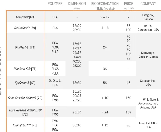
Table 1.3 – Synthetic biodegradable marketed membranes, with respective constituent polymer, dimensions in mm, biodegradation time in weeks, price in Euros per unit and produced company.
POLYMER DIMENSION
(mm) BIODEGRADATION TIME (weeks)1
PRICE (€/unit) COMPANY
M
AR
KETE
D ME
M
BRA
NES
Artisorb® [69] PLA - 9 – 12 - Citagenix, Canadá
BioCellect™ [70] PLA 15x20 20x30 4 – 8 100 67 Corporation, USA IMTEC
BioMesh® [71] PLGA PGA PLLA 15x12 17x17 25x17 30X24 40X30 25X20 24 70 70 70 70 106 92 Samyang’s, Daejeon, Coreia BioMesh-S® [71] PLGA PGA
PLLA 36 -
EpiGuide® [69] D, D-L, L- PLA 18x30 56 46 Curasan Inc., USA
Gore Resolut Adapt® [72] TMC PGA
15x20 20x25
25x20 > 10 150 W. L. Gore &
Associates, Inc., Arizona, USA Gore Resolut Adapt LT®
[72] TMC PGA 25x30 > 24 158
Inion® GTR™ [73] TMC PLA
PGA 30x40 > 12 96
Inion Ltd, UK e USA
In order to avoid these undesirable characteristics, maintaining the desirable ones, synthetic materials have been more frequently used, predominantly the poly(α-hydroxyesters) family. [72] These materials are the gold standard in applications of biodegradables in medicine. The chemical properties of these polymers allow its hydrolytic degradation and removing by natural pathways. [68] Moreover their processing is easy compared to other polymers and the variety of existent molecular weights and copolymers permits a wide range of physical, mechanical and degradation rate related adjustments. Epi-Guide®, Gore Adapt®, Inion®, BioMesh® and BioCellect® are examples of current available marketed GTR membranes of this specific type, among many others described and analyzed in Table 1.3.
5| Biocomposite Materials
First generation of biomaterials was developed to achieve exclusively bioinert tissue response. The forward generation emerged as bioactive biomaterials. Bioactivity is described as the capacity of the material to elicit a controlled action and reaction in the physiological environment. [74]
Reinforcement of biodegradable polymers matrices to produce composites of tailored surface, chemical and mechanical properties is a desirable approach for potential biomedical applications, such as in the regeneration of hard or even soft tissues. [75] The increasing research efforts worldwide for bone tissue regeneration [73;76-88] are trying to fulfill as many requirements as possible. For example, inorganic phases of tricalcium phosphate [89], hydroxyapatite [90-92] or bioactive glasses [79; 93; 94] are included into biodegradable poly(α-hydroxyester)-based matrices, such as poly(lactic acid), poly(glycolic acid) and their copolymers, as a viable way to improve the abovementioned properties and enhance bioactivity. Actually, the possibility of counteracting the acidic degradation of biodegradable polymer by the use of bioactive glasses is another reason for the use of composites. [95-97]
Consequently, composite systems combining advantages of biodegradable polymers and ceramics seem to be a really promising choice.
5.1 Biodegradable Polymers, the Poly(α-hydroxy-esters)
There are two types of biodegradable polymers taking into account their origin. The natural-based materials are one category, including starch, chitosan, alginate, hyaluronic acid derivatives (common polysaccharides) or collagen, fibrin gels, silk and soy (proteins). [98; 99] Synthetic biodegradable polymers constitute the second category: in general they may be obtained with high purity and
CHAPTER I. General Introduction
exhibiting predictable and reproducible mechanical and physical properties (elastic modulus, compressive or tensile strength and degradation rate) due to their under controlled production. [100-103]
Poly-α-hydroxy-esters are the most widely used biodegradable polymers for tissue engineering and regeneration inclusively have been approved by the US Food and Drug Administration for several applications. In these range are included poly(lactic acid) (PLA) and poly(glycolic acid) (PGA), as well as
poly(lactic-co-glycolide) (PLGA) copolymers. PLA exists in three forms: L-PLA (PLLA), D-PLA (PDLA), and
racemic mixture D,L-PLA (PDLLA). [68; 100; 104; 105] Table 1.4 describes some of these parameters
for the cited polymers, which may however vary with molecular weight and crystallinity [106]:
Table 1.4 – Physical properties of synthetic, biocompatible and biodegradable polymers from saturated aliphatic polyesters family used as scaffold materials. (Adapted from [75])
Tm (°C) Tg (°C) BIODEGRADATION
TIME (months)1
COMPRESSIVE*/TENSILE
STRENGTH (MPa) MODULUS (GPa)
POL
YM
ERS
PDLLA Amorphous 55-60 12-16 Pellet: 35-150*
Film/disk: 1.9-2.4 Film/disk: 29-35 PLLA 173-178 60-65 >24 Pellet: 40-120* Film/disk: 1.2-3.0 Film/disk: 28-50 Fibre: 870-2300 Fibre:10-16
PGA 225-230 35-40 6-12 Fibre: 340-920 Fibre: 7-14
PLGA Amorphous 45-55 Adjustable:1-12 41.4-55.2 1.4-2.8
PCL 58 -72 >24 - -
1 Until complete bioresorption
The chemical nature of these polymers allow hydrolytic degradation. Human body already contains highly regulated mechanisms for the complete removing of monomeric components of lactic and glycolic acids, i.e., once degraded, they are removed by natural pathways. PLA is eliminated through the tricarboxylic acid cycle, while PGA is converted in metabolites or cleared by other mechanisms. [68] The in vivo degradation of these poly(α-hydroxy esters) occurs by hydrolysis, releasing the lactic or
glycolic acids which are metabolized through the Krebs cycle into carbon dioxide (CO2) and water (H2O),
basic human body elements. [72] Even though, the degradation kinetics is affected by different factors, such as: chemical composition and configurational structure, molar mass (Mw), polydispersity (Mw/Mn), environmental conditions, stress and strain, crystallinity, morphology (e.g. porosity) and
chain orientation, distribution of chemically reactive compounds within the matrix, additives, presence of original monomers, overall hydrophilicity and their processing history. [95; 96]
In general, their processing is easy compared to other polymers and the various existent molecular weights and copolymers permit a wide range of adjustments, in respect to degradation rate, physical and mechanical properties. PLA is more hydrophobic than PGA due to the additional methyl group in the structure of PLA; therefore PGA degrades much more quickly (a few weeks [107; 108]) than PLA, which can remain stable for over 1 year [109], or more depending on its degree of crystallinity. Albeit, these polymers still have some drawbacks. Sometimes their bulk erosion can result in scaffolds premature fails and, for example, an abrupt release of these acidic degradation products can cause strong inflammatory responses, being the typical pH drop associated with PLA and PGA implants one of its major negative aspects. [110; 111]
5.1.1 Poly (D,L-Lactic Acid)
Poly (D,L-lactic acid), PDLLA, is the racemic polymer of the poly(lactic acid) family – see Figure 1.3, general chemical structure. It is originated by the chirality of carbon α that allows the synthesis of enantiomers composites: L and D. Due to the random distribution of L and D unities on the polymeric chain, PDLLA does not have crystalline domains, being an amorphous material with lower stiffness as compared to the semi-crystalline PLLA. Therefore, the hydrolysis of this amorphous polymer is faster due to the lack of crystalline regions. [112-114]
Figure 1.4 – General chemical structure of poly(lactic acid). (Adapted from [112])
Li et al [115] studied a series of PDLLA copolymers, concluding that after 12 weeks of in vitro
degradation in PBS, the material bulk suffers significant mass decrease, being these results concomitants with the in vivo results among other studies. [115]
PDLLA shows excellent biocompatibility in vivo and high mechanical stability. [116; 117] Such excellent features of PDLLA with respect to implant performance have made itself an extensively investigated biomedical coating orthopedic material. [100; 118] Much more attention has been also
CHAPTER I. General Introduction
paid to PDLLA for applying it as a scaffold material for tissue engineering and regeneration. Desirable features such as the ability to combine with drugs like growth factors, antibiotics or thrombin inhibitor, establishing a locally acting drug-delivery system, are in the center of this interest. [118]
5.2 Bioactive Ceramic phases
Bioactive glasses and calcium phosphates have been largely applied in bioactive composite materials. Their excellent biocompatibility, as well as the possibility of counteracting the acidic degradation the ability of bone bonding are essential attributes for their choice. [119]
After implantation in vivo or contact with biological fluids, bioactive glasses and ceramics develop on the surface a biologically active hydroxy-carbonate apatite (HCA) layer which provides the bonding interface with the local tissue. The HCA phase is structural and chemically equivalent to the bone mineral phase, providing interfacial bonding (bridging host tissue with implants [120]). This procedure is achievable in vitro using a protein-free and acellular simulated body fluid (SBF), which nearly has the exact ion concentration of human blood plasma. [121; 122] This time-dependent kinetic modification of the surface is a bon-bonding behavior referred to as bioactivity, the main characteristic of this kind of materials. [119; 121; 123-126]
Table 1.5 – Mechanical properties of dense and highly porous 45S5 Bioglass®, hydroxyapatite, A/W glass-ceramic, and human cortical bone. (Adapted from [75])
COMPRESSIVE STRENGTH (MPa) TENSILE STRENGTH (MPa) ELASTIC MODULUS (GPa) FRACTURE TOUGHNESS (MPa )
CERA
M
ICS
45S5Bioglass® ≈500 42 35 0.5 – 1 Hydroxyapatite (HA) >400 ≈40 ≈100 ≈1.0 Glass-ceramic A/W 1080 215 118 2.0Porous bioactive glass70S30C
(82%) 2.25 - - -
Porous Bioglass® - derived
glass-ceramic (>90%) 0.2 – 0.4 - - -
Porous HA (82-86%) 0.21 – 0.41 - 0.83 – 1.6 x 10⁻³ -
Cortical Bone 130 – 180 50 – 151 12 – 18 6 – 8
Table 1.5 gives summarized information related to the typical mechanical properties of different bioactive glasses and ceramics. Bioactive glass-ceramics exhibit better mechanical performance compared to amorphous glass and calcium phosphate. Nonetheless their low fracture toughness and mechanical strength are still a drawback, specially comparing to cortical and cancellous bone (Table 1.5). [123; 127-130]
It is recognized that a layer of biologically active HCA must form to occur the bonding with the bone tissue; in fact, this is the only common characteristic of all the known bioactive implant materials. [74] Starting in 1967, Hench [121; 122] has extensively studied bioactive glasses as well as bioactive glass-ceramics. Although he could summarize the stages that are involved in the bone-bonding formation, some details remain yet indefinite. On the other hand, Hench was able to clearly define three classes of bioactive materials (A, B and C) characterized by the rate of bone regeneration and repair: materials that lead to both osteoconduction (the growth of bone along the bone-implant interface) and osteoproduction, as a result of the fast reactions on the implant surface, constitute the class A [131; 132]; alternatively, class B bioactivity takes place when just osteoconduction occurs [133; 134]; and, finally, materials that are resorbed within 10-30 days in tissue are included in class C. [121]
To emphasize the relevance of bioactive glasses there is the fact that they also have the ability to support enzyme activity [135-137], vascularization [138; 139], promote osteoblast adhesion, growth and differentiation, and induce mesenchymal cells differentiation into osteoblasts [85; 140; 141]. Furthermore, particularly for 45S5 Bioglass® composition, their dissolution products upregulate the gene expression that control osteogenesis and the growth factors production [142].
5.2.1 Bioglass®
Bioglass® is a proven osteoconductive material.[121] Since it belongs to the bioactive glasses family, it encloses a reactive silicate surface which, when in contact with biological fluids, forms a layer of carbonated HA, a strong and adherent bond with bone. Particularly for 45S5 Bioglass® composition,
this complex multi-stage process occurs very rapidly. [143; 144] Additionally, Wilson et al [126]
demonstrated that further than its excellent bone-bonding properties, Bioglass® also forms a bond with soft connective tissues.
In general, bioactive glasses contain SiO2, Na2O, CaO and P2O5. Specifically, for 45S5 Bioglass®, 45
represents 45 wt% SiO2, S is the network former and 5 corresponds to the ratio of CaO to P2O5, with the
rest weight percentage (around 24.5 wt%) correspondent to the Na2O portion. [75; 143; 144] This was
CHAPTER I. General Introduction
ternary eutectic showed by the equilibrium phase diagram
Na2O–CaO–SiO2. However, further researches have been
trying different wt% proportions, in order to improve the bioactivity of these bioactive glasses. For example, 45S5
Bioglass® (see Figure 1.4), with 55% SiO2, exhibit a high
bioactivity index and bond to both soft and hard tissues. [75]
Bioglass® has been used as a reinforcing agent within polymer matrices, being expected to increase also mechanical properties of the composite. Stiffness and microhardness of high-density polyethylene composites
was amplified with a reinforcement of filler volume fraction in Bioglass® particulates (melt blending, compounding, powdering and comprehension moulding techniques involved). [145] On the other hand, that cited positive reinforcing effect was not replicated in composites of poly(α-hydroxyesters). [93] The
addition of bioactive glass (53 wt% SiO2) as a filler for poly(D,L-lactide) composites may reduce its
modulus and strength in specimens assessed, both dry and wet conditions. [80; 93] Rich et al [97]
evaluated the influence of the incorporation of small particles of bioactive glass into poly(ε-caprolactone–co–D,L-lactide 96/4), concluding that it might accelerate the degradation of the composite. Although, it is important to state that high-temperature processing methods were involved, remaining to the possibility of the processing-temperature influence and consequent favor of the composite debilitation and faster degradation. Moreover, further studies have shown that the use of low-temperature processing for poly(α-hydroxyesters) with Bioglass® may circumvent these problems [79; 146], actually very recently, Blaker et al (2010) [144] proved it as well.
The addition of Bioglass® to bioresorbable polymers allows a rapid exchange of protons in water for alkali in the glass, providing a pH buffering effect at the polymer surface and consequently modifying the acidic polymer degradation. [83; 147] Thus it has been proved that bioactive glasses interfere on the polymer degradation behavior. [148] Actually, the pH is just one among other adjustable parameters controlling the degradation kinetics, by the integration of bioactive glasses in polymer scaffolds. These materials also modify the surface and inclusively the bulk properties of composite scaffolds, by increasing the hydrophilicity and water absorption of the hydrophobic polymer matrices. In particular, 45S5 Bioglass® particles were found to increase water absorption when included in PDLLA [149] and PLGA [83; 147] polymer foams, compared to the pure ones.
Figure 1.5 – SEM micrographs illustrating the typical ―cauliflower‖ morphology of hydroxyapatite formed on the surface of a 45S5 Bioglass® based foam after immersion in SBF for 28 days. (Adapted from [151])
Depending on the particle size and processing technique, the surface reactions on bioactive glasses can release critical concentrations of soluble Si, Ca, P and Na ions, inducing intra and extracellular responses. [142; 150] Ideally, the degradation and resorption of composite scaffolds are designed to allow cells to proliferate and secrete their own extracellular matrix, while the scaffolds gradually vanish, leaving space for new cell and tissue growth. [75] Stimulation of neo-vascularization is also appointed as an ability of 45S5 Bioglass®. In some studies, scaffolds containing controlled concentrations of
Bioglass® showed to increase secretion of vascular endothelial growth factor (VEGF) in vitro and to
enhance vascularization in vivo. [75; 138]
The entire aforementioned characteristics engaged by 45S5 Bioglass® made out it like a successful material for tissue regeneration. Their application extends since as scaffold materials, either as filler or coating of polymer structures [147; 151], or even as porous material involving melt-derived and sol-gel-derived glasses [125]. Bioglass® implants have been applied in the replacement of damaged middle ear bones and restoring hearing to patients [125]. Concretely, the main applied area is the Dentistry, in which there are already marketed products, such as Perioglass™ for clinical treatment of periodontal disease and Novabone™ used as bone filler. [121]
5.3 Bioactive Composites Processing Techniques
Bioactive composites can be processed by different techniques. Injection or compression moulding and twin-screw extrusion, are typical melt-based processes. To obtain porous composite scaffolds, molding with leachable particulates, sintering of composite microspheres and gas foaming are also melt-based techniques that can be used. Contrasting, there are also low-temperature processes, such as thermally induced phase separation (TIPS), combined solvent casting and porogen leaching, solid freeform methods and certain gas foaming methods. [144]
5.3.1 The Solvent Casting Method
Solvent casting is used to process biocomposite materials. This processing involves the dissolution of the polymer in a solvent and the consequent casting of the solution into a predefined mold. Particles may be added to the solution, with specific dimensions. The mixture is molded in accordance with the pretended final geometry, which means that this solution can be either converted in membranes using a flat glass plate, or in 3D structures using an appropriate mold. Subsequently, the solvent evaporates
![Figure 1.1 – Tooth anatomy and different distinguished structures. (Adapted from [9])](https://thumb-eu.123doks.com/thumbv2/123dok_br/17919513.850296/21.892.483.786.364.610/figure-tooth-anatomy-different-distinguished-structures-adapted.webp)


![Figure 1.4 – General chemical structure of poly(lactic acid). (Adapted from [112])](https://thumb-eu.123doks.com/thumbv2/123dok_br/17919513.850296/33.892.341.561.781.904/figure-general-chemical-structure-poly-lactic-acid-adapted.webp)