Universidade de Lisboa
Faculdade de Farmácia
Development and pre-clinical evaluation of a new HIV-1
vaccine concept
Rita Diogo de Almeida Calado
Orientador: Prof. Doutor Nuno Eduardo Moura dos Santos da Costa Taveira Co-orientador: Prof. Doutor José António Frazão Moniz Pereira
Tese especialmente elaborada para obtenção do grau de Doutor em Farmácia, especialidade Microbiologia
Universidade de Lisboa
Faculdade de Farmácia
Development and pre-clinical evaluation of a new HIV-1 vaccine
concept
Rita Diogo de Almeida Calado
Orientador: Prof. Doutor Nuno Eduardo Moura dos Santos da Costa Taveira Co-orientador: Prof. Doutor José António Frazão Moniz Pereira
Tese especialmente elaborada para obtenção do grau de Doutor em Farmácia (Microbiologia)
Júri:
Presidente: Doutora Matilde da Luz dos Santos Duque da Fonseca e Castro, Professora Catedrática e Diretora da Faculdade de Farmácia da Universidade de Lisboa
Vogais:Doutor Nuno Eduardo Moura dos Santos Costa Taveira, Professor catedrático, Instituto Superior de Ciências da Saúde Egas Moniz, orientador
Doutora Patrícia Maria Cavaco Silva de Sá Montez, Professora associada, Instituto Superior de Ciências da Saúde Egas Moniz
Doutor Miguel Agostinho Sousa Pinto Torres Fevereiro, Investigador Principal, Instituto Nacional de Investigação Agrária e Veterinária, I.P.
Doutor João Manuel Braz Gonçalves, Professor associado com agregação, Faculdade de Farmácia da Universidade de Lisboa
Doutora Helena Paula Lopes Henriques Rebelo de Andrade, Professora auxiliar convidada, Faculdade de Farmácia da Universidade de Lisboa
Rita Calado teve o apoio financeiro da Fundação para a Ciência e Tecnologia através de uma bolsa de Doutoramento (SFRH/BD/70715/2010)
Todas as afirmações efetuadas no presente documento são da exclusiva responsabilidade do seu autor, não cabendo qualquer responsabilidade à Faculdade de Farmácia, Universidade de Lisboa, pelos conteúdos nele apresentado.
Rita Calado teve o apoio financeiro da Fundação para a Ciência e Tecnologia através de uma bolsa de Doutoramento (SFRH/BD/70715 /2010)
“The important thing is to never stop questioning” -Albert Einstein
i
It has been a long journey full of obstacles and achievements.
I would like to acknowledge all the people that accompanied and contributed to make this work possible.
First of all I would like to thank to Professor Doutor Nuno Taveira for accepting me as part of his team and for believing in me. Thank you for your excellent supervising, guidance and support and for being always so optimistic. Thank you for helping me keeping moving forward and for all the scientific advices. Thank you so much for all the opportunities you gave me to work in several projects which allowed me to expand my scientific knowledge. With you I’ve learned that achievements are in direct proportion to the effort that we apply.
I would like to thank to Professor José Moniz Pereira, director of the Department of Microbiology at Faculty of Pharmacy for receiving me and allow me to develop my work at this unit and also for co-supervising the work developed.
I would also like to thank to all the teachers that work in this unit, particularly Prof. Quirina, Prof. João Gonçalves, Prof. Isabel Portugal and Prof. Aida Duarte. Also I would like to thank to Prof. Jorge Vítor for all the support and for teaching me so many things and also to Lavinia who provided me samples so many times so I can proceed with my work.
To José Maria Marcelino thank you for all your support and guidance.
To Fundação para a Ciência e Tecnologia thank you for the financial support that allowed me to develop my work.
Thank you to Prof. Doutor Luis Graça and Sílvia Almeida from the Laboratory of Lymphocyte Regulation at Instituto de Medicina Molecular, Faculdade de Medicina da Universidade de Lisboa for all the colaboration in this work. Thank you also to Dra. Belmira Carrapiço, Inês Dias and Frederico Aires da Silva from FMV for your collaboration.
ii
I would like to thank to all the people that contribute to my work and accommodation: Paula, Cleida, Teresa and São for providing me proper working conditions; Ofélia and Vera for taking care of the paperwork and of course Sr. Joaquim and D. Marilia for all the snacks.
Thank you to all my lab colleagues and friends for such an amazing work environment. Thank you Joana Duarte, Andreia, Cheila, Inês Bártolo, Pedro, Carla, João Perdigão, Francisco, Marta Calado, Marta Gíria, Joana Vital, Ana Rita, Inês Figueiredo, Inês Moranguinho, Rita Mateus e Jaciara for your friendship, advices, support and crazy moments that made me so happy. Thank you all for the dinners, lunches, picnics and amazing events that contribute greatly to my accommodation (and make me gain some weight). Without you it wouldn’t be the same. Also I would like to thank to all the other colleagues with whom I´ve shared the P3, corridors and other facilities.
A special thanks to Joana Duarte for your collaboration and devotion and of course for your friendship. Also thank you for your immunology explanations through whatsupp. I never thought I could learn to like cellular immunology. Francisco and Figueiredo thank you so much for your collaboration and support. João Perdigão thank you for always solving my computer problems and Pedro thank you so much for always being there and helping me whenever I needed. Carla thank you for being so kind and helpful. Andreia and Inês B. thank you for friendship and good laughs. Marta Gíria thank you for your friendship and for always be there.
Thanks to all my friends outside the lab, specially, Raquel, Daniela, MT, Fábio and Carlos for our great moments and friendship.
Thank you Xana for being there since always. There´s no such place as far away.
To my family, thank you for all the support and understanding and for always believing in me.
Mãe, tia, tio e avó, obrigada por TUDO.
João, António e Amélia. Vocês são o melhor do mundo. Obrigada por existirem e tornarem os meus dias mais felizes ∞
iii
v
Preface
All experimental work was performed at HIV Evolution, Epidemiology and Prevention laboratory, Research institute for Medicines (iMed.ULisboa), Faculty of Pharmacy, Universidade de Lisboa under the supervision of Prof. Doutor Nuno Taveira and the co-supervision of Prof. Doutor José Moniz Pereira.
The results obtained in this thesis were described in the following publications:
1. Bartolo I*, Calado R*, Borrego P, Leitner T, Taveira N. Rare HIV-1 Subtype J Genomes and a New H/U/CRF02_AG Recombinant Genome Suggests an Ancient Origin of HIV-1 in Angola. AIDS Res Hum Retroviruses. 2016 Aug;32(8):822-8. * These authors have contributed equally to the work.
2. Calado R, Duarte J, Borrego P, Marcelino J.M, Wilton J, Bártolo I, Martin F,
Figueiredo IB, Almeida SCP, Vítor J, Graça L, Taveira N. A prime-boost immunization strategy with Vaccinia virus expressing novel envelope gp120 glycoproteins from non-B subtypes induces cross-clade tier 2 HIV-1 neutralizing antibodies in mice (Manuscript submitted).
3. Calado R, Martin F, Figueiredo IB, Vítor J, Carrapiço B, Dias I, Taveira N. Induction of tier 2 cross-clade neutralizing antibodies in rabbits by a prime-boost immunization strategy with Vaccinia virus expressing novel HIV-1 gp120 glycoproteins from clade CRF02_AG (Manuscript in preparation).
vi
Other publications
The research produced during the PhD, were presented in the form of research papers and oral and poster communications in international and national meetings.
Rocha C, Duarte J, Borrego P ,Calado R, Marcelino JM, Tendeiro R, Valadas E, Espada AE, Taveira N. Potency of HIV-2-specific antibodies increase in direct association with loss of memory B-cells. AIDS. 13;31(17):2431-2433. November 2017
Calado R, Duarte J, Borrego P, Marcelino JM, Wilton J, Bártolo I, Martin F, Barroso H,
Almeida SCP, Graça L, Taveira N. Novel HIV-1 gp120 glycoproteins derived from non-B subtypes induces tier 2 cross-clade neutralizing antibodies in mice. 4º Congresso Nacional de Medicina Tropical.1º encontro lusófono de SIDA, Tuberculose e Doenças oportunistas. Instituto de Higiene e Medicina Tropical. 19-21 April 2017. Oral presentation.
Calado R, Duarte J, Diniz AR, Borrego P, Marcelino JM, Bártolo I, Clemente S, Taveira
N. Expression and antigenicity of gp120 and C2-V3-C3 polypeptides from different HIV-1 genetic forms. 6th imed meeting.Faculdade de Farmácia da Universidade de Lisboa, 2 July 2014. Oral presentation.
Calado R, Duarte J, Borrego P, Marcelino JM, Wilton J, Bártolo I, Martin F, Almeida
SCP4, Barroso H, Graça L, Taveira N. A prime-boost immunization strategy with Vaccinia virus expressing novel HIV-1 gp120 glycoproteins induces tier 2 neutralizing antibodies in mice. 9th iMed.ULisboa Postgraduate Students Meeting and 2nd i3DU Meeting.13-14 July 2017. Faculty of pharmacy. Lisbon. Poster presentation.
Calado R, Duarte J, Borrego P, Marcelino JM, Wilton J, Bártolo I, Martin F, Almeida
SCP4, Barroso H, Graça L, Taveira N. Induction of tier 2 cross-clade neutralizing antibodies in mice by a prime-boost immunization strategy with Vaccinia virus expressing novel HIV-1 gp120 glycoproteins from non-B subtypes. Ciência 2017. 3-5 July 2017. Centro de Congressos de Lisboa. Poster presentation.
Calado R, Duarte J, Borrego P, Marcelino JM, Wilton J, Bártolo I, Martin F, Almeida
SCP4, Barroso H, Graça L, Taveira N. Induction of tier 2 cross-clade neutralizing antibodies in mice by a prime-boost immunization strategy with Vaccinia virus expressing novel HIV-1 gp120 glycoproteins from non-B subtypes. 3ª Semana da
vii
inovação. 3-9 May 2017. Reitoria da Universidade de Lisboa (ULisboa). Poster presentation.
Calado R, Duarte J, Borrego P, Marcelino JM, Wilton J, Bártolo I, Martin F, Clemente
S, Almeida SCP, Barroso H, Graça L, Taveira N. A novel prime-boost immunization strategy with Vaccinia virus expressing HIV-1 gp120 derived from non-B subtypes induces autologous and heterologous Tier 2 HIV-1 cross-clade neutralizing antibodies in mice. Poster number 2058. Keystone Symposia, HIV Vaccines, Keystone, March, 2017.
Calado R, Duarte J, Borrego P, Marcelino JM, Wilton J, Bártolo I, Clemente S, Almeida
SCP, Graça L, Taveira N. A novel prime-boost immunization strategy with Vaccinia virus expressing gp120t-AG elicit a neutralizing response in mice against autologous and heterologous tier 2 virus. Ciência 2016 - Science and Technology in Portugal Meeting, Lisboa Congress Centre, 4-6 July, 2016. Poster presentation.
Bártolo, I *, Calado, R*, Borrego, P, Leitner, T,Taveira, N. Rare HIV-1 subtype J genomes and a new H/U/CRF02_AG recombinant genome suggests an ancient origin of HIV-1 in Angola. Ciência 2016 - Science and Technology in Portugal Meeting, Lisboa Congress Centre, 4-6 July, 2016.*These authors have contributed equally to the work. Poster presentation.
Calado R, Duarte J, Borrego P, Marcelino JM, Diniz R, Bártolo I, Wilton J, N Taveira;
Clemente S, Taveira N. Envelope C2-V3-C3-specific antibodies correlate with neutralization activity in plasma from HIV-1 infected patients from Angola. Poster number 1022. Keystone Symposia, HIV Vaccines, Keystone, 22–27 March, 2015.
Other publications
Martin F, Palladino C., Mateus R, Diniz AR, Calado R, Clemente S, Taveira N. Neutralizing antibody response in HIV-1 infected patients from Angola. 9th iMed.ULisboa Postgraduate Students Meeting and 2nd i3DU Meeting.13-14 July 2017. Faculty of pharmacy. Lisbon. Poster number 92.
Martin F, Palladino C, Mateus R, Diniz AR, Calado R, Clemente S, Taveira N. Neutralizing antibody response in HIV-1 infected patients from Angola. Ciência 2017. 3-5 July 2017. Centro de Congressos de Lisboa.
Figueiredo IB, Calado R, Martin F, Borrego P, Cardoso F, Taveira N, Barroso H. Antibody response to an HIV-1/HIV-2 chimeric envelope glycoprotein in mice. 3ª
viii
Semana da inovação. 3-9 May 2017. Reitoria da Universidade de Lisboa (ULisboa). Poster presentation.
Martin F, Palladino C, Mateus R, Diniz AR, Calado R, Clemente S, Taveira N. Characterization of the neutralizing antibody responses in HIV-1 infected patients from Angola and their impact on disease progression. 4º Congresso Nacional de Medicina Tropical. Instituto de Higiene e Medicina Tropical. 19-21 April 2017. Poster presentation.
Figueiredo IB, Calado R, Martin F, Borrego P, Cardoso F, Taveira N, Barroso H. Antibody response to an HIV-1/HIV-2 chimeric envelope glycoprotein in mice. Keystone Symposia, HIV Vaccines, Keystone, March, 2017. Poster presentation.
Martin F, Mateus R ,Calado R, Diniz AR, Palladino C, Clemente S, Taveira N. Characterization of the neutralizing antibody responses in HIV-1 infected patients in Angola. 7th imed meeting held on Faculdade de Farmácia da Universidade de Lisboa, 15-16 July 2015. Poster presentation.
Rocha C, Calado R, Borrego P, Marcelino JM, Bártolo I, Rosado L, Cavaco-Silva P, Gomes P, Família C, Quintas A, Skar H, Leitner T, Barroso H, Taveira N. 2013. Evolution of the human immunodeficiency virus type 2 envelope in the first years of infection is associated with the dynamics of the neutralizing antibody response. Retrovirology, 2013.10:110.
Borrego P, Calado R, Marcelino J, Pereira P, Quintas A, Barroso H, Taveira N. 2013. An ancestral HIV-2/SIV peptide with potent HIV-1 and HIV-2 fusion inhibitor activity. AIDS, 2013 April 24; 27(7):1081-90
Taveira N, Rocha C, Calado R, Borrego P, Barroso H. X4 primary isolates of HIV-2 are less susceptible to antibody neutralization that R5 isolates. Poster 3026. Keystone Symposia, HIV Vaccines, Keystone, Colorado, EUA, February 2013. Poster presentation.
Rocha C, Marcelino JM, Borrego P, Calado R, Tendeiro R, Foxall RB, Valadas E, Sousa AE, Taveira N. Potent and broadly neutralizing antibodies are produced in chronic HIV-2 patients despite evidence of marked memory B cell depletion. Keystone Symposia, HIV Vaccines, Keystone, Colorado, EUA, February 2013. Poster presentation.
Borrego P, Calado R, Marcelino J, Bartolo I, Rocha C, Cavaco-Silva P, Doroana M, Antunes F, Maltez F, Caixas U, Barroso H, Taveira N. 2012. Baseline susceptibility of primary HIV-2 to entry inhibitors. Antivir Ther 17:565-70.
Rocha C, Borrego P, Calado R, Skar H, Marcelino JM, Bártolo I, Leitner T, Barroso H, Taveira N. Evolution of the autologous neutralizing antibody response in early HIV-2
ix
infection. Keystone Symposia, HIV Vaccines, Keystone, Colorado, EUA, 21-26 March 2012. Poster presentation.
Taveira N, Barroso H, Bártolo I, Borrego P, Calado R, Cavaco Silva P, Marcelino J, Rocha C. “Susceptibility of HIV-2 primary isolates to fusion and entry inhibitors”. Keystone Symposia, 20-25 March 2011. Poster presentation.
Borrego P, Barroso H, Calado R, Marcelino J, Quintas A, Taveira, N Cavaco-Silva P, Bártolo I. “Design and evaluation of a new HIV-1 and HIV-2 fusion inhibitor peptide”. Keystone Symposia, 20-25 March 2011. Poster presentation.
xi
A SIDA continua a ser uma prioridade em saúde pública com o número de pessoas infetadas com o HIV-1 a aumentar. Apesar dos progressos feitos na área da prevenção, o controlo da infeção por HIV-1 dependerá do desenvolvimento e aplicação de uma vacina segura e eficaz contra este vírus. Os anticorpos neutralizantes de largo espetro (bNAbs) são considerados um elemento crucial numa vacina preventiva para o HIV sendo capazes de se ligar ao vírus bloqueando a sua entrada na célula hospedeira. Assim, o desenvolvimento de imunogénios que induzam a produção de bNAbs contra as diversas estirpes de HIV-1 constitui a maior prioridade na área das vacinas. A maior parte dos regimes vacinais usados atualmente para a indução de bNAbs consiste na administração combinada e sequencial de diferentes imunogénios. O uso de vetores virais recombinantes como primeira imunização (designada “priming”) seguido de reforços (“boosts”) com proteínas do invólucro do HIV constitui uma das abordagens mais usadas uma vez que induzem no hospedeiro um largo espetro de respostas imunes incluindo anticorpos e células T. Baseado numa estratégia semelhante, o ensaio clínico RV144 foi até à data o único em que se observou algum grau de proteção, embora modesto, contra a infeção por HIV reforçando a ideia de que o desenvolvimento de uma vacina preventiva contra o HIV-1 é uma tarefa concretizável. Contudo, com a exceção dos recentes resultados observados em vacas imunizadas com o trímero BG505 SOSIP, nenhum candidato a vacina foi capaz de induzir de forma consistente bNAbs contra vírus tier 2 heterólogos de diferentes subtipos [1].
Num estudo recente, a imunização de ratinhos BALB/c com vírus da vacina recombinante a expressar a glicoproteína de superfície do invólucro juntamente com um reforço com polipéptidos recombinantes constituídos pela loop V3 e pelas regiões circundantes C2 e C3 do invólucro de um isolado HIV-2 com tropismo R5 induziu a produção de bNAbs contra diversos isolados de HIV-2 com tropismo R5 [2]. Este trabalho demonstrou pela primeira vez que, com uma estratégia de vacinação relativamente simples e os antigénios adequados, é possível obter uma resposta neutralizante de largo espetro. Assim, o principal objetivo deste trabalho foi investigar se uma estratégia vacinal semelhante induzia a produção de bNAbs contra o HIV-1 em ratinhos BALB/c e em coelhos. Para aumentar as hipóteses de sucesso no contexto da enorme diversidade do HIV-1,
decidiu-xii
se utilizar glicoproteínas do invólucro representativas dos genótipos mais comuns a nível mundial e provenientes de vírus característicos de uma epidemia antiga e estável como é o caso de Angola. Neste contexto, o primeiro objetivo específico desta tese foi caracterizar a nível genómico diferentes isolados de HIV-1 provenientes de Angola. Outros objetivos específicos foram expressar genes env dos isolados virais em vírus da vacina, produzir gp120 solúvel e polipéptidos recombinantes contendo as regiões C2, V3 e C3 dos diferentes isolados, caracterizar a antigenicidade e imunogenicidade, em particular a resposta em anticorpos neutralizantes, dos novos antigénios em ratinhos BALB/c e coelhos com diferentes estratégias vacinais e, finalmente, caracterizar as respostas celulares envolvidas na neutralização, nomeadamente, células T foliculares auxiliares (Tfh) e reguladoras (Tfr).
Angola tem uma epidemia antiga de HIV-1 que data da primeira metade do século 20 e terá desempenhado um papel crucial na disseminação inicial do HIV-1 a nível regional e mundial. Para melhor compreender a origem e dinâmica de transmissão dos subtipos e formas recombinantes do HIV-1 presentes neste país, sequenciou-se e analisou-se filogeneticamente os genomas completos de três vírus isolados em 1993 provenientes de três indivíduos originários de Cabinda (capítulo 2). Descobriu-se que um dos isolados era do subtipo J, outro era maioritariamente do subtipo J mas tinha uma pequena região de classificação incerta no local gag/pol e o último era um recombinante H/U/CRF02_AG nunca anteriormente descrito. Os resultados sugeriram ainda que o subtipo J de Angola poderá estar na origem mundial do subtipo J. Estes resultados confirmam que a epidemia de HIV-1 é antiga em Angola e contribuem para a expansão das bases de dados mundiais de sequências do HIV-1 dos subtipos mais raros (H e J).
O objetivo do capítulo 3 foi o de investigar no modelo murino se a estratégia semelhante à usada previamente contra o HIV-2 induzia a produção de anticorpos neutralizantes contra o HIV-1. Um imunogénio vacinal ideal contra o HIV-1 deverá induzir no hospedeiro uma resposta em anticorpos neutralizantes de largo espetro, ou seja, anticorpos que neutralizem vírus contemporâneos de todos os genótipos. A estratégia adotada para gerar uma vacina que fosse de largo espetro foi recorrer aos vírus em circulação nas epidemias Angolana (vírus não-B) e Portuguesa (subtipo B) entre 1993 e 2008. A hipótese que se colocou foi a de que os invólucros dos vírus ancestrais presentes nestas duas epidemias exporiam epítopos conformacionais conservados ao longo da evolução devido a restrições de natureza funcional e induziriam de forma eficaz a
xiii
produção de anticorpos capazes de neutralizar todo o tipo de vírus contemporâneos. Assim, os genes env de isolados dos genótipos B, C, CRF02_AG, G, H e J foram amplificados, sequenciados e clonados em vetores de expressão do vírus da vacina. Todos os isolados possuíam tropismo para o co-receptor CCR5 indicando que seriam aptos para vacinação uma vez que a maioria dos vírus transmitidos usa este co-receptor. Foram produzidos com sucesso vírus da vacina recombinantes a expressar glicoproteínas gp120 dos subtipos B, C, CRF02_AG e J. Adicionalmente produziram-se polipéptidos recombinantes contendo as regiões C2V3C3 e a proteína gp120 solúvel de cada um dos subtipos. Todas as novas proteínas reagiram positivamente em ELISA e Western Blot com anticorpos presentes no soro de indivíduos infetados com HIV-1 indicando que apresentavam uma conformação apropriada. Por outro lado, o anticorpo monoclonal humano 447-52D, direcionado contra o motivo GPGR da região V3, reagiu fortemente em ELISA e Western Blot com a proteína gp120 do subtipo CRF02_AG, enquanto o anticorpo monoclonal HJ16, específico para a região de ligação ao CD4, reagiu contra todas as proteínas embora apenas em ELISA. Estes resultados demonstram que os novos imunogénios apresentam adequadamente o epítopo neutralizante presente no loop da V3 e, ainda, algum grau de exposição dos epítopos presentes no local de ligação ao CD4.
Ratos BALB/c foram imunizados com três combinações vacinais incluindo vírus da vacina a expressar as glicoproteínas gp120 dos subtipos B, C, CRF02_AG e J, polipéptidos recombinantes contendo as regiões C2V3C3 e a proteína gp120 solúvel de cada um dos subtipos. Em geral todos os ratos imunizados desenvolveram anticorpos de ligação contra as proteínas gp120 autólogas e heterólogas sendo que apenas alguns desenvolveram anticorpos de ligação contra a C2V3C3. Os animais inoculados com imunogénios derivados do genótipo CRF02_AG produziram uma boa resposta neutralizante (>50%) contra seis pseudovírus heterólogos de tier 2 e três isolados primários de diferentes subtipos de HIV-1. A produção de bNAbs foi observada apenas nos grupos de animais infetados inicialmente com vírus da vacina recombinantes suportando, por um lado, a importância do uso de vetores vacinais replicativos como componente de uma vacina para o HIV-1 e, por outro, o potencial do uso de monómeros da gp120 quando usados em regimes de combinação.
O alvo dos anticorpos neutralizantes não foi identificado formalmente. No entanto, uma vez que a gp120 do isolado vacinal CRF02_AG se ligou a um anticorpo monoclonal neutralizante que têm por alvo a região V3, juntamente com o facto do isolado
xiv
neutralizado por todos os grupos imunizados com este imunogénio ter o motivo GPGR na V3 (PX2278) e os não neutralizados terem um motivo diferente (GPGQ), sugere que os anticorpos neutralizantes induzidos nos ratos têm sobretudo por alvo o motivo GPGR da região V3. No entanto, a ligação, embora fraca, do anticorpo monoclonal HJ16 a todos os imunogénios gp120 em ELISA juntamente com a indução de bNAbs contra pseudovírus e isolados primários de subtipos B e não B (que não possuem o motivo (GPGR) indica que outros epítopos possam estar envolvidos. Assim, embora os novos imunogénios utilizados neste estudo não pareçam originar epítopos conformacionais, os resultados demonstraram que as respostas induzidas contra os epítopos na V3 foram suficientes para neutralizar vários isolados tier 2 de HIV-1 suportando a importância da inclusão da região V3 numa vacina contra o HIV-1.
Ao contrário do que tinha sido observado previamente para o HIV-2, verificou-se que os polipéptidos recombinantes C2V3C3 não induzem uma resposta neutralizante contra o HIV-1 provavelmente porque os epítopos neutralizantes da V3 não são apresentados de forma correta por estes polipéptidos. Nos ratos imunizados com vírus da Vacina recombinante a expressar gp120 truncada dos subtipos C, CRF02_AG e J seguidos de um reforço com polipéptidos recombinantes C2V3C3 e a proteína gp120 solúvel dos mesmos isolados (ensaio piloto) o número de células Tfh correlacionou-se de forma positiva com o número e níveis de anticorpos de ligação contra os imunogénios gp120 e a frequência de células Tfr correlacionou-se negativamente com as respostas neutralizantes. Estes resultados constituem um suporte adicional para o uso de vírus da vacina replicativos como componente de uma vacina para o HIV uma vez que estes são capazes de induzir respostas celulares específicas envolvidas na indução de anticorpos anti-HIV.
No capítulo 4 efetuou-se um novo estudo vacinal em coelhos com o objetivo de investigar se o nosso protótipo de vacina baseado em imunogénios de genótipo CRF02_AG, também induzia bNAbs num modelo animal distinto e mais próximo dos primatas. Os coelhos foram imunizados com vírus da vacina recombinante a expressar a proteína gp120 truncada derivada do subtipo CRF02_AG seguido de reforço com a proteína gp120 truncada do mesmo isolado. Todos os coelhos imunizados desenvolveram elevados títulos de anticorpos de ligação contra proteínas do invólucro autólogas e heterólogas tendo um dos animais neutralizado a mais de 50% a maioria (13/16) dos vírus testados. Estes resultados confirmaram os observados previamente em ratos (capítulo 3), reforçando o potencial desta estratégia vacinal.
xv
Em conclusão, o trabalho desenvolvido nesta tese demonstrou que uma estratégia vacinal baseada no uso de um vetor viral replicativo a expressar uma proteína do invólucro derivada de um isolado ancestral de subtipo não-B juntamente com um reforço com a proteína gp120 autóloga induziu a produção de bNAbs contra diversos subtipos de HIV-1 em dois modelos animais distintos sendo este o tipo de resposta pretendido com uma vacina preventiva contra este vírus.
Palavras-chave: diversidade genética do HIV, vacinas contra HIV/SIDA, imunogénios
xvii
New immunogens that elicit the production of broadly neutralizing antibodies (bNAbs) are needed to prevent and control HIV-1 epidemic. However, their induction by vaccination is still a difficult task. Prime-boost immunization strategies combining poxvirus with envelope glycoproteins constitutes a promising approach for an HIV-1 preventive vaccine as they provide strong immune responses. Recently, bNAbs against HIV-2 were elicited in mice using a Vaccinia vector-prime C2V3C3 polypeptide boost vaccination strategy. Thus, the main goal of this thesis was to determine if a similar strategy would elicit the production of bNAbs against HIV-1. The general aims of this thesis were: obtain and examine HIV-1 samples derived from Angolan isolates as a paradigm of the ancestral viruses we intended to use in a new type of vaccine, express envelope genes from Angolan and Portuguese isolates in Vaccinia virus and produce the autologous C2V3C3 recombinant polypeptides, investigate the immunogenicity of these immunogens in mice and rabbits using different regimens and quantify the respective cellular immune responses. In chapter 2, three full-length genomes from Angolan patients were sequenced and analyzed in order to better understand the origin and dynamics of HIV-1 in Angola. A pure subtype J, a subtype J with a small uncertain region and the first H/U/CRF02_AG recombinant were identified. Overall, these results supported the extraordinary genetic diversity of HIV-1 and confirm the ancestral presence of this subtypes in Angola. In chapters 3, gp120 glycoproteins expressed in Vaccinia virus, soluble gp120 and C2V3C3 polypeptides derived from several HIV-1 isolates from Angola and Portugal (clades B, C, CRF02_AG and J) were produced and used as immunogens in mice and rabbits (chapter 4). CRF02_AG based immunogens were able to elicit bNAbs against several heterologous HIV-1 tier 2 viruses and V3 region was found to be one of the main target of this immunogen. Antibody responses were associated with adequate Tfh and Tfr responses indicating that this strategy targeted the cellular subsets required for the induction of an effective NAb response. In conclusion, the results obtained suggest that the novel CRF02_AG based immunogens and prime-boost immunization strategy may be able to induce the type of response intended in a preventive HIV-1 vaccine.
Keywords: HIV genetic diversity, HIV/AIDS vaccines, Envelope based immunogens,
xix
Ab Antibody
Ad Adenovirus
ADCC Antibody-dependent cell-mediated cytotoxicity
ADCVI Antibody-dependent cell-mediated virus inhibition
aLRT Approximate likelihood-ratio test
AIDS Acquired Immunodeficiency Syndrome
APC Antigen presenting cells
ART Antiretroviral therapy
BI Branching index
BCG Bacille Calmette-Guerin
BSA Bovine serum albumin
BSL-2 Biosafety level 2
bNAb Broadly neutralizing antibody
CA Conic shaped viral capsid
CD4 Cluster of differentiation
CD4bs CD4 binding site
CCR5 C-C chemokine receptor type 5
CDC Center for disease control and prevention
CDR H3 Third heavy chain complementary determining regions
CLR C-type lectin receptors
CMV Cytomegalovirus
CO2 Carbon dioxide
xx
CTL T cytotoxic lymphocytes
DC Dendritic cell
DMEM Dulbecco´s minimal essential medium
DNA Deoxyribonucleic acid
dsDNA Double stranded DNA
DRC Democratic Republic of Congo
ELISA Enzyme-Linked Immunosorbent Assay
Fab Antigen-binding fragment
FBS Fetal bovine serum
Fc Fragment crystallizable region
FcRs Fc receptors
FP Fusion peptide
GALT Gut associated lymphoid tissue
GC Germinal Centers
GPCR G-protein-coupled receptor
HIV-1 Human immunodeficiency virus type 1
HIV-2 Human immunodeficiency virus type 2
HLA Human leukocyte antigen
HR1 Heptad Region 1
HR2 Heptad region 2
HTLV Human T-cell leukaemia viruses
Hu-BLT Humanized mice
HuMAbs Human Monoclonal antibodies
ICOS Inducible T cell co-stimulator
xxi
IFN Type I interferon
IgG Immunoglobulin
IN Integrase
IQR Interquartile range
jpHMM Jumping profile Hidden Markov Model
KIR Killer immunoglobulin receptor
LCMV Lymphocytic choriomeningitis virus
LTR Long terminal repeat
MA Matrix protein
MALT Mucosa-associated lymphoid tissue
mDC Myeloid dendritic cell
ML Maximum likelihood
MPER Membrane proximal external region
mRNA Messenger RNA
MRCA Most recent common ancestor
MSM Men who have sex with men
MVA Modified Vaccinia virus Ankara
NAb Neutralizing antibody
NC Nucleocapsid protein
NHPs Non-human primates
NK Natural killer cells
NYVAC Vaccinia virus derived from the Copenhagen Vaccinia strain OD Optical density
PAMPs Pathogen-associated molecular patterns
xxii
PBS Phosphate buffered saline
PCR Polymerase chain reaction
pDC Plasmacytoid dendritic cell
PEP Post-exposure prophylaxis
PIC Pre-integration complex
PNGS N-linked glycosylation sites
PrEP Pre-exposure prophylaxis
PRRs Pathogen-recognition receptors
PR Protease
RC Republic of Congo
RIP Recombinant Identification Program
RLU Relative light units
RNA Ribonucleic acid
RRE Rev Response element
RT Reverse transcriptase
RTC Reverse transcriptase complexes
SD Standard deviation
Sgp120 Supernatant containing gp120 protein
SHM Somatic Hypermutation
SHIV Simian-Human Immunodeficiency Virus
SIV Simian immunodeficiency virus
SLPC Short-lived plasma cells
SPF Specific pathogen free
SU Surface glycoprotein
xxiii
TB Tuberculosis
TD Transmembrane domain
Tfh T Follicular helper cells
Tfr T Follicular regulatory cells
TI Thymus-independent antigens
TLR Toll-like receptor
TM Transmembrane glycoprotein
TNF- α Tumor necrosis factor α
URF Unique recombinant form
USA United States of America
VACV Vaccinia virus
VLPs Virus-like particles
VSV Vesicular stomatitis virus
VV Vaccinia virus
VVWR Western Reserve strain of Vaccinia virus
WHO World Health Organization
Units ºC Celsius degrees kb Kilobase kDa kilodalton ml milliliter nm nanometers µg micrograms µl microliter
xxv Acknowledgements ... i Preface ... v Resumo ... xi Abstract ... xvii Abbreviations ... xix Chapter 1... 1 1. General introduction ... 3 1.1. Discovery of HIV ... 3 1.2 Origin and Genetic diversity of HIV-1 ... 3 1.3 HIV-1 Genome and structure ... 6 1.4 HIV-1 life cycle ... 7 1.5 HIV-1 transmission ... 9 1.6 HIV-1 prevention ... 11 1.7 HIV-1 pathogenesis ... 12 2. HIV-1 envelope ... 15 2.1 Molecular and structural organization ... 15 2.2 HIV-1 entry and Interaction between the Env protein and the cell ... 17 3. Immune responses against HIV-1 ... 18 3.1 Innate immune responses ... 19 3.2 Adaptive immune responses ... 22 3.2.1 Cellular responses ... 22 3.2.2 Humoral responses ... 27 3.2.2.1 B cells ... 28 3.2.2.3 Neutralizing antibodies ... 33 3.2.2.4 Neutralizing epitopes in Env ... 36 3.3 Search for an HIV-1 vaccine ... 42 3.3.1 Primer of vaccinology ... 42 3.3.2 Recombinant vaccines ... 45 3.3.2.1 Recombinant protein vaccines... 47 3.3.2.2 Live recombinant bacterial or viral vector vaccines... 47 3.3.2.3 Recombinant DNA vaccine vectors ... 52 3.3.2.4 Prime-boost strategies ... 52 3.3.3 HIV-1 completed efficacy trials ... 53
xxvi
3.3.4 Vaccination strategies to induce bNAbs ... 56 Aims and work plan ... 59 Chapter 2 Rare HIV-1 subtype J genomes and a new H/U/CRF02_AG recombinant genome suggests an
ancient origin of HIV-1 in Angola ... 63
Chapter 3 A prime-boost immunization strategy with Vaccinia virus expressing novel envelope gp120
glycoproteins from non-B subtypes induces cross-clade tier 2 HIV-1 neutralizing antibodies in mice ... 77 Chapter 4 Induction of tier 2 cross-clade neutralizing antibodies in rabbits by a prime-boost immunization strategy with Vaccinia virus expressing novel HIV gp120 glycoproteins from clade CRF02_AG ... 121 Chapter 5 General Discussion and Conclusions ... 143 References ... 153
Chapter 1
3
1. General introduction
1.1. Discovery of HIV
The discovery of the Human Immunodeficiency virus type 1 (HIV-1) as the causative agent of the Acquired Immunodeficiency Syndrome (AIDS) constitutes one of the major scientific discoveries in recent history. AIDS was first described in 1981, when several cases of Pneumocystis carinii pneumonia and Kaposi´s Sarcoma were observed in healthy men who have sex with men (MSM) in Los Angeles, New York and California[3, 4]. After one year, there were 593 reported cases of AIDS of which 243 resulted in death (41%)[5]. Between 1982 and 1983 the disease was also reported in patients from Haiti, Africa and Europe [6-11] and in other risk groups like injection drug users, hemophiliacs, heterosexual women and infants (vertical transmission) [5, 12-14]. In 1983, Luc Montaigner and Françoise Barré-Sinoussi from the Pasteur Institute in France isolated a new retrovirus from an AIDS patient belonging to the family of the human T-cell leukemia viruses (HTLV) that could be the causative agent of AIDS[15]. In 1984, a group of American investigators announced the discovery of a similar retrovirus (HTVL-III) which later in 1986 was officially called HIV and considered to be the causative agent of AIDS [16-20]. Soon after this discovery, a new retrovirus distinct from HIV-1 that caused clinical symptoms similar to AIDS was isolated in patients from Guinea-Bissau and Cape Verde Islands that were admitted in a hospital in Lisbon, Portugal. Later, this new virus was classified as Human Immunodeficiency virus type 2 (HIV-2) [21, 22].
1.2 Origin and Genetic diversity of HIV-1
HIV-1 and HIV-2 are of zoonotic origin and result from multiple transmission events from SIVs (Simian immunodeficiency virus) of non-human primates (NHPs) to humans[23]. Although it is not known how humans acquired the virus it is believed that transmission must have occurred as a result of the frequent contact with primates in many parts of Africa due to primate hunting and meat consumption[24, 25]. SIV infection have been documented in more than 40 different primate species from Africa with a
4
seroprevalence that can be close to 50% in some species[26]. The great majority of primate species harbor a single strain of SIV[27].
HIV-1 was originated from SIVcpz that infects West Central African chimpanzees (Pan troglodytes troglodytes) and from SIVgor that infects Western lowland gorillas (Gorilla gorilla gorilla) [28-30] whereas HIV-2 descend from SIVsmm which is found in sooty mangabey monkeys (Cercocebus atys)[31-34]. The four groups (M, N, O, P) that constitutes HIV-1 are the result of four independent cross-species transmissions from chimpanzees and gorillas. HIV-1 groups M and N descend from SIVcpzPtt that was transmitted from chimpanzees to humans in southern Cameroon whereas HIV-1 groups O and P descend from SIVs (SIVgor) infecting western lowland gorillas [29, 35-38]. Phylogenetic analysis of divergent HIV-2 strains demonstrated that at least nine cross-species transmissions have been described, resulting in nine HIV-2 groups (A-I)[39].
Transmission from simians to humans is estimated to have occurred between the end of the 19th century in Kinshasa (now Democratic Republic of Congo, DRC) and the beginning of the 20th century [40-46]. In fact, HIV-1 strains were identified in a blood sample from DRC and in a biopsy in 1960 and they already presented a high genetic diversity [40, 42]. Molecular epidemiological studies demonstrated that HIV-1 group M started to spread in humans around 1908 (1884-1924) and that the epicenter was the western part of the DRC where ancestors have been identified [37, 46-49]. For HIV-1 group O radiation is estimated to be around 1920 (1890-1940) [43, 45, 50] and for group P the estimated date is between 1845 and 1989[51]. Group N presents the lower intragroup genetic diversity suggesting a more recent introduction in human population (1948-1977)[45].
The extreme genetic diversity of HIV-1 is the result of several mechanisms as the lack of proofreading activity of the reverse transcriptase, host immune pressure and high recombination rate [35, 52-54]. HIV-1 group M is responsible for the pandemic being the only HIV-1 group that has spread across Africa and all the globe [35, 49, 55-57]. The remaining groups are restricted to West Africa and are much less prevalent [58-61]. HIV-1 M group is divided in nine different subtypes or clades (A, B, C, D, F, G, H, J, K) which have arisen from just one cross-species transmission event[34]. Subtypes E and I have been described but further analysis revealed that they have a mosaic structure [62, 63].
5
Within group M the intersubtype genetic variability is 15% for the gag gene and 25% for the env gene[64]. Within a subtype it is still possible to identify several subsubtypes which are phylogenetically more related to each other than to other subtypes. For instance, clades A and F can be separated in subsubtypes A1-A5 and F1-F2, respectively[63].
HIV-1 group M also includes more than 70 circulating recombinant forms (CRFs) and many unique recombinant forms (URFs)[62, 65, 66]. Of note, a recombinant form is named CRF if it is documented in at least three individuals that are not epidemiological related and URF if it is restricted to a limited number of individuals. When three or more subtypes are involved the term cpx is used to refer a complex mosaic structure. CRFs can recombine and in that case the viruses are called second or even third generation CRFs[35]. On a global perspective, the most prevalent subtypes are C (50%), A (12%), B (11%), followed by CRF02_AG (8%), G (5%), CRF01_AE (5%) and D (2%). The remaining subtypes and recombinant strains represent less than 1% of all HIV-1 infections (Figure 1)[35]. In fact, there are less than nine full-length genomes of subtypes J and H available in the Los Alamos sequence database[65]. Geographic distribution of HIV-1 subtypes is illustrated in figure 1.
Figure 1- Global distribution and prevalence of HIV-1 (adapted from[64]) .
Because HIV-1 pandemic originated in West Central Africa, the highest genetic diversity is observed in DRC and surrounding countries. For instance, in Angola, a country surrounded by Namibia, Zambia, DRC and Republic of Congo (RC) all subtypes except B and many CRFs and URFs have been detected[66-68]. In Portugal, the most prevalent subtypes are B, G and CRF14_BG [69-71].
6
1.3 HIV-1 Genome and structure
HIV-1 and HIV-2 belong to the Retroviridae family, Orthoretrovirinae subfamily and Lentivirus genus[72]. HIV is a spherical enveloped retrovirus with 120nm in diameter[73]. HIV particles are surrounded by an envelope that is composed of a lipid bilayer where a transmembrane glycoprotein (TM) with approximately 41kDa is inserted.
Figure 2- Schematic structure of HIV-1 particle (adapted from[74] ).
Anchored to the TM there is an outer surface glycoprotein (SU) with approximately 120kDa[75]. In the mature virion, glycoproteins TM and SU are associated as trimers in the cell surface by non-covalent interactions [76]. The envelope is surrounded internally by matrix proteins (MA) and within the matrix there is a conical shape viral capsid (CA) which includes two identical copies of a positive sense single stranded ribonucleic acid (RNA) associated with nucleocapsid proteins (NC)[75]. HIV-1 capsid is formed by approximately 1500 CA subunits and provides structural stability to the virion[77]. Inside the particle there are the viral enzymes reverse transcriptase (RT), integrase (IN), protease (PR) and the accessory proteins Nef, Vif, Vpr and Vpu[75].
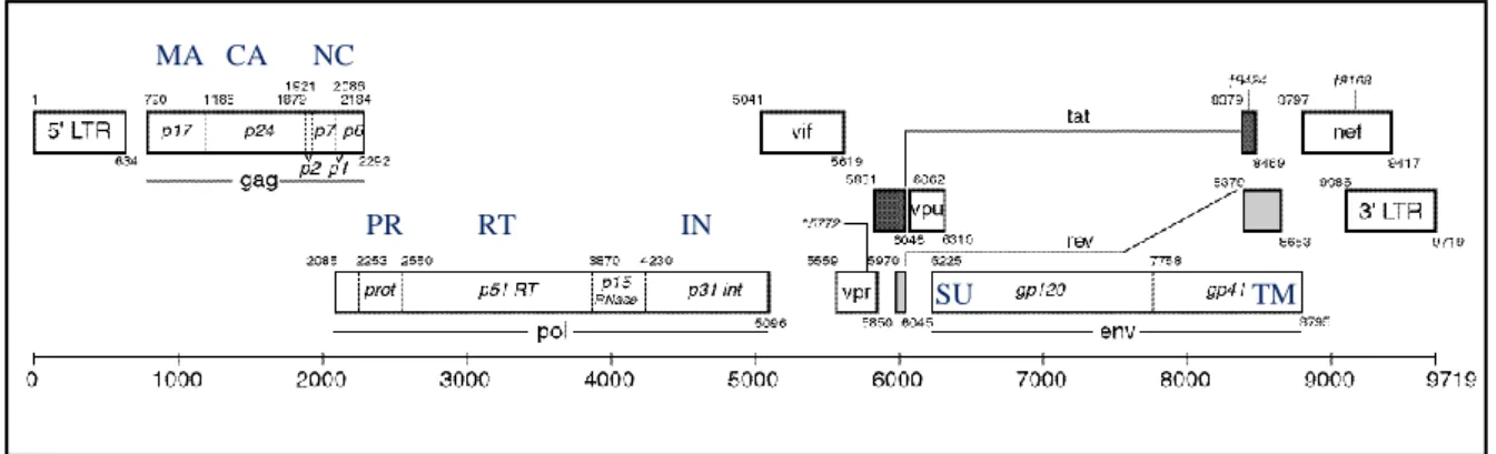
Each RNA molecule that constitutes HIV-1 genome consists of approximately 9800 nucleotides long and is flanked by long terminal repeats (LTR) at both ends (5´-3´) (Figure 3). HIV genome contains nine genes that encode major structural, regulatory and accessory proteins in the mature virion[75]. Structural proteins and viral enzymes are essential components for the retroviral particle whereas regulatory proteins modulate transcriptional and posttranscriptional steps of gene expression being also essential for
7
virus propagation. Although not necessary for viral propagation in tissue culture, accessory proteins play an important role in vivo[78].
Figure 3- Genomic organization of HIV-1. MA- matrix proteins); CA- capsid; NC-nucleocapsid proteins;
PR-protease; RT- reverse transcriptase; IN-integrase; SU- surface glycoprotein; TM-transmembrane glycoprotein (adapted from https://www.hiv.lanl.gov/content/sequence/HIV/MAP/landmark.html).
Genes gag, pol and env encode for structural or enzymatic proteins, tat and rev for regulatory proteins and finally nef, vif, vpr and vpu for accessory proteins[75, 79, 80]. The gag encodes the polyprotein precursor P55Gag that is posteriorly cleaved by the viral protease into the MA (p17), CA (p24), NC (p7), p6 proteins and two additionally spacer peptides (p1 and p2)[75]. The pol gene encodes the viral enzymes PR (p15), IN (p31) and RT (p66 and p51 subunits). These enzymes are produced as a Gag-Pol precursor polyprotein (Pr160GagPol) that is processed by PR. The env gene encodes for a polyprotein precursor Pr160Env which is processed by furin into the SU (gp120) and TM (gp41) glycoproteins [75, 81].
1.4 HIV-1 life cycle
The initial step of the HIV life cycle consists of viral entry into the host cells in order to initiate infection. Host cells (e.g. T-helper cells, monocytes, macrophages and dendritic cells) which expresses the CD4 (cluster of differentiation 4) glycoprotein receptor on its surface are the main target of HIV[78]. Binding of HIV Env (SU, gp120) to CD4 receptor present in the host cell surface induces conformational changes in the SU and a second receptor (co-receptor) belonging to the chemokine receptor family, CXCR4 and CCR5 becomes exposed [82, 83]. Binding of gp120 both to CD4 and co-receptor triggers new
MA CA NC
PR RT IN
8
conformational changes in TM glycoprotein that result in the insertion of gp41 fusion peptide into the host cellular membrane leading to the fusion of viral and cellular membranes and the release of the viral core into the cytoplasm[75, 84]. After uncoating of the virus in the cytoplasm, the viral RNA is reverse transcribed by the viral RT enzyme generating a linear double-stranded DNA molecule (dsDNA)[84]. Uncoating of the viral core leads to the establishment of the reverse transcriptase complexes (RTCs) and pre-integration complexes (PICs)[75, 78, 84]. PIC, which includes dsDNA, MA, NC, IN, RT and Vpr, is then conducted to the nucleus using the cytoplasmatic microtubules network and this process is mediated by IN and Vpr [75, 78]. Still outside the nucleus, IN binds to each end of the newly formed cDNA and removes 2 nucleotides at each 3´end of both DNA strands originating two recessive ends[75, 78]. Later in the nucleus, PIC-dsDNA is inserted into an open region of the host chromosomal genome. Finally, the unpaired regions between HIV and host dsDNA are repaired through cellular cofactors and a provirus is generated[78]. In productively infected cells, integrated provirus serves as a template for the transcription of both viral mRNA and genomic RNA. Transcription of proviral DNA is mediated by the promotor region within the 5´LTR. However, when 5´LTR is defective, 3´LTR activation can occur [85]. Successful transcription leads to the generation of HIV viral transcripts which are derived from a single full-length transcript by alternative splicing, generating messenger RNA (mRNA) with common 5´and 3´ends [86]. HIV-1 transcripts can further be grouped into three different classes: completely
spliced mRNA or early transcripts (encoding early regulatory proteins as Tat, Nef and
Rev), incompletely spliced mRNA or late transcripts (encoding Env, Vif, Vpr and Vpu) and unspliced and complete mRNA that encode for the polyprotein precursors Gag and Gag-Pol[75, 86]. Unspliced mRNAs are later incorporated in the viral particles as genomic RNA. To complete the expression of the later transcripts proteins from the early transcripts (Tat and Rev) are necessary. Tat binds to a secondary structure located in the R region of the 5’ LTR, named the trans-acting response (TAR) element resulting in an increased processivity of RNA polymerase. Rev is responsible for the transport of unspliced and incompletely spliced mRNA outside the nucleus to the cytoplasm to be translated. This process is mediated by the binding of Rev to the Rev response element (RRE), a 240 base region of complex RNA secondary structure [75, 87].
9
Figure 4- Life cycle of HIV-1 (adapted from www.niaid.nih.gov and [88])
The Env precursor polyproteins are glycosylated in the Golgi apparatus before they oligomerize in trimers. Then, polyproteins are cleaved into the SU and TM glycoproteins and conducted to the cytoplasmatic membrane in order to initiate assembly process. The assembling virion includes all of the components necessary for infectivity, i.e., two copies of viral RNA, cellular tRNA, molecules to prime cDNA synthesis, Env, Gag, PR, RT and IN. HIV-1 Gag and Gag-Pol precursor polyproteins mediate the virion assembly including the generation of spherical particles and genomic RNA packaging[80]. Immature viral particles bud from the cell by gemulation of the cytoplasmatic membrane and acquire the lipid envelope containing the TM/SU trimers. The last step of HIV-1 life cycle consists in the release of new HIV mature particles and cleavage of the Gag and Gag-Pol precursors into the functional proteins by PR [75, 80].
1.5 HIV-1 transmission
In 2015, about 2.1 million (1.8 million-2.4 million) new HIV cases were reported resulting in a total of 70 million infected people since the beginning of the HIV epidemic [89]. Although this represent a reduction in new infections (2.6 million new infections in 2009), the reduction has not been uniform across different regions and group risks. Thus, HIV infection continues to be a major global public health issue [90, 91].
10
HIV-1 transmission mainly results from sexual contact across mucosal surfaces, maternal-infant exposure (during pregnancy, delivery and breastfeeding) and percutaneous inoculation (through contaminated blood or blood products)[92] . Once a person is infected, HIV is present in semen, vaginal fluids, breast milk, blood and rectal secretions. Sexual contact with an infected person constitutes the most frequent mode of transmission of HIV and the majority of HIV transmissions worldwide occur through heterosexual contacts [93]. The risk of transmission of HIV-1 depends largely on the susceptibility of the uninfected host but also on the viral features. HIV viral load (i.e. amount of HIV in a body fluid) in the transmitting partner seems to affect the efficiency of the infection and varies according to the stage of disease [94]. In fact, HIV-1 transmission is likely to occur with more probability during the acute stage of HIV infection (earliest days) and in the latest stage of the disease (AIDS) where intense viral replication is observed [90, 94-96]. Moreover, taking antiretroviral therapy (ART) can reduce the risk of an HIV-infected person transmitting the infection to another by as much as 96%[97] . HIV transmission is also influenced by the presence of co-infection with other STDs (Sexual Transmitted Diseases) such as syphilis and herpes simplex-2 (HSV-2) [90, 98, 99]. Also, genital ulcers and inflammation can contribute to enhance HIV-1 sexual transmission with ulcerative STDs presenting an additional entry point for HIV [90, 99].
Despite the high diversity of HIV variants observed in infected patients, HIV transmission involves a limited number of variants (bottleneck effect) that are not necessarily the dominant variant in the donor [90, 100]. Thus, while chronically infected patients present a set of viral quasispecies that are genetically diverse, acutely infected patients frequently present a more “homogenous” set of viral variants that result from the transmission of one or few closely related viruses [90, 100, 101]. This is observed both in sexual and percutaneous routes of infection albeit at different levels [100, 101]. Transmission of multiple and more heterogeneous variants can be observed in injecting drug users (IDUs) probably due to the lack of a mucosal barrier and the associated protective cells (e.g. Langerhans cells) and innate immune response (e.g. production of IFNɣ by local macrophages) that play an important role in reducing HIV-1 transmission [101, 102]. Because the majority of HIV variants that are transmitted present a strong preference for CCR5 co-receptor, the capacity of HIV to establish an efficient infection also depends on the availability of target CD4 cells expressing CCR5 co-receptors [101, 103]. In fact,
11
the defective expression of CCR5 in humans (Δ32 mutation) is known to confer protection from HIV infection[104].
1.6 HIV-1 prevention
In the absence of a cure, the control of HIV/AIDS epidemic requires the implementation of several prevention measures. There are several options to reduce the risk of acquisition and transmission of HIV. This includes behavioral interventions (e.g. counseling and sex education of susceptible populations, use of condoms, needle and syringe programs) and biomedical interventions (e.g. male circumcision, HIV testing, antiretroviral drugs, pre and post-exposure prophylaxis[105].
Because globally only 54% of individuals infected with HIV-1 are aware of their status, HIV testing should be considered one of the first prevention methods. Awareness of HIV status is crucial to identify HIV infected individuals in order to establish the appropriate measures to get HIV treatment and also to prevent HIV transmission to others [92]. In fact, the WHO established with the 90-90-90 objectives that by 2020, 90% of all people living with HIV will know their status, 90% of all people with diagnosed HIV infection will receive sustained antiretroviral therapy and 90% of all people receiving ART will have viral suppression[106].
Besides their role in the improvement of health quality and survival rate in HIV-1 infected individuals, antiretroviral therapy is also crucial in preventing HIV transmission and infection. In fact, ART contributes crucially to lower viral load to undetectable levels in blood and genital secretions in HIV-1 infected individuals thereby reducing HIV transmission to negligible levels (< 1%) and can also be used to prevent infection very efficiently in uninfected (PreP, Pre-Exposure Prophylaxis) or recently infected individuals (PEP, Post-Exposure Prophylaxis) [97, 107-110]. Currently, oral PrEP consists of a combination of tenofovir disoproxil fumarate and emtricitabine and is able to reduce the risk of getting HIV by more than 90% if taken consistently everyday [111-114]. More recently, topical microbicides have been proposed to prevent HIV sexual transmission by directly inactivate or prevent HIV entry or replication in susceptible target cells present in the vagina and/or rectum. Topical microbicides can be delivered
12
over a prolonged period of time (e.g. intravaginal ring) or applied on a daily basis (e.g. gel)[115]. Recent results from the ASPIRE study conducted in Malawi, South Africa, Uganda and Zimbabwe demonstrated that the use of a vaginal ring continuously releasing dapivirine provided a modest degree of protection against HIV particularly in subgroups with evidence of increased adherence[116]. Currently, there is still no safe and effective microbicide available to the public.
Despite the important role of ART and PrEP in lowering and prevent HIV infection, in 2015 less than 50% of HIV infected adults and children were accessing treatment and 2.1 million people became newly infected with HIV[117]. Therefore, control and ultimately eradication of HIV will depend on the development of a safe and effective HIV vaccine accessible to all. Despite several years of investigation and important advances in vaccine field there is still no effective vaccine available for HIV. Currently, vaccine trial HVTN 702, based in the optimization of the RV144 trial[118], is being conducted among South African adults in order to test if an experimental vaccine regimen safely prevents HIV infection[119]. Vaccination including HIV vaccines will be explained in more detail in chapter 3.
Other biological interventions like male circumcision also contributes to lower HIV infection. In fact, this method reduces the risk of infection up to 60% by eliminating many cellular targets of HIV (e.g.Langerhans' cells) that are present in the penile foreskin [120, 121]. Nonetheless, behavioral interventions (e.g. sexual abstinence, reduced number of sexual partners, use of condoms, implementation of needle and syringe cleaning practices among IDUs) and better access to healthcare services are also important strategies to reduce the risk of HIV infection[105, 122]. Because no single intervention measure has been found to be completely effective it is believed that the combination of different prevention approaches that integrate behavioral and biomedical strategies will lead to an important reduction in new HIV infections [123].
1.7 HIV-1 pathogenesis
The first stage of HIV infection (acute phase or primary infection) is characterized by an intense viral replication that, in the absence of antiretroviral therapy, declines after three to four weeks after initial exposure (Figure 5) [103]. The first symptoms occur two to
13
four weeks after transmission and includes flu-like clinical manifestations, fatigue, myalgia, fever and lymphadenopathy[124]. During several months plasma viremia continues to gradually decline before reaching a steady state (viral set point)[103]. Viral set point constitute an important determinant on the rate of disease progression because it points out the beginning of the chronic stage of HIV infection[103].
CD4 T cells are the first targets of the virus. Along with the high replication of HIV in the mucosa, submucosa and draining lymphoreticular tissues there is a dramatic loss of CD4+T cells in the peripheral blood that is associated with the depletion of CCR5+ memory CD4+ T cells in the gut associated lymphoid tissue (GALT)[103, 125]. In fact, GALT harbors the majority of lymphoid tissue being an important site for viral replication and interactions with the host immune system during HIV infection[126]. This condition is followed by the dissemination of the virus to peripheral lymphoid tissue, particularly lymph nodes, and the establishment of persistent lymphoid tissue viral reservoirs[103]. The decline of CD4+ T cells is closely related with the direct effect of the viral infection of these cells together with the host cellular responses (e.g. host cytotoxic responses and natural killer cells)[127].
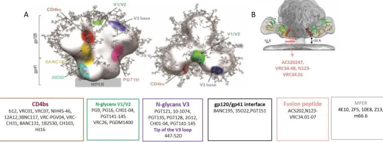
Seroconversion, with detection of HIV specific antibodies, occurs normally after three to 12 weeks after HIV exposure [103, 128]. The first antibodies detected are those directed against p24 antigen followed by antibodies directed to gp120 and gp41 (envelope)[128]. Approximately 12 weeks after transmission neutralizing antibodies (NAbs) start to arise and evolve but this response seems not to be enough to clear the virus. The first NAbs are directed against the autologous virus and are not able to neutralize more divergent virus (heterologous viruses) [128-132].
The chronic stage of HIV-1 infection (second stage) is asymptomatic (or latent) and lasts between eight to ten years (Figure 5). It is characterized by low but persistent levels of viral replication in the lymph nodes and constant antigen stimulation by the host immune system [103, 127, 133]. During this period, the immune system becomes activated by several factors that include viral proteins, microbial products that are translocated from the GALT and host responses. As cells are activated, they produce a set of proinflammatory cytokines which in addition with viral replication will ultimately lead to chronic immune activation[103]. This persistent immune activation is seen by an increased T cells turnover, monocytes and natural killer cells (NK), high levels of CD4
14
and CD8 T cell apoptosis and polyclonal B cell activation that can lead to hypergammaglobulinemia [103, 127, 134-136].
Figure 5- Natural course of untreated HIV-1 infection. Representation of the relation between HIV viral
load (red) and CD4 counts (blue) during the course of HIV infection (adapted from [137] and [138].
Together, these changes within each cell population greatly affect the overall immunologic competence leading to the exhaustion of the immune system. Moreover, in the absence of ART the majority of HIV infected patients become susceptible to the occurrence or reactivation of opportunistic infections (e.g. candidiasis, pneumonia, tuberculosis) as well as the development of virus induced tumors (e.g. Epstein-Barr virus related lymphomas, Kaposi´s sarcoma and cervical cancer caused by Human Papillomavirus [103, 134]). These symptoms define the early symptomatic stage of HIV chronic phase. In untreated patients, the progressive loss of CD4+ T cells can lead to an increased state of immunodeficiency that mark the onset of the last stage of HIV-1 infection: AIDS. According to Center for disease control and prevention (CDC), CD4 counts less than 200 cells/ul associated with opportunistic infections is a criterion that defines the AIDS stage [139]. Most untreated HIV-1 infected patients develop AIDS and eventually die [140]. A small minority (<5%) of individuals termed long-term nonprogressors remains healthy for several years in the absence of treatment [140, 141].
CD4 + ce ll count (c el ls/ m m 3 ) H IV RN A co p ie s p e r m l
Viral set point
15
2. HIV-1 envelope
2.1 Molecular and structural organization
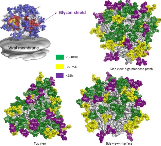
HIV-1 entry to host cells is mediated by interactions between the virus envelope (SU and TM glycoproteins) and the host cell receptors. The HIV-1 envelope glycoprotein (Env) is a trimer composed of three copies of non-covalently associated heterodimers of gp120 (SU) and gp41 (TM) that represent several spikes in the viral surface. These glycoproteins result from a gp160 precursor glycoprotein that is cleaved by cellular proteases and remain non-covalently associated on the cell or viral surface [142, 143]. Whereas gp120 interacts with cellular receptors and co-receptors, gp41 mediates fusion between viral and cellular membranes [142]. In this context, the main exposed surface of HIV-1 envelope is composed of gp120 while gp41 is mostly shielded. HIV-1 gp120 envelope include five conserved regions (C1-C5) and five variable regions (V1-V5) (Figure 6)[144]. Whereas the five conserved regions compose the structural core of gp120, the five variable regions are highly glycosylated and protect the core from neutralizing antibodies [143, 145]. Four of these hypervariable regions (V1-V4) tend to form loops through disulfide bonds that are exposed on the outer surface of the viral Env [146, 147].
The V1/V2 loop is involved in Tat binding and Tat-mediated viral entry, a previously unknown mechanism[148], whereas V3 loop plays an essencial role in coreceptor binding and viral entry[83]. In addition, both regions represent a target for antibodies, including neutralizing antibodies, when accessible on the surface of the virion [149, 150].
Figure 6- S linear representation of the surface and transmembrane envelope glycoproteins. Surface
glycoprotein (SU, gp120) includes five conserved (C1-C5) and five variable regions (V1-V5). Transmembrane glycoprotein (TM, gp41) contains the fusion peptide (FP), two heptad regions (HR1 and HR2), the membrane proximal external region (MPER), one transmembrane domain (TD) and a cytoplasmatic domain (adapted from [75] and [151]).
In its native trimeric conformation, SU has one inner and outer domain (Figure 7). The outer domain is highly glycosylated and is involved in the interaction between SU and
16
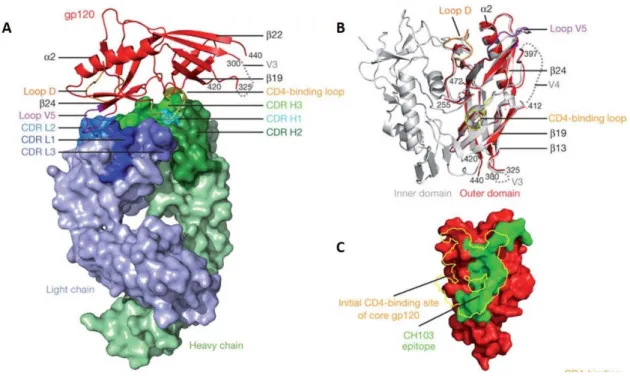
the cellular receptors and co-receptors. The outer domain is the region more exposed to the host immune responses and has most of the antigenic determinants in Env including, for example, the potent neutralizing antibody epitopes in V1/V2 and V3 loop [150, 152-155]. The inner domain is hydrophobic and essential for the association between SU and TM. Between the inner and outer domain there is another domain named bridging sheet (Figure 7). This bridging sheet is formed between V1/V2 stem and β sheets 20 and 21 of C4 as the result of conformational changes that occur after gp120-CD4 binding [75, 146, 156, 157]. After binding of the viral receptor and co-receptor by gp120 the fusion machinery in the gp41 subunit is readily activated.
HIV gp41 glycoprotein is divided in three major domains: one extracellular domain or ectodomain (512-683 in HXB2), one transmembrane domain (TD) that inserts in the host membrane cell (683-707 in HXB2) and one cytoplasmatic domain (708-856 in HXB2).
Figure 7- HIV-1 gp120 structure (adapted from [158, 159]). Monomeric (A) and trimeric (B) gp120
core with inner (black) and outer domain (red) and the bridging sheet (violet). Secondary structure of gp120 (C);
The extracellular domain, which mediates the major functions of TM, can be subdivided in five functional regions: a fusion peptide (FP, 512-534 in HXB2) followed by two α-helices containing leucine-zipper motifs designated heptad repeat 1 (HR1, 542-591 in HXB2) and heptad repeat 2 (HR2, 623-661 in HXB2), the loop region (593-622 in HXB2) that separate the HR1 and HR2 and finally the membrane proximal external region
17
(MPER, 662-683 in HXB2)[160]. Both fusion peptide and HR1/HR2 have a crucial role on the fusion of the virus to the host cell. The cytoplasmatic domain of TM binds to the matrix protein during the assembly of new viral particles [75, 160, 161].
2.2 HIV-1 entry and interaction between the Env protein and the cell
HIV-1 entry into host cells represents the first step in the viral infection cycle and is mediated by the Env glycoprotein. This process involves three major steps: 1) adhesion of the virus to the host cell and binding of the SU glycoprotein to the CD4 receptor, 2) binding of the SU glycoprotein to the cell co-receptor (CCR5 and/or CXCR4) and 3) fusion of the viral envelope with the cell membrane (Figure 8) [83].
Adhesion of the virus to the host cell brings Env glycoprotein into close proximity with the CD4 receptor. CD4 is a 60 kDa membrane glycoprotein belonging to the immunoglobulin superfamily and is expressed in functionally mature T cells, macrophages, dendritic cells, and monocytes [83, 162]. After binding to CD4 in the host cell, SU suffers major conformational changes that include rearrangements and exposure of V1/V2, V3 and C4 regions and formation of a bridging sheet as described previously. These conformational changes result in the approximation between the viral envelope and the cellular membranes which consequently leads to the interaction of the SU (namely V3 region) with the host cell co-receptors [83]. CCR5 and CXCR4 are considered the major HIV-1 co-receptors in vivo [75, 83, 163]. The chemokine receptor CCR5 is predominantly expressed on memory CD4+ T lymphocytes, activated T lymphocytes (mainly Th1 CD4+T cells) and macrophages whereas CXCR4 is mainly found on CD4+ and CD8+ T lymphocytes, monocytes, dendritic cells and B lymphocytes[104, 164, 165].
Viruses that infect preferentially macrophages typically use the CCR5 coreceptor (R5 viruses) mainly during the initial and asymptomatic stage of infection while viruses that infect mainly lymphocytic cell lines use the CXCR4 coreceptor (X4 viruses) preferentially later in the infection (AIDS stage)[104, 166].There are also viruses (the R5/X4 viruses) that are able to use indifferently the both co-receptors producing an effective infection [104, 165, 167, 168]. Moreover, R5 viruses seems to replicate more efficiently in CD4+ T cells compared with X4 viruses contributing to the R5 viral
![Figure 1- Global distribution and prevalence of HIV-1 (adapted from[64]) .](https://thumb-eu.123doks.com/thumbv2/123dok_br/19186404.947764/37.892.226.662.633.940/figure-global-distribution-prevalence-hiv-adapted.webp)
![Figure 4- Life cycle of HIV-1 (adapted from www.niaid.nih.gov and [88])](https://thumb-eu.123doks.com/thumbv2/123dok_br/19186404.947764/41.892.209.654.107.442/figure-life-cycle-hiv-adapted-www-niaid-nih.webp)
![Figure 5- Natural course of untreated HIV-1 infection. Representation of the relation between HIV viral load (red) and CD4 counts (blue) during the course of HIV infection (adapted from [137] and [138]](https://thumb-eu.123doks.com/thumbv2/123dok_br/19186404.947764/46.892.259.614.227.489/figure-natural-untreated-infection-representation-relation-infection-adapted.webp)


![Figure 12- Envelope features defense mechanism against antibody recognition (adapted from [298])](https://thumb-eu.123doks.com/thumbv2/123dok_br/19186404.947764/67.892.242.634.120.402/figure-envelope-features-defense-mechanism-antibody-recognition-adapted.webp)