Ana Carla David Pereira
Maio de 2012
Role of the medial Prefrontal Cortex
in nociception: functional characterization of
Prelimbic and Infralimbic nuclei
UMinho|20 12 Ana Carla Da vid P er eira R ole of t he medial Prefront al Cor te x in nocicep
tion: functional characterization of Prelimbic and Infralimbic nuclei
Trabalho realizado sob a orientação da
Professora Doutora Filipa Pinto-Ribeiro
e co-orientação do
Professor Doutor Armando Almeida
Ana Carla David Pereira
Maio de 2012
Dissertação de Mestrado
Mestrado em Ciências da Saúde
Role of the medial Prefrontal Cortex
in nociception: functional characterization of
Prelimbic and Infralimbic nuclei
iii
Agradecimentos
Gostaria de agradecer a todos aqueles que contribuíram para este trabalho e sem o qual não seria possível:
Ao Professor Armando Almeida, pela oportunidade que me ofereceu e por todo o apoio e orientação.
À Professora Filipa Pinto-Ribeiro, pelo apoio, orientação, paciência e confiança e por ser não só uma orientadora exemplar mas também uma amiga para a vida.
À minha mãe, pelo apoio incondicional e confiança inabalável.
À Diana, pela amizade, companheirismo e por estar sempre pronta a ajudar. À Bárbara, à Carina e à Joana, pelos momentos de conversa e alegria. À Carmo, por partilhar trabalho e histórias comigo.
Ao Professor Patrício Costa por toda a ajuda no tratamento estatístico. A todos os NeRDs.
v
The role of the medial Prefrontal Cortex in nociception: functional characterization of Prelimbic and Infralimbic areas in the rat
Abstract
Pain is a vital defense mechanism that triggers evasive, damage controlling reactions. However, when it becomes persistent, it loses its biological value and becomes a disease. Nociceptive modulation has been extensively studied in an attempt to increase the efficacy of pain management therapies and reduce the suffering of chronic pain patients. Multiple brain areas and circuits have been studied, but in the last decade brain imaging technologies have highlighted the importance of the limbic-cognitive component of pain and especially of the role of the prefrontal cortex (PFC) in this disorder. Imaging studies in chronic pain patients have shown dramatic changes in several areas of the PFC such as the dorsolateral PFC, the orbitomedial PFC and the anterior cingulate cortex (ACC). The ACC has been the most extensively studied when compared to the adjacent prelimbic (PrL) and infralimbic cortices (IL), two other components of the medial PFC (mPFC). In the present work, we proposed to clarify the role of these two PFC areas in pain modulation by using behavioural and electrophysiological techniques to (i) characterize the electrophysiological response of PrL and IL neurones to peripheral noxious and innocuous stimulation and to assess the influence of these areas upon (ii) animal nociceptive behaviour and (iii) the activity of rostral ventromedial medulla (RVM) pain modulatory cells, a major area involved in descending pain control.
Our results showed that both PrL and IL neurones responded to peripheral stimulation, although PrL cells are stimuli specific whereas IL cells responded to more than one modality of stimulation. Moreover, in the PrL, neurones responded primarily to noxious heat in detriment of other stimuli. Interestingly, IL neurones responsive to noxious mechanic stimulation showed a decrease in response intensity to noxious heat. The pharmacological activation/inhibition of the IL and PrL by glutamate and lidocaine, respectively, demonstrated that both the areas (i) participate in the modulation of peripheral noxious heat inputs, (ii) promote descending antinociception by mainly (iii) decreasing the activity of RVM pronociceptive ON-like cells.
In conclusion, we demonstrated that both the PrL and IL participate in the modulation of nociception and that their descending antinociceptive-drive inhibits the activity of RVM ON pronociceptive cells.
vii
O papel do Córtex Pré-frontal medial na nocicepção: caracterização funcional das áreas Prelímbica e Infralímbica no Rato
Resumo
A dor é um mecanismo essencial de defesa que induz comportamentos evasivos com vista à sobrevivência do organismo. No entanto, quando se torna persistente perde o seu valor biológico e torna-se uma doença. Muitas áreas e circuitos supraespinhais têm sido avaliados, sendo o sistema límbico um componente essencial do processamento emocional e cognitivo da dor. Dentro deste sistema, o papel do córtex pré-frontal (PFC) na dor é ainda pouco conhecido, tendo-se verificado em doentes com dor crónica alterações basais na actividade de áreas como o PFC dorsolateral, PFC orbitomedial e o córtex cingulado anterior (ACC). A maioria dos estudos que correlacionam dor e o PFC no Rato têm-se concentrado no ACC, havendo muito menos informação acerca dos córtices prélimbico (PrL) e infralimbico (IL), dois outros componentes do PFC medial (mPFC). O objectivo deste trabalho é clarificar qual o papel destas duas áreas do mPFC na modulação da dor, (i) caracterizando a resposta electrofisiológica de neurónios do PrL e do IL à estimulação periférica nóxica e inócua e verificando a influência destas áreas (ii) sobre o comportamento nociceptivo de animais e (iii) a actividade das células do bolbo rostral ventromedial (RVM), uma das principais áreas supraespinhais responsáveis pelo controlo de dor. Os nossos resultados a presença de neurónios no PrL e no IL que respondem a estimulação periférica, mas com diferentes caracteristicas funcionais: os neurónios do PrL respondem exclusivamente a cada um dos estímulos aplicados enquanto as células do IL respondem a mais do que uma modalidade de estimulação. Verificamos também que os neurónios do IL que respondem a estímulos mecânicos nóxicos têm uma resposta menos intensa a estímulos de calor nóxicos. Através da activação/inibição farmacológica do PrL e do IL demonstramos também a sua participação na modulação descendente de estímulos de calor nóxico, com ambas as áreas a promoverem efeitos antinociceptivos. Mais ainda, a acção modulatória descendente das áreas PrL e IL é, pelo menos parcialmente, mediada por neurónios pronociceptivos do RVM do tipo ON.
Em conclusão, com este trabalho mostramos que as áreas do PFC em estudo, o IL e o PrL, estão implicados no processamento nociceptivo, verificando-se a existência de uma acção descendente principalmente inibitória sobre as células ON pronociceptivas do RVM.
ix
Index
1. Chapter 1: Introduction ... 1
1.1. Nociceptive transmission ... 3
1.2. Central processing ... 4
1.3. The Prefrontal Cortex and pain ... 6
1.3.1. The Prelimbic cortex ... 8
1.3.2. The Infralimbic cortex ... 8
1.4. Descending pain control ... 9
2. Chapter 2: Objectives ... 13
3. Chapter 3: Materials and methods ... 17
3.1. Animals and ethical issues ... 19
3.2. Anaesthesia and euthanasia ... 19
3.3. Procedures for intracerebral microinjections ... 20
3.4. Behavioural assessment of nociception ... 21
3.5. Drugs ... 21
3.6. Course of the behavioural study ... 22
3.7. Course of the electrophysiological study ... 22
3.7.1. Electrophysiological characterization of the PrL and IL neurones receiving nociceptive input ... 23
3.7.2. Modulation of the activity of RVM pain modulatory cells by the PrL and IL cortices ... 23
3.8. Statistics ... 24
4. Chapter 4: Results ... 27
4.1. Histological confirmation of the injection and recording sites ... 29
4.2. Influence of the anaesthesia method upon the total number of PrL and IL neurones receiving sensory inputs ... 30
4.3. Electrophysiological activity of medial PFC neurones ... 32
4.3.1. Characterization of the neuronal population in the PrL ... 32
x
4.4. Effect of the pharmacological activation and inhibition of the mPFC upon nociceptive behaviour in the rat and its correlation with the activity of RVM pain modulatory cells during
peripheral innocuous and noxious stimulation ... 37
4.4.1. Nociceptive behaviour ... 37
4.4.1.1. Role of the PrL upon acute nociceptive behaviour ... 37
4.4.1.2. Role of the IL upon acute nociceptive behaviour ... 38
4.4.2. Modulation of the activity of RVM pain modulatory cells neurones modulation by the medial PFC ... 39
4.4.2.1. Role of the activation and inhibition of PrL upon the activity of RVM pain modulatory cells during peripheral innocuous and noxious stimulation ... 40
4.4.2.2. Role of the activation and inhibition of IL upon the activity of RVM pain modulatory cells during peripheral innocuous and noxious stimulation ... 42
5. Chapter 5: Discussion ... 45
5.1. Scope and limitations of the experimental design ... 48
5.1.1. Animal model ... 48
5.1.2. Anaesthesia ... 49
5.1.3. Acute noxious stimulation ... 50
5.1.4 Single-cell extracellular recordings ... 51
5.2. Characterization of medial PFC neurones ... 51
5.3.1. Influence of the PrL upon nociceptive behaviour and RVM cell activity ... 52
5.3.2. Influence of IL over nociceptive behaviour and RVM cell activity ... 53
5.4. Classification of supraspinal neurones as NS-like vs WDR-like cells ... 55
5.5. Possible pathways mediating the descending modulatory effect of the PrL and IL... 55
6. Chapter 6: Conclusions and future perspectives ... 59
xi
Abbreviations ACC – anterior cingulate cortex
ANOVA – analysis of variance ANOVA1W – ANOVA one-way ANOVA2W – ANOVA two-way ANOVARM – repeated measures dlPFC – dorsolateral prefrontal cortex DRt – Dorsal reticular nucleus i.p. – intraperitoneal injection IL – infralimbic cortex
IMS – innocuous mechanical stimulation mGluR(s) – metabotropic glutamate receptor(s) mPFC – medial prefrontal cortex
NHS – noxious heat stimulation NMS – noxious mechanical stimulation NON-N – non-nociceptive neurones NR – non-responsive neurones NS – nociceptive specific neurones PAG – periaquedutal grey matter PFC – prefrontal cortex
PrL – prelimbic cortex
PWL – paw-withdrawal latency R – responsive neurones
RVM – rostral ventromedial medulla SEM – standard error of the mean WDR – wide-dynamic range neurones
1
Chapter 1: Introduction
3
1. Introduction Pain is an experience that is difficult to quantify as it is biased by the individual’s cognitive, emotional and social background. In an attempt to incorporate all the components that modulate pain perception, the International Association for the study of Pain (IASP) has defined pain as “an unpleasant sensory and emotional experience associated with actual or potential tissue damage, or described in terms of such damage” (Merksey and Bogduk 1994; Loeser and Treede 2008). In terms of preclinical research it is also important to emphasize that nociception, however, differs from pain as it comprises only “the neural processes of encoding and processing noxious stimuli” (Loeser and Treede 2008).
1.1. Nociceptive transmission
Upon noxious peripheral stimulation, the nociceptors (sensory fibres - primary afferents) are activated and the nociceptive input is transduced and transmitted to neurones in the superficial dorsal horn of the spinal cord (D'Mello and Dickenson 2008). Cutaneous primary afferents comprise three types of fibres (i) Aβ-fibres [large (4–8 µm in diameter), myelinated, fast conduction of action potencial transmission (24–48 ms-1)], (ii) Aδ-fibres [medium (2 – 6 µm in diameter), myelinated, with intermediate velocity (12–30 ms-1)] and (ii) C-fibres [thin (0.4–1.2 µm in diameter) unmyelinated and slow-conducting (0.5–2.0 ms-1)] that respond to mechanical, thermal and/or chemical noxious and innocuous stimulation and whose level of activation reflect the stimulus intensity (Julius and Basbaum 2001).
More specifically, nociceptors include only (i) Aδ-fibres that respond mainly to a single type of stimulation and whose activity is responsible for the “first pain” which evokes protective reflexes, and (ii) C-fibres that are mainly polymodal and are responsible for the more prolonged “second pain” (Craig 2003).
4
In the spinal cord, nociceptors synapse in neurones (second order neurones) of laminae I and II of the superficial dorsal horn (D'Mello and Dickenson 2008). These spinal neurones can be classified as non-nociceptive (NON-N), nociceptive-specific (NS) and wide-dynamic range (WDR) neurones. NS neurones are distributed more superficially and receive inputs exclusively from Aδ and C-fibres whereas WDR neurones additionally also receive inputs from Aβ-fibres (touch – innocuous mechanical stimuli) (D'Mello and Dickenson 2008).
1.2. Central processing
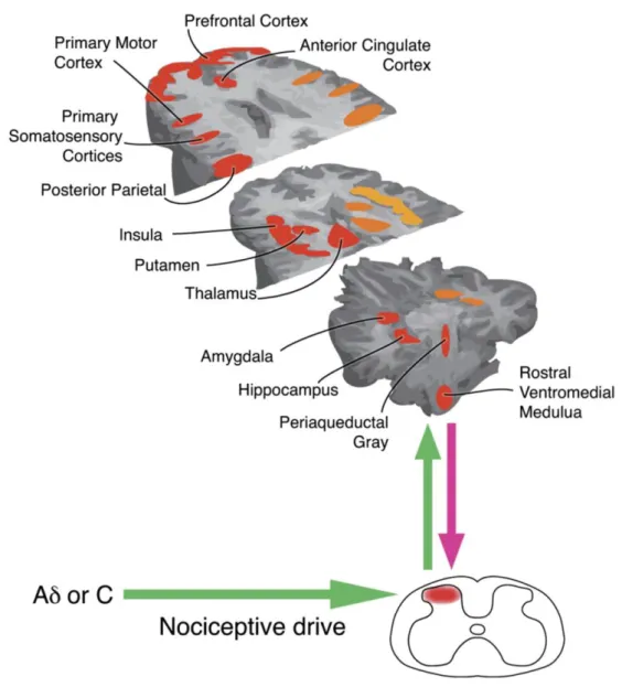
Nociceptive information is forwarded to the brain by spinal projection neurones along spinofugal pathways, the anterior lateral and posterior tracts. The anterior lateral system is the most important for somatic pain and comprises mainly five ascending tracts: the spinothalamic, spinoreticular, spinomesencephalic, spinohypothalamic and spinoreticular-thalamic tracts. These pathways distribute nociceptive inputs mainly to the brainstem nuclei (autonomic responses to pain) and to higher-level circuits (processing of the sensory, emotional and cognitive components of pain) that together constitute the pain matrix (Melzack 1999) (Fig. 1).
Third order thalamocortical fibres that receive inputs from the medial and lateral subdivisions of the spinothalamic tract project to somatosensory I and II, the posterior insula and medial prefrontal cortices (Loewy 1990). The spinoreticular tract transmits sensory information to the brainstem reticular formation, namely to the dorsal and caudal medullary reticular formation and to the medial and intralaminar nuclei of the thalamus that forward it bilaterally to the prefrontal cortex (Willis and Westlund 1997). The spinomesencephalic tract targets brainstem nuclei such as the periaqueductal gray matter (PAG) and locus coeruleus but also more caudal areas like the dorsal reticular nucleus (DRt) in the medulla (Willis and Westlund 1997). The spino-hypothalamic tract targets the amygdala, the medial thalamus, hypothalamus and other areas within the limbic system (Willis and Westlund 1997).
The posterior system comprises three ascending tracts, the first order dorsal column neurones, the post-synaptic dorsal column pathway and the spinocervical tract and has been demonstrated to convey visceral nociceptive information (Al-Chaer et al. 1999). Although there are extensive bidirectional projections between the areas within the pain matrix, the lateral circuits are mainly
5
dedicated to the sensory-discriminative component of pain while the medial circuits are mostly involved in the cognitive and emotional components of pain (Tracey and Mantyh 2007).
Figure 1 – Representation of the main brain regions that are activated during the application of a noxious stimuli (areas in orange represent areas that during the activation of nociceptors are activated bilaterally in the brain, areas in red represent areas activated contralaterally to the site of noxious stimulation and areas in yellow represent areas that activated ipsilaterally) (Tracey and Mantyh, 2007).
The sensory-discriminative component of pain enables the identification of the location, duration and intensity of the stimulus according to a somatotopically organized map in the SI cortex (contralateral), the SII cortices (bilateral), and the posterior insula (bilateral) (Treede et al. 1999; Craig 2003). The activity in SI region is correlated to the intensity of noxious stimuli (Timmermann et al. 2001), the SII participates in the learning and memory of pain in addition to
6
pain–motor integration (Schnitzler and Ploner 2000) and the posterior insula in the quality of the pain perception (e.g. burning) (Ostrowsky et al. 2002; Brooks et al. 2002).
The cognitive and emotional components of pain deal with the unpleasantness of the stimulus which in association with pain memories and mood results in pain expression. Imaging studies have contributed greatly to the understanding of the dynamic mechanisms underlying pain modulation as it enabled us to visualize how a painful stimulus extensively alters the neuronal activity of circuits mediating emotions. These studies have demonstrated that the parallel activation of several nuclei within the limbic system (Bushnell and Apkarian 2006) is at the base of the complex emotional responses to pain.
1.3. The Prefrontal Cortex and pain
The use of human imaging studies has fostered the interest on the role of the prefrontal cortex (PFC) in pain modulation. While studies on placebo analgesia have shown that PFC activation is directly correlated not only to a decrease in the activity of the thalamus, the posterior insula and the anterior cingulate cortex (ACC) but also to an increase of periaqueductal gray matter (PAG)-mediated spinal opioid release (Wager et al. 2007; Petrovic et al. 2002; Wager et al. 2003). In chronic pain disorders this area is strongly involved in catastrophizing (Seminowicz and Davis, 2006). The modalities of noxious peripheral stimuli known to activate areas within the medial PFC (mPFC) include electric intra-epidermal (Tanaka et al. 2008), chemical (Porro et al. 2002), mechanical (Zhang et al. 2004), and thermal (Matre et al. 2010; Tran et al. 2010) stimulation. In humans, each subregion of the PFC plays a role in acute pain, with the mPFC and the ACC involved in unpleasantness of pain (Rainville et al. 1997) and anticipation (Eisenberger and Lieberman 2004), the ACC alone mediating the affective component of pain (Phan et al. 2002) and the regulation of the autonomic responses to pain (Craig 2003a), the ACC and the dorsolateral prefrontal cortex (dlPFC) mediating the detection of conflicting information (Medalla and Barbas 2009) and the insular cortex also implicated in autonomic regulation (Augustine 1996; Verbene and Owens 1998) and intensity coding (Craig et al. 2000, Brooks et al. 2005).
7
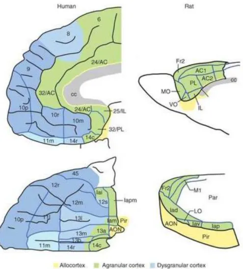
It is important to note that while rodents and humans both have frontal cortices, common homologous areas are restricted to the agranular part (Wise 2008). Hence, the absence of granular areas in the PFC of rodents limits the establishment of homologies between these areas in the rat and the primate dorsolateral prefrontal cortex (Ongür and Price 2000). Consequently, only the rodent infralimbic (IL), prelimbic (PrL), agranular insular and orbital and ACC areas have been attributed homologue areas in primate brains (Wise 2008) (Fig. 2).
Figure 2 – Schematic representation of the citoarchitectonics of the prefrontal cortices in humans and in the rat. Homologous areas between the rat and man are coloured in green and yellow while blue areas correspond to areas existing in humans alone. (AC, anterior cingulate area; AON, anterior olfactory nucleus; c, caudal; cc, corpus callosum; Fr2, second frontal area; I, insula; i, inferior; Ia, agranular infralimbic cortex; IL, infralimbic cortex; l, lateral; LO, lateral orbital area; m, medial; M1, primary motor area; MO, medial orbital area; o, orbital; p, posterior; Par, parietal cortex; Pir, Piriform cortex; PL, prelimbic cortex; r, rostral; s, sulcal; v, ventral; VO, ventral orbital area. Numbers indicate cortical fields, except that after certain areas, such as Fr2 and AC1, they indicate subdivisions of cortical fields) (Wallis 2011)
8
In acute conditions, the chemical/electrical stimulation of the ACC facilitates nociceptive responses to heat (Calejesan et al. 2000). In addition, in pathological pain conditions, such as inflammatory pain, an upregulation of NMDA receptors is observed in the ACC (Wu et al. 2005) and in neuropathic pain this area undergoes profound morphological or functional changes (Metz et al. 2009). In an interesting anatomical study by Jasmim and colleagues (2004) puts forward a possible role for the agranular insular cortex in multiple aspects of pain behaviour owing to its efferent projections to the medial thalamic nuclei (motivational/affective), the mesolimbic/mesocortical ventral forebrain networks (sensorimotor) and the brainstem (descending pain).
More recently and due to its anatomical and functional association with the limbic system, the PrL and the IL have also been implicated in emotional and cognitive functions (Vertes 2004), but the literature available is scarce.
1.3.1. The Prelimbic cortex
Anatomical studies show that efferent projections from the PrL target dorsally the ACC, and perirhinal area; laterally, the dorsomedial and ventral striatum, the amygdala, lateral hypothalamus, several thalamic nuclei, and, ventrally, the PAG, ventral tegmentum, and raphe nuclei (Sesack et al. 1989). Studies involving the lesion of the PrL demonstrated that this nucleus is necessary for the extinction of conditioned fear, enhances anxiety-related behaviours, participates in working memory processes and in the ability to learn from cues. In terms of pain, the PrL responds to visceral stimuli in both humans and rats, but only in females (Wang et al. 2009), and to mechanical innocuous and noxious stimulation in the tail (Ji et al 2010; Nakamura et al. 2010)
1.3.2. The Infralimbic cortex
Axonal projections from the IL ascend dorsally to innervate the PrL and ACC; laterally to the insular cortex, the perirhinal cortex, and the piriform cortex, and ventrally to the hypothalamus, the amygdala, the bed nucleus of the stria terminalis, the brainstem (mainly to the PAG,
9
parabrachial nucleus, nucleus of the solitary tract and ventrolateral medulla) and to the superficial dorsal horn of the spinal cord (Hurley et al. 1991; Floyd et al. 2001).
The right IL appears to be specifically related to anxiety or aversion behaviours (Jinks and McGregor 1997) and to behavioural flexibility (Delatour and Gisquet-Verrier 2000). Wang and colleagues (2009) verified that in male rodents, similarly to what is observed for humans, the IL responds to noxious visceral stimulation.
In light of the little literature available for the PrL and the IL in pain processing, we believe that studies focusing on their functional characterization in pain modulation are strongly needed. In fact, very little is known about the nociceptive processing of noxious-evoked activation of nociceptive neurones in these areas.
1.4. Descending pain control
Descending pain control results from a dynamic balance between the activation and inhibition of many areas that are part of the pain matrix (Fields and Basbaum 1999; Pertovaara and Almeida 2006), yet its mechanisms are not fully understood. Literature shows that a number of brain regions such as the diencephalon, hypothalamus, amygdala, ACC, insular, and prefrontal cortices are key players (Tracey and Mantyh 2007) in inhibiting or facilitating nociception through the brainstem (Vanegas and Schaible 2004; Almeida et al. 2006).
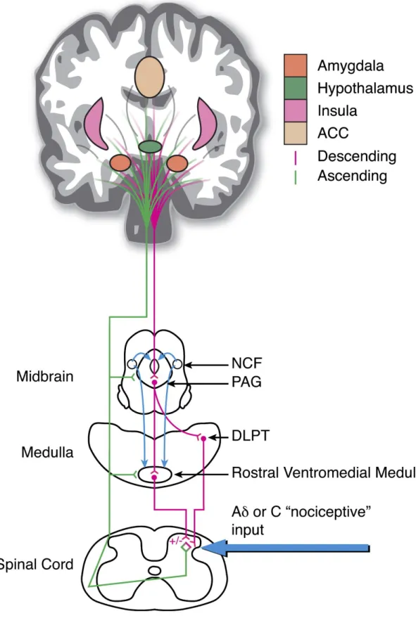
Several interconnected networks are described to modulate pain at different levels, the frontal-cortical-limbic-brainstem circuit (Mayer et al. 2005; Price 2000), the PAG-rostral ventromedial medulla (RVM) circuit (Fields et al. 1995; Heinricher et al. 2009), the spino-bulbo-spinal loop through the dorsal reticular nucleus (DRt) (Lima and Almeida 2002) and interneurones in the spinal cord. Considering the PFC, it has been shown in the rat that the nociceptive facilitating action of the ACC is mediated through a medullary relay in the DRt (Zhang et al. 2005), but again nothing has however been published about the participation of the PrL and IL in pain modulation.
10
Figure 3 – Schematic representation of the areas involved in top-down pain modulation (NCF - nucleus cuneiformis; PAG - periaqueductal gray matter; DLPT - dorsolateral pontine tegmentum; ACC - anterior cingulated cortex; +/− indicates both pro- and anti- nociceptive influences, respectively; green line – ascending tracts; lilac line – descending tracts). (Tracey and Mantyh 2007)
11
Of the areas mentioned above, the RVM is of particular interest as it is considered the effector nucleus of supraspinal descending pain modulation. It has been shown that descending inhibition or facilitation results from a fine balance between the activation of its ON- and OFF-cells (Heinricher et al. 2009). ON-cells are associated to the increase of pain perception (pronociceptive) as their firing activity increases immediately before there is a behavioural withdrawal response to a noxious stimulus applied to the periphery, whereas OFF-cells are considered to inhibit pain perception (antinociceptive) as their activity decreases immediately before a behavioural response to noxious peripheral stimulation is observed (Fields and Heinricher 1985; Gebhart 2004; Heinricher and Neubert 2004; Ren and Dubner 2002). Several anatomical studies further support this theory as it has been clearly demonstrated that these cells share reciprocal projections with neurones in the laminae I and II of the superficial dorsal horn (Urban and Gebhart 1999). Interestingly, cells with identical functional profiles have also been described for the PAG (Heinricher et al. 1987) and the caudal ventrolateral medulla (CVLM) (Pinto-Ribeiro et al. 2011; Heinricher et al. 2009).
In the spinal cord, pain inhibition is achieved either by direct inhibition of projecting neurones, desinhibition of relaying inhibitory interneurones (Melzack and Wall 1965; Todd and Koerber 2006) or by inhibition of primary afferents (Pertovaara and Almeida, 2006), while pain facilitation involves the activation of projecting neurones, excitatory interneurones (D'Mello and Dickenson 2008) and primary afferents (Le Bars 2002; Millan 2002) that modulate the activity of NS and WDR neurones (Millan 2002).
13
Chapter 2: Objectives
15
2. Objectives Human imaging and some animal studies indicate that the prefrontal cortex (PFC) is involved in pain modulation. However, most studies have focused on the anterior cingulate cortex (ACC) and our knowledge on the contribution of other PFC areas towards endogenous pain control if far from complete. Axonal projections arising from the prelimbic (PrL) and infralimbic cortices (IL) and targeting the periaqueductal gray matter (PAG) and the rostral ventromedial medulla (RVM) support the hypothesis that these areas directly participate in pain modulation. Further evidence comes from works demonstrating the remodelling of these prefrontal areas in pain pathological conditions (Metz et al. 2009; Devoize et al. 2011).
Taking the above mentioned into account, this work was aimed at evaluating the involvement of the PrL and IL subnuclei of the PFC in the processing of different types of stimulation through the use of behavioural and electrophysiological approaches to address three specific goals:
1. To characterize in rodents how PrL and IL cells respond to different modalities of acute peripheral noxious stimulation;
2. To investigate the phasic and tonic role of the PrL and IL in pain processing by evaluating the effect of pharmacological activation (glutamate) and inhibition (lidocaine) of these areas upon the behavioural nociceptive responses in a rodent model of acute pain; 3. To further extend the results of Aims 1 and 2 by analysing the functional connectivity
between the prefrontal cortex and the RVM, an area mediating descending pain modulation. Based on the previous results, this analysis was used to assess whether the PFC modulates the activity pattern of RVM neurones and whether these changes can be correlated to the behavioural data described in Aim 2.
17
Chapter 3: Materials and methods
19
3. Materials and methods 3.1. Animals and ethical issues
The experiments were performed in fifty two adult male Wistar han rats weighting between 250-350g (Charles Rivers, Barcelona, Spain). The experimental protocols were approved by the Institutional Ethical Commission and followed the Directive 2010/63/EU of the European Parliament and of the Council concerning the use of animals for scientific purposes. All efforts were made to minimize animal suffering and to use only the number of animals necessary to produce reliable scientific data.
3.2. Anaesthesia and euthanasia
During intracerebral cannulae implantation, anaesthesia was induced through the intraperitoneal (i.p.) administration of a mixture of ketamine (0.75 mg/kg, i.p.; Imalgene, Merial Lyon, France), a NMDA receptor agonist, and medetomidine (0.5 mg/kg, i.p.; Dorbene, Esteve Veterinaria, Léon, Espanha), an α2-adrenergic agonist. After the surgical procedures, the anaesthesia was reverted with atipamezole hydrochloride (1mg/kg, i.p.; Antisedan, Orion Pharma, Orion Corporation, Espoo, Finland), a synthetic α2-adrenergic antagonist, and the animals were monitored until they were fully recovered.
In order to assess the best anaesthesia to perform recordings in the medial prefrontal cortex (mPFC) during peripheral stimulation, a pilot study involving the comparison of the effect of urethane and pentobarbitone anaesthesia upon the total number of PrL and IL cells recorded was performed. After the animals (n=10) were anaesthetised either through the injection of a urethane solution (n=7) (1.5-1.8 g/kg i.p.; Sigma-Aldrich, Lisbon, Portugal) or a pentobarbitone solution (n=3) (Eutasil, CEVA, Saúde Animal, Algés, Portugal), a total of 10 systematic dorso-ventral recordings were done in each animal. Cells were classified into different categories
20
according to their response to innocuous mechanical (IMS), noxious mechanical (NMS) and noxious heat stimulation (NHS).
During the remaining electrophysiological sessions, anaesthesia was induced through the injection of a urethane solution (1.5-1.8 g/kg i.p.; Sigma-Aldrich). A warming blanket was used to maintain the body temperature within physiological range.
After the completion of either the behavioural tasks or the electrophysiological sessions, animals received a lethal dose of pentobarbitone (80 mg/kg, i.p.; Eutasil, CEVA) and the brains were removed and fixed in a 4% paraformaldehyde solution. Afterwards, brains were sliced (50µm sections) using a vibratome, mounted on a slide, counterstained, dehydrated, covered in
mounting media (Entellan New, Merck, Darmstadt, Germany) and cover slipped for histological confirmation of cannula placement or electrode recording site.
3.3. Procedures for intracerebral microinjections
For intracerebral drug administration, cannulae were implanted as described by Pinto-Ribeiro and colleagues (2008). Briefly, the rats were placed in a stereotaxic frame, a longitudinal incision was made in the scalp, which was retracted as well as the subcutaneous fascia, and a sterilized stainless-steel guide cannula (26 gauge; Plastics One, Roanoke, Virginia, USA) was implanted in the brain. The tip of the guide cannula was positioned 1mm above the desired injection site in the PFC (PrL: RC: +2,76mm; LM: 0.6mm; DV: 3.5mm; IL: RC: +2,76mm; LM: 0.6mm; DV: -4.9mm; RC – rostro-caudal to the bregma; LM – latero-medial to the sagital suture; DV – dorso-ventral to the brain surface), according to the coordinates of the atlas by Paxinos and Watson (2007). The guide cannula was fixed to the skull with two screws and dental acrylic cement and the skin sutured around it. A dummy cannula (Plastics One) was inserted into the guide cannula to prevent contamination and the animals were allowed to recover from the surgery for at least one week.
Test drugs were administered either in the PrL or the IL cortices through a 33-gauge injection cannula (Plastics One) protruding 1 mm beyond the tip of the guide cannula. The microinjection was performed using a 5.0 µL Hamilton syringe connected to the injection cannula by a
21
polyethylene catheter (PE-10; Plastics One). The injection volume was 0.5 µL and therefore, the spread of the injected drugs within the brain was expected to have a diameter of 1 mm (Myers 1966). The efficacy of injection was monitored by watching the movement of a small air bubble through the tubing. The injection lasted at least 20 seconds and the injection cannula left in place for additional 30 seconds to minimize the return of drug solution back to the injection cannula. 3.4. Behavioural assessment of nociception
Prior to performing the behavioural tests, rats were habituated to the experimental conditions (i) by allowing them to spend 1 – 2 hours daily in the testing room during the week preceding any testing, and (ii) by performing daily handling sessions. For assessing nociception in unanaesthetized animals, the latency of hindpaw withdrawal following radiant heat stimulation (Hargreaves test; Plantar Test Device Model 37370, Ugo Basile, Comerio, Italy) was determined. In each behavioural session, the withdrawal latency was assessed prior to drug administration and at various intervals following the intracerebral injections (Fig. 4). At each time point, the measurements were repeated twice at an interval of 1 minute (except for glutamate due to its fast effect) and the mean of these values was used in further calculations. The cut-off time for radiant-heat exposure was set at 15 seconds in order to avoid any damage to the skin.
3.5. Drugs
Glutamate (Merck, Darmstadt, Germany) solution for intracerebral drug injection in the PFC was prepared with sterilized saline solution 0.9% (Unither, Amiens, France; pH 7,2). Lidocaine was acquired as a solution (B. Braun Medical, Barcarena, Portugal). Each injection had the volume of 0.5 µL and contained 50 nmol of glutamate or lidocaine 2%. The doses were chosen according to previous studies (Pinto-Ribeito et al., 2008; Ansah et al., 2009). Control injections with saline solution were performed as control values, in order to avoid any bias that might result from injecting the solution itself.
22
3.6. Course of the behavioural study
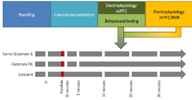
At least one week after the insertion of the guide cannula in the PFC, the phasic and tonic action of PrL and IL upon nociceptive behaviour was determined in un-anaesthetized animals through the assessment of changes in paw-withdrawal latency (PWL) after the injection of glutamate or lidocaine, respectively. Withdrawal latencies were assessed 30 seconds, 5 , 10 , 20 and 30 minutes following the intracerebral injections (Fig. 4). The interval between behavioral assessments of different drugs was of at least two days. The order for testing each different drug varied amongst animals.
Figure 4 – Schematic representation of the experimental design. Rats used to evaluate the action of PrL and IL upon descending pain modulation were accustomed to the room for 5 days, after which animals were implanted with a cannula in the area of study and allowed to recover for a week. Animals included in the behavioural study were trained in the behavioural apparatus for one week, while animals destined to electrophysiological studies were immediately analysed. Pharmacological tests were performed at the same time-points for both the behavioural and the electrophysiological studies and adjusted to the time of action of each drug. (PI – pre-injection;IL – Infralimbic cortex; PrL – Prelimbic cortex; mPFC – medial prefrontal cortex; RVM – rostral ventromedial medulla).
3.7. Course of the electrophysiological study
In order to assess the involvement of the PrL and the IL in nociceptive processing, two electrophysiological approaches were used: (i) the systematic recording of PrL and IL neurones with further characterization of cells which responded to peripheral stimulation and assess responses to various modalities of peripheral innocuous and noxious stimulation and (ii) the
23
evaluation of changes in the activity of RVM pain modulatory ON- and OFF-like cells (spontaneous and innocuous- and noxious-evoked activity) during the pharmacological activation and inhibition of the PrL or IL cortices.
3.7.1. Electrophysiological characterization of PrL and IL neurones receiving nociceptive input In urethane anaesthetized animals, the skull was exposed as described in Section 3.3. to allow the placement of a recording electrode in the PrL (RC: +2,76mm; LM: -0.6mm; DV: -3.5mm) and in the IL (RC: +2,76mm; LM: -0.6mm; DV: -4.9mm). During PFC recordings, the response properties of neurones were assessed by determining its spontaneous activity and its response to (i) innocuous brushing of the back (IMS), (ii) noxious heating of the tail (NHS) and (iii) noxious tail pinching (NMS). The search and recording of neurones was performed dorso-ventrally in the PrL and IL.
3.7.2. Modulation of the activity of RVM pain modulatory cells by the PrL and IL cortices
Under urethane anaesthesia, a guide cannula was placed 1 mm above the desired injection site either in the PrL or the IL and a recording electrode was placed in the RVM (RC: -10.92mm; LM: 0mm; DV: -10.4mm). The response properties of RVM neurones were assessed as described for the PFC neurones, by assessing the spontaneous, innocuous and noxious-evoked activity of RVM neurones (Fig. 5). Accordingly, these parameters were reassessed after the microinjection of glutamate and lidocaine into the PrL or the IL. If necessary, the search for another recording site started about 30 min after the previous recording and only if no neuronal activity could be recorded meanwhile.
24
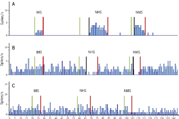
Figure 5 - Example of an output from software Spike 2. A – Example of a recording of a ON-WDR-like neurone discharge frequency. This cell is activated not only by innocuous peripheral stimulation (IMS) but immediately before the withdrawal reflex this cell increases its firing rate and return to basal levels after stimulation is over; B – Example of a recording of an OFF-NS-like neurone discharge frequency. Although no response was observed during the innocuous stimulation, immediately before the withdrawal reflex this cell decreases its firing rate, which only return to normal activity after stimulus is over; C – Example of a recording of a neutral-cell discharge frequency. This cell does not display any changes in its firing rate during either innocuous or noxious stimulation. Green lines – beginning of stimulation; black lines – beginning of the response; red lines – end of stimulation (IMS – innocuous mechanical stimulation; NHS – noxious heat stimulation; NMS – noxious mechanical stimulation).
3.8. Statistics
Using the IBM SPSS Statistics 19 and GraphPad Prism 5 software, K-means test was applied in PFC neurone division by clusters according to response properties to peripheral stimulation (Tables 1 and 2, Fig. 10 and 12). Z-values represent how many standard deviations a value is above or below the mean of the population and was used to detect outliers in neuronal populations. Two-way analysis of variance (ANOVA) followed by t-test with a Bonferroni correction for multiple comparisons was used to compare cell number in pentobarbitone and urethane anaesthetized animals (Fig. 8 and 9) and evaluation of IL neurones division (Fig. 14). T-tests were used for comparison of two groups (Tables 1 and 2, Fig. 11 and 13). ANOVA repeated-measures followed by t-test with a Bonferroni correction for multiple comparisons was used to compare results from the behavioural test (Fig. 15 and 16) and RVM neuronal alterations after drug
25
injection in the PrL or the IL (Fig. 17 to 24). P<0.05 was considered to represent a significant difference.
27
Chapter 4: Results
29
4. Results 4.1. Histological confirmation of the injection and recording sites
The recording sites of PrL and IL neurones during its characterization are represented in Figure 6. Most recording sites were present in the target areas (PrL and IL) and recordings perfomed outside, either in the anterior cingulate cortex (above) or in the dorsal peduncular cortex (below), were not considered for the present work.
30
4.2. Influence of the anaesthesia method upon the total number of PrL and IL neurones receiving sensory inputs
The results concerning the total number of PrL neurones, divided by cell type, recorded in animals under urethane (n=7) and pentobarbitone (n=3) anaesthesia is presented in Figure 7. The number of each type of neurones recorded was significantly higher when urethane was used as an anaesthetic (innocuous mechanical stimulation (IMS): ANOVA2W, F(1,24)=40.29, p<0.0001; noxious heat stimulation (NHS): ANOVA2W, F(1,24)=24.82, p<0.0001; noxious mechanical stimulation (NMS): ANOVA2W, F(1,24)=31.05, p<0.0001).
As shown in figure 8, identical results were obtained for the IL (IMS: ANOVA2W, F(1,24)=43.09, p<0.0001; NHS: ANOVA2W, F(1,24)=22.84, p<0.0001; NMS: ANOVA2W, F(1,24)=14.35, p<0.0001).
31
Similarly, the spontaneous activity of PrL (Fig. 9A) and IL (Fig. 9B) neurones was also higher when urethane was used as an anaesthetic instead of pentobarbitone (PrL: t-test, t(438)=5.726, p<0.0001; IL: t-test, t(265)=3.871, p=0.0001).
32
4.3. Electrophysiological activity of mPFC neurones
4.3.1. Characterization of the neuronal population of the PrL
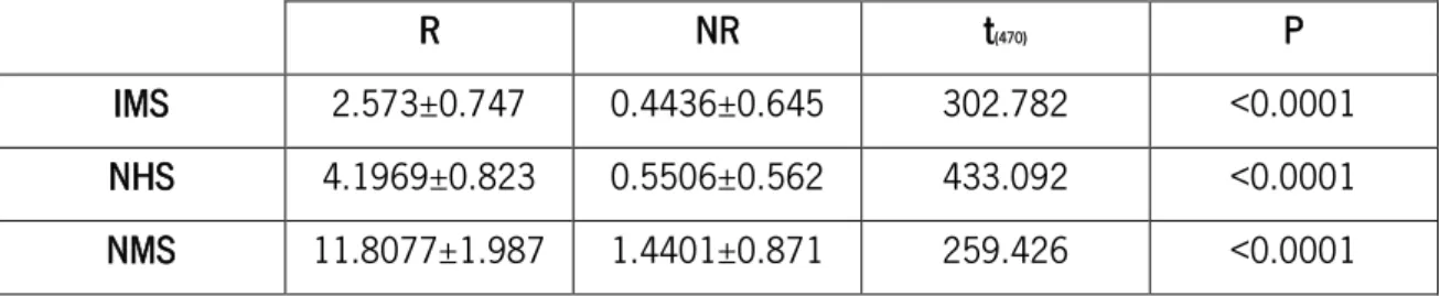
A total of 478 neurones were recorded in the PrL, 7 of which were excluded for having significantly higher spontaneous activity (Z≥3). In order to analyse exclusively the responsiveness of these neurones to each modality of noxious stimulation, only the absolute value of the difference between the activity during stimulation and the spontaneous activity was considered. Using the K-means clustering test, neurones were firstly grouped according its absolute response to evoked-activity upon peripheral stimulation into two categories: responsive (R) and non-responsive (NR). The results of the comparison between each cluster are summarized in Table 1 and show that the evoked activity is significantly different between R and NR cells.
Table 1 – Evoked activity of R and NR PrL neurones during peripheral stimulation. The mean and standard deviation of each cluster are presented for each R and NR pair. A t-test was used to compare means of evoked activity of each cluster. (IMS – innocuous mechanical stimulation; NHS – noxious heat stimulation; NMS – noxious mechanical stimulation; NR – non-responsive; PrL – prelimbic cortex; R – responsive).
R NR t(470) P
IMS 2.573±0.747 0.4436±0.645 302.782 <0.0001 NHS 4.1969±0.823 0.5506±0.562 433.092 <0.0001 NMS 11.8077±1.987 1.4401±0.871 259.426 <0.0001 R neurones were clustered together and further divided using the K-means clustering method (Fig. 10). Only 6% of the recorded cells responded to peripheral stimulation and of these, 41% only respond to IMS while the remaining 59% responds only to noxious stimulation (NS). From the nociceptive specific-like neurones (NS-like), 71% respond to mechanical stimuli (NMS) and 29% to heat (NHS). Neurones activated by both innocuous and noxious stimuli [wide-dynamic range-like (WDR-like) neurones] were absent in the PrL.
33
The spontaneous activity of cells from the groups presented in Figure 10 reveals that there are significant differences between the spontaneous activity of responsive (higher) and non-responsive (lower) cells (t-test, t(470)=93.264, p>0.0001) (Fig. 11A) but not in the responses between NS-like and NON-N-like groups (t-test, t(27)=2.189, p=0.151) (Fig. 11B) nor NHS and NMS NS-like groups (t-test, t(13)=1.329, p=0.270) (Fig. 11C).
34
4.3.2. Characterization of the neuronal population in the IL
In the IL, of the 302 neurones recorded 20 were excluded due to their high spontaneous activity (Z≥3). Similarly to what was observed in the PrL, the analyses of neuronal responsiveness to peripheral innocuous and noxious stimulation was performed using the absolute value of the difference between the activity during peripheral stimulation and the spontaneous activity of the cell. The analysis of IL neurones followed the same steps as for the PrL neuronal analysis: the K-means clustering test was used to successively divide neurones into two clusters based on the response to each modality of peripheral stimuli applied (Table 2).
35 Table 2 – Evoked activity of R and NR IL neurones during peripheral stimulation. The mean and standard deviation of each cluster are presented for each R and NR pair. A t-test was used to compare means of evoked activity of each cluster. (IMS – innocuous mechanical stimulation; NHS – noxious heat stimulation; NMS – noxious mechanical stimulation; NR – non-responsive; R – responsive)
R NR t(281) P
IMS 2.0517±0.740 0.4530±0.337 554.618 <0.0001 NHS 2.2200±0.669 0.3601±0.314 714.141 <0.0001 NMS 5.4874±1.565 1.1817±0.886 845.969 <0.0001
66% of the neurones recorded were classified as NR to any type of stimuli applied, and the remaining R cells (34%) were divided in three groups: 48% of cells were NS-like, 34% of cells were WDR-like and 18% of cells were NON-N-like neurones. NS-like and WDR-like neurones were then divided by responses to a specific noxious stimulation. Of the NS-like neurones, 57% responded to NMS, 26% to NHS and 17% to both. Of the WDR-like neurones, 27% responded to NMS, 55% to NHS and 18% to both stimuli (Fig. 12). The comparison of the spontaneous activity showed significant differences between R and NR cells (t-test, t(280)=20.783, p<0.0001) (Fig. 13A). In addition, although NS-like cells display lower spontaneous activity when compared to WDR-like
36
and NON-N-like neurones, this difference does not reach statistical significance (ANOVA1W, F(2,96)=2.726, p=0.071) (Fig. 13B).
Within WDR-like neurones the spontaneous activity of NHS and NMS cells is not different (ANOVA1W, F(2,46)=0.469, p=0.705) (Fig. 13C). In NS-like neurones however, although there is no statistically significant difference, NMS neurones spontaneous activity is almost significantly lower than that of NHS and NMS/NHS (ANOVA1W, F(2,46)=3.030, p=0.059) (Fig. 13D).
Taking these results into account, we evaluated if intensity of the response to noxious heat (NHS) would vary between WDR-like and NS-like neurones that respond to noxious mechanical stimulation. This analysis revealed that (i) neurones that respond to noxious mechanical stimulation (NMS) usually display a lower response to noxious heat (ANOVA2W, F(1,79)=31.417,
37
p<0.0001) (Fig. 14); (ii) the intensity of the response to noxious heat is independent of the cell type (WDR-like or NS-like) (ANOVA2W, F(1,79)=1.264, p=0.264); and (iii) the intensity of the response to noxious mechanical stimulation is also independent of the cell type (WDR-like or NS-like) (ANOVA2W, F(1,79)=0.773, p=0.382).
4.4. Effect of the pharmacological activation and inhibition of the mPFC upon nociceptive behaviour in the rat and its correlation with the activity of RVM pain modulatory cells during peripheral innocuous and noxious stimulation
4.4.1. Nociceptive behaviour
The administration of a control saline solution in the PrL and IL did not alter the nociceptive behaviour of rats [PrL: (ANOVARM, F(5,41)=1.388, p=0.2567, n=7) (Fig. 15A) and IL: ANOVARM, F(5,47)=1,498, p=0,2156, n=8) (Fig. 16A)], confirming that the injection of a solution in the PrL and the IL by itself does not have an effect upon paw-withdrawal latency (PWL).
4.4.1.1. Role of the PrL upon acute nociceptive behaviour
Glutamate significantly increased PWL 30 seconds after its microinjection (ANOVARM, F(2,23)=3.944, p=0.0139, n=8), indicating that the PrL has a phasic descending antinociceptive action (Fig. 15B).
38
Concomitantly, the microinjection of lidocaine significantly decreased PWL (ANOVARM, F(4,34)=7.023, p=0.0007, n=7) (Fig. 15C), with a maximum effect at 20 minutes and recovery at 30 minutes, indicating this area also has a tonic descending antinociceptive action.
4.4.1.2. Role of the IL upon acute nociceptive behaviour
Glutamate microinjection showed the existence of a delayed pronociceptive phasic effect of the IL as PWL decreased only 10 minutes after administration (ANOVARM, F(5,71)=9.82, p<0.0001, n=12) and recovering 30 minutes after injection (Fig. 16B). Lidocaine administration in the IL significantly decreased PWL, which peaked at 20 minutes and recovered 30 minutes after injection (ANOVARM, F(4,29)=10.18, p<0.0001, n=6) (Fig. 16C), showing that the IL also has a antinociceptive descending drive.
39
4.4.2. Modulation of the activity of RVM pain modulatory cells neurones modulation by the medial PFC
The number RVM OFF-like cells recorded was very low, so the data presented in this work will focus only on the spontaneous and NHS evoked fire ratings of ON-like cells in the rostral ventromedial medulla (RVM). Evoked activity is presented as mean firing rate during peripheral stimulation.
The administration of a saline solution in the PrL did not alter either the spontaneous (NS-like: ANOVARM, F(5,64)=1.333, p=0.2756; WDR-like: ANOVARM, F(5,84)=0.9168, p=0.4383) or NHS-evoked firing rate (NS-like: ANOVARM, F(5,59)=0.4289, p=0.7333; WDR-like: ANOVARM, F(5,69)=1.140, p=0.3417) of ON-like cells in the RVM. Similarly, saline injection did not alter either the spontaneous (NS-like: ANOVARM, F(5,119)=1.087, p=0.3778; WDR-like: ANOVARM, F(5,67)=0.2815, p=0.8385) or NHS-evoked activity (NS-like: ANOVARM, F(5,119)=1.403, p=0.2313; WDR-like: ANOVARM, F(5,67)=0.6946, p=0.5626) of ON-like cells in the RVM.
40
4.4.2.1. Role of the activation and inhibition of PrL upon the activity of RVM pain modulatory cells during peripheral innocuous and noxious stimulation
Glutamate in the PrL did not alter the spontaneous activity of ON-NS-like cells in the RVM (ANOVARM, F(2,29)=3.020, p=0.0740) (Fig. 17A) although it significantly decreased the spontaneous activity of ON-WDR-like neurones (ANOVARM, F(2,44)=4.281, p=0.0239) (Fig. 17B).
Additionally, glutamate in the PrL significantly decreased NHS evoked activity in both ON-NS-like cells (ANOVARM, F(2,29)=5.713, p=0.0120) (Fig. 18A) and ON-WDR-like cells (ANOVARM, F(2,44)=8.969, p=0.0010) (Fig. 18B) 30 seconds after its administration.
41
Lidocaine administration in the PrL failed to alter the spontaneous activity of ON-NS-like (ANOVARM, F(3,27)=1.886, p=0.1618) cells (Fig. 19A) but induced a significant decrease of the spontaneous activity of ON-WDR-like neurones (ANOVARM, F(3,95)=4.018, p=0.0107) (Fig. 19B).
Concurrently, heat-evoked activity of ON-NS-like (ANOVARM, F(3,27)=0.8132, p=0.5031) (Fig. 20A) was not altered by lidocaine microinjection in the PrL although it significantly decreased NHS evoked activity of ON-WDR-like neurones 20 minutes after administration (ANOVARM, F(3,95)=3.629, p=0.0171) (Fig. 20B).
42
4.4.2.2. Role of the activation and inhibition of IL upon the activity of RVM pain modulatory cells during peripheral innocuous and noxious stimulation
Administration of glutamate in the IL significantly altered the spontaneous firing rate of ON-WDR-like cells (ANOVARM, F(5,101)=2.435, p=0.0416) (Fig. 21B) but had no effect upon ON-NS-like cell spontaneous activity (ANOVARM, F(5,41)=1.727, p=0.1589) (Fig. 21A).
The same effect was observed during NHS-evoked activity with glutamate having no effect upon NHS-evoked ON-NS-like cells activity (ANOVARM, F(5,41)=0.7914, p=0.5643) (Fig. 22A) although significantly decreasing the NHS-evoked ON-WDR-like cells activity 10 minutes after it injection in IL (ANOVARM, F(5,89)=2.429, p<0.0434) (Fig. 22B).
43
When lidocaine was injected in the IL, no differences were found in the spontaneous firing rates of ON-NS-like (ANOVARM, F(3,15)=0.7101, p=0.5700) (Fig. 23A) and ON-WDR-like cells (ANOVARM, F(3,71)=0.4553, p=0.7147) (Fig. 23B).
However, even though NHS-evoked activity of ON-NS-like neurones did not change (ANOVARM, F(3,15)=0.5914, p=0.6361) (Fig. 24A), the NHS-evoked activity of ON-WDR-like neurones significantly decrease 10 minutes after lidocaine microinjection in the IL (ANOVARM, F(3,71)=3.240, p=0.0295) (Fig. 24B).
45
Chapter 5: Discussion
47
5. Discussion
A growing amount of evidence obtained mainly from imaging studies and clinical observations suggests that the prefrontal cortex (PFC) is directly involved in pain modulation. However, in terms of single cell electrophysiology of PFC cells, very little information is available. So far, it has only been demonstrated that the anterior cingulate cortex (ACC) is activated by noxious acute stimulation, with no studies analysing the involvement of the infralimbic cortex (IL) and only two reports published for the prelimbic cortex (PrL). In the present work, by performing systematic recordings of medial PFC (mPFC) neurones during noxious peripheral stimulation, we demonstrated the existence of a population of nociceptive neurones in the PrL and IL areas of the PFC and characterized their response properties to innocuous and noxious-evoked peripheral stimulation. The difference in the degree of functional specialization between PrL and IL neurones suggests that these areas are involved in the processing of distinct components of nociception, with the PrL neurones displaying a greater discriminatory ability for pain modality. In addition, the functional contribution of each region towards pain modulation was further explored by assessing the impact of its activation/inhibition upon nociceptive behaviour and the neuronal activity of an area implicated in pain modulation, the rostral ventromedial medulla (RVM). Behaviourally, our pharmacological data showed that both the PrL and the IL display a phasic and tonic modulatory action, mainly inhibitory (antinociceptive), upon nociception. Additionally, the early onset of the PrL-mediated modulatory drive following peripheral noxious stimulation contrasts with the later IL-mediated effect, suggesting that different receptors or circuits are mediating their nociceptive modulatory actions. Finally, a direct involvement of the mPFC in brainstem circuits involved in descending pain modulation was also achieved by correlating PrL and IL-mediated behavioural responses with changes in the activity of RVM nociceptive modulatory cells. Our findings confirm that the PrL and the IL play a crucial role in the integration and mediation of nociception at the cortical level.
48
5.1. Scope and limitations of the experimental design
The study of pain mechanisms presents a major challenge not only for ethical limitations but also because under sedation the emotional and cognitive functions are greatly altered and, finally, because of the difficulty in extrapolating results from animal models to humans studies.
The single cell extracellular electrophysiology technique provides insight on nociceptive processing in the areas recorded by allowing a real-time measurement of neuronal activity and its modulation by drug administration. The use of animals in research is an important tool to study in vivo bio/physiological effects of an intervention in addition to being a valuable instrument in the design and development of pharmacological agents with potential clinical use. The use of heat noxious stimulation is a selective way to stimulate both thermo-sensitive and nociceptive fibres (Millan 2002), while the application of a sharp noxious mechanical stimulus has been demonstrated to evoke a spinal activation pattern in rodents that is identical to what is observed in humans (Cervero et al. 1988).
5.1.1. Animal model
One of the first questions asked when working with animal models is whether the model is suitable for addressing the question at hand. In this study the experimental work was performed on rats whose PFC varies significantly in terms of size and differentiation from primates. First it should be noted that although homologies between rats and primates were firstly based solely in its architectural similarities, more recently the criteria evolved and is now based on pattern and density of neuronal connections, functional proprieties of neurones, presence/absence of specific molecular markers and embryologic origin in addition to citoarchitecture alone (Willis 2011). Hence, it has been demonstrated that although less specialized, the rat’s mPFC anatomically and functionally retains the PFC dorsolateral-like features observed in primates (Uylings et al. 2003). Also interesting is the fact that there isn’t an accepted PFC nomenclature for the rat as significant differences can be found between the most commonly used rat atlases of Paxinos and Watson (2007) and Swanson (1992), as these atlases differ not only in the nomenclature adopted but also in the delineations of limits between different areas.
49
5.1.2. Anaesthesia
It is important to note that part of the experimental procedures were performed in animals under anaesthesia, which may interfere with brain function and bias the results due to possible species-specific effects of the sedation. As anaesthesia often interferes with neuronal activity, at the beginning of the experiment a preliminary study was performed to test whether pentobarbitone or urethane would be adequate to study the PFC. We verified that with urethane anaesthesia not only the total number of cells but also their spontaneous activity was higher (Section 4.2. – Fig. 7-9) when compared with the results on cells of animals under the pentobarbitone anaesthesia. In addition, by using urethane we were able to maintain the animals stably sedated for over 4 hours, which allowed for the effective repetition of electrophysiological recordings on the same animal as described previously by other authors (Maggi and Meli 1986; Koblin 2002). On the other hand, the maintenance of an anaesthetized state using pentobarbitone depended either on an hourly intraperitoneal injection, compromising the stability of the anaesthesia level (as it could change from high to deep anaesthesia), or on its intravenous continuous administration (Cleary et al. 2008), which was not accessible in our lab due to technical reasons.
It is possible that the differences at the cellular level are due to a greater enhancement of the GABAergic tone by pentobarbitone when compared to urethane. In anaesthetic concentrations, urethane does not alter the uptake or release of GABA on the nervous system significantly and has been considered appropriate for use in physiopharmacological research (Maggi & Meli 1986). Previous studies also showed that in urethane anesthetized rabbits, hypothalamic neurones respond to a variety of thermal, painful and auditory stimuli, (Cross & Silver 1963) and mPFC neurones in the rat respond to mechanical stimulation (Zhang et al. 2004; Nakamura et al. 2010), highlighting that peripheral stimulation is still capable of activating central areas. Changes in cardiovascular conditions are minimal at anaesthetic concentrations (Koblin 2002), reducing the chances of death by respiratory deficiencies and interference of this parameter with our results.
On the other hand, urethane is a mutagenic, carcinogenic and hepatotoxic substance (Koblin 2002) that can only be administered in terminal experiments and requires careful manipulation,
50
specifically during the pre-administration period, as afterwards it is metabolized into ethanol and carbamic acid (Maggi and Meli 1986). Additionally, since the administration of urethane induces a deep anaesthetised state, motor responses are lost, making it impossible to correlate neuronal activity with motor responses. Finally, we are uncertain whether the low number of OFF-like cells in the RVM might reflect a cell-specific effect of the use of urethane anaesthesia, and further studies are needed to assess it.
5.1.3. Acute noxious stimulation
One of the main disadvantages of using acute experimental pain models is the fact that the short duration of the acute stimuli may fail to evoke a sustained cognitive-emotional component of pain, hence failing to reproduce clinical pain. In an attempt to counteract this limitation we opted for a multimodal approach to evoke neuronal responses from the mPFC.
The use of radiant heat has several disadvantages, like being poorly absorbed by the skin while being greatly reflected. To overcome some of its limitations we opted to (i) use a strain of albino animals to extend the level of penetration of the rays beneath the skin surface, (ii) heat the testing surface of the apparatus in order to prevent changes in skin temperature, (iii) maintain the intensity of radiation throughout the experimental period and (iv) test animals while they rested in order stabilize the plantar pressure on the apparatus surface during the application of the noxious heat stimuli. As the evoked activity of noxious mechanical stimulation depends on the intensity and duration of the stimulus and this type of stimulation is prone to cause tissue damage, we opted to apply a single sharp stimulus at a constant force such that it could evoke changes in neuronal activity. The application of an innocuous stimulus through brushing allowed us to distinguish between cells that receive input from neurones that encode solely noxious information (nociceptive specific – NS) from those receiving input from neurones able to transmit innocuous and noxious information simultaneously (wide-dynamic range – WDR). This distinction is pertinent as WDR neurones alone are able to evoke sensory and affective responses to pain (Coghill et al. 1993).
51
5.1.4. Single-cell extracellular recordings
In this study we used a single-cell recording technique to study nociceptive-evoked activity of mPFC neurones as well as to assess its top-down modulatory effect upon RVM pain modulatory cells in anaesthetized animals. While this technique allows the recording of a few cells simultaneously in every session, the area recorded is rather small when compared with capability of other electrophysiological techniques, such as the whole-field. On the other hand, the latter methodology quantifies changes in neuronal activity in a nuclei/area as a whole leaving out the possible subtle contribution of individual cells so often described to be involved in the modulation of nociception (Heinricher et al 2009). Ideally, multi-cell continuous recordings should be performed in order to increase our perception on how each individual neurone responds to the dynamics of PFC impinging during the processing of nociception. Additionally, future studies should also evaluate simultaneous recording of PrL and IL in order to determine the time-course of temporal/functional relationship between these two areas.
5.2. Characterization of mPFC neurones
The electrophysiological characterization of the response of PrL and IL neurones to the application of acute peripheral stimuli clearly showed the existence of a minority of neuronal clusters (6% and 34% of all cells, respectively) within these areas that code mechanical and thermal peripheral information. The percentage of cells activated by peripheral stimulation in either nucleus is slightly different from what was described by Zhang and colleagues (2004) for the ACC, in which 20% of all cells were responsive to mechanical stimulation. Comparatively, the PrL appears to be much less involved in the coding of nociception than the IL. It should however be noted that we did not quantifie all the nociceptive responsive neurones as we did not apply either chemical or visceral stimuli. However, we expect that the proportions of cells would remain identical at least in what concerns the multireceptive IL.
In terms of PFC activation by peripheral stimuli, our results are in accordance with data from human studies where the existence of neurones that responded to noxious cutaneous (Snow et al. 1992) and visceral (Yang and Follet 1998) stimulation was described for the orbitofrontal cortex and with data from animal studies where noxious and innocuous stimulation of the tail
52
activated the ACC (Zhang et al. 2004). In addition, two recent publications reported the activation of neurones within the PrL and IL during noxious visceral stimulation (Wang et al. 2009) and of the PrL during noxious mechanical cutaneous stimulation (Nakamura et al. 2010).
Our most interesting data is the difference in functional specificity between neurones of the PrL and IL. While PrL neurones respond exclusively to a single type of peripheral stimuli, either innocuous or noxious, IL neurones are multireceptive, responding to more than one single modality of stimuli. Firstly, although the existence of “functionally distinct subsets of neurones” within the PrL had already been proposed by Ji and colleagues (2010), no reference to a similar classification for IL neurones could be found. Secondly, the responsiveness profile of PrL neurones suggests that these cells encode stimulus intensity whereas IL neurones not only discriminate noxious intensity but, as suggested by Ji and colleagues (2010) for multireceptive neurones, also encode the temporal duration of stimulus intensity.
5.3.1. Influence of the PrL upon nociceptive behaviour and RVM cell activity
Our behavioural data shows that the PrL has a main inhibitory drive upon thermociception, since its activation by glutamate increased paw-withdrawal latencies while lidocaine had the opposite effect. This assumption is partly corroborated by the electrophysiological data on the PrL-RVM circuit, although the inability to record RVM OFF-cells limits our analysis. In fact, glutamate activation of the PrL decreased both the spontaneous activity of pronociceptive ON-WDR-like cells and the evoked activity of pronociceptive ON-NS-like and ON-WDR-like neurones (Heinricher et al. 2009). Additionally, our data suggests that PrL glutamate activation is probably mediated by ionotropic receptors as glutamate effects were observed as soon as 30 seconds after its microinjection.
The data concerning the lidocaine effect upon RVM cells does not however fit the behavioural results, since the decrease in spontaneous and heat-evoked activity of ON-WDR-like cells would by itself promote facilitation but we observed antinociception. It is possible the tonic PrL action upon ON-WDR-like cells is not sufficient to explain the behavioural results and that this facilitatory effect could be inhibited by a parallel circuit involving antinociceptive OFF-like cells (Heinricher et al. 2009), or even inhibitory actions from nuclei outside the RVM such as the amygdala, which is