Abstract
Since FV-Leiden polymorphism was first described in 1994, a growing number of polymorphic loci have been identified in association with increased genetic risk for thrombophilia. Often however, these risk factors have been studied in isolation of the remaining known phe-notype linked polymorphisms. This fact has, at least in part, been justified by the laborious techniques tradi-tionally used in the genotyping studies, as well as its relatively high costs. Another major problem concern-ing these studies has been the non-negligible incidence of dubious genotypes, resulting from the manual, labour intensive techniques applied, and their some-times difficult to read output’s. These difficulties have also hampered the widespread use of genotyping data in the clinical assessment of the genetic risk levels both in patients and their relatives, leaving some clinicians less than convinced about its clinical usefulness. Recently however, the introduction of new genetic tech-niques in the clinical genetics laboratory has started to change this picture. Most notably, the advent of Real-time-PCR has brought the possibility of genotyping patients and controls at a large scale, with increased specificity, automation and speed. Moreover, the use of these techniques in the clinical genetics setting has not only increased the quality of the results, but most
importantly has also increased our capability of answer-ing questions at a deeper level. Among the new ques-tions that can now be answered without increased costs and uncertainty is the study of the association of genet-ic risk factors in thrombophilia. Our results show that indeed even common polymorphic loci may increase our ability to further discriminate the genetic thrombo-sis risk of individual patients and relatives.
It must however be noted that the innovation level in the clinical genetics lab is just starting to grow. In fact we haven’t even started to experience the advantages brought about by the genome program, and its massive identification of SNP’s. The technology to test these is also presently being refined, and is expected to go from research to the clinical lab in the near future. Only then, can we expect to define with high certainty the com-bined genetic risks for such complex pathologies as the thrombophilias.
© 2002 S. Karger AG. Basel 1424-8832/02/0326-0235$18.50/0
Prof. Manuel Campos
Servicio de Hemotologia - Hospital de Santo Antonio Largo Prof. Abel Salazar - 400 Porto, Portugal
Copyright © 2002 S. Karger AG. Basel
Advances in the genotyping of thrombosis
genetic risk factors: clinical and laboratory
implications
José Manuel Cabeda
1,2,3, Mónica Pereira
1, José Miguel Oliveira
1, Alexandra Estevinho
1,
Irene Pereira
2, Sara Morais
2, Benvindo Justiça
2, Manuel Campos
21Molecular Biology Unit and 2Clinical Haematology, Santo António General Hospital, Porto, Portugal; 3Faculdade de Ciências da Saúde-Universidade
Fernando Pessoa, Porto, Portugal
Materials and methods
A total of 863 individuals (226 healthy blood donors, 253 arterial thrombosis patients and 384 venous thrombosis patients were included in the study. Arterial thrombosis events included 16 patients with ischemic thrombosis, 157 with stroke, 59 with myocardial infarction, 9 with retinal artery thrombosis, 6 with pulmonary artery thrombosis and 6 with other arterial thrombosis. Venous thrombosis events included 13 patients with venous cerebral thrombosis, 338 with deep venous thrombosis, 2 with portal vein thrombosis, and 31 other venous thrombosis events.
Factor V-Leiden and Prothrombin G20210A polymor-phisms were tested by Real-time-PCR in the LightCycler (Roche, Germany) using commercially available kits from the same supplier. Previously, Factor V-Leiden and Prothrombin G20210A had been tested by PCR-ASO using kits from Vienna Labs (Austria). PCR-RFLP was also used in the past to test for Factor-V-Leiden using a previously published protocol (1). Methylenetetrahydrofolate Reductase (MTHFR) C677T polymorphism was tested using previously described primers and probes (2) with the DNA FastStart Hybridization probes kit (Roche) optimised at 2.5 mM McCl2 and the following thermal cycling protocol: Enzime activation at 94°C for 8 min-utes followed by 40 round of amplification consisting on denaturation at 94°C for 0 seconds, annealing at 55°C for 10 seconds and polymerisation at 72°C for 15 seconds. Acquisition was performed during the annealing step.The melting profile included an initial denaturation at 94°C for 0 seconds, a stabilization at 40°C for 5 seconds, and the melting step with 80°C target temperature and stepwise acquisition at 0.6°C/second.
Serum levels of Protein C, Protein S and Antithrombin III were assayed using the respective IL test on the ACL 9000 sys-tem (Instrumentation Laboratory, USA), as recommended by the manufacturer.
Comparison of polymorphism frequencies between groups was done by the chi square test using the Statistica for Windows software package (Statsoft).
Introduction and results
Thrombosis is a haematological disorder with variable interacting causes that range from acquired to hereditary risk factors (3-6). Over the last decade an increasing number of genetic polymorphic markers associated with thrombosis have been described – a simple search on NCBI nucleotide data-base (www.ncbi.nih.gov) with the words “thrombosis AND Homo sapiens” returned at writing time 102 different entries, corresponding to a total of 39 different genes. This list howev-er does not include some genes with polymorphisms that rep-resent weak risk factors for thrombosis such as the MTHFR
gene. In fact, a quick browse of the literature has allowed us to generate a list of another 20 loci (3-7). In addition to coagula-tion risk factors, some immunological related polymorphic loci may also mediate thrombosis risk, by modulating the pres-ence of auto-antibodies that in turn modulate the plasma levels of coagulation factors. Therefore, this already long list of genetic risk factors for thrombosis should be regarded as a still incomplete and evolving list.
Not only has this list of polymorphic loci modulating the risk of thrombosis been growing, as the technical tools to study them have been changing dramatically in the same period. These improvements have enabled clinically useful genetic testing for the diagnosis of diverse pathological situations such as infectious, oncologic, and genetic diseases be them of a monogenic or a multifactorial trait such as thrombosis.
The technical genotyping advances, although greatly increasing speed, accuracy and volume, have also increased the range of techniques used at any single moment in diverse labs to test for the same parameter. This, in turn, has increased the possibility that different laboratories testing the same sam-ple would return divergent results.
Genotyping experiments have evolved from the PCR-RFLP techniques from late 1990’s to ARMS-PCR, PCR-ASO, and more recently Real-Time-PCR with melting profile analysis. Although the first techniques used are less expensive and eas-ier to implement in a molecular biology lab, they are also more prone to error, laborious, and far less convenient when a large number of samples has to be studied. Despite these inconve-nients, economical issues still make them a convenient choice for many labs.
One of PCR-RFLP’s most serious problems affecting result fidelity is the occurrence of incomplete reactions, which should be checked, using an internal universal restriction site. However, this internal control is not always possible to imple-ment, leaving some room for result uncertainty.
ARMS-PCR is prone to false negative amplification reac-tions caused by sample-to-sample variareac-tions in inhibitor con-centrations. For this reason, an internal control is mandatory in this procedure, and can only be substituted by the universal use of an extra inhibition control reaction for each of the amplifi-cation reactions.
PCR-ASO is a useful and widely used technique due to the possibility of automation in the microtiter plate based forms, using the common and largely available ELISA automation equipments. However, automation cannot by itself prevent variations in PCR kinetics between different reactions (or between different DNA templates in the same reaction). Since genotyping usually relies on the comparison of end-point sig-nal intensity, these variations can significantly influence the results. In fact, in our lab, we have found that inter-reaction variability can vary on average 13-26% (ranging from 0 to 173%). In order to control this variation we have implemented as routine, the use of triplicates for any genotyping experiment
by this method. As a result, from a total of 2840 reactions per-formed in triplicate (a total of 8520 assays for wild-type hybridisation probes and an equal amount with mutant hybridisation probes), we found 275 reactions (9.7%) with at least one discordant genotyping result. Of these, only 10 (0.4%) were completely discordant whereas the correct geno-type of the remaining reactions could be deduced from the 2 concordant results. Nevertheless, if single reactions had been performed, which is common practice in most labs for eco-nomical reasons, up to 10% false results would be expected from this procedure.
The advent of Real-Time-PCR, in its several probe flavours
(mainly molecular beacons, Taqman, Eclipse and FRET) has allowed much faster amplification reactions (particularly with the Smartcycler[Cepheid] and most notably with LightCycler[Roche] technologies). Furthermore, because PCR product detection is a part of the PCR reaction, the overall time from PCR run to mutation detection can be greatly reduced (9-11). Moreover, since PCR detection does not require PCR product handling, nucleic acid contaminations attributable to PCR products are virtually eliminated.
Regardless of these advantages, Real-Time-PCR amplifica-tion also presents an addiamplifica-tional feature, much valuable for genotype analysis – the opportunity to perform melting curve analysis, and therefore to determine the melting temperature of a specific nucleic acid sequence (10-11). The melting point determination is based on the fact that probe binding is a tem-perature and sequence dependent process. Thus, temtem-perature ramping monitored by optical reading allows the machine to detect a drop in signal that will occur when the probe specifi-cally reaches its sequence-dependent melting temperature. This variation in the fluorescence signal can be visualised by plotting the negative derivate of the fluorescence intensity. In this plot, a sudden decrease in signal strength is observed as a peak (fig.1), making possible a clear discrimination between homozygous wild-type, heterozygous and homozygous mutant samples (fig.1).
Over the last 2 years, we moved from microplate PCR-ASO hybridisation to a FRET based Real-Time-PCR procedure on the LightCycler (Roche, Germany) for the genotyping of Factor-V-Leiden, Prothrombin G20210A, and MTHFR C677T polymorphisms.
A total of 863 individuals (226 normal controls, 253
arteri-Fig. 1. Negative derivative of the melting profile for Factor V
Leiden. Shown are the plots for homozygous wild-type (WT/WT), heterozygous (WT/M) and homozygous mutant (M/M) samples. Note that the observed peaks for the two alleles are well separated, making genotype discrimination simple and unambiguous.
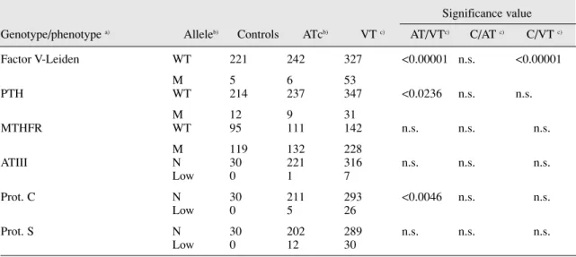
Table 1. Frequency of Wild-Type and mutated alleles in patients and controls
Significance value Genotype/phenotype a) Alleleb) Controls ATcb) VT c) AT/VTc) C/AT c) C/VT c)
Factor V-Leiden WT 221 242 327 <0.00001 n.s. <0.00001 M 5 6 53 PTH WT 214 237 347 <0.0236 n.s. n.s. M 12 9 31 MTHFR WT 95 111 142 n.s. n.s. n.s. M 119 132 228 ATIII N 30 221 316 n.s. n.s. n.s. Low 0 1 7 Prot. C N 30 211 293 <0.0046 n.s. n.s. Low 0 5 26 Prot. S N 30 202 289 n.s. n.s. n.s. Low 0 12 30
a) Factor V leiden, Prothrombin and MTHFR refer to the respective genotypes, whereas ATIII, Prot.C and Proc.S refer to the relative protein levels and are therefore phenotype determinations. All data available for each genotypic marker was included.
b) WT=Wild-type; M=mutated
al thrombosis patients [AT] and 384 venous thrombosis patients [VT]) were studied by genotyping procedures. In a subgroup of 547 individuals (40 normal controls, 204 AT and 303 VT patients) serum levels of Protein C, Protein S and Antithrombin III were also studied. As can be seen in table 1 the frequency of genetic polymorphisms/mutations found in the 863 individuals tested confirms that Factor V-Leiden is individually the genetic marker with the highest value in the discrimination of thrombotic genetic risk among venous thrombosis patients. Two other markers were found to be sig-nificantly different among venous and arterial thrombosis patients (Prothrombin G20210A, and reduced levels of protein C). In both cases, abnormalities were more frequent among venous thrombosis patients than among arterial thrombosis patients. In addition, none of the studied markers was found to individually show correlation to arterial thrombosis.
Although there is no significant correlation between most of the studied parameters alone and thrombosis, we were able to find an association between combined factors and increased thrombosis risk.
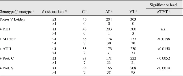
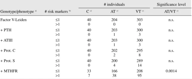
As can be seen in table 2, indeed when taken together, the studied risk factors are found to better discriminate between patients and controls, and between patient groups than when used alone. Interestingly, even modest thrombosis risk factors such as MTHFR C677T polymorphisms were found to con-tribute significantly to the global risk factor in the patient group. In fact, when the MTHFR polymorphism is only included in the last step (table 3), the increase in the number of patients identified by the inclusion of each additional factor is very small when compared to the observed when MTHFR is included (13 versus 78 in VT and 4 versus 34 in AT).
Therefore, our results show that even common polymorphisms cannot be excluded from investigation as they can be found to contribute significantly to the clarification of the global throm-bosis genetic risk factor in any individual patient.
Discussion
The growing list of polymorphic loci relevantly influencing the genetic risk of thrombosis raises the discussion to the point of discriminating the most relevant loci for the study (4,7). This is due to the economical and technical burden of studying an elevated number of genetic polymorphisms, each with a small contribution to the global risk. In this regard, it must be stressed that although this approach seems wise under the present technological tools available in the clinical laboratory, it is an ever-changing situation as recent history is showing.
From the techniques already available to the routine clini-cal laboratory, Real-Time-PCR with melting profile analysis seems to be the one that presents the best characteristics in terms of cost, reproducibility, speed, sensitivity and specifici-ty. Its level of automation, and the absence of PCR products handling greatly reduce the human expertise and the laborato-ry constrains necessalaborato-ry to its implementation. Also, the speed at which each reaction can be performed allows a much high-er numbhigh-er of reactions phigh-er patient to be phigh-erformed on the same day, thus increasing the feasibility of multi-locus typing. This can be even easier when machines such as the SmartCycler series (Cepheid, USA) are used, as they allow the simultane-ous run of different protocols for the same sample in separate reaction vessels.
238 Pathophysiol Haemost Thromb, 2002;32:235-240 Manuel Campos
Table 2. Frequency of associated polymorphic risk markers of thrombosis
Significance level Genotype/phenotype a) # risk markers b) C c) AT c) VT c) AT/VT c)
Factor V-Leiden ≤1 40 204 303 >1 0 0 0 + PTH ≤1 40 203 300 n.s. >1 0 1 3 + MTHFR ≤1 33 174 233 <0.0198 >1 7 30 70 + ATIII ≤1 33 173 230 <0.0150 >1 7 31 73 + Prot. C ≤1 33 171 222 <0.0052 >1 7 33 81 + Prot. S ≤1 33 166 208 <0.0014 >1 7 38 95
a) Factor V leiden, Prothrombin and MTHFR refer to the respective genotypes, whereas ATIII, Prot.C and Proc.S refer to the relative protein levels and are therefore phenotype determinations. Only data of individuals with all genotyping/phenotypic markers tested were included
b) Number of risk markers refers to the total number of risk markers found in each patient when considering only the genotypes/phenotypes from top of the table to each line. Note that for genotype markers each allele was considered one marker, thus an homozygous mutant individual would be con-sidered to have 2 risk markers.
Thus, at present, with the availability of Real-Time-PCR with melting profile analysis, the study should in our opinion include, not only the very strong risk factors such as Factor V-Leiden, but also some weaker factors, and common polymor-phisms such as Prothrombin G20210A and MTHFR C677T at a minimum. The conjugated study of these factors, not only allows the clarification of the genetic risk of more patients, as allows a clearer stratification of the genetic risk, due to associ-ation of several risk factors (12-13). Another complementary approach may be to study, at the protein level, some other genetic risk factors such as Proteins C and S, Antithrombin III and PAI-1, as these are more difficult to study at the genetic level (due to the nature or number of mutations to be screened). The results here described do indeed suggest that the con-jugation of these strategies allows a clearer elucidation of the genetic risk of each patient, and also a clearer stratification of the genetic risk differences between patients, with potential clinical benefits in terms of differential patient management.
Real-Time-PCR is so far the best of the genotyping tech-niques available in the clinical molecular biology laboratory. However, this is about to change in the near future, as prob-lems with the miniaturized devices being developed over the last decade are solved, and these devices approach the com-mercial status, with costs reaching affordable values. Miniaturized devices are being developed for nucleic acid extraction, capillary electrophoresis, PCR, multiplex-PCR, degenerate oligonucleotide primed PCR, Ligase chain reac-tion, RFLP and hybridisation assays, as well as combinations of these (14-16). Many of these devices are still in an early development stage, and for this reason can only be used in the research setting (16). However, some of these devices have
already matured enough to be released commercially, such as the Lab-on-a-chip (Agilent) for capillary electrophoresis and multiple examples of hybridisation chips. For the most part, the major obstacle to the use of these devices in current diag-nostic procedures is cost, and this is expected to drop signifi-cantly as their use increases, and production and development costs dilute with quantity. Also, as these devices become more widely used, the lab and personel constraints characteristic of any clinical molecular biology lab nowadays are expected to diminish, making these techniques available in most clinical laboratories. Also, as specific devices for disease risk factor genotyping become available, the number of tested risk-factors per patient is also expected to climb (current commercial ver-sions of DNA chips hold up to 20,000-45,000 probes in a 1.28 cm2 area [16]), making conclusions on a patient basis clearer, as more of the combined risk factors get tested.
In conclusion, as a fast evolving discipline, clinical molecu-lar genetics is quickly changing, both at the laboratory desk and at the clinical setting. In these changing times, care must be taken to ensure not only adequate testing, but also correct clin-ical interpretation of the available, but incomplete data. However, as miniaturization proceeds, it is expected that both completeness of the data, as well as reproducibility and stan-dardization at the lab desk results in data easier to obtain and to interpret.
Table 3. Frequency of associated polymorphic risk markers of thrombosis
# individuals Significance level Genotype/phenotype a) # risk markers b) C c) AT c) VT c) AT/VT c)
Factor V-Leiden ≤1 40 204 303 n.s. >1 0 0 0 + PTH ≤1 40 203 300 n.s. >1 0 1 3 + ATIII ≤1 40 203 300 n.s. >1 0 1 3 + Prot. C ≤1 40 202 295 n.s. >1 0 2 8 + Prot. S ≤1 40 200 289 n.s. >1 0 4 14 + MTHFR ≤1 33 166 208 0.0014 >1 7 38 95
a) Factor V leiden, Prothrombin and MTHFR refer to the respective genotypes, whereas ATIII, Prot.C and Proc.S refer to the relative protein levels and are therefore phenotype determinations. Only data of individuals with all genotyping/phenotypic markers tested were included
b) Number of risk markers refers to the total number of risk markers found in each patient when considering only the genotypes/phenotypes from top of the table to each line. Note that for genotype markers each allele was considered one marker, thus an homozygous mutant individual would be con-sidered to have 2 risk markers.
References
1. Koeleman BP, Reitsma PH, Allaart CF, Bertina RM: Activated Protein C Resistance as an additional risk factor for thrombosis in protein C-deficient families. Blood 1994; 84:1031-1035.
2. Ahsen N, Shütz E, Armstrong VW, Oellerich
M: Rapid detectiopn of Prothrombotic
Mutations of Prothrombin (G20210A), Factor V (G1691A) and Methylenetetrahydrofolate Reductase (C677T) by Real Time Fluorescence PCR with the LightCycler. Clinical Chemistry 1999; 45:694-696 3. Lane DA, Grant P: Role of hemostatic gene
polymorphisms in venous and arterial throm-botic disease. Blood 2000; 95:1517-1532. 4. Franco RF, Reitsma P: Genetic risk factors of
venous thrombosis. Hum.Genet. 2001; 209:369-384.
5. Franco RF, Reitsma PH: Gene polymorphisms of the haemostatic system and the risk of arte-rial thrombotic disease. Brit.J.Haematol. 2001; 115:491-506.
6. Chak M, Wallace GR, Graham EM, Stanford MR: Thrombophilia: genetic polymorphisms and their association with retinal vascular
occlusive disease. Br.J.Ophtalmol 2001; 85:883-886.
7. Murin S, Marelich GP, Arroliga AC:
Hereditary Thrombophilia and venous throm-boembolism. Am.J.Respir.Crit.Care Med. 1998; 258:1369-1373.
8. Cabeda J.M: Genética Molecular da
Trombose. Mundo Médico 2000; 2:70-71. 9. Wittner C. Rapid Cycle Real-Time PCR:
Methods and Apllications in Meuer S., Wittwer C., Nakagawara K. (ed): Rapid Cycle Real-Time PCR: Methods and Apllications Berlin, Springer, 2001
10. Bernard PS, Reiser A, Pritham GH. Mutation Detection by Fluorescent Hybridization Probe Melting Curves in Meuer S., Wittwer C., Nakagawara K. (ed): Rapid Cycle Real-Time PCR: Methods and Apllications Berlin, Springer, 2001
11. Landt O. Selection of Hybridization Probes for Real-Time Quantification and Genetic
Analysis in Meuer S., Wittwer C.,
Nakagawara K. (ed): Rapid Cycle Real-Time PCR: Methods and Apllications Berlin, Springer, 2001
12. Nowak-Göttl U, Junker R, Kreuz W,
Eckardstein A, Kosch A, Nohe N, Schobess R, Ehrenforth S: Risk of recurrent venous throm-bosis in children with combined prothrombot-ic risk factors. Blood 2001; 97:858-862. 13. Salomon O, Steinberg DM, Zivelin A, Gitel S,
Dardik R, Rosenberg N, Berliner S, Inbal A, Many A, Lubetsky A, Varon D, Martinowitz U, Seligsohn U: Single and combined prothrom-botic factors in patients with idiopathic venous thromboembolism: prevalence and risk assess-ment. Arterioscler.Thromb.Vasc.Biol. 1999; 19:511-518.
14. Kricka LJ: Miniaturization of analytical sys-tems. Clinical Chemistry 1998; 44:2008-2014. 15. Munro NJ., Snow K, Kant JA, Landers JP: Molecular Diagnostics on Microfabricated Electrophoretic devices: from slab gel- to cap-illary- to Microchip-based assays for T- and B-cell lymphoprolipherative disorders. Clinical Chemistry 1999; 45:1906-1917.
16. McGlennen RC.: Miniaturization technologies for molecular diagnostics. Clinical Chemistry 2001; 47:393-402.