UNIVERSIDADE DA BEIRA INTERIOR
Ciências
Biorecognition by amino acid-based affinity
chromatography for RNA purification
Ana Rita Nunes Martins
Tese para obtenção do Grau de Doutor em
Bioquímica
(3º ciclo de estudos)
Orientadora: Prof. Doutora Fani Pereira de Sousa
Co-orientador: Prof. Doutor João António de Sampaio Rodrigues Queiroz
iii
“Nothing is impossible; the word itself says
‘I'm possible’!”
Audrey Hepburnv
Dedication
Esta Tese de Doutoramento é dedicada de forma muito sentida ao meu Pai e à minha Mãe. Ao meu Pai pelo exemplo que sempre foi para mim, pelo seu dinamismo, força de vontade e boa disposição que sempre o acompanharam ao longo da vida, e por me ter ensinado que as palavras “Não” e “Impossível” não existem. À minha Mãe, por me ter incutido a sua generosidade, a sua paciência e a sua forma subtil e desembaraçada de enfrentar a vida.
Todos esses ensinamentos e atitudes definiram a minha personalidade e fizeram-me encarar a vida com uma motivação adicional e com uma perspectiva lutadora, o que me ajudou a alcançar sempre todos os objectivos a que me propus, como é o exemplo desta Tese de Doutoramento.
vii This work was financed by the Portuguese Foundation for Science and Technology (SFRH/BD/64100/2009) under the program QREN - POPH - Type 4.1 – Advanced Training, co-founded by the European Social Fund and by national funds from the MCTES and also under a research and development project (PTDC/EBB-BIO/114320/2009) and strategic programs (PEst-C/SAU/UI0709/2011) co-founded by the operational program factors of competitiveness – COMPETE and (EXPL/BBB-BIO/1056/2012) co-founded by FEDER funds (FCOMP-01-0124-FEDER) of COMPETE.
ix
Acknowledgments
I wish to thank all those who directly or indirectly contributed for the completion of this doctoral thesis, including those who I will not specifically acknowledge here, but that were also very important in realizing this goal.
First of all, I would like to express my gratitude to my supervisors. To my supervisor Prof. Fani Sousa for her professionalism, dedication and all knowledge provided, for believing in me, for all encouragement, support, and especially for her friendship, my very thank you! To my co-supervisor Prof. João Queiroz, my sincerely appreciation for his geniality and inspiration, for his friendship and availability, as well as the valuable scientific knowledge that he conveyed me, all of which had a remarkable influence in the course of my academic career and in the achievement of this thesis.
I would also like to thanks to Professor Cláudio Maia for his valuable contribution to this work through his expertise in genetics and molecular biology. I am really thankful for the availability and interest he demonstrated for my work through the great scientific discussions and the knowledge transmitted.
To the Portuguese Foundation for Science and Technology (FCT) I want to acknowledge the financial support through my doctoral fellowship (SFRH/BD/64100/2009) that allowed the execution of all my research in Health Science Research Centre at University of Beira Interior (CICS-UBI).
In addition, thanks to CICS-UBI for the host and to the technicians, Ana Sofia Duarte e Margarida Carrilho, for their precious contribution in the organization of my laboratory tasks and for the willingness that they responded to all my requests.
An appreciative note to Professor Luís Passarinha, for his friendship and support, and for allowing me to extend my scientific knowledge and experience by participating in his scientific projects and contributing with my slight experience for the formation of other colleagues.
Moreover, an enormous thanks to my laboratory colleagues for the good working environment, helpfulness and all the excellent moments of scientific discussions but also of fun. I wish them very success in their research projects and their future lives. It was a pleasure working with all of them. However, I could not let to name those who, in addition to co-workers, have become great friends. Indistinctly, my deeply thankful to Ana Martinho, Ângela Sousa, Catherine Nunes, Diogo Maio, Eduarda Coutinho, Fátima Santos, Filomena Silva, Marta Silva and Patrícia Pereira.
To you, Miguel, I am deeply grateful for all the affection, caring, unconditional support and patience throughout these months. You entered my life in the most difficult, demanding and delicate phase of my existence, by coincidence or fate (I do not know!!), but you really demonstrated to be a true companion for life.
Por último, gostaria de agradecer de uma forma muito especial e sentida à minha família pelo amor, confiança, estímulo e apoio incondicional que sempre me deram. Aos meus Pais fica a sentida dedicação desta Tese de Doutoramento. To my sister, I express my thanks for all her caring and for being present at every stage of my life. The complicity that we still have, even at distance, continues to keep us together and encouraged me to move forward, knowing that she will always be there for me. E para o meu cunhado preferido, deixo o meu Obrigado pelos momentos divertidos que em muito me ajudaram a manter o vigor mental.
xiii
Resumo
A sequenciação completa do genoma humano ofereceu novos horizontes relativamente à prevenção, diagnóstico e tratamento de doenças humanas. Para além disso, com o avanço da engenharia genética, têm surgido novas tecnologias terapêuticas, entre as quais se destacam a terapia génica utilizando ácidos nucleicos. Apesar destas estratégias génicas se terem iniciado com o DNA (ácido desoxirribonucleico), estudos recentes têm avaliado o potencial interesse terapêutico do RNA (ácido ribonucleico).
O RNA foi recentemente reconhecido como uma molécula fundamental nos processos celulares, com implicações fundamentais na evolução dos organismos, na hereditariedade e na regulação de vários genes, o que destacou o seu vasto potencial terapêutico e conduziu ao aparecimento de várias terapias baseadas em moléculas de RNA. Os resultados promissores dessas novas abordagens terapêuticas têm vindo a reforçar a investigação relacionada com as moléculas de RNA, a avaliar pelo número, cada vez mais elevado, de estudos estruturais, biofísicos e biomédicos presentes na literatura. Além disso, a indústria biotecnológica e farmacêutica começam a visar as moléculas de RNA como uma nova classe de produtos bioterapêuticos.
Um requisito fundamental em todos esses estudos é a obtenção de grandes quantidades de RNA isolado e puro e de integridade assegurada. Como por exemplo, em biologia molecular a purificação de RNA é o primeiro passo chave para avaliar a expressão de um gene, uma vez que a realização bem como a reprodutibilidade e relevância biológica dessa experiência está dependente da quantidade e qualidade das preparações de RNA. Por outro lado, as terapias promissoras e revolucionárias baseadas em RNA, como a vacinação ou utilização de biofármacos recombinantes envolvem formulações de RNA que devem satisfazer critérios de qualidade rigorosos recomendados por agências reguladoras internacionais. No entanto, o processo de purificação das moléculas de RNA pode ser um passo bastante limitante para o sucesso da sua aplicação terapêutica.
O RNA tem uma série de características químicas únicas que se reflectem na sua conformação estrutural. Apesar do RNA ser uma molécula de cadeia simples na sua base, ele tem uma elevada propensão para formar estruturas secundárias e terciárias bastante complexas. São estas compactações próprias das moléculas de RNA que definem as suas importantes funções biológicas. A elevada reactividade química é outra característica própria do RNA e com grande relevância biológica a nível regulatório, por proporcionar mais instabilidade à molécula aumentando a sua susceptibilidade à degradação. Num contexto laboratorial, estas características moleculares do RNA são enormes desafios para a sua extracção e purificação,
pois a sua actividade biológica e integridade podem ser facilmente comprometidas durante os procedimentos devido à presença ubíqua de enzimas que o degradam.
Várias técnicas têm sido desenvolvidas para superar os desafios inerentes ao isolamento e purificação de moléculas de RNA, tais como a extracção com fenol e clorofórmio ou as extracções em fase sólida empregando colunas ou esferas de sílica (SPE), bem como algumas técnicas de purificação com cromatografia líquida de alta eficiência (HPLC) de fase reversa e troca iónica. No entanto, estas ainda apresentam várias limitações nomeadamente em relação ao elevado tempo despendido e à necessidade do uso de solventes tóxicos e condições desnaturantes durante os procedimentos. Por todos estes motivos, torna-se evidente o crescente interesse na avaliação e melhoria das metodologias actualmente utilizadas para o isolamento e purificação de RNA de modo a satisfazer os requisitos necessários à sua aplicação.
A cromatografia é um dos métodos mais diversos e potentes em biotecnologia, tanto a nível analítico como preparativo devido à sua simplicidade, robustez, versatilidade e alta reprodutibilidade. Por sua vez, a cromatografia de afinidade é reconhecida como uma técnica poderosa apresentando grande aplicabilidade na purificação de muitas biomoléculas, incluindo o DNA plasmídico (pDNA) e proteínas, porque explora o reconhecimento biomolecular, ou seja, a capacidade de uma macromolécula biologicamente activa formar complexos específicos e reversíveis com ligandos de afinidade.
Posto isto, o trabalho desenvolvido no âmbito desta tese incide na crescente necessidade de desenvolver novas estratégias de isolamento e purificação para moléculas de RNA de modo a ultrapassar as limitações ainda existentes nas metodologias actuais, contribuindo para a evolução e sucesso da investigação e aplicações terapêuticas do RNA. Para isso, a potencialidade da cromatografia de afinidade foi considerada nesta temática e foi explorada a aplicação de aminoácidos como ligandos de afinidade. Este trabalho foi baseado em vários estudos de reconhecimento molecular e atómico que descrevem a existência de diferentes interacções entre proteínas e ácidos nucleicos nos sistemas biológicos, especialmente com aminoácidos básicos como a histidina e a arginina, e também na hipótese de existirem interacções preferenciais entre os aminoácidos e as bases nucleotídicas. Além disso, estudos recentes de processos de purificação mostraram grande aplicabilidade destes aminoácidos em isolar e purificar moléculas de pDNA biologicamente activas para aplicação em terapia génica e vacinas.
Assim sendo, novas metodologias preparativas e analíticas foram alcançadas ao longo deste trabalho, ou seja, foi possível obter preparações de RNA a partir de diferentes fontes biológicas e de reacções sintéticas com elevados rendimentos e grau de pureza e de integridade preservada, bem como também foi possível o desenvolvimento de um método
xv A potencial aplicabilidade da cromatografia de afinidade, usando histidina como ligando de afinidade, na purificação de moléculas de RNA foi demonstrada pela primeira vez com a purificação do RNA 6S, um RNA regulatório não codificante presente nos procariotas, nomeadamente em Escherichia coli (E. coli), que tem uma função reguladora importante no processo de transcrição deste organismo. Nas estratégias de purificação com histidina foram usados gradientes de sulfato de amónio devido à presença do anel de imidazol na cadeia lateral aromática do aminoácido, o que permitiria explorar interacções maioritariamente hidrofóbicas entre o RNA e a matriz, quer por interacção com o anel ou por pontes de hidrogénio. Assim, foi utilizado um gradiente decrescente em concentração de sulfato de amónio em três etapas que revelou um reconhecimento bioespecífico com RNA 6S, permitindo a sua purificação de uma mistura complexa de outras moléculas de RNA de baixo peso molecular (sRNA).
Uma segunda nova estratégia desenvolvida com a matriz de histidina permitiu obter simultaneamente o isolamento e purificação das classes de sRNA e RNA ribossómico (rRNA) a partir de lisados celulares de E. coli. Neste estudo, ambas as classes foram separadas das impurezas do hospedeiro (DNA genómico e proteínas) com elevado rendimento e grau de pureza quando comparadas com amostras preparadas com métodos de isolamento convencionais baseados na extracção com fenol e clorofórmio. No entanto, a metodologia baseada na cromatografia com histidina demonstrou a vantagem de evitar o uso de produtos químicos tóxicos durante o processo.
A versatilidade da matriz de histidina na purificação de RNA, tanto de moléculas específicas (RNA 6S) como das classes (sRNA e rRNA) sugeriu que o mecanismo de interacção envolveria não só interacções hidrofóbicas, mas também um bioreconhecimento das bases do RNA pela histidina. Apesar das preparações de RNA obtidas com estes métodos necessitarem de caracterização funcional adicional para provar a sua aplicabilidade, o uso da cromatografia de afinidade com histidina representa um grande avanço no isolamento de moléculas de RNA, uma vez que as técnicas tradicionais não têm a capacidade de fraccionar RNA ao nível de um tipo de molécula ou de isolar sRNA e rRNA num só processo. Todavia, a necessidade de aplicar elevadas concentrações de sal nestas metodologias pode ser visto como uma desvantagem, principalmente no que diz respeito à aplicação biotecnológica, pois acarreta maiores custos de processo e tem maior impacto ambiental.
O uso de arginina como aminoácido imobilizado na cromatografia de afinidade foi utilizado na perspectiva de melhorar as técnicas anteriores uma vez que este aminoácido se apresenta sobretudo carregado positivamente, pelo que poderiam ser exploradas interacções electrostáticas para a purificação de RNA utilizando condições de eluição moderadas. Além disso, as interacções de arginina têm sido reconhecidas como as mais prevalentes nos complexos RNA-proteína, aumentando a potencialidade de purificação por um maior bioreconhecimento entre as moléculas de RNA e a matriz de arginina. Deste modo, um
gradiente com o aumento gradual da concentração de cloreto de sódio permitiu o isolamento e purificação do RNA total de extractos celulares eucarióticas. O suporte de arginina demonstrou uma aplicabilidade excepcional para interagir com todas as classes funcionais do RNA apesar da sua diversidade estrutural e as suas diferentes conformações em condições nativas. Essas interacções mais fortes e selectivas parecem advir da cadeia lateral do aminoácido de arginina que apresenta uma multiplicidade para interacções, podendo promover um contacto múltiplo com o RNA, através da sua estrutura açúcar-fosfato ou com as suas bases, atendendo ao seu estado conformacional. Embora as interacções electrostáticas entre os grupos fosfato do RNA e os ligandos de arginina possam desempenhar uma função importante na retenção do RNA na coluna, as interacções com as bases também estão envolvidas e modulam de alguma forma a interacção favorecida e a especificidade encontradas na cromatografia com arginina. Assim, neste processo de purificação verificou-se um elevado rendimento de recuperação do RNA total e pelas análises de controlo de qualidade efectuadas mostrou-se que este apresentava uma elevada integridade bem como uma boa pureza, demonstrada pela difícil detecção de proteínas nas amostras purificadas e pela redução de DNA genómico para concentrações residuais. A eficiência desta técnica de purificação e a aplicabilidade do RNA por ela obtido foi demonstrada num procedimento habitualmente usado em biologia molecular para a análise de expressão génica. As amostras de RNA total foram usadas com sucesso como moldes na reacção em cadeia da polimerase em tempo real (qPCR) para a avaliação da expressão de dois genes controlo geralmente empregues nestes procedimentos.
Reunindo os resultados anteriormente descritos, o isolamento e purificação de RNA de amostras biológicas complexas usando as técnicas de cromatografia de afinidade com aminoácidos imobilizados demostrou vários benefícios em relação a métodos de isolamento actualmente empregues, como a extracção de fenol e clorofórmio ou de SPE, uma vez que simplificam a integração do processo e minimizam o manuseamento das amostras, tornando a cromatografia baseada em aminoácidos útil no desenvolvimento de metodologias em condições não desnaturantes, livres de RNases e solventes orgânicos, particularmente importantes em vários estudos estruturais e funcionais bem como de aplicabilidade terapêutica.
Com os resultados positivos destas metodologias de afinidade na purificação de RNA foi também desenvolvido e validado (de acordo com a legislação internacional e europeia para métodos bioanalíticos) um método analítico para a quantificação e monotorização de RNA. Com a crescente importância, a nível terapêutico, do desenvolvimento de novas ferramentas analíticas para avaliar o RNA, uma vez que ainda existem várias lacunas nas técnicas actuais de quantificação de RNA, tais como a falta de selectividade, tornou-se imperativo avaliar a potencialidade dos suportes de afinidade neste campo. A versatilidade da metodologia foi demonstrada pela sua aplicabilidade na quantificação de RNA de diferentes fontes
xvii eucarióticas e também em amostras complexas de RNA quimicamente sintetizado, o que demonstrou a usa utilidade em múltiplas áreas de investigação.
Desenvolveu-se ainda mais um estudo no isolamento e purificação de RNA com base nas matrizes de aminoácidos, em particular com matriz de arginina, de modo a aproximar a aplicabilidade desta técnica à prática terapêutica. Neste caso, o novo objectivo foi explorar o seu potencial na purificação de moléculas de RNA mensageiro (mRNA) não a partir de células, mas a partir de reacções sintéticas de transcrição in vitro, com o interesse de aplicar as moléculas em terapias de vacinação com mRNA no cancro do colo do útero. As moléculas de mRNA que codificavam para as proteínas E6 e E7 do vírus do papiloma humano (VPH) 16 foram purificadas com sucesso de uma série de impurezas próprias das reacções de transcrição in vitro, com são o pDNA molde, as enzimas, nucleótidos, sais e tampões, e a sua caracterização em termos de rendimento, pureza e integridade foi avaliada. Neste trabalho, a cromatografia de arginina voltou a demonstrar a sua capacidade singular em melhorar os processos de purificação, pelas vantagens de eliminar passos adicionais e melhorar a economia global do processo de produção.
Em suma, esta tese permitiu o desenvolvimento de novas metodologias para a purificação e quantificação de RNA revelando várias características interessantes das moléculas, incluindo o seu comportamento cromatográfico e interacções naturais que podem ocorrer com os suportes de aminoácidos. Por conseguinte, estes métodos mostraram uma potencial aplicabilidade polivalente, contribuindo para o progresso das investigações fundamentais e terapêuticas baseadas no RNA, suportando a utilização da cromatografia de afinidade baseada em aminoácidos no desenvolvimento futuro de novos processos de preparação de RNA.
Palavras-chave
xix
Abstract
Following the decoding of the human genome, a new era was opened for developing new gene therapy strategies employing nucleic acids. Recently, RNA was renowned a central molecule in cellular processes with implications in many diseases as well as in understanding of evolution, becoming one of the most exciting research areas of molecular biology. From basic to applied research, many procedures employ pure and intact RNA molecules. On one hand, RNA purification is a first critical step of a number of molecular biology procedures and its quality is crucial to ensure reproducibility and biological relevance of an experiment. On the other hand, the promising and revolutionary RNA-based therapies of RNA vaccination, gene therapy or recombinant biopharmaceuticals involves RNA formulations which should fulfill rigorous quality criteria recommended by international regulatory agencies. However, the isolation and purification of RNA are critical steps because of the easy degradability of RNA, which can impair chemical stability and biological functionality essential for analysis. Many techniques have been development to overcome the challenges of purifying RNA molecules; nonetheless they still have several limitations in regard to time demanding and the requirement of toxic solvents and denaturing conditions. Therefore, there is a growing demand for the evaluation and improvement of the methodologies currently used for RNA isolation and purification.
Chromatography is undoubtedly one of the most diverse and potent methods in biotechnology, both at analytical, preparative and industrial level due to its simplicity, robustness, versatility and high reproducibility. Affinity chromatography is recognized as a powerful technique with great applicability in the purification of many biomolecules, including plasmid DNA and proteins because it exploits the principle of biomolecular recognition. The work that we have been developing considers new chromatographic strategies for RNA purification, using amino acids as affinity ligands. These studies are based on the fact that many different interactions exist between proteins and nucleic acids in biological systems, involving in particular basic amino acids such as histidine or arginine. New methodologies were accomplished that allowed obtaining RNA preparations from different sources with high recovery yields, purity and integrity. A new analytical method for RNA quantification was also developed in this work.
The applicability of histidine-based affinity chromatography in the purification of RNA molecules was first demonstrated in the separation of 6S RNA, a regulatory non-coding RNA of the prokaryotic Escherichia coli (E.coli). A specific recognition between the histidine support and 6S RNA allowed its selective purification from a complex mixture of other small RNAs (sRNA). In another strategy, the simultaneous isolation of sRNA and ribosomal RNA from E.coli cell lysates, eliminating host DNA and proteins, was also attained by a histidine
chromatographic-based method. Furthermore, arginine matrix was employed in RNA purification from eukaryotic cells demonstrating an exceptional ability to interact with all functional classes of RNA, despite their structural diversity and different folding states, enabling their isolation from impurities of eukaryotic crude cell extracts. Moreover, an analytical technique based on arginine affinity support for quantification and quality assessment of total RNA from different eukaryotic cells and synthetic RNA samples was also developed and validated, according to international and European legislation for bioanalytical methods.
More efforts into RNA purification were developed with amino acid-based matrices, in particular with arginine-agarose matrix, in order to approach this technique to therapeutic application of RNA. The new goal was to exploit its applicability in purifying messenger RNA (mRNA) molecules not from cells, but from synthetic crudes of in vitro transcription reactions, pursuing mRNA vaccination for cervical cancer. In this work, arginine-based chromatography also showed its singular capability to improve purification processes, showing the advantages of eliminating additional steps and improving global economics of the production process.
The development of these new methodologies revealed several interesting characteristics of RNA molecules, including their chromatographic behavior and natural interactions that can occur between amino acids-based supports and RNA molecules. Accordingly, these methods demonstrated a potential multipurpose applicability by aiding in molecular biology RNA-based analysis and RNA therapeutics, which support the interest in applying amino acid-based affinity chromatography for the future development of new RNA isolation, purification and quantification processes.
Keywords
xxiii
Thesis Overview
This thesis is structured in four main chapters.
The first chapter includes an introduction that is divided in two sections. One section explains the interest on RNA molecules to be potentially applied in novel therapies such as gene therapy and RNA vaccination and the second section presents a brief explanation about the relevance of purifying RNA molecules, as well as the main challenges and concerns regarding RNA purification. In addition, a brief review of the literature related to the techniques used in RNA purification regarding their advantages and disadvantages is discussed together with the improvements that have been done lately. This second section is presented as a publisher paper review form (Paper I).
The second chapter presents the main purpose and the specific goals that were established for the development of this research work.
In the third chapter, the results obtained during this work are presented and discussed in the form of original research papers organized as follows:
Paper II - A new affinity approach to isolate Escherichia coli 6S RNA with histidine- chromatography
Paper III - Histidine affinity chromatography-based methodology for the simultaneous isolation of Escherichia coli small and ribosomal RNA
Paper IV - A new strategy for RNA isolation from eukaryotic cells using arginine affinity chromatography
Paper V - New approach in RNA quantification using arginine-affinity chromatography: potential application in eukaryotic and chemically synthesized RNA
Paper VI - Arginine-affinity chromatography for mRNA vaccines purification
Finally, the fourth chapter outlines the concluding remarks about this work, regarding the initial hypothesis of using affinity chromatography with immobilized amino acids in the purification of RNA molecules. Furthermore, some future work are suggested to complement the important findings achieve in this study.
xxv
Index
List of Figures xxvii
List of Tables xxix
List of Scientific publications xxxi
List of Scientific communications xxxv
Chapter 1 1
1. Gene therapy and vaccination with RNA 3
1.1. Introduction 3
1.2. Overview of gene therapy 4
1.3. RNA-based therapies 11
1.3.1. Catalytic RNAs: Ribozymes 11
1.3.2. Antisense oligoribonucleotides 13
1.3.3. Agents of RNA interference 13
1.3.4. Aptamers 16
1.3.5. mRNA vaccination 17
References 19
2. Current issues in RNA preparation: approaching affinity chromatography
into RNA purification challenges (Paper I) 23
Chapter 2 67
Aims of the thesis 69
Chapter 3 73
Paper II. A new affinity approach to isolate Escherichia coli 6S RNA with
histidine-chromatography 75
Paper III. Histidine affinity chromatography-based methodology for the
simultaneous isolation of Escherichia coli small and ribosomal RNA 85 Paper IV. A new strategy for RNA isolation from eukaryotic cells using arginine
affinity chromatography 97
Paper V. New approach in RNA quantification using arginine-affinity
chromatography: potential application in eukaryotic and chemically
synthesized RNA 111
Paper VI. Arginine-affinity chromatography for mRNA vaccines purification 125
Chapter 4 151
Concluding remarks 153
xxvii
List of Figures
Figure 1 – Compiled data on gene therapy clinical trials provided by regulatory
agencies 5
Figure 2 – The in vivo and ex vivo paths used in gene therapy 6
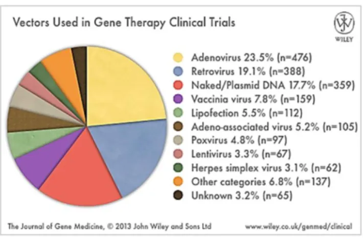
Figure 3 – Vector systems commonly used in gene therapy clinical trials 7
Figure 4 – Ribozymes activities 12
Figure 5 – miRNAs, shRNAs and siRNAs pathways for RNAi in mammalian cells 15
xxix
List of Tables
Table 1 - Viral vectors use for gene therapy 7
xxxi
List of Scientific publications
Papers related to this Thesis
Current issues in RNA preparation: approaching affinity chromatography into RNA purification challenges
R. Martins, J. A. Queiroz, F. Sousa Submitted for publication (2013)
Arginine-affinity chromatography for mRNA vaccines purification R. Martins, C. J. Maia, J. A. Queiroz, F. Sousa
To submit (2013)
New approach in RNA quantification using arginine-affinity chromatography: potential application in eukaryotic and chemically synthesized RNA
R. Martins, J. A. Queiroz, F. Sousa
Analytical and Bioanalytical Chemistry. 2013. 405(27): 8849-8858
A new strategy for RNA isolation from eukaryotic cells using arginine affinity chromatography
R. Martins, C. J. Maia, J. A. Queiroz, F. Sousa
Journal of Separation Science. 2012. 35(22): 3217-3226
Histidine affinity chromatography-based methodology for the simultaneous isolation of Escherichia coli small and ribosomal RNA
R. Martins, J. A. Queiroz, F. Sousa
Biomedical chromatography. 2012. 26(7): 781-788
A new affinity approach to isolate Escherichia coli 6S RNA with histidine-chromatography
R. Martins, J. A. Queiroz, F. Sousa
Papers not related to this Thesis
Performance of hydrophobic interaction ligands for human membrane-bound catechol-O-methyltransferase purification
F. M. Santos, A. Q. Pedro, R. F. Soares, R. Martins, M. J. Bonifácio, J. A. Queiroz, L. A. Passarinha
Journal Separation Sciences. 2013. DOI: 10.1002/jssc.201300010
Screening of gellan gum as an ionic and hydrophobic chromatographic matrix for biomolecule purification
L. A. Rocha, A. Gonçalves, F. Silva, R. Martins, A. Sousa, L. A. Passarinha Submitted for publication (2013)
Matriz cromatográfica baseada no polímero polissacárido gelana L. A. Rocha, F. M. Santos, F. Silva, R. Martins, A. Sousa, L. A. Passarinha Portuguese Patent 106446. Sep 7, 2012
Characterization of polyplexes involving small RNA
P. Pereira, A. F. Jorge, R. Martins, A. A. Pais, F. Sousa, A. Figueiras Journal Colloid Interface Science. 2012. 387 (1):84-94
xxxv
List of Scientific communications
Oral communications related to this Thesis
Affinity-based method for RNA purification pursuing mRNA vaccination R. Martins, C. J. Maia, J. A. Queiroz, F. Sousa
19th ISSS - International Symposium on Separation Sciences: New achievements in
Chromatography 2013. Poreč, Croatia
A new strategy for RNA isolation from eukaryotic cells using arginine affinity chromatography.
R. Martins, C. J. Maia, J. A. Queiroz, F. Sousa
32th ISPPP - International Symposium on the Separation of Proteins, Peptides and
Polynucleotides 2012. Istanbul, Turkey
A new effective method for purifying Escherichia coli small and ribosomal RNA using histidine affinity chromatography
R. Martins, J. A. Queiroz, F. Sousa Affinity 2011. Tavira, Portugal
RNA purification by histidine affinity chromatography R. Martins, J. A. Queiroz, F. Sousa
IV National Meeting of Biochemistry Students 2009. Covilhã, Portugal
Oral communications not related to this Thesis
Evaluation of human membrane-bound catechol-O-methyltransferase purification by hydrophobic interaction chromatography
F. M. Santos, A. Q. Pedro, R. F. Soares, R. Martins, M. J. Bonifacio, J. A. Queiroz, L. A. Passarinha
32th ISPPP - International Symposium on the Separation of Proteins, Peptides and
Poster communications related to this Thesis
Arginine based-chromatography as a new approach for prokaryotic and eukaryotic RNA isolation
R. Martins, J. A. Queiroz, F. Sousa
7º National Meeting of Chromatography 2012. Porto, Portugal
Isolation of RNA from cell lysates using histidine affinity chromatography. International
R. Martins, J. A. Queiroz, F. Sousa
30th ISPPP -Symposium on the Separation of Proteins, Peptides and Polynucleotides
2010. Bologna, Italy
Ribosomal RNA isolation from cell lysates by histidine affinity chromatography R. Martins, J. A. Queiroz, F. Sousa
12th SBCN - International meeting and workshop of the Society for
Biochromatography and Nanoseparations 2010. Lyon, France
A new affinity approach to isolate RNA species with histidine-chromatography R. Martins, J. A. Queiroz, F. Sousa
Affinity 2009. Reykjavik, Iceland
Poster communications not related to this Thesis
Structural and functional characterization of polyplexes for small RNA delivery P. Pereira, A. F. Jorge, R. Martins, A. A. Pais, F. Sousa, A. Figueiras
8th World Meeting on Pharmaceutics, Biopharmaceutics and Pharmaceutical Technology 2012. Istanbul, Turkey
Development of a Gellan Gum stationary phase as a new support for biomolecules purification
L. A. Rocha, A. Gonçalves, F. Silva, R. Martins, A. Sousa, L. A. Passarinha
32th ISPPP - International Symposium on the Separation of Proteins, Peptides and
Chapter 1
1. Gene therapy and vaccination with RNA
1.1. Introduction
In recent decades, the advances in molecular biology combined with the culmination of the decoding of the human genome have provided a genetic understanding of cellular processes and disease pathogenesis. Numerous genes involved in disease have been identified as targets for therapeutic approaches and a new era was opened for developing new gene therapy strategies employing nucleic acids. Although the gene-based therapeutic strategies started to be developed using DNA, a large number of studies are in progress in which the therapeutic potential of RNA is evaluated.
The concept of using RNA molecules as therapeutic agents rose from a variety of newly scientific discoveries that revealed RNA to be a versatile biological macromolecule fundamental in mobilizing and interpreting genetic information and essential in cellular processes of all living systems. The research in this area has been fuelled with the exploitation of the inherent properties of RNAs with the purpose to interfere with or repair dysfunctional nucleic acids or proteins and to stimulate the production of therapeutic gene products in a variety of pathological situations. The simplicity of RNA engineering combined with its versatility in structure and function has highlighted the use of RNA-based strategies for therapy. The first generation of RNA therapeutics is now being evaluated in clinical trials, raising significant interest in this emerging area of medical research.
1.2. Overview of gene therapy
Gene therapy is a highly promising therapeutic method to treat various diseases, including both genetic and acquired disorders. In principle, gene therapy uses genetic information for the treatment or prevention of a disease. It involves the transfer of a therapeutic genetic material into specific cells of an individual in order to repair a defective gene or to introduce a new gene whose function is to cure or to favourably modify the clinical course of a condition (Verma and Weitzman, 2005).
Virtually all cells in the human body contain genes, making them potential targets for gene therapy. Nevertheless, cells can be divided into two major categories: somatic cells (most cells of the body) or cells of the germ-line (eggs or sperm). In theory it is possible to transform either somatic cells or germ cells. However, somatic cells are non-reproductive and therefore somatic cell therapy is viewed as a more conservative, safer approach, because it affects only the targeted cells in the patient and is not passed on to future generations. All gene therapy to date on humans has been directed at somatic cells, whereas germ-line engineering in humans remains controversial and prohibited in for instance the European Union (Wivel, 2002).
Historically, treating diseases by genetic engineering is an original conceptualization of the investigators Avery, MacLeod and McCarthy that pioneered the notion and demonstrated that genes could be transferred within nucleic acids in the early 1940s (Wolff and Lederberg, 1994). Soon after, viruses were envisioned as potential tools for human’s benefit, in theoretical studies in somatic-cell genetics or possibly in gene therapy. Viral genomes were then used for the development of the first relatively efficient methods for gene transfer into mammalian cells in culture. In the late 1970s, the discovery of recombinant DNA technology provided the tools to efficiently develop gene therapy. In the decades that followed, tremendous advances in this technology enabled the manipulation of viral genomes, isolation of genes, identification of mutations involved in human diseases, characterization and regulation of gene expression, and engineer various delivery systems. In the early 1990s, the first human gene therapy clinical trial was finally approved for treating a form of immune deficiency called adenosine deaminase deficiency. Within a short time period, gene therapy has moved from the conceptual stage to technology development and laboratory research to clinical translational trials, which is clearly demonstrated by the increased number of gene therapy clinical trials approved since 1989 as well as the widened range of diseases for which gene therapy trials have been approved and the various gene types that have been used over recent years (figure 1).
Figure 1 – Compiled data on gene therapy clinical trials provided by regulatory agencies. Gene
therapy trials are categorised according to: (A) annual number of trials approved/initiated 1989-2013; (B) diseases addressed and (C) gene types transferred.
(Available at: www.wiley.com/legacy/wileychi/genmed/clinical/, October 2013).
Technically, a gene therapy procedure encompasses genetically altering or modifying cells or tissues with a composition of exogenous genetic materials. This composition is an active substance which consists of recombinant nucleic acids used in administration to human beings with a view to regulate, repair, replace, add or delete a genetic sequence, and its therapeutic, prophylactic or diagnostic effect relates directly to the recombinant nucleic acid sequence it contains, or to the product of genetic expression of this sequence ( as o n et al., 2013). This therapeutic product can be introduced and expressed in the cells of a patient by two major general approaches: transfer of genes into patient cells inside the body (in vivo), often with the goal of targeting particular tissues (or organs), or outside of the body (ex vivo), where the patients’ ells are isolated, expanded and modified ex vivo before being reintroduced into the same subject (O'Connor and Crystal, 2006). Figure 2 demonstrates the general process of both strategies used in transfer of genes for gene therapy treatments.
Figure 2 – The in vivo and ex vivo paths used in gene therapy. The left side of the illustration shows
the in vivo approach to gene therapy where the therapeutic nucleic acid is directly delivers to the patient. The gene can be delivered into the target cell by several delivery systems, commonly called as gene therapy vectors. On the right side is the representation of the ex vivo approach that involves the transfer of genes into cultured cells which were previously isolated from the patient or other donors. These genetically altered cells are proliferated or cultured in vitro and subsequently implanted into the patient. Gene transfer in vitro can be performed by the same delivery systems as those used in in vivo. (Adapted from Kaji and Leiden (2001)).
In general, a therapeutic gene is delivered to the cell using a carrier, or vector, rather than dire tly inserted into patient’s ells, due to the redu ed uptake into the ells of naked therapeutic nucleic acids. A key factor in the success of gene therapy is the development of delivery systems that are capable of efficient gene transfer in a variety of tissues, without causing any associated pathogenic effects. A gene transfer system can be considered ideal if the following aspects have been satisfied: specificity and efficiency of gene transfer; magnitude and duration of expression; immunogenicity and manufacturing (Verma and Weitzman, 2005). To make gene transfer more efficient, specific and safe, a variety of different vectors and delivery techniques have been developed and studied, and applied in gene therapy trials (Figure 3). Generally, these methods can be divided into two categories, viral gene delivery and non-viral gene delivery, depending on the vectors involved.
Figure 3 – Vector systems commonly used in gene therapy clinical trials.
(Available at: www.wiley.com/legacy/wileychi/genmed/clinical/, October 2013).
Currently, the most common type of vectors are viruses that have been genetically altered to carry normal human nucleic acids (Ginn et al., 2013). To date, five main classes of viral vectors have been tested for clinical applications. These include adenoviruses, adeno-associated viruses, retroviruses, lentiviruses and herpes simplex viruses (table 1). Viral vectors are in fact the most effective because they offer higher transduction efficiency and long-term gene expression, but their application can be limited by their immunogenicity, oncogenicity and the small size of the nucleic acids they can transport (Walther and Stein, 2000).
Table 1 - Viral vectors use for gene therapy. (AAV, adeno-associated viruses; dsDNA, double-stranded
DNA; ssDNA/RNA, single-stranded DNA/RNA). (Adapted from Sheridan (2011)).
Adenovirus AAV Retrovirus/Lentivirus Herpesvirus Family Adenoviridae Parvoviridae Retroviridae Herpesviridae
Genome dsDNA ssDNA ssRNA+ dsDNA
Infection/tropism Dividing and non-dividing cells Dividing and non-dividing cells Dividing cells Dividing and non-dividing cells Host genome
interaction Non-integrating Non-integrating Integrating Non-integrating Transgene
expression
Transient
Potential Long lasting Long lasting
Potential long lasting
Packaging
capacity 7.5 kb 4.5 kb 8 kb >30 kb
Over the past decade, numerous non-viral methods for gene transfer have been proposed, including physical methods and the employment of chemical vectors (table 2). These non-viral vectors offer several advantages over viral vectors: simplicity of large scale production, low immunogenicity, low toxicity and potential for more tissue specificity. The simplest non-viral gene delivery system uses ‘naked’ nu lei a ids, su h as plasmid DNA, whi h an be delivered into cells or tissues by physical methods such as electroporation, gene gun delivery, sonoporation or hydrodynamic injection. Meanwhile, chemical vectors can be used to encapsulate nucleic acids, exerting a protective effect. Currently in use, include inorganic nanoparticles, as calcium phosphate, and synthetic or natural biodegradable particles such as cationic lipids (forming lipoplexes upon mixing with nucleic acids) or cationic polymers (forming polyplexes upon mixing with nucleic acids) ( as o n et al., 2013). However, despite recent technological advances, the main limitation of non-viral systems is their low transfection efficiency, although it has been improved by different strategies and the efforts are still ongoing (Wang et al., 2013).
Table 2 – Non-viral delivery systems used in gene therapy approaches. (Adapted from as o n et al.
(2013)).
Category System for gene delivery
Physical methods
Needle injection
Balistic DNA injection
Electroporation Sonoporation Photoporation Magnetofection Hydroporation Inorganic particles Calcium phosphate Silica Gold Magnetic Synthetic or natural biodegradable particles Polymeric-based vectors Poly(lactic-co-glycolic acid)
Poly lactic acid
Poly(ethylenimine) Chitosan Dendrimers Polymethacrylates Cationic lipid-based vectors Cationic liposomes Cationic emulsions
Solid lipid nanoparticles
Cationic liposomes
Peptide-based vectors
Poly-L-lysine
Other peptides to functionalize other delivery systems: cell-penetrating peptides, protamine.
Gene therapy is a relatively new paradigm in medicine with enormous therapeutic potential. A major motivation for gene therapy has been the need to develop novel treatments for diseases for which there is no effective conventional treatment. As previous stated, the spectrum of gene therapy applications has now broadened considerably to every area of molecular medicine offering new possibilities of mitigating, and even curing, numerous of medical conditions ranging rare inherited monogenic disorders, metabolic diseases, infections and even complex disorders such as cancer (Ginn et al., 2013). The traditional gene therapy was focused on delivery of DNA encoding therapeutic proteins into cells. Depending on purpose and delivery method, successful gene transfer could have several outcomes: to modify defective host genes, to replace deficient host genes, to insert into the host genome or to stay in the nucleus without integration into the host genome. The subsequent transgene expression could restore normal cellular processes or induce new cellular responses. With the research in recent years, current gene therapy is only restricted to deliver DNA. The delivery of any other therapeutic nucleic acid materials, such as RNA who interferes with gene expression by regulating post-transcription or translation, could also be included into the concept of gene therapy (Strachan and Read, 1999 , Wang et al., 2013).
1.3. RNA-based therapies
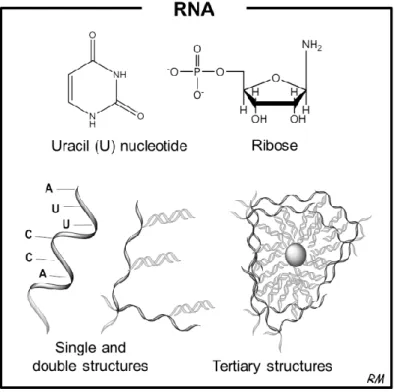
RNA was once considered to be just an intermediate molecule in taking genetic information from the genome to the ribosome, but that view has been changing rapidly by the recent knowledge coming from basic RNA research. Recently, RNA was renowned as a central molecule in cellular processes and gene regulation. This centrality of RNA reflects its unprecedented biochemical properties. The linear sequence of RNA makes it a simple source of genetic information. The property of RNA to form secondary structure, shielding some sequences while exposing others for recognition, facilitates its interactions with other molecules. In a more complex way, RNA can assume tertiary structures that present surfaces for interactions and contain internal environments that create binding sites for metal ions that can promote catalytic reactions (Sharp, 2009).
It is now clear that RNA is a versatile molecule that play key roles in many important biological processes like splicing, editing, protein export and others, and it can also act catalytically, like enzymes (Soll et al., 2001) which underscore the therapeutic potential of RNA as a new gene therapy tool. RNA-based therapeutics make use of the mechanism of activity of the various RNA molecules, which include catalytically active RNA molecules (ribozymes), inhibitors of mRNA translation (antisense oligoribonucleotides), the agents of RNA interference in gene expression (small RNAs), and RNAs that bind proteins and other molecular ligands (aptamers) (Burnett and Rossi, 2012).
1.3.1. Catalytic RNAs: Ribozymes
The discovery that RNA can act as an enzyme changed the paradigm of the central dogma of molecular biology (Lehman, 2010), and led to the development of a new class of therapeutics based on RNAs enzymes. Ribozymes, or RNA enzymes, are RNA molecules that can mediate their own cleavage or splicing or act as enzymes to promote reactions on substrate RNA molecules (Khan, 2006) (figure 4).
Two types of RNA enzymes - hammerhead and hairpin ribozymes – have been the main focus of efforts towards assessing the potential therapeutic utility of ribozymes. These ribozymes were found to mediate inhibition of gene expression through the binding of messenger RNA (mRNA) by complementarity and inducing its site-specific cleavage (figure 4a) with the particularity that ribozymes recycle themselves ready to repeat this process multiple times. Early preclinical works showed that such RNA enzymes could repeatedly cleave practically any pathogenic transcript, which supported their use as therapeutic tools for manipulation of gene expression (Hauswirth et al., 2001).
Several phase I and II clinical trials have been initiated using trans-cleaving ribozymes in a small number of patients with infectious diseases or cancer. In these studies the ribozymes
have been delivered to the patients either by delivery systems or by direct injection of a syntheti ribozyme that ontains hemi al modifi ations that greatly in rease the ribozyme’s stability in biological fluids. However, the gene therapy-based trials have focused upon developing ribozyme-based treatments for individuals infected with the human immunodeficiency virus (HIV) (Burnett and Rossi, 2012).
Recently, the ribozymes with self-splicing ability were exploited to trans-splice RNA targets in order to repair mutant mRNA molecules giving rise to genetic diseases (figure 4b). RNA repair is an alternative way to control gene expression at the mRNA level, repairing mutant mRNAs rather than destroying them. This method uses the splicing and editing processes that create mRNAs from DNA to replace mutant regions in mRNA. The initial studies focused on RNA repair used a trans-splicing version of a group I ribozyme to repair mutant lacZ transcripts in bacteria and mammalian cells. These studies showed that the ribozyme was able to repair the mutant RNA by recognizing the target transcript by base pairing with it, cleaving off mutant sequences and linking a wild-type sequence onto the cleaved product (Phylactou et al., 1998).
Figure 4 – Ribozymes activities. (a) Trans-cleaving ribozymes can bind pathogenic mRNAs through
base-pairing interactions and perform sequence-specific cleavage by phosphodiester isomerization, releasing the reaction products. Ribozymes can repeat this process with multiple turnover. (b) Trans-splicing ribozymes can repair a mutant RNA by recognizing the target transcript upstream of a mutation site (Xm) by base pairing with it. The mutant sequence is cleaving off and an exon with a wild-type sequence (Xwt) is ligated onto the cleavage product to generate a corrected transcript. (mRNA, messenger RNA). (Adapted from Sullenger and Gilboa (2002)).
1.3.2. Antisense oligoribonucleotides
Antisense RNAs are small, diffusible, highly structured RNAs that act via sequence complementarity on target RNAs called sense RNAs. In eukaryotes, some processes like splicing or editing make use also of complementary small RNAs (Brantl, 2002). Antisense RNA has long been thought of as a promising technique for disease therapy. The concept underlying antisense technology is relatively straightforward: the use of a sequence, complementary by virtue of Watson-Crick base pair hybridization, to a specific mRNA can inhibit its expression and then induce a blockade in the transfer of genetic information from DNA to protein. Conceptual simplicity, the possibility of rational design and relatively inexpensive cost has led to the widespread use of these short fragments of RNA as therapeutic agents (Dias and Stein, 2002). The success on clinical trials of this RNA-based therapy has led to the first antisense RNA approved by the Food and Drug Administration (FDA) for commercialization in 1998. Vitravene (Isis Pharmaceuticals/Novartis) is the brand name for Fomivirsen, an antiviral drug used in the treatment of cytomegalovirus retinitis (CMV) in immune compromised patients, including those with acquired immunodeficiency syndrome. Fomivirsen is a synthetic 21-nucleotide sequence with phosphorothioate linkages (which are resistant to degradation by nucleases), which blocks translation of viral mRNA by binding to the complementary sequence of the mRNA transcribed from the coding segment of a key CMV gene (Grillone and Lanz, 2001). Since then, the significant advances in RNA chemistry led to the creation of second-generation antisense technology that was expected to overcome many of the limitations of the original approaches and expand the use of this technology to other diseases. In fact, this has been successful and in the beginning of this year FDA approved an injectable antisense RNA, called KYNAMROTM (mipomersen sodium) from Genzyme Company,
which is an oligoribonucleotide inhibitor of apolipoprotein B-100 synthesis indicated as an adjunct to lipid-lowering medications and diet to reduce low density lipoprotein-cholesterol, apolipoprotein B, total cholesterol, and non-high density lipoprotein-cholesterol in patients with homozygous familial hypercholesterolemia (Genzyme, 2013).
1.3.3. Agents of RNA interference
The Nobel-prize winning in 1998 for the discovery of the mechanism of activity of a class of small RNA (sRNA) molecules produced by eukaryotes aroused a novel therapeutic approach to treat human diseases, called RNA interference (RNAi) technology. The cellular process of RNAi uses sRNAs to mediate resistance to both endogenous parasitic and exogenous pathogenic nucleic acids, and regulates the expression of protein-coding genes through post-transcriptional gene silencing (PTGS). PTGS is regulated by two distinct mechanisms: sequence-specific cleavage of perfectly complementary mRNAs and translational repression and degradation of mRNAs with imperfect complementarity. Small interfering RNA (siRNA) and microRNA (miRNA) are the biological agents of RNAi, a family of regulatory non-coding
RNAs of 19-28 nucleotides in length (figure 5) (Burnett and Rossi, 2012, Kim, 2005). siRNAs are short double-stranded RNA (dsRNA) with 2 nu leotides overhangs at the 3′- ends. In the cytoplasm, siRNAs are loaded into a protein complex called the RNA-induced silencing complex (RISC). The loaded RISC complex then scans all intracellular mRNA for a target mRNA with a complementary sequence to the loaded siRNA. If a target mRNA is found by the loaded RISC, the target mRNA is cleaved and degraded, successfully inhibiting the translation of the target gene. siRNAs can be generated in several ways. In some cases, long dsRNA is introduced in the cell, either by a virus or endogenous RNA expression (microRNA). The enzyme Dicer cleaves the long duplex RNAs into siRNAs (Guo et al., 2010).
On the other hand, miRNAs are originated from endogenous genome DNA sequence and are first transcribed in the nucleus as parts of long primary miRNA transcripts (referred to pri-miRNA) with 5′- aps and 3′- polyA tails. miRNAs with hairpin structures are then processed into pre-miRNAs by the ribonuclease Drosha. The pre-miRNAs are subsequently transported out of the nucleus to cytoplasm by the dsRNA-binding protein Expotin-5, and processed to mature miRNAs by the endoribonuclease Dicer. Similar to siRNA-mediated silencing pathway, miRNA is then loaded into RISC. However, its mode of action is dependent on the extent of sequence complementarity between the miRNA and the target mRNA. When a miRNA matches the sequence of the mRNA completely, the miRNA/RISC complex mediates the cleavage of the mRNA using the same mechanism as siRNA. For miRNAs that only partially match the mRNA's sequence, the miRNA/RISC complex induces translational inhibition and subsequent mRNA degradation. miRNAs silencing is arguably more complex than siRNA silencing, owing to the fact that miRNAs only require partial sequence complementarity to silence genes (Guo et al., 2010, Lin et al., 2006). The miRNA mechanism is not fully understood and some diseases are suggested to be linked to aberrant miRNA expression and function (Soifer et al., 2007).
For therapeutic purposes, siRNA has been the focused molecule in the RNAi pathway. In fact, siRNAs are even being used to interfere with aberrant miRNAs. PTGS may be efficiently induced by delivering exogenously synthetic siRNA molecules to cells. Chemically synthesized siRNA duplex or siRNA molecules prepared in vitro from dsRNAs by incubating with recombinant Dicer protein, are commonly used in research for gene silencing. In this last strategy, Dicer-processed siRNA products simply bypass the Dicer cleavage step. Another way to introduce siRNA into cells is to express short hairpin RNAs (shRNA) genes in plasmid vectors (Guo et al., 2010). shRNA is a sequence of RNA which is created in the cell from a DNA construct encoding a sequence of single stranded RNA and its complement, separated by a stuffer fragment, allowing the RNA molecule to fold back on itself, creating a dsRNA molecule with a hairpin loop. In the cell, it is transcribed under the control of RNA Polymerase-II or Polymerase-III promoters and folds into a structure resembling a siRNA duplex. shRNAs are then processed by Dicer into siRNAs (figure 5) (Guo et al., 2010, Tokatlian and Segura, 2010, Xiang et al., 2006).
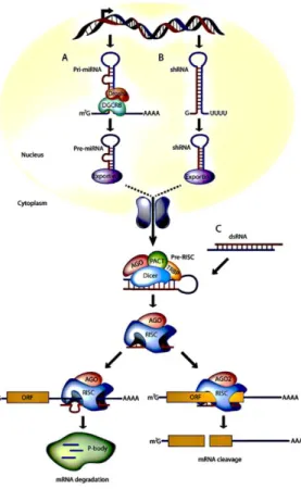
Figure 5 – miRNAs, shRNAs, and siRNAs pathways for RNAi in mammalian cells. (A) miRNA genes are
transcribed by RNA polymerase II to generate the primary transcripts (pri-miRNAs) and processed into stem-loop precursor miRNAs of 70 nucleotides by the Drosha-DGCR8 complex, which are then exported by Exportin 5 to the cytoplasm. Upon export, Dicer participates in the second step of processing to produce miRNA duplexes of 22 nucleotides. The imperfectly complementary miRNA duplexes are associated to the AGO protein and are loaded into RISC, where the passenger strand is removed and the guide strand remains to target mRNA for silencing. The final products (RISC complex) act as guide molecules in translational control or cleavage of certain mRNAs. (B) Like miRNAs, engineered shRNAs are transcribed from DNA and undergo similar processing. However, the perfect Watson-Crick base-pairing between the guide strand and the target mRNA triggers AGO2-mediated cleavage of the mRNA target. shRNA expression cassette can be delivered by viral vectors such as retroviral vector, lentiviral vector, and adenoviral vector or it can be expressed in the nucleus from expression plasmids. (C) In contrast to miRNA and shRNAs, siRNAs are processed in the cytoplasm. But, all steps of siRNA and shRNA are the same after processing by Dicer/TRBP. siRNA can be artificially introduced into the cytoplasm in RNAi-based therapeutics either as a chemically synthesized siRNA duplex or Dicer-processed siRNAmolecules. Viral and non-viral delivery systems are used for siRNA transfer into cells. (miRNAs, micro RNA; shRNAs, small hairpin RNA; siRNAs, small interfering RNA; RNAi, RNA interference; RISC, RNA-induced silencing complex; mRNA, messenger RNA). (Adapted from Burnett and Rossi (2012)).
The RNA interference technology is one of the most exciting biotechnology advances in the last decade. It has revolutionized biology research, including drug target discovery and revitalized interest in the clinical development of nucleic acid-based gene inhibition approaches. Theoretically, RNAi can silence the expression of mRNA for any gene, including growth factors, viral genes, or oncogenes and genes that were once considered therapeutically unreached by small molecule inhibitors. The great effectiveness and the simplicity of the design of a therapeutic siRNA, which only requires knowledge of the target gene's sequence, have contributed for the increased development and success of this RNA-based therapeutic approach. Moreover, the fact that siRNA-mediated RNAi mechanism takes place in the cytoplasm is a potential advantage over other gene regulation mechanisms that require penetrating the nucleus (Tokatlian and Segura, 2010). Therefore, several pharmaceutical companies are focusing on the development of RNAi-based therapeutics for the treatment of a wide range of diseases (Burnett and Rossi, 2012, Melnikova, 2007).
1.3.4. Aptamers
Many small RNAs can fold into three-dimensional structures that allow them to bind target proteins with high affinity and specificity. This additional feature of RNAs makes them tempting to consider as a therapeutic agent since they can bind to proteins and inhibit them in an analogous manner to protein antagonists. This idea of ‘de oy’ RNAs has been shown in preclinical work to slow down HIV replication, where trans-activation response region (TAR) and rev response element (RRE), two decoy RNAs, could be used to competitively inhibit viral protein function and replication. This suggested that other small-structured RNA molecules might be able to bind pathogenic target proteins and inhibit their activity (Kohn et al., 1999). Meanwhile, synthetic short oligoribonucleotides sequences that bind to a specific target molecule with high affinity, called aptamers, emerged as potential molecules for both basic research and clinical purposes as macromolecular drugs. Aptamers are highly specific, relatively small in size, and non-immunogenic. Aptamers are essentially a chemical equivalent of antibodies. However, aptamers can be chemically synthesized to produce large quantities of these compounds for in vivo experimentation and clinical trials. Aptamers are usually created by iterative in vitro selection methods that isolate high-affinity RNA ligands from large pools of randomized RNA sequences (vast RNA shape libraries) that could bind to proteins and small molecules. The selection process was named SELEX (systematic evolution of ligands by exponential enrichment) (Ellington and Szostak, 1990), but natural aptamers also exist as part of a nucleic acid-based genetic regulatory element named riboswitch (Tucker and Breaker, 2005). Since the discovery of aptamers in the early 1990s, great efforts have been made to make them clinically relevant for diseases like cancer, HIV, and macular degeneration. For therapeutic purposes, aptamers can be used to bind and inhibit harmful molecules or serve as targeting ligands for nanomedical constructs. RNA aptamers, simplify the need for chemical conjugation or mixing with other moieties. Aptamers have been used as
ligands for specific delivery of siRNA to prostate cancer cells and lymphocytes (Ni et al., 2011). The first aptamer based therapeutic was FDA approved in 2004 for the treatment of age-related macular degeneration and several other aptamers are currently being evaluated in clinical trials (Burnett and Rossi, 2012).
1.3.5. mRNA vaccination
Coding mRNA is emerging as a particularly attractive option in the development of new approaches for the treatment of cancer or infection diseases focusing on immunotherapies (Kreiter et al., 2011). The use of genetic vaccination, in which a genetically engineered nucleic acid encoding an antigen is administered to an organism in order to stimulate an immunological response against the antigen (Tang et al., 1992), is not a new therapeutic strategy. Vaccines based on nucleic acids are already known to stimulate all effectors of the adaptive immune response: B lymphocytes, cytotoxic T cells, and T helper cells have been exploited for the creation of prophylactic vaccines for infectious diseases and for cancer immunotherapy. However, vaccination approaches involving nucleic acids have focused mostly on DNA-based strategies due to the instability and rapid degradation of RNA in the human body, since RNA is prone to hydrolysis by ubiquitous ribonucleases. Nevertheless, researchers have been overcoming these limitations by developing strategies for stabilization and delivery of mRNA (Pascolo, 2008).
mRNA is a large family of RNA molecules that plays a fundamental and integral role in every living cell. mRNAs are carriers of genetic information from DNA to the ribosome, where they specify the amino acid sequence of the protein products of gene expression. Following transcription of mRNA by RNA polymerase, the mRNA is translated into a polymer of amino acids: a protein. Therefore, the concept of mRNA vaccination is to carry the information of an antigenic protein to be translated in the cell cytoplasm and therefore generate an immunological response. As an alternative to DNA-based vaccines, mRNA-based vaccines present additional safety features including no persistence, no integration in the genome and no autoimmune response. Moreover, mRNA which are generated by in vitro transcription, are easy to produce in large amounts and with very high purity. This feature facilitates the good manufacturing practices process and guaranties reproducibility (Pascolo, 2006).
Vaccination with mRNA can be achieved by several delivery methods, including direct injection of naked mRNA, injection of mRNA encapsulated in liposomes, gene gun delivery of mRNA loaded on gold beads, or in vitro transfection of the mRNA in cells followed by re-injection of the cells into the patients (Pascolo, 2008). This last strategy is generating a lot of interest in cancer immunotherapy. The approach uses tumour mRNA isolated from the patient by biopsy, amplified and ombined with the immune ells from the patient’s bone marrow called dendritic cells (DCs). These cells internalize the mRNA and present the proteins encoded by the mRNA as an antigen on the cell surface, which ensures the stimulation of the
immune response to the tumour (figure 6). The preclinical experience suggests that vaccination with tumour RNA-transfected DCs may constitute a highly effective and broadly applicable treatment for patients with recurring cancer (Sullenger and Gilboa, 2002). Other technologies of direct injection of globin-stabilized mRNA are also being evaluated in human clinical trials (Pascolo, 2006).
Figure 6 - Treatment of cancer patients with tumour RNA-transfected DCs. Tumour cells are removed
from the patient and used to isolate, and if necessary amplify, tumour RNA. Blood cells are obtained from the patient and monocytes are isolated and their proliferation induced to immature DCs. Immature DCs are transfected with RNA and cultured to mature. The antigen-loaded DCs are then infused into the patient. (DCs, dendritic cells). (Adapted from Sullenger and Gilboa (2002)).