UNIVERSIDADE DA BEIRA INTERIOR
Ciências da Saúde
Development of a novel hydrogel for skin
regeneration
Sónia Alexandra Pereira Miguel
Dissertação para obtenção do Grau de Mestre em
Ciências Biomédicas
(2
ºciclo de estudos)
Orientador: Ilídio Joaquim Sobreira Correia, Ph.D.
Coorientador: Maximiano José Prata Ribeiro, MSc.
UNIVERSIDADE DA BEIRA INTERIOR
Ciências da Saúde
Desenvolvimento de um novo hidrogel para a
regeneração da pele
Sónia Alexandra Pereira Miguel
Dissertação para obtenção do Grau de Mestre em
Ciências Biomédicas
(2
ºciclo de estudos)
Orientador: Ilídio Joaquim Sobreira Correia, Ph.D.
Co-orientador: Maximiano José Prata Ribeiro, MSc.
iv
List of Publications
Articles in peer reviewed international journals:
Ribeiro, M., Morgado, P., Miguel, S., Coutinho, P.and Correia, I.; Dextran based-hydrogel containing chitosan microparticles loaded with growth factors to be used in wound healing. Materials Science and Engineering: C. (2013). 33 (5): 2958-2966.
Poster communications:
Patrícia I. Morgado, Maximiano P. Ribeiro, Sónia P. Miguel, Ilídio J. Correia, Ana Aguiar-Ricardo, Development of poly(vinyl alcohol) and chitosan wound dressings using supercritical fluids technology, 11º Encontro Nacional de Química-Física, 9 – 10th of May, Faculty of Sciences, University of Porto, Portugal.
Oral presentations:
M.P. Ribeiro, P.I. Morgado, S.P. Miguel, P. Coutinho, I.J.Correia.,Dextran-based hydrogel containing chitosan microparticles loaded with growth factors to be used in wound healing, VI Symposium on Technology and Health, Instituto Politécnico da Guarda (IPG), 3rd of May 2013, Guarda, Portugal.
vi
“Para ser grande, sê inteiro; nada teu exagera ou exclui; sê todo em cada coisa; põe
quanto és no mínimo que fazes; assim em cada lago, a lua toda brilha porque alta
vive.” Fernando Pessoa
viii
Acknowledgments
At the end of this master thesis, there are a lot of people that I would like thank, by the support and courage. One way or another, they all contributed to this work has been done. First, I would like to thank to my supervisor Professor Ilídio Correia for the unconditional support, guidance, patient, and help during my master’s degree. Their wise advices help me to grow as an investigator and person. I also feel the need to acknowledge him for having believed in my capabilities and for making me believe in myself. It has been a privilege working with him.
I would like thank to my co-supervisor Maximiano Ribeiro, for all guidance and support in progress of my work. His friendship and advices were, without any doubt, very important to overcome the difficulties faced.
I also would like to thank to Eng. Ana Paula from the Optical Center of Universidade da Beira Interior for the help in acquiring scanning electron microscopy images of the hydrogel produced.
I would like to thank my entire colleagues group for their support, knowledge sharing, force and courage in moments less good, but also for all the moments of fun and happiness that we had together, without a doubt, I have been blessed with a friendly and cheerful group. To my closest friends that have always accompanied me in these last 5 years. They always believed in me and they helped me to always have a smile in my face. “In difficult times, is in the hug of a friend that we find the strength to continue”. Thank you all from the bottom of my heart.
Lastly, and most importantly, I would also like to express my sincere thanks to my parents, António and Helena and my sister, Telma. Thanks for the unconditional support and for always believing in me when I doubted about myself. They are truly important in all of my victories; your love is enough to make me feel stronger. To them I dedicate this thesis.
x
Abstract
Skin lesions are traumatic events that lead to increased fluid loss, infections, scars formation and the appearance of immunocompromised regions. The loss of skin integrity can result in significant physiological imbalances and disability or even death. Skin functionality must be restored quickly in order to maintain homeostasis. Researchers have been developing new systems to accelerate the healing process. Although the many skin substitutes available in the market, there is none that promotes full restoration of the native structure of skin. Among the various materials used to cover the wound immediately after injury, hydrogels are the most studied. The hydrogels are made of a highly hydrophilic polymeric network forming a three dimensional structure very similar to the extracellular matrix, that allows cell growth. Moreover, hydrogels are biocompatible, biodegradable and have porous structures that allow cell internalization and proliferation within its structure and promote the diffusion of gases, nutrients and waste products. Currently, new hydrogels that respond to external stimuli such as pH, and temperature have been extensively studied in tissue engineering. Thus, the work plan of this master thesis had as main goal to produce a hydrogel composed of deacetylated chitosan and agarose, formed at body temperature (37°C), in order to verify their applicability in the treatment of wounds. The hydrogel structure was initially characterized by Fourier transform infrared spectroscopy. Its inner and surface morphology was characterized by scanning electron microscopy. Cellular adhesion and internalization into the porous structure of the hydrogel was visualized by confocal and scanning electron microscopy. The cytotoxicity profile of the hydrogel was characterized through cell viability assays, and the results obtained confirmed the biocompatibility of the hydrogel. The antimicrobial activity of the hydrogel was also evaluated and the results showed that the hydrogel inhibits the growth, at the surface, of the most common microorganism in skin infection. The results obtained demonstrated that this 3D network has the suitable properties for improving the healing process of cutaneous wounds.
Keywords
xii
Resumo
As lesões na pele são acontecimentos traumáticos que levam ao aumento da perda de fluidos, a infecções, à formação de cicatrizes e ao aparecimento de regiões imunocomprometidas. A perda da integridade da pele pode resultar em desequilíbrios fisiológicos e incapacidade significante ou mesmo até a morte do paciente. A funcionalidade da pele deve ser restaurada rapidamente, de forma a manter a homeostase. Os investigadores têm procurado desenvolver novos substitutos de pele que permitam acelerar o processo de cicatrização. Apesar de já existirem muitos substitutos de pele disponíveis no mercado, ainda não existe nenhum que promova o restabelecimento da estrutura nativa da pele, na sua totalidade. Entre os mais variados materiais utilizados para cobrir a ferida, imediatamente após a lesão, os hidrogéis são a classe de materiais mais estudada. Os hidrogéis são constituídos por uma rede polimérica altamente hidrofílica, que forma uma estrutura tridimensional muito semelhante à matriz extracelular. Os hidrogéis promovem a adesão e o crescimento celular. Normalmente, os hidrogéis apresentam estruturas porosas que possibilitam a internalização e proliferação das células no seu interior, a difusão de gases, nutrientes e resíduos. Alem disso, os hidrogéis são biocompatíveis e biodegradáveis. Atualmente, os hidrogéis que respondem a estímulos externos como o pH, temperatura têm sido amplamente estudados na engenharia de tecidos. Assim, o plano de trabalhos deste mestrado teve como objetivo a produção de um hidrogel constituído por quitosano desacetilado e agarose, o qual se forma à temperatura corporal (37°C), para ser aplicado no tratamento de feridas cutâneas. A composição química do hidrogel foi analisada por espectroscopia de infravermelho com transformada de Fourier e a morfologia da superfície e do interior foi caraterizada por microscopia electrónica de varrimento. A visualização da adesão e internalização celular no hidrogel foi conseguida através de imagens de microscopia electrónica de varrimento e confocal. O perfil citotóxico do hidrogel foi caraterizado através de testes de viabilidade celular, os quais confirmaram a biocompatibilidade do hidrogel. A atividade antimicrobiana do hidrogel foi também avaliada e os resultados obtidos confirmaram que o hidrogel inibe à sua superfície, o crescimento do microorganismo mais comum em infecções cutâneas (Staphylococcus aureus). Os resultados obtidos neste trabalho demonstram que o hidrogel desenvolvido possui as propriedades adequadas para ser usado na regeneração das feridas cutâneas.
xiii
Palavras-chave
Actividade antimicrobiana, Biocompatibilidade, Cicatrização de feridas, Estudos in vitro, Hidrogel, Internalização celular.
xv
Resumo alargado
A pele é o maior órgão do corpo humano, podendo pesar entre 6 a 10 kg. Em adultos, apresenta uma área aproximada de 2m2. Está envolvida na preservação da homeostase dos fluidos do corpo, na regulação térmica e na protecção contra agentes infecciosos. Diariamente, a pele está sujeita à perda da sua integridade devido a uma doença ou trauma. As lesões na pele são descritas como resultado da disrupção da estrutura anatómica e funcional da pele. As lesões cutâneas podem ser causadas por uma interrupção precisa de tecido pelo bisturi do cirurgião (incisão), por grandes danos no tecido (por exemplo, trauma, queimaduras), ou ainda resultado de uma contusão, hematoma, laceração ou uma resistência à abrasão. Consoante, o tipo e duração do processo de cicatrização das feridas, estas podem ser classificadas como agudas ou crónicas e que podem resultar num desequilíbrio fisiológico, numa deficiência significativa ou até mesmo na morte do indivíduo.
Desta forma, a funcionalidade da pele deve ser restaurada rapidamente, de forma a manter a sua homeostase. A cicatrização de feridas é um processo dinâmico e complexo que inclui uma série de fases como, coagulação, inflamação, síntese e deposição da matriz extracelular, angiogénese, fibroplasia, epitelização, contração e remodelação. Estas fases do processo cicatricial ocorrem em cascata e envolvem a migração de vários tipos de células para o local da lesão. Após uma lesão, a pele necessita ser revestida de forma a diminuir a dor, a contaminação, o risco de infeção e, por outro lado promover o restabelecimento da integridade da pele. Esta necessidade fez com que nas últimas décadas um grande número de
substitutos de pele fosse desenvolvido e introduzido no mercado. Os substitutos de pele, estabelecem uma barreira mecânica contra microorganismos e previnem a desidratação; promovem a entrega de componentes da matriz extracelular (colagénio, proteínas de adesão), citocinas e fatores de crescimento que contribuem para melhorar o processo de cicatrização normal; e ainda, fornecem uma estrutura de suporte para incorporação de células e moléculas bioativas, as quais são aplicadas no local da lesão e auxiliam o processo de regeneração.
Muitos substitutos de pele têm sido aplicados no tratamento de feridas cutâneas, como é o caso dos auto-, alo- e xeno- enxertos. No entanto, estas abordagens terapêuticas apresentam inúmeras limitações tais como, a rejeição por parte do paciente, o risco de transmissão de doenças e, ainda a disponibilidade limitada dos recursos em relação à procura. De modo a suplantar estas limitações, investigadores têm procurado desenvolver novos substitutos de pele à base de biomateriais que permitam acelerar o processo de cicatrização. Existem já substitutos de pele disponíveis no mercado, classificados de acordo com a camada da pele que pretendem substituir (Epidérmicos, Dérmicos e Dermo-epidérmicos), os quais são constituídos à base de polímeros naturais/ sintéticos e, alguns deles possuem células encapsuladas (fibroblastos e/ou queratinócitos).
xvi Entre os mais variados tipos de materiais utilizados para a produção de uma estrutura que promova a cicatrização de uma ferida cutânea, os hidrogéis surgem como os substitutos mais estudados na engenharia de tecidos devido ao seu caráter hidrofílico, que forma uma rede tridimensional muito semelhante à matriz extracelular. O seu alto conteúdo em água dos hidrogéis torna-os biocompatíveis para a maioria dos tecidos vivos e a sua natureza viscoelástica diminui o dano do tecido circundante, quando implantado no hospedeiro.
Os hidrogéis fornecem um microambiente que contém poros suficientemente grandes para permitir a adaptação das células, as quais podem entrar e proliferar no interior da rede polimérica. Adicionalmente, estes poros permitem também, a difusão de gases, nutrientes e resíduos. Polímeros naturais como agarose e o quitosano são dos mais usados na produção de hidrogéis pois não induzem uma resposta imunológica crónica no hospedeiro, uma vez que estes são biodegradáveis e biocompatíveis.
Os métodos tradicionais usados para a produção de hidrogéis incluem a co-polimerização e a reticulação dos percursores poliméricos. Estes métodos de síntese de hidrogéis não permitem um controlo preciso da sua estrutura, produzindo hidrogéis com deficientes propriedades mecânicas. Por conseguinte, os hidrogéis que respondem a estímulos externos como o pH e temperatura têm sido amplamente estudados na engenharia de tecidos. Hidrogéis que são produzidos apenas pela alteração da temperatura, no local da lesão oferecem diversas vantagens, uma vez que, não necessitam de solventes orgânicos ou agentes de co-polimerização para reticular, permitem que o processo seja mais simples e rápido. Além disso, o hidrogel que é polimerizado na lesão torna o método menos invasivo, aumenta a viabilidade e melhora o conforto ao paciente, reduzindo-lhe a dor associada ao processo de cicatrização de feridas. No entanto, nenhum destes substitutos de pele promove o restabelecimento da estrutura nativa da pele, na sua totalidade.
Assim, o plano de trabalhos deste mestrado teve como objetivo avaliar a aplicabilidade de um hidrogel, constituído por quitosano desacetilado e agarose no tratamento de lesões de pele. A composição química do hidrogel foi analisada por espectroscopia de infravermelho com transformada de Fourier e a morfologia do hidrogel foi caracterizada através de imagens de microscopia electrónica de varrimento e microscopia confocal. A biocompatibilidade deste biomaterial foi avaliada através de ensaios in vitro. Fibroblastos humanos, foram usados como modelos nos ensaios in vitro, de modo a avaliar a citotoxicidade do hidrogel. A atividade antimicrobiana do hidrogel foi também avaliada, através de ensaios microbiológicos com Staphylococcus aureus. Verificou-se que agarose e quitosano desacetilado estabeleciam pontes de hidrogénio entre si e formavam um hidrogel poroso. Os fibroblastos aderiram e proliferaram na presença do hidrogel em estudo, o que permitiu concluir a ausência de citotoxicidade deste, assim como dos produtos resultantes da sua degradação. Os resultados obtidos dos ensaios de microbiologia confirmaram que o hidrogel inibe à sua superfície, o crescimento do Staphylococcus aureus. Os resultados obtidos sugerem que o hidrogel desenvolvido possui as propriedades adequadas para ser usado na regeneração das feridas cutâneas.
xviii
Table of Contents
Chapter I- Introduction
1.Introduction ... 1
1.1 Skin ... 2
1.1.1 Functions and structure ... 2
1.1.2 Epidermis ... 4
1.1.3 Dermis ... 5
1.1.4 Hypodermis ... 6
1.1.5 Skin appendages ... 6
1.2 Wounds ... 7
1.2.1 Types of wound healing ... 8
1.2.2 The wound healing mechanism ... 9
1.2.2.1 Haemostasis ... 10
1.2.2.2 Inflammation ... 10
1.2.2.3 Cell migration and proliferation ... 11
1.2.2.4 Remodelling (maturation) ... 13
1.3 Tissue Engineering ... 14
1.3.1 Tissue engineering: skin substitutes ... 18
1.4 Hydrogels ... 24
1.5 Chitosan ... 28
1.6 Hydrogels stimulus-sensitive: agarose... 31
1.7 Main Goals ... 34
Chapter II- Material and Methods
2.Material and Methods ... 362.1 Materials ... 36
2.2 Methods ... 36
2.2.1 Deacetylation of Chitosan ... 36
xix
2.2.3 Fourier transform infrared spectroscopy analysis ... 37
2.2.4 Study of water uptake ability ... 37
2.2.5 Proliferation of fibroblast cells in the presence of CAH ... 37
2.2.6 Characterization of the cytotoxic profile of the hydrogel produced-MTS assay 38 2.2.7 Evaluation of antimicrobial activity of CAH ... 38
2.2.8 Scanning electron microscopy analysis ... 39
2.2.9 Confocal microscopy analysis ... 39
2.2.10 Statistical analysis of the results ... 39
Chapter III- Results and Discussion
3.Results and Discussion ... 413.1 Characterization of the morphology of CAH ... 41
3.2 FTIR-analysis of CAH ... 45
3.3 Swelling behavior of CAH ... 47
3.4 Evaluation of viability and cell proliferation in contact with CAH ... 48
3.5 Characterization of the cytotoxic profile of CAH ... 54
3.6 Evaluation of the antimicrobial activity of the CAH ... 56
Chapter IV- Conclusion
4.Conclusion ... 60Chapter V- Bibliography
5.Bibliography ... 63Chapter VI- Appendix
6.Appendix ... 80xxi
List of Figures
Chapter I- Introduction
Figure 1: Scheme of skin structure ... 3 Figure 2: Representation of the skin components and layer. ... 5 Figure 3: Phases of wound healing ... 9 Figure 4: Representation of the inflammatory phase after a skin injury ... 11 Figure 5: Cell migration and proliferation phase ... 13 Figure 6: Remodelling phase ... 14 Figure 7: Representation of a tissue engineering concept that involves seeding cells within
porous biomaterial scaffolds ... 16
Figure 8: Schematic representation of the production of a skin substitute at the laboratory. 17 Figure 9: Schematic description of the strategies used to control hydrogel loaded with cells
immunogenicity ... 27
Figure 10: Representation of chitosan structure ... 29 Figure 11: Schematic illustration of chitosan's solubility. ... 30 Figure 12: Schematic representation of agarose ... 32
Chapter III- Results and Discussion
Figure 13: Macroscopic images of CAH ... 41 Figure 14: SEM images of CAH. ... 43 Figure 15: CLSM images of CAH. ... 44 Figure 16: FTIR spectra of the produced CAH and the compounds used for their production. 46 Figure 17: Swelling profile of the produced CAH... 48 Figure 18: Microscopic images of human fibroblast cells after being seeded in the presence of
the CAH ... 50
Figure 19: SEM images of human fibroblast cells in contact with CAH. ... 51 Figure 20: 3D visualisation of the cell localization within CAH. ... 52 Figure 21: 3D visualisation of the cell internalization in the CAH ... 53 Figure 22: Cellular activities measured by the MTS assay after 24h and 72h ... 55 Figure 23: Representation of the reduction of the resazurin by viable bacteria and MIC values
of CAH ... 58
xxiii
List of Tables
Chapter I- Introduction
Table 1: Example of commercially available epidermal skin substitutes. ... 21 Table 2: Example of commercially available dermal skin substitutes.. ... 22 Table 3: Example of commercially available dermo-epidermal skin substitutes. ... 23 Table 4: Advantages and disadvantages of hydrogels for being used in TE ... 24
xxv
List of Abbreviations
3D Three-dimensional
Auto Autologous
BSA Bovine Serum Albumin
CAH Chitosan-Agarose Hydrogel
CFU Colony-Forming Unit
CSLM Confocal Laser Scanning Microscopy
DD Degree of Deacetylation
DEJ Dermal-Epidermal Junction
DMEM Dulbecco’s Modified Eagle’s Médium
ECM Extracellular Matrix
EDTA Ethylenediaminetetraacetic Acid
EtOH Ethanol
FBS Fetal Bovine Serum
FGF Fibroblast Growth Factor
FTIR Fourier-Transform Infrared Spectroscopic
GAGs Glycosaminoglycans
HA Hyaluronic Acid
K- Negative Control
K+ Positive Control
MIC Minimum Inhibitory Concentration
MMP Metalloproteinases
MMW Medium Molecular Weight
MTS
(3-(4,5-dimethylthiazol-2-yl)-5-(3-carboxymethoxyphenyl)-2-(4-sulfophenyl)-2H-tetrazolium
P(PF-co-EG) Poly (Propylene Fumarate- co- Ethylene Glycol)
PAA Poly (Acrylic Acid)
xxvi
PDFs Platelet-Derived Growth Factors
PEO Poly (Ethylene Oxide)
PGA Polyglycolic Acid
PI Propidium Iodide
PLA Polylactic Acid
PVA Poly (Vinyl Alcohol)
RGD Arginine–Glycine–Aspartic
SEM Scan Electron Microscopy
Synth Synthetic
TE Tissue Engineering
TGF Transforming Growth Factors
TIMP Tissue Inhibitors of Metalloproteinases
US United States
VEGF Vascular Endothelial Growth Factor
Chapter I
Introduction
1
1. Introduction
The skin is the largest organ of the human body and plays highly specialized functions. It protects against toxins and microorganisms from the environment and prevents the body from dehydration. It is also involved in other functions, such as immune surveillance, sensory detection, and self-healing. Overall, skin is an indicator of general well-being and health state of each individual (Holbrook and Wolff, 1993, Clark et al., 2007, Menon and Kligman, 2009, Lanza et al., 2007).
Skin injuries have a high impact on the life quality of patients. In fact, the World Health Organization (WHO) estimates that, annually, over 300 000 millions suffer from a partly devastating physical and emotional consequences thereof (Yildirimer et al., 2012). Skin injuries are caused by burns, accidents and diseases. Depending on the severity of the wound, loss of skin integrity may result in substantial physiologic imbalance and ultimately in significant disability or even patient death (Zhang and Michniak-Kohn, 2012).
The most common cause of significant skin loss is thermal injury, which accounts for approximately one million of emergency visits per year to the health care system, just in the United States (US) (Association, 2005). Depending on the location and depth of the burn, a victim may experience a wide number of potentially fatal complications including shock, infection, electrolyte imbalances and respiratory failure. Beyond physical complications, burns can also result in severe psychological and emotional distress due to long-term hospitalization, scarring and deformity (Evers et al., 2010). For a complete restoring of both skin structure and function, successful wound healing must occur. The healing process of an adult skin is complex, requiring the collaborative efforts of several tissues and cell lineages, as well as both extracellular and intracellular signals (Kirker et al., 2002, Gurtner et al., 2008, Sun et al., 2011).
In order to solve the problems associated with re-establishment of skin native structure and to promote the mechanisms responsible for improving the healing, some research groups have investigated the development of wound dressings that could protect the wound from bacterial infection, dehydration and absorption of the wound exudate, in order to enhance the healing process (Kirker et al., 2002). Skin substitutes have a high demand for clinical uses. Actually, they represent approximately 50% of tissue engineering and regenerative medicine market revenues. By the year of 2019, the total potential target population for the use of tissue-engineered skin replacements and substitutes is expected to increase to 6.4 million, resulting in a potential market just in US of approximately 24.3 billion dollars (Zhang and Michniak-Kohn, 2012).
In a near future, Tissue Engineering (TE) purposes to produce a biodegradable wound dressing that promotes the re-establishment of skin’s native structure (epidermis, dermis and skin
Chapter I- Introduction
2 appendages). Furthermore, these skin substitutes are expected to reduce costs and pain associated with skin regeneration (Lanza et al., 2007).
The introduction section of this MSc thesis aims to describe the problems and strategies that are currently being developed to improve skin regeneration. The others sections describe the methods, results and conclusions obtained in this study.
1.1 Skin
1.1.1 Functions and structure
Skin occupies almost 2m2 of surface area and accounts for 8% of the body’s mass (Hunt et al., 2009, Bhat and Kumar, 2012, Marieb and Hoehn, 2010). Skin is an anatomical barrier between the organism and environment, which aims to protect it from toxic substances, pathogens and microorganisms (Hunt et al., 2009, Zhang and Michniak-Kohn, 2012, Holbrook and Wolff, 1993). Essentially, skin is involved in four main functions, such as (Young, 2006, Hunt et al., 2009, Marieb and Hoehn, 2010).
Protection: the skin provides protection against ultraviolet light, mechanical,
chemical and thermal insults. Moreover, its relatively impermeable surface prevents patient dehydration and acts as a physical barrier to avoid microorganisms invasion.
Sensation: the skin is largest sensory organ in the body and contains a variety of
receptors for touch (Meissner’s Corpuscle), pressure (Pacini Corpuscle), pain (nociceptors) and temperature (thermo-receptors).
Thermoregulation: in humans, skin is a major organ of thermoregulation. The body is
insulated against heat loss by presence of hairs and subcutaneous adipose tissue. Heat loss is facilitated by evaporation of sweat from the skin surface and increased blood flow through the rich vascular network of the dermis.
Metabolic functions: subcutaneous adipose tissue constitutes a major store of
energy, mainly in the form of triglycerides. Vitamin D, which is responsible by maintenance of calcium and phosphorus concentration in blood, is synthesised in the epidermis.
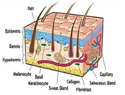
3 Human skin also provides a potential path for the transport of functional active therapeutic substances such as drugs/reagents/biomolecules into blood stream (topical delivery) and/or the body (transdermal delivery) (Guy and Hadgraft, 2003). Skin is structurally complex and highly specialized consisting of two intimately associated layers known as the epidermis and dermis (Lodish et al., 2001, Hunt et al., 2009). Some authors refer a third layer, the hypodermis or subcutaneous layer, that is mainly composed of fat (Metcalfe and Ferguson, 2007a). The epidermis is mainly responsible for providing a barrier to prevent water loss and infection (Elias, 2007). Epidermis and dermis are separated by the extracellular matrix (ECM) also known as the basal lamina (Ajani et al., 2007, Horch et al., 2007). Certain skin appendages such as hair follicles and sweat glands, span from the epidermis to dermis and penetrate into the subcutaneous adipose tissue below the dermis, also known as hypodermis (Lodish et al., 2000, Metcalfe and Ferguson, 2007b). The main functions of the hypodermis comprises the providing of blood vessels and nerves to the skin and also allow its connection to the underlying bones and muscles (Seeley et al., 2005).
So, three main cell populations are found in the skin: epithelial cells covering the surface as the epidermis, but also forming the structures of the hair follicles, sweat and sebaceous glands; the fibroblasts, which are found in the dermis, give shape and maintain skin structure; and the endothelial cells that are found in the blood vessels (Shakespeare, 2001).
Chapter I- Introduction
4
1.1.2 Epidermis
The outermost layer of the skin, known as the epidermis, is mainly responsible for providing a barrier function to prevent water loss and infections (Brohem et al., 2011). The epidermis is mainly constituted by keratinocytes (90-95%), which proliferate in the stratum basale and are responsible for the resistance as well as the structural characteristics and impermeability of the skin (Seeley et al., 2005, Scheuplein, 2011). Notwithstanding, this layer also contains other cell types such as Langerhans cells, which have an essential role in the skin immunological defense (Regnier et al., 1998), melanocytes that contribute for the color of the skin and Merkel cells, that are thought to play a sensory role (Boyce and Warden, 2002). The epidermis does not contain any blood vessels and, as a result, it is usually possible to rub off dead cells from the epidermis without bleeding (Marieb and Hoehn, 2010, Hunt et al., 2009). The continuous process of all proliferation, differentiation and ultimately, death and shedding, allow the compartmentalization of this layer of skin into a number of sub- layers representing different stages of keratinocyte maturation.
Cells produced by proliferation of the germinal basal layer adjacent to the dermis undergo maturational changes related with the production of keratin. The outer keratinised layer is removed continuously and is replaced by the progressive movement and maturation of cells from the germinal layer. The rate of the mitosis in the germinal layer generally equals the rate of desquamation from the outer surface. As shown in Figure 2, the stages of this dynamic process are represented on four morphological sub-layers. The stratum basale is the germinal layer of the epidermis and its mitotic activity provides a constant supply of new keratinocytes to replace those lost by normal wear and tear. The stratum spinosum contains cells that are in the process of growth and early keratin synthesis. The stratum granulosum is characterized by intracellular granules that are involved in the process of keratinisation. The stratum corneum consists of flattened, fused cell remnants are mainly composed by fibrous protein, keratin (Zhang and Michniak-Kohn, 2012, Young, 2006, Scheuplein, 2011, Balasubramani et al., 2001).
5
Figure 2: Representation of the skin components and layers. Epidermis contains melanocytes and
keratinocytes that are able to differentiate and form the different strata (corneum, luccidum,
granulosum, spinosum and basale). Dermis contains fibroblasts embedded in a matrix. Hypodermis is
composed by the adipose tissue (adapted from (Brohem et al., 2011)).
The epidermis is bound tightly to the underlying dermis through the basement membrane at the Dermal-epidermal junction (DEJ). The basement membrane can be divided into lamina lucida (the layer closer to the epidermis that is made of laminin, integrins, entactins, and dystroglycans) and lamina densa (a sheet-like structure composed mainly collagen type IV). The whole basement membrane is involved in the mechanically stabilization of the epidermis (Zhang and Michniak-Kohn, 2012, Seeley et al., 2005, Inoue et al., 2005).
1.1.3 Dermis
The dermis lies below the epidermis and constitutes the main part of the skin (Brohem et al., 2011). The cellular components of dermis include fibroblasts (as shown in Figure 2), endothelial cells, smooth muscle cells, macrophages and adipocytes. Fibroblasts are responsible for the production of collagen, elastic fibers, glycosaminoglycan and glycoproteins, that comprise the ECM (Zhang and Michniak-Kohn, 2012, Seeley et al., 2005). The expression and function of these ECM proteins and their receptors are highly regulated spatially and temporally during wound healing and tissue remodelling. Inappropriate deposition of ECM components will impair the normal healing and function of this tissue. The ECM components play important roles in every step of wound healing processes by providing
Chapter I- Introduction
6 both scaffold support and playing signaling roles, promoting cell adhesion and migration during wound repair. Furthermore, they also mediate interactions between cells and matrix and act as reservoir and modulator for growth factors. Such is fundamental for connecting epidermis to dermis and also for restoring the integrity and function of the skin (Hunt et al., 2009, Seeley et al., 2005, Li and Kirsner, 2005).
The dermis is composed by the papillary and the reticular dermis. The papillary dermis contains more cells than fibers. It also holds numerous blood vessels that provide nutrients to the overlying epidermis, remove waste products and even help to regulate the body temperature (Seeley et al., 2005, Zhang and Michniak-Kohn, 2012). On the other hand, the reticular dermis is mainly composed by dense collagen and elastic fiber matrix, being the main source of strength and flexibility of the skin (Eckhart et al., 2003, Inoue et al., 2005, Seeley et al., 2005, Zhang and Michniak-Kohn, 2012).
1.1.4 Hypodermis
Despite being recognized by the majority of authors as a subcutaneous tissue, the hypodermis is composed by collagen and elastin fibers. Fibroblasts, adipose cells (as seen in Figure 2) and macrophages can also be founded in hypodermis. Approximately half of the fat stored in the body can be found in this layer, although their amount and location depend of the age, sex and diet. The main functions of the hypodermis are supplying of energy and also assure mechanical strength to skin (Böttcher-Haberzeth et al., 2010, Seeley et al., 2005, Metcalfe and Ferguson, 2007b).
1.1.5 Skin appendages
Skin has a variety of appendages, principally hairs, sebaceous and sweat glands. In most areas of the skin, the sweat glands are simple, coiled tubular glands that secrete a watery fluid onto the skin surface, by the process of exocrine secretion. The coiled, secretory portions of these glands are an important component of the thermoregulatory mechanism in humans. For instance, sebaceous glands secrete sebum to moisturize the skin and hair and even the hair follicles, which are a source of proliferation of keratinocytes during epithelialization, performing an important role in wound healing. So, the sebum acts as a waterproofing and moisturising agent for the hair and skin surface (Lanza et al., 2007, Seeley et al., 2005, Zhang and Michniak-Kohn, 2012, Young, 2006). Moreover, there are nerve fibers that can be both myelinated and unmyelinated. The unmyelinated fibers go from the epidermis up to the granular layer. Myelinated fibers are distributed regularly in the upper dermis with terminations to Meissner corpuscles and Merkel complexes. These nerve fibers are responsible for cutaneous sensations and prevent skin from injury due to heat, cold, pain and pressure. The smooth muscle of the skin, the arrector pili muscle, helps in the erection of the hair
7 (goose pimples) in cold weather, thereby trapping warm air near the skin, to protect against the cold (Zaidi and Lanigan, 2010).
1.2 Wounds
Wound healing is a highly regulated process of cellular, humoral and molecular mechanisms that starts immediately after an injury (Lazarus et al., 2002, Enoch and Leaper, 2005). According to the US Wound Healing Society, a wound can be described as a result of the “disruption of normal anatomic structure and function” of the skin, resulting from physical or thermal damage or as a consequence of the presence of an underlying medical or physiological condition (Boateng et al., 2008). Wounds can be classified based on the nature of the repair process, on the number of skin layers and area of skin affected. Based on the type of the repair process, wounds can be classified as acute or chronic wounds (Boateng et al., 2008, Li et al., 2007).
Acute wounds are characterized by their ability to heal completely with minimal scarring within the expected time frame, normally 8-12 weeks (Boateng et al., 2008). The newly formed tissue has a similar structure and comparable functions to native skin (Li et al., 2007). Mechanical injuries, due to external factors such as abrasions, caused by frictional contact between the skin and hard surfaces, are the primary causes of acute wounds (Li et al., 2007, Boateng et al., 2008). Other examples of mechanical injuries include penetrating wounds caused by knives, gun shots and surgical procedures. Burns and chemical injuries (caused by radiation, electricity, corrosive chemicals and thermal sources) are another categories of acute wounds (Boateng et al., 2008).
Chronic wounds represent a different kind of challenge for TE (Supp and Boyce, 2005). These are characterized by their slow healing, i.e., that do not heal after 12 weeks of injure occur and often reoccur. Repeated tissue insults or underlying physiological conditions such as diabetes and other malignances, persistent infections, poor primary treatment and other patient related factors, contributes to the disability of these wounds to heal faster (Boateng et al., 2008). These wounds involve a large surface area and have a high incidence in general population, featuring an enormous medical and economic impact (Supp and Boyce, 2005). Pressure and leg ulcers (venous, ischaemic or of traumatic origin) are known as the most common chronic wounds (Boateng et al., 2008, Supp and Boyce, 2005).
Some authors have described wounds both acute and chronic that are difficult to heal as “complex wounds” with unique characteristics, such as: extensive loss of the integument which comprises skin, hair, and associated glands; infection that may result in tissue loss; tissue death or impairment of the signs circulation and presence of pathology (Boateng et al., 2008). There are also anomalies in certain stages of the healing process that can result in
Chapter I- Introduction
8 excessive healing (e.g. hypertrophic scars, keloids) (Enoch and Leaper, 2005, Reinke and Sorg, 2012).
Skin functionality must be quickly restored, in order to maintain homeostasis. The scarring of acute wounds involves a complex process comprising a series of dynamic events (Enoch and Leaper, 2005). Healing of acute wounds occurs as a carefully regulated cascade of overlapping processes, that require the coordination of a variety of cellular activities, including phagocytosis, chemotaxis, mitogenesis, and synthesis of components of the ECM (Enoch and Leaper, 2005).
1.2.1 Types of wound healing
In each healing process, there are several mechanisms involved. The severity of the wound, number of skin layers affected and the occurrence or absence of bacterial infection allows us to classify the wound healing in different categories (Gurtner et al., 2008, Enoch and Leaper, 2005, Reinke and Sorg, 2012):
Primary healing- occurs when a wound is closed within 12-24 hours after its occurrence (e.g. clean surgical incision, clean laceration). The incisions cause only a localized disruption in the basal membrane and the death of some cells of the underlying connective tissue. In this type of healing, epithelial regeneration predominates over fibrosis (Ladin, 2001, Enoch and Leaper, 2005, Gurtner et al., 2008).
Delayed primary healing- occurs in a contaminated or poorly delineated wound. The skin and subcutaneous tissues are left unopposed and the closured is performed after host defenses have helped to debride the wound. After 3-4 days, the local recruitment of phagocytic cells to the wound occurs and inflammatory cells kill the contaminating bacteria. Collagen metabolism is usually unaffected and the wound retains its tensile strength, as if the wound closure occurred immediately (Enoch and Leaper, 2005, Gurtner et al., 2008, Sun et al., 2011).
Partial-thickness wounds (superficial healing)- are seen in injuries such as superficial burns and abrasions where the injury involves the epithelium and the superficial part of the dermis. The cells migrate towards each other from the basal layer to surround the wound. The healing occurs purely by epithelialization, and the anatomical and physiological restoration is virtually completed (Enoch and Leaper, 2005, Gurtner et al., 2008, Reinke and Sorg, 2012).
9
Full-thickness wounds (secondary healing)- occur in a wound where an extensive loss of soft tissue occurred, as a consequence of a major trauma, severe burns and after some surgical procedures. This type of wounds requires the application of procedures that help the wound contraction and subsequently allows epithelialization. The epithelial cells alone are not able to restore the skin original architecture, so there is ingrowth of granulation tissue from the wound margin, followed by accumulation of ECM with the laying down of collagen (Enoch and Leaper, 2005). Myofibroblasts, which have structural properties similar to that of fibroblasts and a smooth muscle cells, play a crucial role in the healing of this type of wounds (Enoch and Leaper, 2005, Ladin, 2001, Sun et al., 2011, Reinke and Sorg, 2012).
1.2.2 The wound healing mechanism
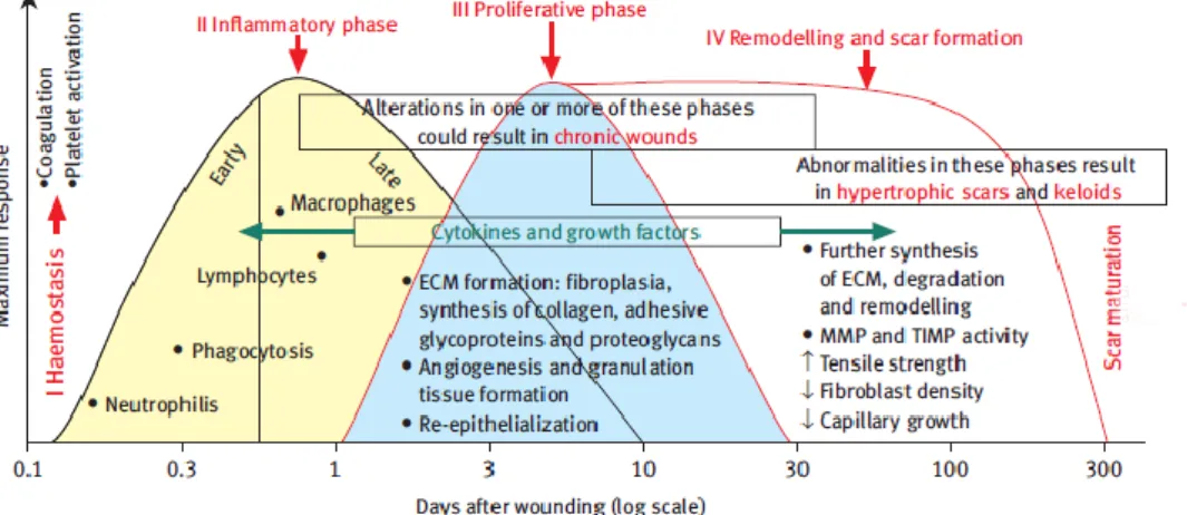
Wound healing is a complex biological process that includes a wide range of mechanisms such as coagulation, inflammation, matrix synthesis and deposition, angiogenesis, fibroplasia, epithelialization, contraction and remodelling (Reinke and Sorg, 2012, Ladin, 2001, Alemdaroğlu et al., 2006). These events occur in cascade and involve the migration of several cell types at the wound site, during the different stages of the healing process (as seen in Figure 3) (Enoch and Leaper, 2005).
The normal mammalian response to wound injury occurs in four overlapping but distinct phases: haemostasis, inflammation, migration-proliferation and remodelling (maturation) (Reinke and Sorg, 2012, Gurtner et al., 2008).
Figure 3 : Phases of wound healing. This mechanism is the result of a complex process, where different
types of cells are involved in the different stages of the healing process. Phase I: Haemostasis which is characterized by coagulation and platelet activation; Phase II: Inflammatory phase where the cells of the immune system are recruited to injury site; Phase III: in Proliferation phase occur the formation of ECM, granulation tissue and also angiogenesis; Phase IV: Remodeling, formation and maturation of the scar. ECM: Extracellular matrix; MMP: Metalloproteinases; TIMP: Tissue inhibitors of metalloproteinases (adapted from (Enoch and Leaper, 2005)).
Chapter I- Introduction
10
1.2.2.1 Haemostasis
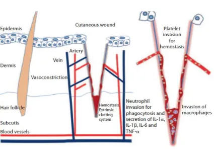
The first stage of wound healing process is dedicated to haemostasis and the formation of a provisional wound matrix. Such event occurs immediately after injury and is completed after some hours (as depicted in Figure 4) (Reinke and Sorg, 2012, Enoch and Leaper, 2005, Gurtner et al., 2008). It begins with the formation of a platelet plug, followed by a fibrin matrix deposition which becomes the scaffold for cell infiltration. There is an invasion of inflammatory cells such as leukocytes, macrophages and neutrophils at the wound site. These cells and platelets release cytokines and growth factors in order to activate the inflammatory process (Reinke and Sorg, 2012).
1.2.2.2 Inflammation
The inflammatory phase of the wound healing cascade occurs immediately after tissue damage. In this stage, components of the coagulation cascade, inflammatory pathways and immune system are all needed for preventing the ongoing blood and fluid losses, to remove dead and devitalized tissues and also avoid infection (Gurtner et al., 2008, Reinke and Sorg, 2012). This phase can roughly be divided into an early phase with neutrophil recruitment and a late phase with the appearance and transformation of monocytes (as shown in Figure 4) (Grose and Werner, 2004, Gurtner et al., 2008). Then, neutrophils are recruited to the wound, and the degranulation of platelets occurs. Moreover, once at the wound site, neutrophils perform their function of killing and phagocytising bacteria and damaged matrix proteins within the wound bed. The role that neutrophils play is crucial within the first days after injury, due to their ability to perform and also secrete proteases, which kills the local bacteria and helps to degrade the necrotic tissue (Reinke and Sorg, 2012). After 2-3 days, monocytes appear at the wound site and differentiate into macrophages. Macrophages get into the injury and support the ongoing process, by performing phagocytosis of pathogens and cell debris, as well as the secretion of growth factors, chemokines and inflammatory cytokines (IL-6, TNF-α) (Gurtner et al., 2008, Reinke and Sorg, 2012, Profyris et al., 2012). Macrophages are involved in host defence, resolution of inflammation, removal of apoptotic cells and support cell proliferation and tissue restoration after injury (Koh and DiPietro, 2011, Reinke and Sorg, 2012).
11
Phase 2: Inflammatory phase (days 1-3)
Figure 4: Representation of the inflammatory phase after a skin injury; haemostasis and invasion of
inflammatory cells (adapted from (Reinke and Sorg, 2012)).
1.2.2.3 Cell migration and proliferation
Cell migration occurs 2-10 days after injury and is characterized by the migration of different cell types. The migration phase involves the movement of epithelial cells (such as keratinocytes) and fibroblasts to the injured area, in order to replace damaged or lost tissue (Gurtner et al., 2008, Strodtbeck, 2001). New blood vessels are formed, in a process known as angiogenesis (as illustrated in Figure 5) and the sprouts of capillaries associated with fibroblasts and macrophages replace the fibrin matrix by a granulation tissue (Strodtbeck, 2001, Bauer et al., 2005, Reinke and Sorg, 2012).
In the proliferation phase (3-10 days after injury) the main focus of the healing process relies on covering of the wound surface. In this stage, the formation of a granulation tissue occurs and the vascular network is restored (Robson et al., 2001, Strodtbeck, 2001, Bauer et al., 2005, Gurtner et al., 2008). After fibroblasts migrate along the fibrin network, the reepithelialisation starts from the wound edges, neovascularization and angiogenesis processes get activated (Bauer et al., 2005, Strodtbeck, 2001, Reinke and Sorg, 2012, Enoch and Leaper, 2005). Fibroblasts are stimulated by macrophages and some of them are differentiated into myofibroblasts (Opalenik and Davidson, 2005). These are contractile cells that bring the edges of a wound together. Fibroblasts and myofibroblasts interact and produce ECM, mainly collagen type III (Werner et al., 2007). The synthesis of collagen increases throughout the wound, while the proliferation of fibroblasts decreases successively, adjusting a balance between synthesis and degradation of the ECM (Strodtbeck, 2001). The formation of the ECM represents another important step, since it provides support for cell
Chapter I- Introduction
12 adhesion, regulates and organizes cell growth, and also allows their displacement and differentiation within it (Reinke and Sorg, 2012, Barker, 2011). The last step of the proliferation phase is the development of the acute granulation tissue, which is characterized by a high density of fibroblasts, granulocytes, macrophages, capillaries and loosely organized collagen bundles (as depicted in Figure 5) (Gurtner et al., 2008, Reinke and Sorg, 2012, Nauta et al., 2011).
At the end of this stage, the fibroblasts assume a phenotype of myofibroblast characterized by large bundles of microfilaments actin disposed along the cytoplasmic face of the plasma membrane of the cells and by linkages cell-cell and cell-matrix. The appearance of myofibroblasts corresponds to the initiation of connective-tissue compaction and the contraction of wound (Hinz, 2007, Singer and Clark, 1999).
The regeneration of any type of tissue requires an interaction between different types of cells, local and systemic mediators such as GFs and hormones, and the ECM where these events occur. GFs are proteins that induce a change in the cellular function by inducing proliferation or differentiation (Werner and Grose, 2003). They promote healing by stimulating the production of components of the basement membrane, preventing dehydration, increasing inflammation and the formation of granulation tissue (Werner and Grose, 2003, Li et al., 2007).
Multiple studies have demonstrated a benefic effect of many of GFs, e.g., platelet-derived growth factors (PDGFs), serine protease thrombin, transforming growth factors (TGF) –α and – β, and epidermal and endothelial growth factors in the wound healing process. Fibroblasts produce vascular endotelial growth factor (VEGF) and fibroblast growth factor (FGF) (Li et al., 2007, Werner and Grose, 2003).
PDGF was the first growth factor shown to be chemotactic, since it induces cells migrating into the healing skin wound, such as neutrophils, monocytes, and fibroblasts. In addition, it enhances proliferation of fibroblasts and production of extracellular matrix by these cells. Finally, it stimulates fibroblasts to contract collagen matrices and induces the myofibroblast phenotype in these cells (Heldin and Westermark, 1999, Whitney, 2005). PDGF has been suggested to have two major, but distinct, roles in wound repair: an early function to stimulate fibroblast proliferation and a later function to induce the myofibroblasts phenotype (Whitney, 2005).
Furthermore, FGFs are mitogenic for several cell types that are present at the wound site, including fibroblasts and keratinocytes (Werner and Grose, 2003). In addition to their mitogenic effects, FGFs also regulate migration and differentiation of their target cells, and some FGFs have been shown to be cytoprotective and to support cell survival under stress conditions (Ornitz and Itoh, 2001). Numerous in vivo effects of FGFs have been demonstrated, which suggest a role of these growth factors in wound repair. In particular, FGF1 and FGF2
13 were shown to stimulate angiogenesis in various assay systems (Werner and Grose, 2003, Whitney, 2005).
The TGF-β superfamily encompasses a range diverse of proteins, many of which play important roles during development, homeostasis, disease, and repair. Furthermore, TGF-βs are very potent stimulators of the expression of extracellular matrix proteins and integrins. Thus they possess the properties expected of wound cytokines and indeed are among the most studied molecules for wound healing (Whitney, 2005, Li et al., 2007, Werner and Grose, 2003). For example, TGF-β1, TGF-β2, and TGF-β3 strongly promote the migration of fibroblasts and endothelial cells and the deposition of extracellular matrices by fibroblasts, during granulation tissue formation (Li et al., 2007).
Figure 5: Cell migration and proliferation phase. Fibroblasts and myofibroblasts produce mainly collagen
at the beginning of the angiogenesis (adapted from (Reinke and Sorg, 2012)).
1.2.2.4 Remodelling (maturation)
Remodelling is the last phase of the wound healing and occurs from day 21 to up to 1 year after injury. The formation of granulation tissue stops through cells’ apoptosis (Enoch and Leaper, 2005, Nauta et al., 2011). During this stage, most of the endothelial cells, macrophages and myofibroblasts undergo apoptosis or exit from the wound, leaving a mass that contains few cells, which is mainly composed by collagen and other ECM proteins (Reinke and Sorg, 2012). During the maturation of the wound, ECM components suffer certain changes (as it is depicted in Figure 6). Collagen type III that was produced in the proliferative phase is now replaced by collagen I. Later on, the myofibroblasts promote wound contraction, through their multiple attachment to collagen and help to decrease the surface of the developing scar (Reinke and Sorg, 2012, Profyris et al., 2012). Finally, the angiogenic
Chapter I- Introduction
14
Phase 4: Remodelling phase (days 21 to 1 year)
response decreases, the wound blood flow decreases and the acute wound metabolic activity slow down and stop. Subepidermal appendages such as hair follicles or sweat glands are not re-established after serious injury (Reinke and Sorg, 2012, Gurtner et al., 2008, Nauta et al., 2011).
Figure 6: Remodelling phase ; reorganization of the connective tissue and contractile response (adapted
from (Reinke and Sorg, 2012)).
1.3 Tissue Engineering
One therapeutic approach of particular relevance to wound healing is tissue engineering (TE), a concept described 20 years ago as “an interdisciplinary field that applies the principles of engineering and life sciences toward the development of artificial tissues. A variety of materials have been investigated to be used as matrices including autologous, allogenic, and xenogenic tissues for tissue engineering purposes (Nair and Laurencin, 2006). To date, the materials used in TE can be natural or synthetic origin: examples of natural materials include alginate and chitosan, collagen, fibronectin, glycosaminoglycans (GAGs), hyaluronan, hydroxyapatites, polypeptides. Such materials have the advantage of be nontoxic and trigger a low chronic inflammatory response. On the other hand, the synthetic materials include polyglycolide, polylactide, polylactide coglycolide and others, that are used for sutures and meshes (Vats et al., 2003, Metcalfe and Ferguson, 2007b). Polytetrafluoroethylene, polyethylene terephthalate and their copolymers are other synthetic materials used for producing new tissues or restore the existing ones (Metcalfe and Ferguson, 2007b). Moreover, materials such as polyurethanes (Fromstein and Woodhouse, 2002, Skarja and Woodhouse, 2000, Guan et al., 2005), poly(diol citrates) (Yang et al., 2004, Yang et al., 2006), polyhydroxyalkanoates (Martin and Williams, 2003, Williams et al., 1999),
poly(е-15 caprolactone) copolymers (Matsumura et al., 2003, Lee et al., 2003, Jeong et al., 2004), poly(1,3-trimethylnecarbonate)copolymers (Pêgo et al., 2003, Pego et al., 2003) and poly (glycerol sebacate) (Wang et al., 2002) have also been used for developing biodegradable scaffolds with elastic properties in order to produce elastomers that mimic the soft tissue properties. The synthetic nature of materials used in TE has some distinct advantages and disadvantages when compared to natural biological structures.
TE emerged as an essential tool to fill all the problems that exist in current skin substitutes. The application of TE includes three basic elements, i.e., appropriate cells to synthetize the matrices of a new tissue, a three-dimensional (3D) polymeric matrix that provides the suitable environment for the cells to adhere, and growth factors that stimulate cells to regenerate the damage tissue (Lanza et al., 2007, Nerem and Sambanis, 1995, Elias, 2007). Over the last 25 years, TE of the skin has been based on a strong background of material technologies and cell molecular biology (Böttcher-Haberzeth et al., 2010).
The composition and properties of the skin substitute can be much more precisely controlled. Various biomolecules such as GFs and matrix components can be added to skin substitutes for improving their suitability for this particular biomedical application. However, these synthetic skin substitutes generally lack basement membrane and their architecture does not reproduce the native skin structure. The use of non-biological components can be problematic when trying to produce a biologically compatible material (Halim et al., 2010). Moreover they have some limitations, such as lack of cell recognition signals. The synthesis of such materials requires complex procedures, which become expensive for commercial and clinical purposes. Besides, synthetic polymers induce some inflammatory response after implantation (Yang et al., 2006).
One of TE's primary objectives is to recreate an appropriate cellular environment that supports the control and regulation of cells’ functions. The best approach is to design a scaffold able to mimic the functions and structure of the naturally existing ECM, in order to promote cell adhesion, proliferation and differentiation to allow the formation of the desired tissue (Lanza et al., 2007, Mohamed and Xing, 2012, Böttcher-Haberzeth et al., 2010). The biomaterials used for the production of these scaffolds must have properties such as:
I. Similar mechanical properties to the injured tissue;
II. Enable or support cell metabolism that build up a new tissue with suitable surface chemistry for cell attachment, proliferation and differentiation;
III. A three dimensional structure with interconnected pores for cellular adhesion, growth, ECM secretion, revascularization and adequate nutrient and oxygen supply;
IV. It must be biodegradable along with the reconstruction of the newly build tissue and the products resultant from its degradation must not affect the tissue regeneration and remodelling process;
Chapter I- Introduction
16 V. It must be biocompatible. This property is dependent on the site of implantation, the function and size of the implant and the duration of implantation with a key issue being the time-scale required for material-host tissue interactions to become established.
The scaffold must provide an adequate 3D support for tissue regeneration. While the cells proliferate and differentiate, they produce ECM and scaffolds suffer degradation. Such events can eventually result in the formation of functional tissues (Zhang and Michniak-Kohn, 2012, Böttcher-Haberzeth et al., 2010).
Figure 7: Representation of a tissue engineering concept that involves seeding cells within porous
biomaterial scaffolds: a) Cells are isolated from the patient. b) Cell culture in vitro. c) Cells are seeded in porous scaffolds together with growth factors, small molecules, and micro- and/or nanoparticles. The scaffolds serve as a mechanical support and a shape-determining material, and their porous nature provides high mass transfer and waste removal. d) Cell constructs are further cultivated in bioreactors to provide optimal conditions for organization into a functional tissue. e) Once a functioning tissue has been successfully engineered, the construct is transplanted into the defect to restore tissue function (adapted from (Dvir et al., 2010)).
The ultimate purpose of tissue-engineered skin grafts is to enable a complete wound regeneration. A 3D supporting framework should serve as a template for tissue regeneration, while simultaneously prevents wound bed contraction, throughout the first stages of healing (Chevallay and Herbage, 2000). These scaffolds mimic the structural environment and serve as systems to simultaneously deliver multiple components, present in the wound microenvironment (e.g. GFs, bioactive molecules and stem cells) in hybrid systems. These 3D structures are generally extracellular-like matrices that may contain cells (fibroblasts and keratinocytes). Another strategy to promote wound regeneration is the exogenous delivery of cytokines (as GFs) and other ligands, for example an arginine–glycine–aspartic (RGD) peptide
17 matrix has been utilized, underscoring the importance of cell-matrix adhesion in skin repair (Wong and Gurtner, 2012).
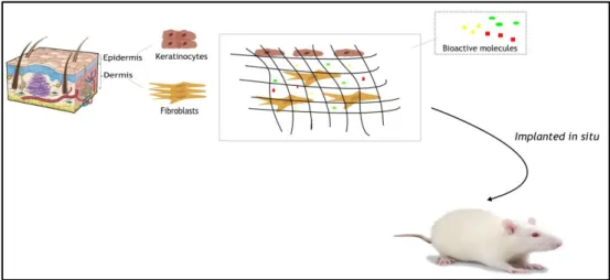
Figure 8 shows one potential approach for creating skin constructs in the laboratory. Such technique consists in isolating viable cells from the epidermis and dermis of the human body (keratinocytes and fibroblasts), expand the cell population through in vitro culture and then culture these cells on a polymeric matrix (scaffold) that provides a favourable environment for accelerating the wound healing process and prevent the wound from infection. Then, the resulting tissue engineered construct is placed into the animal for in vivo assays (Dvir et al., 2010, Lanza et al., 2007). The 3D scaffold should be biodegradable and act like an ECM, since it will temporarily serve as a template for cell adhesion, unhindered proliferation and tissue development. The scaffold should also have an interconnected macroporous network that allows cell penetration and nutrients, oxygen and waste products exchange (Lanza et al., 2007).
Figure 8: Schematic representation of the production of a skin substitute at the laboratory. After a skin
damage occur, the reconstruction of the main layers of the skin (epidermis and dermis) is a key factor for an effective wound healing. Keratinocytes and fibroblasts are fundamental cells in the process of regeneration of the skin. Their incorporation into a polymeric matrix with bioactive molecules will promote the wound healing, in an animal model.
There has been progress in the area of skin TE research and it is anticipated that further novel biomaterials combined with nanotechnology will be applied in this research area. Skin tissue engineering's primary goal is the production of a skin substitute that will support the complete regeneration of a functional tissue with all the appendages and the different layers. Moreover, the reestablishment of the vascular and nervous network in the host at the scar site is other key step in skin regeneration (Mohamed and Xing, 2012).
The ultimate goal of the tissue engineer is to satisfy most, if not all, of these criteria when producing novel, smart skin replacement therapies. Such matrices should attempt to
Chapter I- Introduction
18 reproduce the properties of ECM, in order to promote an optimal tissue repair and regeneration of the full thickness wounds (Metcalfe and Ferguson, 2007b, Halim et al., 2010).
1.3.1 Tissue engineering: skin substitutes
Nowadays, the loss or failure of a tissue or organ is the most significant problem in healthcare (Lanza et al., 2007). There are several full-thickness skin injuries resulting from burns, soft tissue trauma and diseases leading to skin necrosis. This represents a significant problem that is far from being solved (Böttcher-Haberzeth et al., 2010). For burns affecting large areas, there is a considerable loss of skin, the development of temporary or permanent skin substitutes appears to be the solution, since the patient's own body is not able to immediately restablish skin arquitecture (Metcalfe and Ferguson, 2007a, Atiyeh and Hayek, 2005).
In the past decades, many skin substitutes such as autografts, allografts and xenografts have been employed for wound healing (Garfein et al., 2003). Autograft skin is harvested from the patient and then placed in the excised areas of the wounded skin of the same individual. Autograft does remain the most frequently used grafting method since it does not induce an adverse immunological reaction from the host (Atiyeh and Hayek, 2005, Babensee et al., 1998). However, there are some disadvantages associated with skin autografts which are the limited availability and the donor site morbidity and scarring (Zhang and Michniak-Kohn, 2012, Babensee et al., 1998). Allograft skin is harvested from an organism of the same species, prior to placement. The use of allograft skin is limited since there is a great risk of disease transmission, eventual immune rejection and difficulties associated with its storage (Babensee et al., 1998). The demand for tissues and organs seriously exceeds the supply, creating a substantial waiting list. Moreover, the immune system tends to reject the foreign tissue or organ (Zhang and Michniak-Kohn, 2012). Xenograft skin is harvested from a different species and the majority of xenograf tissues are excluded due to a vigorous immune response of the patient, that may be caused upon the implantation process, thus leading to a high failure rate (Babensee et al., 1998, Garfein et al., 2003).
Therefore, there is a significant demand of tissues and organs that can be used for regenerative purposes and for being implanted in the human body. In the literature, the skin substitutes have been referred as 3D skin, reconstructed skin, skin equivalents, artificial skin, organotypic culture of skin, or skin grafts (Brohem et al., 2011).
Skin substitutes are a heterogeneous group of wound coverage materials that aid in wound closure and replace the functions of the skin, either temporarily or permanently, depending on the product characteristics (Shores et al., 2007, Halim et al., 2010). There are several important factors that are taken into consideration in the decision to use the skin substitutes in wound management. These include the depth of injury, availability of donor site, presence of wound infection, sites of wound, likelihood of contracture, aesthetic outcome, relative