Própolis: teor em fenóis totais e actividades antimicrobiana e
inibitória da enzima hialorunidase
João Carlos Sousa da Silva
Dissertação apresentada à Escola Superior Agrária de Bragança para obtenção do Grau de Mestre em Qualidade e Segurança Alimentar
Orientado por
Professora Doutora Maria Leticia Miranda Fernandes Estevinho
Agradecimentos
Este trabalho foi possivel atravéz do esforço de várias pessoas, às quais humildemente e com o maior pazer, gostaria de agradecer.
Em primeiro lugar, à Professora Doutora Letícia Estevinho, pela dedicação, pelos conhecimentos transmitidos, pela amizade,orientação deste trabalho e acima de tudo por acreditar nas minhas capacidades.
Aos colegas de laboratório, Leandro Moreira e Ana Paula Pereira e às funcionárias do Laboratório de Microbiologia, Dª Arminda e Dª Fátima por toda a ajuda na elaboração deste trabalho e por tornarem o Laboratório de Microbiologia a minha segunda casa. Aos amigos Cátia Silva, Linda Santos, Ana Bela Martins, Rafaela Fonseca, Sandra Luzio, Carla Nogueira por todos os bons momentos proporcionados.
À minha familia, especialmente aos meus pais, pelo apoio incondicional na realização deste trabalho, por acreditarem no meu trabalho, e por todos os sacrifícios que fizeram. À Patrícia, pelo carinho, pela paciência, pelas noites acordadas, por me tornar melhor trabalhador.
ii ÍNDICE
Resumo ... » v
Abstract ... » vi
Índice de figuras ... » vii
Índice de tabelas ... » viii
Capítulo I: Introdução 1.- Qué é o propolis ... » 2
2.- Características e composição química ... » 4
3.- Propiedades bioactivas do própolis ... » 5
3.1. Atividade antitumoral ... » 6
3.2. Atividade antiviral ... » 6
3.3. Atividade cariostatica ... » 7
3.4. Atividade hepatoprotetora ... » 8
3.5. Actividade antioxidante ... » 9
3.6. Atividade antimicrobiana ... » 10
3.7. Atividade anti-inflamatória ... » 11
iii
1.- Introduction ... » 29
2.- Material and methods ... » 31
2.1. Chemicals and Reagents ... » 31
2,2 Propolis samples ... » 31
2.3 Palynological identification ... » 31
2.4 Extraction procedure ... » 32
2.4.1. Aqueous extract ... » 32
2.4.2. Methanolic extract ... » 32
2.4.3. Hydro-alcoholic extract ... » 32
2.5 Total Phenolics and Flavonoids ... » 33
2.6 UV-Visible Absorption Spectroscopy ... » 33
2.7 Anti-inflammatory activity – Hyaluronidase assay ... » 34
2.8 Antimicrobial activity ... » 34
2.9 Statistical analysis ... » 35
3.- Results and disussion ... » 36
3.1. Palynological identification ... » 36
3,2 Total Phenolics and Flavonoids ... » 37
3.3 UV-Visible Absorption Spectroscopy ... » 39
iv 4.- References ... » 45 Capitulo III: Consideraciões finais
v Hoje em dia na literatura está disponível uma grande quantidade de informação sobre os aspetos químicos e biológicos dos produtos apícolas, no entanto a informação científica fundamentada sobre a sua utilização terapêutica é limitada. O objetivo deste estudo foi avaliar o perfil fenólico, a atividade antimicrobiana in vitro e o efeito sobre a enzima hialuronidase (amplamente relacionado com o processo de inflamação) do própolis Português. Foi também comparada a eficácia da extração do própolis em três extractos (hidro-alcoólico, metanólico e aquoso). Foi escolhido o extrato hidro-alcoólico, porque foi o mais eficaz na extração de fenóis totais.
Foi analisada a atividade antimicrobiana do própolis contem bactérias Gram-positivas e Gram-negativas e leveduras, isoladas de diferentes fluídos biológicos. Os resultados foram comparados com os obtidos para microrganismos de referência. O própolis de Bragança foi o que possuiu o mais alto teor de fenóis totais. A amostra de Beja evidenciou a inibição menos significativa da enzima hialuronidase.
Em relação à atividade antimicrobiana, Candida albicans foi a mais resistente e Staphylococcus aureus a mais sensível. Os micorganismos de coleção foram mais sensíveis do que os isolados a partir de fluídos biológicos.
vi Nowadays a great amount of information regarding chemical and biological aspects of bee products is available in the literature, but few data on their therapeutic uses are found. The aim of this study was to evaluate the phenolic profile, the in vitro antimicrobial activity and effect in the hyaluronidase enzyme (widely related with the inflammation process) of propolis harvested in Portugal. The efficacy of three extracts (alcoholic, methanolic and aqueous) was also compared. It was chosen the hydro-alcoholic extract, because this was the most effective for extracting phenolic compounds. The antimicrobial activity was accessed in positive and Gram-negative bacteria and yeasts, isolated from different biological fluids and the results were then compared with the obtained for reference microorganisms. The propolis from Bragança was the one that possessed the highest polyphenols’ content. The sample from Beja showed less significant inhibition of the hyaluronidase enzyme. Concerning the antimicrobial activity, Candida albicans was the most resistant and Staphylococcus aureus the most sensitive microganism. The reference microorganisms were more sensitive than the ones isolated from biological fluids.
vii Figure 1 -- Abelha levando própolis à colmeia (Bioapis, Lda.) ……… ... » 2 Figure 2 - Abelha a cubrir uma grelha com própolis (Bioapis, Lda.) ... » 2 Figure 3 - Palynological spectrum of bee pollen samples. DP – Dominant
Pollen (>45%); AP - Acessory Pollen (15%-45%); IP – Isolated Pollen
(<15%) ... » 37 Figure 4 - Absorption spectra of the ethanolic propolis extracts from different
locations ... » 39 Figure 5 - Inhibition of the activity of Hyaluronidase by the propolis extracts
for each concentration. The letters (a,b) represent which samples are different
by Tukey test with significance of p = 0.05. ... » 41 Figure 6 - Minimum Inhibitory Concentration (mg/mL) for each place and
microorganism. The letters (a,b) represent which samples are different by
vii i Table 1 – Microorganisms used in the present study to test antimicrobial
activity of propolis extracts ... » 35 Table 2 – Concentration (mg/g) of total phenolics and flavonoids in propolis
extracts from different locations (n=27) ... » 38 Table 3 – Minimum Inhibitory Concentration (mg/mL) for the studied
microorganisms and relation between the same species (reference culture and
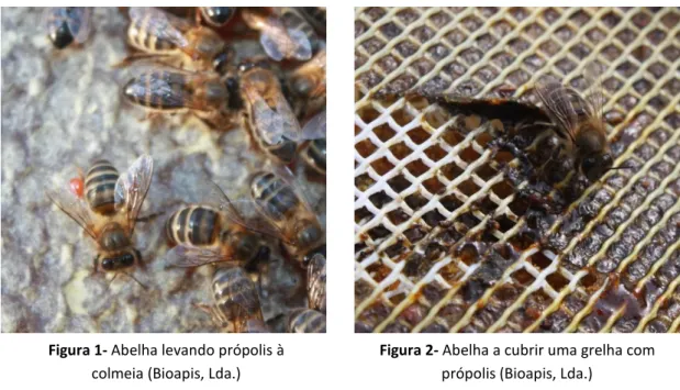
2 Figura 1- Abelha levando própolis à
colmeia (Bioapis, Lda.)
Figura 2- Abelha a cubrir uma grelha com própolis (Bioapis, Lda.)
1.
QUE É O PRÓPOLISEntende-se por própolis o produto oriundo de substâncias resinosas, viscosas e balsâmicas, colhidas pelas abelhas (Apis mellifera), dos brotos, flores e exsudados de algumas árvores e plantas, nas quais as abelhas acrescentam secreções salivares, cera e pólen para elaboração final do produto (Regulamento Técnico para fixação de identidade e qualidade de própolis).
A abelha recolhe os exsudados com as suas mandíbulas, durante este processo, enzimas presentes na saliva das abelhas são misturadas com as resinas recolhidas produzindo-se a hidrólise de alguns compostos. A abelha armazena estas substâncias nas patas traseiras, até chegar à colmeia (Sforcin, 2007). Posteriormente, estas substâncias são modificadas com a adição de ceras.
O termo própolis, do Grego pro = em defesa e polis = cidade ou comunidade,
3 de animais que morrem dentro da colmeia, evitando a sua decomposição (Bankova et al., 2000).
Em geral, o própolis é composto por 50% de resinas e balsamos vegetais, 30% de cera, 10% de óleos essenciais e aromáticos, 5% pólen e 5% de outras substâncias, incluindo restos orgânicos (Burdock, 1998). A cera e os restos orgânicos são removidos no processamento, normalmente a extração etanólica, e a tintura do própolis resultante, contém grande parte dos constituintes bioativos do própolis.
O própolis tem sido utilizado como remédio pelos humanos desde os tempos mais antigos. É usado na medicina popular em muitas partes do mundo. Egípcios, Gregos e Romanos reportam o uso deste produto apícola geralmente pelas suas qualidades curativas. O própolis tem sido reconhecido como um agente anti-inflamatório e curativo de chagas e úlceras. No antigo Egipto era usado para mumificar, e mais recentemente utilizado na Guerra Bôeres como curativo de feridas e regeneração de tecidos (Ghisalberti et al, 1978). Atualmente ainda é utilizado em remédios, produtos higiénicos e outras preparações. É um dos medicamentos mais usado nos Países Balcãs (Wollenweber et al., 1990; Bankova, 2005a). Convém no entanto salientar que o conhecimento sobre estas propriedades é empírico pois só nestas últimas décadas está a ser estudado para conhecer os seus constituintes e suas propriedades.
Vários autores demostraram que a administração do própolis, tanto em humanos como em ratos, não evidenciou efeitos secundários (Kaneeda and Nishina, 1994; Sforcin et al., 1995, 2002b; Jasprica et al., 2007). Ramadan et al. (2012) demostraram que o extrato etanólico de própolis não foi tóxico e não causou mortalidade nem sinais de toxicidade em ratos, quando foi administrado oralmente a doses superiores a 5 g/kg de massa vivo ao nascimento, tendo estabelecido este valor como a sua LD 50.
4
2.
CARACTERÍSTICAS E COMPOSIÇÃO QUÍMICAO própolis é uma substância resinosa e muito adesiva, dai a baixa solubilidade em água. Na sua extração devem utilizar-se solventes orgânicos como éter etilico, acetona, tolueno e tricloroetileno ou álcoois como metanol e etanol que permitem a dissolução da maior parte dos compostos do própolis. Para além disso tem sido desenvolvidas patentes com novos métodos ou solventes para extrair os compostos presentes no própolis, que utilizam óleos vegetais comestíveis, triglicéridos e ácidos gordos(Kasuma and Kenichi, 2001a, 2001b; Namiki et al., 2005).
À temperatura ambiente é um produto de consistência sólida, a partir de 30ºC torna-se maleável e funde entre os 60-70ºC podendo chegar a seu ponto de fusão até aos 100ºC dependendo da sua composição.
A sua coloração pode variar de verde até amarelo, vermelho, castanho, castanho-escuro ou preto (Ghisalberti et al., 1978) dependendo da origem botânica da resina.
O seu odor é característico e extremamente forte devido maioritariamente aos compostos voláteis nele existentes (Anon, 1927)
A composição química do própolis é muito complexa. O própolis bruto contém mais de 300 compostos entre os quais se encontram flavonoides, ácidos fenólicos e seus ésteres, sesquiterpenos, quininas, esterois, vitaminas, aminoácidos, açúcares e proteínas (Kumar et al., 2009; Lotfy, 2006; Markham et al., 1996; Trusheva et al., 2007). Vários destes compostos encontram-se de forma natural nos alimentos ou são utilizados como aditivos alimentares dentro do grupo de sustâncias geralmente reconhecidas como seguras ou sustâncias Generally Recognised as Safe (GRAS) (Burdock et al., 1998). Este facto sugere que o própolis é uma sustância apropriada para ser utilizada como conservante natural em alimentos, satisfazendo a atual procura por parte dos consumidores, de antioxidantes e antimicrobianos naturais. Deste modo é possível obter alimentos minimamente processados, com conservantes naturais ou que utilizam conservantes sintéticos em concentrações muito baixas (Chaillou and Nazareno, 2009; Han and Park, 1995; Oldoni et al., 2011; Tosi et al., 2007).
5 2011; De Vecchi and Drago, 2007; Falcão et al., 2010; Ghassan et al., 2011; Gregoris and Stevanato, 2010; Ilhami et al., 2010; Medana et al., 2008) que são considerados os principais responsáveis pelas suas propriedades biológicas e farmacológicas (Carvalho et al., 2011; De Castro, 2001; Kumazawa et al., 2004; Sforcin, 2007; Nolkemper et al., 2010).
No entanto, o teor nestes compostos e, consequentemente, as suas propriedades bioativas variam segundo a origem geográfica, dependendo da flora local e da fenologia das plantas de origem (Ahn et al., 2007; Kujumgiev et al., 1999; Sforcin, 2007), bem como de outros fatores como a estação do ano (Isla et al., 2009; Teixeira et al., 2010; Valencia et al., 2012). Alguns autores descrevem a presença de sustâncias específicas, nomeadamente os trepenos, em função da origem geográfica do mesmo (Oldoni et al., 2011; Popova et al., 2009; Popova et al., 2010; Trusheva et al., 2003). Esta grande variabilidade constitui um impedimento à aprovação oficial do própolis para uso em preparações farmacológicas ou produtos nutracêuticos, sendo necessária uma estandardização química que garantir-se a sua qualidade, segurança e eficácia (Bankova, 2005a; Marcucci, 1995; De Castro, 2001).
3.
PROPRIEDADES BIOACTIVAS DO PRÓPOLIS6 apiterapia, alimentos saudáveis e em outros fins (Bankova et al., 2000; Banskota et al., 2001; Ghisalberti, 1978).
3.1Atividade anti tumoral
O própolis foi reportado como um agente anti tumoral, pela sua capacidade anti proliferativa das células tumorais, quer in vitro quer in vivo (Banskota et al., 2001; Buriol et al., 2009; Chen et al., 1996; Kim et al., 2008; Rao et al.,1992).O seu principal mecanismo da ação anti tumoral esta relacionado com a inibição do crecimento celular e indutor do processo de apoptose (Sforcin 2007).
Vários estudos demostraram que compostos isolados do própolis como o ácido cafeico e seus esteres são os responsáveis por esta atividade. Lee et al., 2005 observaram que o ácido cafeico interfere com o ciclo celular, a análise do fluxo citométrico demonstrou a interrupção da célula na fase G2/M. Orsolic et al., 2004, verificaram que, compostos polifenólicos como o acido caféico e seus ésteres assim como a quercetina, diminuíram o numero de nódulos tumorais do pulmão. Contudo, o efeito anti metastático observado no própolis inteiro, foi superior aos compostos isolados. Esta ação do própolis pode ser resultado de ações sinergéticas dos compostos polifenólicos (Orsolic et al., 2005).
Carvalho et al., (2011) demonstraram o efeito citotóxico de diferentes extratos de própolis in vivo, através do modelo Sarcoma 180 (tumor frequentemente induzido em ratos para estudos anti tumorais in vivo) transplantado em ratos, em que todos os extratos de própolis demonstraram significativas reduções do crescimento do tumor.
3.2Atividade antiviral
Atividade farmacológica do própolis, foi observada contra várias infeções virais, tais como, o vírus da gripe (Serkedjieva et al., 1992), vírus imunodeficiência humana (Ito et al., 2001) , adenovírus (Amoros et al., 1992a) e herpes simplex vírus(Debiaggi et al., 1990; Amoros et al., 1992b; Amoros et al., 1994; Huleihel e Isanu, 2002).
7 (Chakrabarti et al., 2000). Existem dois tipos, o tipo 1 é transmitido através de gotículas e recorrentemente provoca herpes labiais, ao passo que o tipo 2 é transmitido principalmente por via sexual e é o agente causador do herpes genital (Smith e Robinson, 2002; Stranska et al., 2005).
Galangina e crisina, constituintes do própolis, reduziram a formação de placas de vírus herpes simplex tipo 1 livre em 68,0% e 56,4%, respetivamente, quando comparados com os controlos não tratados.
A sinergia dos compostos do própolis pode ser demonstrada quando combinações binárias flavo-flavonóis foram testadas contra o vírus herpes simplex, isto explica o facto do própolis ser mais ativo do que seus compostos individuais (Scheller et al., 1999).
3.3Atividade cariostática
Vários produtos de higiene oral têm na sua base ativa propriedades cariostáticas. No própolis também foram encontrados compostos que por diferentes mecanismos de ação são capazes de controlar a aparição de cáries.
Analises antimicrobianas realizadas com alguns compostos do própolis revelaram que o sesquiterpeno tt-farnesol é eficaz conta o Streptococcus mutans, microrganismo responsável pela formação caries, com concentrações mínimas inibitórias (MIC) de 14 –28 g/ml e concentrações mínimas bactericidas (MBC) de 56–
112 g/ml (Koo et al., 2002).
Outro composto, o flavonoide apigenina demonstrou ser um potencial inibidor da atividade glucosiltransferase tanto em solução como num ensaio feito com grânulos de hidroxiapatite revestido com saliva (Koo et al., 2000). Esta enzima é responsável pela formação de biofilmes que dão resistência ao Streptococcus mutans tornando-o menos sensível às substâncias antibacterianas.
8 polissacarídica do biofilme do Streptococcus mutans sem causar um grande impacto na viabilidade bacteriana (Koo et al., 2003).
Juntamente com o flureto de sodio, a apigenina e o tt-farnesol são capazes de reduzir a virulência do Streptococcus mutans, e promover o efeito cariostatico do floreto. Neste estudo foi evidente que a combinação destes compostos diminui a quantidade de glucanos insolúveis extracelulares no bio filme do Streptococcus mutans e que esta combinação pode ser uma alternativa aos agentes químicos normalmente utilizados para a prevenção do processo cariostático (Koo et al., 2005).
3.4Atividade hepatoprotetora
As doenças hepáticas são consideradas um dos maiores problemas de saúde, sendo o fígado um órgão importante tanto na detoxicação como deposição de substâncias endógenas e exógenas (Sehrawat et al., 2006)
As lesões provocadas no fígado pelo consumo excessivo de remédios halopáticos nos últimos anos, tem levado a muitas patologias crónicas deste órgão. O tratamento das disfunções hepáticas, sejam funcionais ou lesionais, envolvem substâncias que têm ação hepatoprotetora (que atuam nos hepatócitos protegendo-os dos danos diversos: químicos, microbiológico).
Vários extratos de própolis apresentaram forte atividade hepatoprotetora. Os extratos metanólicos do própolis do Brasil, China, Peru e da Holanda apresentaram grande atividade hepatoprotetora da D-galactosamine (D-GalN)/ fator de necrose tumoral -a (TNF-a) – morte celular induzida em culturas primarias de hepatócitos de ratos (Banskota et al., 2000).
Sugimoto et al. (1999), verificaram que o extrato etanólico (95%) do própolis Brasileiro possui forte atividade hepatoprotetora da D-GalN – lesão hepática induzida em ratos, González et al. (1994, 1995) observaram que o extrato etanólico (95%) do própolis Cubano exibiu forte atividade hepatoprotetora do paracetamol – induzindo danos no fígado em ratos e do CCl4 i - lesão hepática induzida em ratos.
9 A atividade hepatoprotetora do Própolis Brasileiro deve-se principalmente aos compostos fenólicos, tais como os flavonoides.
3.5Atividade antioxidante
Os radicais livres de oxigénio são produtos normais do metabolismo celular provocando o stress oxidativo (Valko et al., 2007). Estes são responsáveis por várias anomalias celulares e o consumo regular de antioxidantes parece limitar ou prevenir os efeitos provocados por estes radicais livres (Kaur and Geetha, 2006).
Embora nosso organismo possua as suas próprias defesas contra estes radicais estas podem ser insuficientes devido às agressões sofridas pelo nosso organismo no dia-a-dia. Assim, o consumo de produtos com capacidade antioxidante como o própolis torna-se um importante fator na defesa do nosso organismo (Mohammadzadeh et al., 2007).
As propriedades antioxidantes do própolis são devidas principalmente aos compostos fenólicos. Estes componentes apresentam um efeito notável de proteção contra as reações de oxidação, devido às suas propriedades redox, que lhes permitem atuar como agentes redutores, doadores de hidrogénio, quelantes de metais e supressores de singletos de oxigénio (Parr and Bolwell, 2000). Própolis também tem sido relatado como um inibidor da formação do anião superóxido. Além disso, o própolis pode reverter o consumo de glutationoxidase, uma das principais enzimas antioxidantes, que é sintetizada no fígado, e apresenta capacidade captadora de radicais libres (Castaldo and Capasso, 2002).
As propriedades antioxidantes do própolis além das suas propriedades antibacteriana e anti fungicida, juntamente com o facto de vários dos seus constituintes estarem presentes nos alimentos ou em aditivos alimentares, e ser reconhecido como GRAS (Burdock, 1998), torna-o bastante atrativo como conservante alimentar natural. De facto, tem-se verificado um aumento da procura de antioxidantes e antimicrobianos naturais, por parte dos consumidores em geral e particularmente por parte dos que preferem alimentos minimamente processados (Han & Park, 1995; Tosi et al., 2007).
10 que é uma das principais razões para a deterioração dos produtos alimentares durante o processamento e armazenamento (Halliwell, 1997; Halliwell and Gutteridge, 1999).
3.6Atividade antimicrobiana
Um dos maiores problemas de difícil resolução ao nível da saúde pública, desde o início do seculo, é o aumento de resistência dos vários microrganismos aos agentes antimicrobianos. A evolução incessante de resistências aliada à diminuição do desenvolvimento de novos agentes antimicrobianos ativos contra patogénicos resistentes conduziu a um aumento do número de casos de microrganismos resistentes à maioria ou a praticamente todos os fármacos disponíveis para uso clinico. Perante esta situação são necessárias alternativas terapêuticas eficazes e se possível que não induzam resistências.
O própolis pode constituir uma destas alternativas ao mostrar efeitos sinergeticos com antibióticos. Oksuz et al. (2005) verificou o efeito sinergetico do propolis com ciprofloxacin no tratamento de Staphylococcus aureus keratitis. Segundo Orsi et al. (2006), o própolis tem a capacidade de diminuir a resistência das paredes da bactéria à entrada dos antibióticos (amoxicilina, ampicilina e cefaloxina) e demonstrou efeitos sinérgicos com antibióticos que atuam no ribossoma (cloranfenicol, tetraciclina e neomicina).
Esta atividade não é útil só na terapia antimicrobiana humana se não também no tratamento de doenças das próprias abelhas. Paenibacillus larvae, o agente responsável pelo Loque Americano, tem vindo a ganhar resistências aos antibióticos convencionais, e os extratos de própolis de vários estados do Brasil inibiram significativamente estes microrganismos (Bastos et al., 2008).
De todas as propriedades que o própolis possui, a atividade antimicrobiana é a mais extensivamente estudada (Chaillou e Nazareno, 2009; Kalogeropoulos et al., 2009; Libério et al., 2009; Petrova et al., 2010; Popova et al., 2009) e tem sido documentada contra diferentes tipos de bactérias, fungos e parasitas (Freitas et al., 2006; Guven et al., 2011; Popova et al. 2009; Sforcin et al., 2000; Sforcin et al., 2001).
11 dos microrganismos. A atividade antimicrobiana do própolis está relacionada principalmente com o seu conteúdo em flavonoides (Cushnie and Lamb, 2005; Salomao et al., 2004) como assim os compostos fenólicos, terpenos e ácidos aromáticos e esteres mostraram também atividade antimicrobiana (Bankova, 2005b; Burdock et al., 1998; Popova et al., 2005).
3.7Atividade anti-inflamatória
O processo inflamatório é desencadeado por diversos produtos químicos e/ou biológicos, incluindo enzimas pró-inflamatórias e citocinas, compostos de baixo peso molecular, tais como eicosanóides ou a degradação enzimática dos tecidos (Dao et al., 2004).
Diversos estudos acordam que a ciclooxigenase-2 (COX-2), uma isoforma da ciclooxigenase (COX), que catalisa a transformação de ácido araquidónico a prostaglandina, é a enzima mais relacionada com o processo inflamatório (Griswold and Adams, 1996; Cho et al., 2004). A outra isoforma é a ciclooxigenase-1 (COX-1), que regula os processos de homeostase (Dao et al., 2004).
Nestes últimos 30 anos, diversos estudos demonstram as propriedades anti-inflamatórias do própolis, essas propriedades devem-se basicamente à presença de flavonoides que inibem o desenvolvimento de inflamações provocadas por diversos agentes (Teixeira et al., 2005; Mani et al., 2006).
Galangina, um composto flavonoide, é capaz de inibir a atividade da COX e lipo-oxigenase, limitar a ação da poligalacturonase e reduzir a expressão da isoforma induzível da COX-2 (Raso et al 2001; Rossi et al 2002a, 2002b).
Crisina, outro composto flavonoide de grande interesse presente no própolis, também mostra atividade anti-inflamatória (Kim et al 2002; Ko et al., 2003).O seu mecanismo de ação esta relacionado com a supressão das atividades pró-inflamatórias da COX-2 e induzível sintase do óxido nítrico (Cho et al., 2004).
12 araquidónico a partir da membrana celular, o que conduz á supressão da atividade de COX-1 e COX-2 e inibe a ativação da expressão génica de COX-2 (Mirzoeva and Calder 1996; Lee et al., 2004).
4.
REFERÊNCIASAhn, M. R., Kumazawa, S., Hamasaka, T., Bang, K. S., Nakayama, T., 2004. Antioxidant activity and constituents of propolis collected in various areas of Korea. Journal of Agricultural and Food Chemistry. 52, 7286–7292.
Ahn, M. R., Kumazawa, S., Usui, Y., Nakamura, J., Matsuka, M., Zhu, F., 2007. Antioxidant activity and constituents of propolis collected in various areas of China. Food Chemistry, 101, 1400–1409.
Amoros, M., Sauvager, F., Girre, L., Cormier, M. 1992a. In vitro antiviral activity of propolis. Apidologie 23, 231–240.
Amoros, M., Simoes, C.M., Girre, L., Sauvager, F., Cormier, M., 1992b. Synergistic effect of flavones and flavonols against herpes simplex virus type 1 in cell culture. Comparison with the antiviral activity of propolis. Journal of Natural Products 55, 1732–1740.
Amoros, M., Lurton, E., Boustie, J., Girre, L., Sauvager, F., Cormier, M., 1994. Comparison of the anti-herpes simplex virus activities of propolis and 3- methyl-but-2-enyl caffeate. Journal of Natural Products. 57, 644–647.
Anon. 1927. The origin of the cooration of bee´s wax and the composition of propolis. Comptes Rendus Hebdomadaires Des Seances De L´Academie Des Sciences, 184, 1134-1136.
Bankova, V., 2005a. Chemical diversity of propolis and the problem of standardization. Journal of Ethnopharmacology 100, 114–117.
13 Bankova, V., Castro, S.L., Marcucci, M.C., 2000. Propolis: recent advances in
chemistry and plant origin. Apidologie 31, 3–15.
Banskota, A. H., Tezuka, Y., Adnyana, I K., Midorikawa, K.,Matsushige, K., Massage, D., Huertas, A. A. G., Kadota, S. 2000. Biological evaluation of propolis from Brazil, Peru, The Netherlands and China. Journal of. Ethnopharmacology ,72, 239–246.
Banskota, A.H., Tezuka, Y., Kadota, S., 2001. Recent progress in pharmacological research of propolis. Phytotherapy Research 15, 561–571.
Basnet, P., Matsushige, K., Hase, K., Kadota, S., Namba, T. 1996. Four di-O-caffeoyl quinic acid derivatives from propolis. Potent hepatoprotective activity in experimental liver injury models. Biological and Pharmaceutical Bulletin , 19, 1479–148.
Bastos, E.M., Simone, M., Jorge, D.M., Soares, A.E., Spivak, A.M., 2008. In vitro study of the antimicrobial activity of Brazilian propolis against Paenibacillus larvae. Journal of Invertebrate Pathology 97, 273–281.
Bufalo, M. C., Candeias, J. M. G., Sforcin, J. M., 2009. In vitro Cytotoxic Effect of Brazilian Green Propolis on Human Laryngeal Epidermoid Carcinoma (HEp-2) Cells. Evidence-Based Complementary and Alternative Medicine. 6, 483–487. Burdock, G. A., 1998. Review of the biological properties and toxicity of bee propolis
(propolis). Food Chemistry and Toxicology, 36, 347–363.
Buriol, L., Finger, D., Schmidt, E. M., Santos, J. M. T., Rosa, M. R., Quináia, S. P., et al.
2009. Composição química e atividade biológica de extrato oleoso de própolis: uma alternativa ao extrato etanólico. Química Nova, 32, 296–302.
Callejo, A., Armentia, A., Lombardero, M., Asensio, T. 2001. Propolis, a new bee-related allergen. Allergy 56, 579.
14 and composition of an oil extract of Brazilian propolis. Food Chemistry, 126, 1239–1245.
Castaldo, S.,Capasso,F. 2002.Propolis,an old remedy used in modern medicine. Fitoterapia, 73, 1–6.
Chaillou, L.L., Nazareno, M.A., 2009. Bioactivity of propolis from Santiago del Estero, Argentina, related to their chemical composition. LWT - Food Science and Technology. 42, 1422–1427.
Chakrabarti S, Pillay D, Ratcliffe D, Cane PA, Collinghan KE, Milligan DW. 2000. Resistance to antiviral drugs in herpes simplex virus infections among allogeneic stem cell transplant recipients: risk factors and prognostic significance. Journal of Infectious Diseases ,181, 2055–2058.
Cho, H., Yun, C.W., Park, W.K., Kong, J.Y., Kim, K.S., Park, Y., Lee, S., Kim, B.K. 2004. Modulation of the activity of pro-inflammatory enzymes, COX-2 and iNOS, by chrysin derivatives. Pharmacological Research, 49, 37–43.
Chunying Luo, Xiaoli Zou, Yuanqian Li , Chengjun Sun, Yan Jiang, Zhiyun Wu. 2011. Determination of flavonoids in propolis-rich functional foods by reversed phase high performance liquid chromatography with diode array detection. Food Chemistry, 127, 314–320.
Cushnie, T.P, Lamb, A.J. 2005. Antimicrobial activity of flavonoids. International Journal of Antimicrobial Agents; 26, 343–56.
Dalepranea, J.B.; Da Silva Freitas,V., Pachecoc,A., Rudnickib, M., Faineb, L.A., Dörrb, F.A., Ikegakid, M., Salazarc, L.A., Prates Onga, T., Parra Abdallab, D.S. 2012. Anti-atherogenic and anti-angiogenic activities of polyphenols from propolis. Journal of Nutritional Biochemistry. 23, 557–566.
15 Debiaggi, M., Tateo, F., Pagani, L., Luini, M., Romero, E. 1990. Effects of propolis
flavonoids on virus infectivity and replication. Microbiologica, 13,207–213. De Castro, S.L. 2001. Propolis: biological and pharmacological activities. Therapeutic
uses of this bee-product. Annual Review of Biomedical Science, 3, 49–83. De Vecchi, E. and Drago, L. 2007. Propolis antimicrobial activity: What´s new? Le
Infezioni in Medicina, 15(1), 7–15.
Falcão,S.I., Vilas-Boas, M., Estevinho, L.M., Barros, C., Domingues, M.R.M., Cardoso, S.M. 2010. Phenolic characterization of Northeast Portuguese propolis: usual and unusual compounds. Analytical and Bioanalytical Chemistry, 396, 887–897. Freitas, S.F., Shinohara, L., Sforcin, J.M., Guimarães, S., 2006. In vitro effects of
propolis on Giardia duodenalis trophozoites. Phytomedicine 13, 170–175.
Ghassan M. Sulaiman, Khulood W. Al Sammarrae, Ali H. Adhiah, Massimo Zucchetti, Roberta Frapolli, Ezia Bello, Eugenio Erba, Maurizio D´Incalci, Renzo Bagnati. 2011. Chemical characterization of Iraqi propolis samples and assessing their antioxidant potentials.Food and Chemical Toxicology, 49, 2415–2421.
Ghisalberti, E.L. Jefferies, P.R., Lanteri, R., Matisons, J. 1978. Constituents of Propolis, Experientia, 34, 158.
González, R., Corcho, I., Remirez, D., Rodriguez, S., Ancheta, O., Merino, N., González, A., Pascual, C. 1995. Hepatoprotective effects of propolis extract on carbon tetrachloride-induced liver injury in rats. Phytochemical Research , 9, 114–17.
González, R., Corcho, I., Remirez, D., Rodriguez, S., González, A. S., Ancheta, O., Merino, N., Pascual, C. 1994. Hepatoprotective effects of propolis extract on paracetamol-induced liver damage in mice. Phytochemical Research, 8, 229–
232.
16 Griswold, D.E., Adams, J.L. 1996. Constitutive cyclooxygenase (COX-1) and inducible
cyclooxygenase (COX-2): rationale for selective inhibition and progress to date. Medical Care Research and Review, 16, 181–6.
Gulbahar, O., Ozturk, G., Erdem, N., Kazandi, A.C.,Kokuludag, A., 2005. Psoriasiform contact dermatitis due to propolis in a beekeeper. Annals of Allergy, Asthma & Immunology 94, 509–511
Guven Kayaoglu, ,Hüma Ömürlü, Gülçin Akca, Mügem Gürel, Omür Gençay, Kadriye Sorkun, Bekir Salih. 2011. Antibacterial Activity of Propolis versus Conventional Endodontic Disinfectants against Enterococcus faecalis in Infected Dentinal Tubules. Journal of Endodontics ,37, 3, 376-381.
Halliwell, B., 1997. Antioxidants in human health and disease. Annual Review of Nutrition 16, 33–50.
Halliwell, B., Gutteridge, J.M.C., 1999. Free Radicals in Biology and Medicine. Oxford University Press, United Kingdom.
Han, S. K. and Park, H. K. 1995. A study on the preservation of meat products by natural propolis: Effect of EEP on protein change of meat products. Korean Journal of Animal Science, 37, 551–557.
Hausen, B.M.,Wollenweber, E., Senff, H., Post, B., 1987. Propolis allergy. (II). The sensitizing properties of 1,1-dimethylallyl caffeic acid ester. Contact Dermatitis 17, 171–177.
Hegyi, E., Suchy, V., Nagy, M., 1990. Propolis allergy. Hautartz 41, 675–679.
Hernandez, J., Goycoolea, F. M., Quintero, J., Acosta, A., Castaneda, M., Dominguez, Z., Robles R, Vazquez-Moreno L, Velazquez EF, Astiazaran H, Lugo E, Velazquez C., 2007. Sonoran propolis: Chemical composition and antiproliferative activity on cancer cell lines. Planta Medica. 73, 1469–1474. Huleihel, M., Isanu, V., 2002. Anti-herpes simplex virus effect of an aqueous extract of
17 Ilhami Gulcin, Ercan Bursal, M. Hilal Sehitoglu, Mine Bilsel, Ahmet C. Goren. 2010.
Polyphenol contents and antioxidant activity of lyophilized aqueous extract of propolis from Erzurum, Turkey. Food and Chemical Toxicology, 48, 2227– 2238.
Isla, M. I., Zampini, I. C., Ordóñez, R. M., Cuello, S., Juarez, B. C., Sayago, J. E., Moreno, M.I.N., Alberto, M.R., Vera, N.R., Bedascarrasbure, E., Alvarez, A. Cioccini, F., Maldonado, L.M., 2009. Effect of Seasonal Variations and Collection Form on Antioxidant Activity of Propolis from San Juan, Argentina. Journal of Medicinal Food. 12, 1334–1342.
Ito, J., Chang, F.R.,Wang, H.K., Park, Y.K., Ikegaki, M., Kilgore, N., Lee, K.H., 2001. Anti-AIDS agents. 48.(1) Anti-HIV activity of moronic acid derivatives and the new melliferone-related triterpenoid isolated from Brazilian propolis. Journal of Natural Products 64, 1278–1281.
Jasprica, I., Mornar, A., Debeljak, Z., Smolcic-Bubalo, A., Medic-Saric, M., Mayer, L., Romic, Z., Bucan, K., Balog, T., Sobocanec, S., Sverko, V. 2007. In vivo study of propolis supplementation effects on antioxidative status and red blood cells. Journal of Ethnopharmacology 110, 548–554.
Jorge, R., Furtado, N., Sousa, J. P. B., da Silva, A. A., Gregorio, L. E., Martins, C. H. G., Soares, A.E.E., Bastos, J.K., Cunha, W.R., Silva, M.L.A., 2008. Brazilian Propolis: Seasonal Variation of the Prenylated p-Coumaric Acids and Antimicrobial Activity. Pharmaceutical Biology. 46, 889–893.
Kalogeropoulos, N., Konteles, S.J., Troullidou, E., Mourtzinos, I., Karathanos, V.T., 2009. Chemical composition, antioxidant activity and antimicrobial properties of propolis extracts from Greece and Cyprus. Food Chemistry. 116, 452–461. Kaneeda, J., Nishina, T. 1994. Safetiness of propolis. Acute toxicity. Honeybee Science
15, 29–33.
18 Kasuma, Y. and Kenichi, I. (2001b). Extraction of propolis extract. JP Pat. 2001017096
(A).
Kaur, I.P., Geetha, T. 2006. Screening Methods for Antioxidants- A Review. Mini Reviews in Medicinal Chemistry, 6, 305-312.
Kim, E.J., Kwon, K.J., Park, J.Y., Lee, S.H,, Moon, C.H., Baik, E.J. 2002. Effects of peroxisome proliferator-activated receptor agonists on LPS-induced neuronal death in mixed cortical neurons: associated with iNOS and COX-2. Brain Research, 941, 1–10.
Kim, D.M., Lee, G.D., Aum, S.H., Kim, H.J., 2008. Preparation of propolis nanofood and application to human cancer. Biological and Pharmaceutical Bulletin 31, 1704–1710.
Ko, H.H., Tsao, L.T., Yu, K.L., Liu, C.T., Wang, J.P., Lin, C.N. 2003. Structure–
activity relation- ship studies on chalcone derivatives: the potent inhibition of chemical mediators release. Bioorganic & Medicinal Chemistry, 11, 105–11. Koo, H.,Hayacibara, M.F., Schobel, B.D., Cury, J.A.,Rosalen, P.L., Park,Y.K.,
Vacca-Smith, A.M., Bowen, W.H., 2003. Inhibition of Streptococcus mutans biofilm accumulation and polysaccharide production by apigenin and tt-farnesol. Journal of Antimicrobial Chemotherapy 52, 782–789.
Koo, H., Pearson, S.K., Scott-Anne, K., Abranches, J., Cury, J.A., Rosalen, P.L., Park, Y.K., Marquis, R.E., Bowen, W.H., 2002. Effects of apigenin and tt-farnesol on glucosyltransferase activity, biofilm viability and caries development in rats. Oral.
Koo, H., Schobel, B., Scott-Anne, K., Watson, G., Bowen, W.H., Cury, J.A., Rosalen, P.L., Park, Y.K., 2005. Apigenin and tt-farnesol with fluoride effects on S. mutans biofilms and dental caries. Journal of Dental Research 84, 1016–1020. Koo, H., Vacca Smith, A.M., Bowen, W.H., Rosalen, P.L., Cury, J.A., Park, Y.K.,
19 Kujumgiev, A., Tsvetkova, I., Serkedjieva, Y., Bankova, V., Christov, R., Popova, S.,
1999. Antibacterial, antifungal and antiviral activity of propolis from different geographic origins. Journal of Ethnopharmacology, 64, 235–240.
Kumar, N., Mueen, A.K.K., Dang, R., Shivananda, T.N., Das, K., 2009. GC–MS analysis of propolis of Indian origin. Pharmacognosy 1, 46–48.
Kumazawa, S., Hamasaka, T., Nakayama, T. 2004. Antioxidant activity of propolis of various geographic origins. Food Chemistry, 84, 329-339.
Lee, K.W., Chun, K.S., Lee, J.S., Kang, K.S., Surh, Y.J., Lee, H.J. 2004. Inhibition of cyclooxygenase-2 expression and restoration of gap junction intercellular communication in H-ras- transformed rat liver epithelial cells by caffeic acid phenethyl ester. Annals of the New York Academy of Sciences, 1030, 501–
7.Lee, Y.T., Don, M.J., Hung, P.S., Shen, Y.C., Lo, Y.S., Chang, K.W., Chen, C.F., Ho, L.K., 2005. Cytotoxic of phenolic acid phenethyl esters on oral cancer cells. Cancer Letters 223, 19–25.
Li, F., Awale, S., Zhang, H. Y., Tezuka, Y., Esumi, H., Kadota, S., 2009. Chemical Constituents of Propolis from Myanmar and Their Preferential Cytotoxicity against a Human Pancreatic Cancer Cell Line. Journal of Natural Products. 72, 1283–1287.
Libério, S.A., Pereira, A.L.A., Araújoa, M.J.A.M, Dutrad, R.P., Nascimentoa, F.R.F., Monteiro-Netoc, V., Ribeiro, M.N.S., Gonçalves, A.G., Guerra, R.M.N., 2009. The potential use of propolis as a cariostatic agent and its actions on mutans group streptococci. Journal of Ethnopharmacology. 125, 1–9.
Lima, B., Tapia, A., Luna, L., Fabani, M. P., Schmeda-Hirschmann, G., Podio, N. S., Wunderlin DA, Feresin GE., 2009. Main Flavonoids, DPPH Activity, and Metal Content Allow Determination of the Geographical Origin of Propolis from the Province of San Juan (Argentina). Journal of Agricultural and Food Chemistry. 57, 2691–2698.
20 Majiene, D., Trumbeckaite, S., Pavilonis, A., Savickas, A., Martirosyan, D. M., 2007.
Antifungal and Antibacterial Activity of Propolis. Current Nutrition & Food Science. 3, 304–308.
Mani, F., Damasceno, H.C.R., Novelli, E.L.B., Martins, E.A.M., Sforcin, J.M. 2006. Propolis: effect of different concentrations, extracts and intake period on seric biochemical variables. Journal of Ethnopharmacology, 105, 95–8.
Marcucci, M.C. 1995. Propolis: chemical composition, biological properties and therapeutic activity. Apidologie 26, 83–99
Markham, K. R., Mitchell, K. A., Wilkins, A. L., Daldy, J. A., Lu, Y. 1996. HPLC and GC–MS identification of the major organic constituents in New Zealand propolis. Phytochemistry, 42, 205–211.
Matsushige K., Basnet P., Kadota S., Namba T. 1996. Potent free radical scavenging activity of dicaffeoyl quinic acid derivatives from propolis. Journal of Traditional Chinese Medicine, 13, 217–228.
Medana, C., Carbone, F., Aigotti, R., Appendino, G. and Baiocchi, C. 2008. Selective analysis of phenolic compounds in propolis by HPLC–MS/MS. Phytochemical Analysis, 19(1), 32–39.
Ministério da Agricultura e do Abastecimento do Brasil. Regulamentos Técnicos de Identidade e Qualidade de Apitoxina, Cera de Abelha, Geléia Real, Geléia Real Liofilizada, Pólen Apícola, Própolis e Extrato de Própolis. Anexo VI: Regulamento Técnico para fixação de identidade e qualidade de própolis. Instrução Normativa nº 3, de 19 de janeiro de 2001.
Mirzoeva, O.K., Calder, P.C. 1996. The effect of propolis and its components on eicosanoid production during the inflammatory response. Prostaglandins Leukot Fatty Acids, 55, 441–9.
21 Namiki, H., Matsuka, M., Nakamura, J., Adachi, S., Watanabe, G., Okazaki, M. 2005.
Propolis extract and method for extracting the same. WO Pat. 2005/094853 (A1).
Nolkemper, S., Reichling, J., Sensch, K.J., Schnitzler, P. 2010. Mechanism of herpes simplex virus type 2 suppression by propolis extracts. Phytomedicine, 17, 132-138.
Oksuz, H., Duran, N., Tamer, C., Cetin, M., Silici, S., 2005. Effect of propolis in the treatment of experimental Staphylococcus aureus keratitis in rabbits. Ophtalmic Research, 37, 328–334.
Oldoni, T.L.C., Cabral, I.S.R., Regitano D´Arce, M.A.B., Rosalen, P. L., et al. 2011. Isolation and analysis of bioactive isoflavonoids and chalcone from a new type of Brazilian propolis. Separation and Purification Technology, 77, 208–213. Orsi, R.O., Sforcin, J.M., Funari, S.R.C., Fernandes Jr., A., Bankova, V., 2006.
Synergistic effect of propolis and antibiotics on the Salmonella typhi. Brazilian Journal of Microbiology 37, 108–112.
Orsolic, N., Knezevic, A.H., Sver, L., Terzic, S., Basic, I., 2004. Immunomodulatory and antimetastatic action of propolis and related polyphenolic compounds. Journal of Ethnopharmacology 94, 307–315.
Orsolic, N., Sver, L., Terzic, S., Basic, I., 2005. Peroral application of water-soluble derivative of propolis (WSDP) and its related polyphenolic compounds and their influence on immunological and antitumor activity. Veterinary Research Communications 29, 575–593.
Paulino, N., Dantas, A. P., Bankova, V., Longhi, D. T., Scremin, A., De Castro, S. L., Calixto JB., 2003. Bulgarian propolis induces analgesic and anti-inflammatory effects in mice and inhibits in vitro contraction of airway smooth muscle. Journal of Pharmaceutical Sciences. 92, 307–313.
22 Petrova, A., Popova, M., Kuzmanova, C., Tsvetkova, I., Naydenski, H., Muli, E.,
Bankova, V., 2010. New biologically active compounds from Kenyan propolis. Fitoterapia. 81, 509–514.
Popova, M., Silici, S., Kaftanoglu, O., Bankova, V., 2005. Antibacterial activity of Turkish propolis and its qualitative and quantitative chemical composition. Phytomedicine. 12, 221–228.
Popova, M.P., Chinou, I.B., Marekov, I.N., Bankova, V.S., 2009. Terpenes with antimicrobial activity from Cretan propolis. Phytochemistry. 70, 1262–1271. Popova, M., Graikou, K., Chinou, I., Bankova, V. 2010. GC–MS profiling of diterpene
compounds in Mediterranean propolis from Greece. Journal of Agricultural and Food Chemistry, 58, 3167–3176.
Ramadan, A., Soliman, G., Mahmoud, Sawsan S., Nofal, Salwa M., Abdel-Rahman, Rehab F. 2012. Evaluation of the safety and antioxidant activities of Crocus sativus and Propolis ethanolic extracts. Journal of Saudi Chemical Society, 16, 13–21.
Raso, G.M., Meli, R., Carlo, G., Pacilio, M., Carlo, R. 2001. Inhibition of inducible nitric oxide synthase and cyclooxygenase-2 expression by flavonoids in macrophage J774A.1. Life Science, 68, 921–31.
Rao, C.V., Desai, D., Kaul, B., Amin, S., Reddy, B.S. 1992. Effect of caffeic acid esters on carcinogen-induced mutagenicity and human colon adenocarcinoma cell growth. Chemico-Biological Interactions, 84, 277-290.
Regulamento Técnico para fixação de identidade e qualidade de própolis. Ministério da Agricultura e do Abastecimento. Secretaria de Defesa Agropecuária. Instrução Normativa nº 3, de 19 de janeiro de 2001.
23 Rossi, A., Ligresti, A., Longo, R., Russo, A., Borrelli, F., Sautebin, L. 2002b. The
inhibitory effect of propolis and caffeic acid phenethyl ester on cyclooxygenase activity in J774 macrophages. Phytomedicine, 9, 530–5.
Rudeschko, O., Machnik, A., Dorfelt, H., Kaatz, H.H., Schlott, B., Kinne, R.W. 2004. A novel inhalation allergen present in the working environment of beekeepers. Allergy 59, 332–337.
Salatino, A., Teixeira, E.W., Negri, G., Message, D., 2005. Origin and chemical variation of Brazilian propolis. Evidence-based Complementary and Alternative Medicine 2, 33–38.
Salomao K, Dantas AP, Borba CM, et al. 2004. Chemical composition and microbicidal activity of extracts from Brazilian and Bulgarian propolis. Letters in Applied Microbiology; 38, 87–92.
Scheller, S., Dworniczak, S.,Waldemar-Klimmek, K., Rajca,M., Tomczyk, A., Shani, J. 1999. Synergism between ethanolic extract of propolis (EEP) and anti-tuberculosis drugs on growth of mycobacteria. Zeitschrift für Naturforschung, 54, 549–553.
Sehrawat A, Khan TH, Prasad L, Sultana S (2006). Butea monosperma and chemomodulation: protective role against thioacetamide mediated hepatic alterations in Wistar rats. Phytomedicine, 13, 157-63.
Serkedjieva, J., Manolova, N., Bankova, V. 1992. Anti-influenza virus effect of some propolis constituents and their analogues (esters of substituted cinnamic acids). Journal of Natural Products, 55, 294–297.
Sforcin, J.M., Fernandes Jr., A., Lopes, C.A.M., Bankova, V., Funari, S.R.C., 2000. Seasonal effect on Brazilian propolis antibacterial activity. Journal of Ethnopharmacology, 73, 243–249.
24 Sforcin, J.M., Funari, S.R.C., Novelli, E.L.B., 1995. Serum biochemical determinations
of propolis-treated rats. The Journal of Venomous Animals and Toxins 1, 31–37. Sforcin, J. M., Kaneno, R., Funari, S. R. C., 2002a. Absence of seasonal effect on the immunomodulatory action of Brazilian propolis on natural killer activity. Journal of Venomous Animals and Toxins, 8, 19-29.
Sforcin, J.M.,Novelli, E.L.B., Funari, S.R.C., 2002b. Seasonal effect of Brazilian propolis on seric biochemical variables. The Journal of Venomous Animals and Toxins 8, 244–254.
Sforcin JM., 2007. Propolis and the immune system: a review. Journal of Ethnopharmacology. 113(1), 1–14.
Silke Nolkemper, Jurgen Reichling, Karl Heinz Sensch, Paul Schnitzler. 2010. Mechanism of herpes simplex virus type 2 suppression by propolis extracts. Phytomedicine 17 (2010) 132–138.
Silvani, S., Spettoli, E., Stacul, F., Tosti, A. 1997. Contact dermatitis in psoriasis due to propolis. Contact Dermatitis 37, 48–49.
Smith, J.S., Robinson, N.J. 2002. Age-specific prevalence of infection with herpes simplex virus types 2 and 1:a global review. Journal of Infectious Diseases, 186, 3–28.
Stranska R, Schuurman R, Nienhuis E et al. 2005. Survey ofacyclovir-resistant herpes simplex virus in the Netherlands: prevalence and characterization. Journal of Clinical Virology, 32, 7–18.
Sugimoto, Y., Tarumi, T., Kaneko, Y., Isayama, S., Kawai, N., Sugimoto, H., Yamada, H., Kamei, C. 1999. Effect of própolis extract on D-galactosamine-induced hepatic injury in rats. Biological and Pharmaceutical Bulletin, 22, 1237–1239. Teixeira, E. W., Message, D., Negri, G., Salatino, A., Stringheta, P. C., 2010. Seasonal
Variation, Chemical Composition and Antioxidant activity of Brazilian Propolis Samples. Evidence-Based Complementary and Alternative Medicine. 7, 307–
25 Teixeira, E.W., Negri, G., Meira, R.M.S.A., Message, D, Salatino, A. 2005. Plant origin
of green propolis: bee behavior, plant anatomy and chemistry. Evidence-Based Complementary and Alternative Medicine, 2, 85–92.
Tosi, E. A., Ré, E., Ortega, M. E. and Cazzoli, A. F. 2007. Food preservative based on propolis: Bacteriostatic activity of propolis polyphenols and flavonoids upon Escherichia coli. Food Chemistry, 104, 1025–1029.
Trusheva, B., Popova, M., Bankova, V., Tsvetkova, I., Naydensky, C., & Sabatini, A. G. 2003. A new type of European propolis, containing bioactive labdabes. Rivista Italiana E.P.P.O.S., 36, 3–7.
Trusheva, B., Trunkova, D., Bankova, V., 2007. Different extraction methods of biologically active components from propolis: a preliminary study. Chemistry Central Journal. 1, 13.
Valencia, D., Alday, E., Robles-Zepeda, R., Garibay-Escobar, A., Galvez-Ruiz, J.C., Salas-Reyes, M., Jiménez-Estrada, M., Velazquez-Contreras, E., Hernandez, J. Velazquez, C., 2012. Seasonal effect on chemical composition and biological activities of Sonoran propolis. Food Chemistry. 131, 645–651.
Vlko, M., Leibfritz, D., Moncol, J., Cronin, M., Mazur, M., Telser, J. 2007. Free radicals and antioxidants in normal physiological functions and human disease. The International Journal of Biochemistry and Cell Biology, 39, 44-84.
Velazquez, C., Navarro, M., Acosta, A., Angulo, A., Dominguez, Z., Robles, R., Robles-Zepeda R, Lugo E, Goycoolea FM, Velazquez EF, Astiazaran H, Hernandez J, 2007. Antibacterial and free-radical scavenging activities of Sonoran propolis. Journal of Applied Microbiology, 103, 1747–1756.
CAPÍTULO II
29 Antimicrobial activity, phenolic profile and role in the inflammation of propolis
Food and Chemical Toxicology 50 (2012) 1790–1795
1. Introduction
Propolis is a beehive product prepared by bees of the Apis mellifera species, using resinous substances collected from various plants. These substances are mixed with β-glycosidase enzyme of their saliva, partially digested and added to bee wax to form the final product (Umthong et al., 2011). Propolis is used, by bees, as a sealing wax for filling cracks in beehives and as a protective barrier against the pathogenic microorganisms. It is considered the most important “chemical weapon” (Falcão et al., 2010).
The composition of this sticky resin and its physico-chemical properties, biological activities and therapeutic uses depend on the vegetation where the hives are placed, the climate and the variety of the queen (Quiroga et al., 2006). According to Kumazawa et al. (2004), in samples from Brazil terpenoids and prenylated derivatives of p-coumaric acids predominated whilst the samples from China and Europe mostly contained phenolic acid esters and flavonoids. In spite of the possible differences in composition, most propolis samples share considerable similarity in their overall chemical nature: 50% resin, 30% wax, 10% essential oils, 5% pollen and 5% of other organic compounds (Gómez-Caravaca et al., 2006). In fact, propolis has more than 300 different compounds identified, such as: aliphatic acids, esters, aromatic acids, fatty acids, carbohydrates, aldehydes, amino acids, ketones, chalcones, dihydrochalcones, terpenoids, vitamins and inorganic substances (Bankova et al., 2000).
30 awakened interest in the pharmaceutical industries, being introduced in products for human consumption, such as drinks, food and cosmetics (Moreira et al., 2008).
In addition, the emergence of antibiotic-resistant microorganisms, that are known by their dangerous action in wounds, decrease the treatment options. This led to an increase research of antimicrobial activity of natural products as possible alternatives (Morais et al., 2011). In fact, this was the most studied biological property of propolis. However, European propolis studies are scarce, and particularly in Portugal are non-existent.
This product is also used is medicine as an anti-inflammatory. Inflammation is a process by which the body’s white blood cells and chemicals protect us from infection and foreign substances such as bacteria and viruses (Park et al., 2002). This process is associated with the liberation of inflammatory mediators, like prostaglandins, through enzimatic reactions, in which are involved: lipoxygenases, cyclooxygenases, phospholipase A2 and hyaluronidase (Braga et al., 2006).
The hyaluronic acid is an important component of articular cartilage and plays an important role in tissues’ renovation. Its degradation, by the hyaluronidase enzyme, may cause bone loss, inflammation and pain (Libby et al., 2002). As consequence, the determination of the hyaluronidase enzyme is an indirect way to assess the anti-inflammatory activity.
Both the anti-inflammatory and antimicrobial activities of Portuguese propolis have never been studied, even though beekeeping has great importance in the economy of this country.
In this context it is necessary to ensure the consistency of pharmacological and clinical research, to understand the biological activity of propolis as well as to achieve a reliable standardization on propolis types and to enhance product quality control.
31 2. Material and Methods
2.1. Chemicals and Reagents
All the reagents were of analytical grade purity. Methanol (CH3OH) and ethanol
(CH3 CH2OH) were supplied by Pronolab (Lisbon, Portugal). The Folin–Ciocalteu
reagent, chloroform (CHCl3), sodium carbonate (Na2CO3), gentaminice and fluconazol
were obtained from Merck (Darmstadt, Germany). Gallic acid and (+)-catechin were purchased from Sigma (St. Louis, MO, USA). The bovine testicular hyaluronidase (350 units) and the potassium salt of human umbilical cord hyaluronic acid were obtained from Sigma (St. Louis, MO, USA). The culture mediums were purchased from Himedia (Mumbai, India). The TTC solution (2,3,5-Triphenyl-2H-tetrazolium chloride) was supplied by Fluka (Buchs, Switzerland). The other chemicals were obtained from Sigma Chemical Co. (St. Louis, MO, USA). High purity water (18 MΩ cm), used in all experiments, was obtained from a Milli-Q purification system (Millipore, Bedford, MA, USA).
2.2. Propolis Samples
Propolis samples were collected by beekeepers in the fall of 2010 from Apis mellifera hives located in different zones of Portugal: Bragança (42º 48’ N; 6º 45’ W); Coimbra (40º 15’ N; 8º 27’ W) and Beja (38º 1’ N, 7º 52’ W). Three samples (n=3) were collected from each place and all the analysises were performed in triplicate. They were obtained after the honey extraction by scratching the hive walls and frames. Upon receipt, each sample was inspected in order to find rests of bees, wood, plant, pupa of moth, among others. The major visible impurities were removed from the samples. Each sample was weighed and frozen at -20°C until analysis.
32 Palynological processing of the samples followed the standard methodology, described in detail previously by Moreira et al. (2008). In briefly, 0.5 g of scraped propolis was extracted overnight with ethanol. Next, the sediment was treated with KOH (10%), sonicated for 15 min. and sieved through a 20 mesh stainless steel screen to eliminate large fragments. In this stage, three propolis microscope slides were mounted with sediment obtained after centrifugation (10000 g for 1 min) for observation of plant trichomes and other organic residues that may be destroyed in sequence. Then acetolysis was applied, and two additional microscope slides were prepared using glycerin jelly, one stained with basic fuchsine and the other without stain. Approximately 300 pollen grains in each sample were counted. Pollen grain identification was performed by optical microscope with total magnification (x400 and x1000). A reference collection of CIMO – Mountain Research Centre (Agricultural College of Bragança) and different pollen morphology guides (CUPOD, Cambridge University Palynological Online Database) were used for the recognition of the pollen types.
2.4. Extraction procedure
2.4.1. Aqueous extract
Propolis (5g) was chopped into small pieces and extracted with 50 mL of water (80ºC) for 3 h (Midorikawa et al., 2001). Afterwards, the resulting mixture was filtered and the residue was re-extracted in the same conditions. The next step was the mixture of both filtrated solutions, which were then frozen at -20ºC.
2.4.2. Methanolic extract
The propolis samples were broken into small pieces and homogenized. The samples were extracted with 80% of methanol/water (1/10, v/v) at 45 °C for 1 h. The mixtures were filtered, and the residue was re-extracted following the same procedure. After, the filtrated solutions were combined, concentrated and frozen at −20 °C.
2.4.3. Hydro-alcoholic extract
33 conditions. After the second extraction, the filtrated solutions were combined, concentrated and frozen at −20 °C.
2.5. Total Phenolics and Flavonoids
The total phenolic content in the extracts were recorded using the Folin– Ciocalteu method as described by Moreira et al. (2008). Briefly, a dilute solution of each propolis in MeOH (MeOH-propolis; 500 μL of 1:10 g/mL) was mixed with 500 μL of Folin-Ciocalteu reagent and 500 μL of Na2CO3 (10% w/v). After incubation in dark
at room temperature for 1 h, the absorbance of the reaction mixture at 700 nm was determined against the blank (the same mixture without the MeOH-propolis) using a Unicam Helios Alpha UV–visible spectrometer (Thermo Spectronic, Cambridge, UK). Galic Acid standard solutions (0.01x10⁻³ - 0.08x10⁻³ M) were used for constructing the calibration curve (y=2.3727x+0.0022; R2=0.9998). Total phenols content were expressed as mg of Galic Acid equivalents per g of propolis (GAEs).
For flavonoids’ contents the aluminium chloride method was used. In briefly, MeOH-propolis (250 μL) was mixed with 1.25 mL of distilled H2O and 75 μL of a 5%
NaNO2 solution. After 5 min, 150 μL of a 10% AlCl3·H2O solution was added. After 6
min, 500 μL of 1M NaOH and 275 μL of distilled H2O were added to the mixture and
vortexed. The solution was well mixed and the intensity of pink colour was measured at 510 nm. Catechin standard solutions (0.022x10⁻³ - 0.34x10⁻³ M) were used for constructing the calibration curve (y = 0.9689x-0.0092; R2 = 0.9987). Total flavonoids content were expressed as mg of catechin equivalents per g of propolis (CAEs).
2.6. UV-Visible Absorption Spectroscopy
34 2.7. Anti-inflammatory activity – Hyaluronidase assay
The inhibition of hyaluronidase activity was determined using de method described by Park et al. (1998). The reaction mixture is constituted by 50 μL of propolis’ extract and 50 μL (350 units) of hyaluronidase enzyme (Type IV-S: bovine testes), was incubated at 37ºC for 20 min. Then, calcium chloride was added (1.2 μL, 2.5x10⁻³ M/L) to activate the enzyme and the mixture was incubated at 37ºC for 20 min. To start the reaction 0.5 mL of hyaluronic acid sodium salt (0.1 M/L) were added. The mixture was incubated at 37ºC for 40 minutes. After this, 0.1 mL of potassium tetraborate 0.8M was added and it was incubated in water-bath at ebullition for 3 minutes. The mixture was placed at 10ºC and 3 mL of p-dimethylaminebenzaldehyde were added. Afterwards, it was incubated at 37ºC for 20 minutes. Finally, the absorbance was measured at 585 nm using water as control.
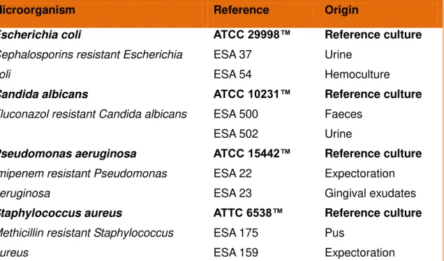
2.8. Antimicrobial activity
35 (DMSO with inoculated medium) was introduced. Antimicrobial activity was detected by adding 20 μL of 0.5% TTC solution.
The Minimum Inhibitory Concentration (MIC) was defined as the lowest concentration of propolis extract that inhibited visible growth, as indicated by the TCC staining (dead cells are not stained by TTC). All the tests were performed in triplicate (n=3). The results are expressed as mg/mL.
Table 1 – Microorganisms used in the present study to test antimicrobial activity of propolis extracts.
2.9. Statistical analysis
Each propolis sample was analysed in triplicate. Results are shown as arithmetic mean values ± standard deviation. In each parameter, the differences between propolis were analysed using one-way analysis of variance (ANOVA) followed by Tukey’s HSD. P values less than or equal to 0.05 were evaluated as statistically significant. This treatment was carried out using SAS v. 9.1.3 program (SAS Inc, New York City, USA).
Microorganism Reference Origin
Escherichia coli
Cephalosporins resistant Escherichia
coli ATCC 29998™ ESA 37 ESA 54 Reference culture Urine Hemoculture Candida albicans
Fluconazol resistant Candida albicans
ATCC 10231™ ESA 500 ESA 502 Reference culture Faeces Urine Pseudomonas aeruginosa
Imipenem resistant Pseudomonas
aeruginosa ATCC 15442™ ESA 22 ESA 23 Reference culture Expectoration Gingival exudates Staphylococcus aureus
Methicillin resistant Staphylococcus
aureus ATTC 6538™ ESA 175 ESA 159 Reference culture Pus Expectoration
36 3. Results and Discussion
3.1. Palynological identification
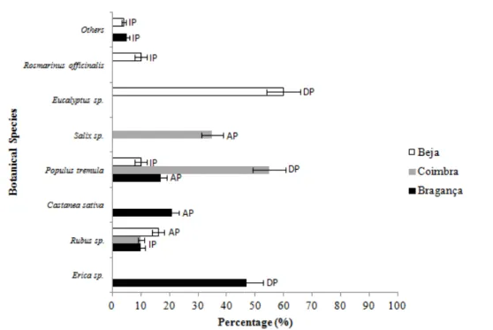
Results of bee pollens’ profile analysis allow scientists to infer the vegetation present in the area and to date and ascertain any biodiversity change, as for example, the presence and distribution of invasive or exotic plants (Morais et al., 2011). The quantification of the pollens’ present in propolis aims to determinate its floral origin. In fact, this origin is one of the factors that influence the bioactive properties of this product. In accordance with melissopalynological criteria (Louveaux et al. 1970), the following designations of pollen frequency were used: PD for dominant (>45%), PA for accessory (15-45%), and PI for isolated pollen loads but important to characterize the phytogeographical origin of the sample (3-15%).
37 Figure 3 - Palynological spectrum of bee pollen samples. DP – Dominant Pollen (>45%); AP - Acessory Pollen (15%-45%); IP – Isolated Pollen (<15%)
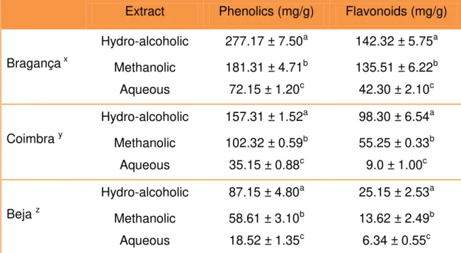
3.2. Total Phenolics and Flavonoids
According to the literature (Bankova et al., 2000; Gómez-Caravaca et al., 2006), the majority of compounds already identified in propolis are polyphenols. These compounds have been extracted using different solvents: water, methanol and ethanol. In this context, the efficiency of these substances was assessed.
38 hydro-alcoholic extract was significantly higher than the amounts extracted by methanol and by water.
Concerning the different places, propolis from Bragança was the one that possessed higher concentration of total phenolics (277.17 mg GAE/g ± 7.50) and flavonoids (142.32 mg GAE/g ± 4.52), followed by Coimbra’s propolis. For a 95% confidence interval (p=0.05), significant differences were found among the samples with different origins (Table 2).
Table 2 – Concentration (mg GAE/g) of phenolics and flavonoids in propolis extracts from different locations (n=9)
Globally, our results are in agreement with the data obtained by Moreira et al. (2008), who studied propolis from the northeast of Portugal. However, Miguel et al. (2010) obtained inferior values when analyzing propolis from the south of the same country. This discrepancy may be due to the great distances between the local of origin and the different apicultural practices. In fact, our data suggest that propolis from different places have different concentrations of polyphenols.
Extract Phenolics (mg/g) Flavonoids (mg/g)
Bragança x
Hydro-alcoholic 277.17 ± 7.50a 142.32 ± 5.75a Methanolic 181.31 ± 4.71b 135.51 ± 6.22b Aqueous 72.15 ± 1.20c 42.30 ± 2.10c
Coimbra y
Hydro-alcoholic 157.31 ± 1.52a 98.30 ± 6.54a Methanolic 102.32 ± 0.59b 55.25 ± 0.33b
Aqueous 35.15 ± 0.88c 9.0 ± 1.00c
Beja z
Hydro-alcoholic 87.15 ± 4.80a 25.15 ± 2.53a Methanolic 58.61 ± 3.10b 13.62 ± 2.49b
Aqueous 18.52 ± 1.35c 6.34 ± 0.55c
39 The values obtained for catechin and gallic acid, which were used as standards, were bellow the concentration obtained in this study for flavonoids. This is in agreement with the reported by Falcão et al. (2010) and Popova et al. (2004) that refer the minor importance of gallic acid in propolis from temperate zones. This phenolic acid is mostly found in tropical samples. In propolis from the Mediterranean region prevailed flavonoids and esters of caffeic and ferulic acids.
Considering that the hydro-alcoholic extract was the most effective, it was used in all the assays performed after.
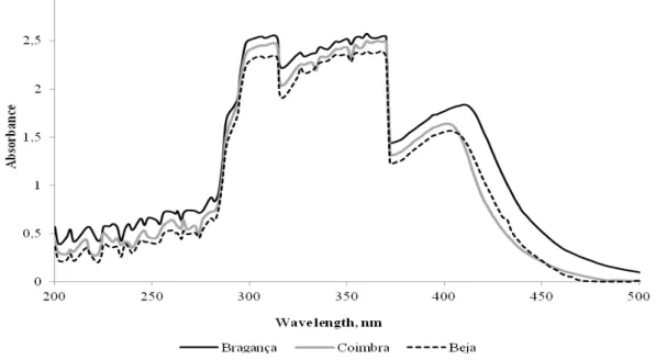
3.3. UV-Visible Absorption Spectroscopy
The absorption spectrum of the hydro-alcoholic extracts is shown in Figure 4. The spectrums of the analysed propolis were similar, with the maximum absorption between 290 nm and 370 nm. In agreement with Castro et al. (2007) the absorption profile between the 270 - 330 nm (wavelength) are attributed to flavonoids and phenolics. This suggests that the polyphenols are the biggest constituents of propolis. The small differences (p=0.103) in absorbance values reflect the concentrations of phenolic and flavonoids present in each propolis: propolis from Bragança possesses the highest amount of polyphenols and also has the highest value of absorbance.
40 3.4. Anti-inflammatory activity
The inflammation process involves production and/or release of mediators from neurons or damaged tissues, which are responsible for different responses including pain. Scavenging of free radicals, generated by neutrophils in inflammatory processes, is the principal mechanism of conventional anti-inflammatory drugs, and is also a known property of propolis (Paulino et al., 2003). In this study, we verified that all the extracts inhibited the hyaluronidase enzyme in a dose-dependent manner (Figure 5). The propolis that showed higher inhibitory activity was the one from Bragança and the product from Beja was the less effective. When the concentration of propolis was 25mg/mL, the percentage of inhibition was 75.79±2.17% (Bragança), 70.48±3.12% (Coimbra) and 53.76±2.87% (Beja).
Concerning the inhibition, it weren’t found significant differences between the samples from Bragança and Coimbra, despite the differences amongst the polyphenols’ concentrations. This suggests that these compounds are not the only factor responsible for the bioactive properties of this beehive product. In fact, other constituents like vitamins and proteins are also involved in this activity (Almeida-Muradian et al., 2005).
The action mechanisms of this product haven’t yet been figured out. However, Hu et al. (2005) claimed that propolis inhibited the increase of prostaglandin E2 and
41 Figure 5 - Inhibition of the activity of Hyaluronidase by the propolis extracts for each concentration. The letters (a,b) represent which samples are different by Tukey test with significance of p = 0.05
3.5. Antimicrobial activity