U
NIVERSIDADE DE
L
ISBOA
F
ACULD ADE DEC
I ÊNCI ASDepartamento de Biologia Vegetal
B cell gene expression in rheumatoid arthritis:
Effect of immunosuppressive treatment
Ana Rita Fernandes Vieira
Dissertação
Mestrado em Biologia Molecular e Genética
U
NIVERSIDADE DE
L
ISBOA
F
ACULD ADE DEC
I ÊNCI ASDepartamento de Biologia Vegetal
B cell gene expression in rheumatoid arthritis:
Effect of immunosuppressive treatment
Ana Rita Fernandes Vieira
Dissertação orientada por:
Doutora Rita Alexandra Pedra Aguiar de Moura Instituto de Medicina Molecular (IMM)
Professora Doutora Maria Margarida BlasquesTelhada Faculdade de Ciências da Universidade de Lisboa
Mestrado em Biologia Molecular e Genética
2014
Todas as afirmações efetuadas no presente documento são de exclusiva responsabilidade do seu autor, não cabendo qualquer responsabilidade à Faculdade de Ciências da Universidade de Lisboa pelos conteúdos nele apresentados.
A
CKNOWLEDGEMENTSWhile writing this thesis I benefited greatly from the discussion I had with a number of people, some of whom also commented parts of it. First, I would like to thank to Professor João Eurico Fonseca and Professor Helena Canhão for receiving me in the Rheumatology Research Unit and for all the ideas brought to the work throughout this year. Also, thanks to Professor Margarida Telhada for having agreed to be my internal supervisor, even in the last minute.
I owe a special debt of gratitude to Rita Moura for accepting me as her student and for the stimulating and fruitful conversations we had, not only on the subject but also in other areas, as well as the encouragement and support she gave me in the preparation of the thesis. Despite being a morning person I learned that the nights in the lab can be less crowded, and, therefore, more quiet than the afternoons. To Claudia because I know now how important it is to have someone in the lab to share our doubts, to help each other, to discuss at length the results while we waited to acquire all events at FACS… as real lab partners do!
A research unit is nothing without its students, whether they are PhD, MSc, graduates or even other people that manage and contribute to the development of it. Our paths were crossed and because of that, I share my ‘thank you’ to all those who had to, somehow, deal with my uncertainties. I might be forgetting someone, but here it is (and the order is arbitrary): Inês, Bruno, Ana and my other mates for creating a wonderful work environment, Mónica, Rita R., Ana D. and Joana S. – thanks y’all!
Because we need those who care about us to provide a mirror for our actions as we step out into the brave new world1, I have to thank my family for the support especially my parents who have been giving me opportunities to pursue my dreams and were always here for every milestone I stepped into.
Finally I would like to thank to my long-time friends that, in their ways, tried to understand my enthusiasm for investigation. Sara thanks for all those years of friendship and for the person who I could always count to get something off my chest; you became almost like the sister I wish to have had.
In this last two years new friendships were made and now the team “Potência máxima” is constantly nagging me (in a good way) about the writing and giving me helpful advices. After all, you already went through the same process and understand better than anyone what this type of endeavour means. For that and much more I will never forget you…
I hope that the project contained in this thesis seems of any merit.
It might be true that there are six billion people in the world, and counting. Nevertheless, what you do makes a difference.Waking life (2001)
1
R
ESUMOA artrite reumatoide (AR) é uma doença autoimune sistémica que se caracteriza pela inflamação crónica das pequenas articulações, particularmente das mãos, bem como articulações maiores, que incluem os joelhos e tornozelos. A prevalência da doença é de cerca de 1% na população mundial adulta e é mais frequente nas mulheres do que nos homens (numa proporção de 3:1). Se não for tratada, a AR origina destruição articular, com erosão óssea e da cartilagem.
O diagnóstico da AR é feito de acordo com critérios definidos pelo American College of Rheumatology (ACR)/ European League Against Rheumatism (EULAR), baseados numa avaliação radiológica e atribuição de pontuação relativa ao número de articulações afetadas; serologia de autoanticorpos IgM específicos para o fragmento Fc de imunoglobulinas IgG (fator reumatoide, RF) ou para péptidos cíclicos citrulinados modificados pós-traducionalmente por incorporação de citrulina por ação das peptidil arginina deaminases (anticorpos anti-proteínas citrulinadas, ACPA ou anti-CCP); quantificação de fatores de inflamação (velocidade de sedimentação eritrocitária e proteína-C reativa); e duração da doença.
A etiologia da doença é desconhecida, mas sabe-se que a influência do genótipo e do ambiente parecem ser fatores essenciais para o início do processo inflamatório. Em termos de predisposição genética, a AR está associada a mutações no gene HLA-DRB1 que pertence ao complexo maior de histocompatibilidade (MHC) classe II, tendo sido identificada uma curta sequência de aminoácidos comum às proteínas codificadas por todos os alelos de risco - o shared epitope. A AR está também associada a outros genes como o PTPN22 e CTLA-4, entre outros. Em relação aos fatores ambientais, a maior associação descrita está relacionada com o tabagismo, sendo que o risco parece ser maior em indivíduos portadores do shared epitope. O efeito da exposição a alguns agentes infeciosos é controverso, mas alguns estudos indicam que poderá levar à formação de complexos imunes, implicados na AR. Também algumas alterações hormonais na mulher, como a gravidez, amamentação e contraceção oral, podem influenciar o desenvolvimento da doença.
No processo inflamatório da AR ocorre uma resposta celular e humoral que contribui para o desenvolvimento da sinovite, ou seja, a inflamação da membrana sinovial, que é a membrana que reveste a articulação. A hiperplasia da sinóvia resulta de uma migração e excessiva infiltração de linfócitos T e B, plasmócitos, células dendríticas, macrófagos, neutrófilos e mastócitos, assim como da ativação dos FLS – fibroblast‐like synoviocytes –
que produzem citocinas pro-inflamatórias, provocando a ativação de outras células. A ativação de macrófagos através dos recetores Fc gama por complexos imunes que se formam a partir de autoanticorpos (RF e ACPA), produzidos por células B autoreactivas, juntamente com estímulos derivados de células T activadas, conduz à libertação de citocinas como a IL-1, IL-6, IL-15, INF-γ e TNF. Algumas destas citocinas ativam células sinoviais residentes a produzir enzimas proteolíticas, como a colagenase e metaloproteinases da matriz que medeiam a destruição da cartilagem. A destruição óssea também ocorre devido ao aumento da atividade dos osteoclastos, ativados através das ligações RANK/ RANK-L.
Nos últimos anos, os linfócitos B têm sido alvo de investigação, particularmente após a eficácia clínica observada com a terapia de depleção de células B com rituximab (anticorpo monoclonal anti-CD20). De facto, os linfócitos B podem contribuir de diversas formas para o desenvolvimento da AR, tais como: 1) produção de autoanticorpos como o RF e os ACPA, que formam complexos imunes que se depositam nas articulações e causam inflamação; 2) apresentação de antigénios e ativação de linfócitos T; 3) secreção de citocinas; 4) ativação de osteoclastos que levam à erosão óssea; 5) imunoregulação por células B reguladoras, que produzem citocinas anti-inflamatórias (IL-10 e TGF-β1) que inibem células T ativadas e restauram o balanço TH1/TH2.
Apesar da AR não ter cura, é fundamental estabelecer um diagnóstico precoce para que se possa proceder ao tratamento e reduzir os sintomas no doente e a progressão da doença o mais rapidamente possivel. O tratamento da AR faz-se com anti-inflamatórios não esteroides (AINES), sendo que estes apenas diminuem a atividade inflamatória; corticoesteróides; DMARDs - agentes antirreumáticos modificadores da doença – sintéticos, onde se encontram o metotrexato (MTX), sulfassalazina (SSZ), hidroxicloroquina (HCQ) e a leflunomida, ou biológicos, que são geralmente anticorpos monoclonais que bloqueiam determinadas citocinas ou recetores celulares. Entre os DMARDs biológicos aprovados para tratamento da AR estão, por exemplo, os anti-TNF (etanercept, infliximab, golimumab, adalimumab e certolizumab), o tocilizumab – TCZ receptor da IL-6) e o rituximab (anti-CD20). Os DMARDs são conhecidos por melhorarem a capacidade funcional global, reduzirem os danos radiológicos e diminuirem os valores clínicos e laboratoriais associados à inflamação. Nem sempre os doentes respondem favoravelmente à terapêutica, podendo apresentar efeitos secundários graves devido ao efeito imunosupressor e/ ou mantendo níveis de atividade de doença elevados, o que obriga muitas vezes a alterações no tratamento, procedendo-se a mudanca entre anti-TNFs, ou mesmo para outros biológicos, como o TCZ e o rituximab.
O principal objetivo deste trabalho consistiu na análise de um grupo de genes relacionados com a ativação e sobrevivência dos linfócitos B (BAFF-R, TACI, BCMA), mudança de classe e hipermutação somática (AID), diferenciação de plasmócitos (BLIMP-1), quimiotaxia (CXCR5), apoptose (BCL- 2), inibição (CD32), inflamação (B2M) e ativação de células B através de Toll-like receptors (TLR7, TLR9, TLR10) em doentes não tratados com AR inicial (ERA) (≤1 ano de duração da doença) e na comparação da expressão dos mesmos genes com doentes com AR estabelecida após tratamento com MTX, MTX pré-biológico (MTX pre-bio, isto é antes do início de terapêutica com agentes biológicos), anti-TNF e TCZ. Para tal, amostras de sangue periférico foram colhidas a doentes ERA (n=13) e AR estabelecidas após tratamento com MTX (n=15) e MTX pre-bio (n=25). No grupo de doentes MTX pre-bio, foi efetuada uma segunda colheita de sangue a um grupo de doentes com AR após 8 meses de tratamento com anti-TNF (n=7) e a outro grupo após 6 meses de tratamento com TCZ (n=9). Amostras de sangue de controlos saudáveis (n=15) com a mesma proporção de idades e género foram também colhidas e processadas para comparação de resultados. Procedeu-se ao isolamento das células mononucleares do sangue periférico (linfócitos B e T, monócitos e granulócitos) e, posteriormente, separam-se os linfócitos B. De seguida, a partir dos linfócitos B isolados com purezas acima dos 90%, extraiu-se o RNA, sintetizou-se o DNA complementar (cDNA) e fizeram-se ensaios de PCR em tempo real para estudar a expressão génica do grupo de genes acima mencionados.
Os resultados da expressão génica revelaram um aumento dos níveis de mRNA do BAFF-R no grupo de doentes com AR estabelecida tratados com MTX (p=0.0212) e anti-TNF (p=0.0025) relativamente aos controlos. Além disso, também se observou um aumento da expressão do BAFF-R no grupo de doentes tratados com anti-TNF quando comparado com os doentes ERA (p=0.0006). Por outro lado, nao foram observadas diferenças significativas na expressão génica de TACI e BCMA em todos os grupos analisados. A análise da expressão génica dos Toll-like receptors revelou diferenças significativas nos grupos de doentes com AR estabelecida em comparação com os controlos. De facto, verificou-se um aumento da expressão de TLR7 e TLR10 no grupo de doentes com AR estabelecida tratados com anti-TNF em relação aos controlos (p=0.0385 e p=0.0097, respetivamente) e, além disso, foi também observado um aumento da expressão do gene TLR10 nos doentes tratados com anti-TNF quando comparados com os doentes ERA (p=0.0801). Adicionalmente, foi observado um aumento da expressão do gene TLR9 nos linfócitos B do grupo de doentes com AR estabelecida após tratamento com MTX relativamente aos controlos (p=0.0043). Verificou-se também uma sobreexpressão do gene B2M em todos os grupos de AR estabelecida quando comparados com os controlos: MTX (p=0.0099), MTX pre-bio (p=0.0116), anti-TNF (p=0.0025) e TCZ (p=0.0112), embora não tenham sido
detetadas diferenças significativas nos doentes ERA. Observou-se ainda que os níveis de expressão do gene BCL-2 estavam significativamente elevados em doentes com AR estabelecida após tratamento com MTX (p<0.0001), MTX pre-bio (p<0.0001), anti-TNF (p=0.0014) e TCZ (p=0.0042) não só em comparação com os controlos, mas também com os doentes ERA (p<0.0001). Além disso, verificou-se um aumento significativo nos níveis de expressão génica de CD32 nos doentes com AR estabelecida após tratamento com anti-TNF quando comparados com os controlos (p=0.0043) e com os doentes ERA (p=0.0015). Observou-se ainda um ligeiro aumento da expressão génica de CD32 em doentes com AR estabelecida após tratamento com MTX pre-bio relativamente aos controlos (p=0.0487). Contudo, foi observado um aumento significativo da expressão de CD32 neste grupo quando comparado com os doentes ERA (p=0.0015). Adicionalmente, não foram observadas diferenças significativas na expressão génica de CXCR5, AID e BLIMP-1 em todos os grupos analisados.
Por fim, quando se procedeu à análise da expressão génica antes e após o tratamento com anti-TNF e TCZ, não foram observadas quaisquer diferenças significativas para todos os genes estudados. Em relação aos dados clínicos dos doentes, verificou-se ainda que o tratamento com TCZ comparativamente com as outras terapêuticas estudadas, parece ser o mais eficaz a reduzir significativamente os valores séricos da proteina-C reativa, a velocidade de sedimentação e a actividade geral da doença indicada pelo DAS28. De salientar, no entanto, que nao foi observada nenhuma correlação significativa entre a idade, os valores da proteína-C reactiva, a velocidade de sedimentação, o DAS28 e o numero de articulações dolorosas e tumefactas com os resultados obtidos da expressão génica para todos os grupos estudados, independentemente do tratamento administrado aos doentes.
Com a realização deste estudo foi possível concluir que em doentes com AR estabelecida existem alterações na expressão de genes associados à ativação dos linfócitos B, quer através do recetor do BAFF (BAFF-R), quer através de TLRs (TLR7, TLR9, TLR10); e/ ou à inibição de linfocitos B (CD32), assim como perturbações na expressão de genes associados à inflamação (B2M) e apoptose (BCL-2). Nos doentes com AR estabelecida, o tratamento com anti-TNF e TCZ parece influenciar a expressão destes genes em comparação com os indivíduos saudáveis, embora nao sejam detectadas diferenças significativas na expressão génica em relação à fase anterior ao início do tratamento. Além disso, a expressão génica em doentes não tratados com AR inicial com menos de 1 ano de duração da doença parece ser semelhante a dos indivíduos saudáveis. Estes resultados sugerem que a expressão génica de linfócitos B em doentes com AR sofre alterações em
diferentes fases de desenvolvimento da doença, podendo ser influenciada pelas opções terapêuticas.
Palavras-chave: Artrite Reumatóide, Linfócitos B, Expressão génica, Metotrexato, Anti-TNF,
A
BSTRACTRheumatoid arthritis (RA) is a systemic autoimmune disease that mainly affects the joints. The clinical success of B cell depletion therapy with rituximab in RA has reinforced the role of B cells in RA pathogenesis. Indeed, recent studies have shown that very early RA patients (with less than 6 weeks of disease duration) have disturbances in circulating memory B cells and increased levels of cytokines directly related with B cell activation and survival, which supports an active role of B cells in RA development since early disease onset.
The main goal of the present study was to analyze the expression of a group of genes directly related with B cell activation, maturation and survival in untreated early RA (ERA) (< 1 year of disease duration) and established RA patients after treatment with methotrexate (MTX), MTX pre-biologic (MTX pre-bio), TNF inhibitors and tocilizumab (TCZ) in order to compare the effects of the therapeutic options on B cell gene expression. For that, blood samples were collected from patients and age and sex-matched healthy donors. B cells were isolated, RNA extracted, cDNA synthesized and gene expression analysis performed by real time PCR.
We found that ERA patients have similar B cell gene expression levels when compared to healthy controls. However, increased gene expression was observed in BAFF receptor (BAFF-R), Toll-like receptors (TLR7, TLR9, TLR10), inhibition marker (CD32) and in genes associated with either active inflammation (B2M) or apoptosis (BCL-2) signaling pathways in established RA patients when compared to controls. No significant differences were observed in TACI, BCMA, CXCR5, AID and BLIMP-1 gene expression in all groups analyzed. Furthermore, the analysis of B cell gene expression levels in established RA patients after treatment with TNF inhibitors and TCZ in comparison with baseline values did not reveal significant differences.
Overall, these results suggest that the expression of genes related with B cell activation and survival are increased in later stages of RA development, which might be influenced by treatment options when compared to healthy individuals. Moreover, B cell gene expression in early RA patients does not seem to significantly change during the first year of RA development when compared to controls.
Keywords: Rheumatoid arthritis, B cells, Gene expression, Methotrexate, TNF inhibitors,
I
NDEX Acknowledgements ... i Resumo ... ii Abstract ... vii Index ... viii List of Abbreviations ... xList of Figures ... xii
List of Tables ... xiii
1. Rheumatoid Arthritis ... 1
1.1 Definition ... 1
1.2 Diagnostic and clinical assessment ... 1
1.3 Etiology and pathophysiology... 3
1.4 Genetic factors and environment ... 5
1.5 Treatment ... 6 1.5.1 Methotrexate ... 7 1.5.2 TNF antagonists ... 7 1.5.3 Tocilizumab ... 7 1.5.4 Rituximab ... 8 2. B cells ... 9 2.1. B cell development ... 9
2.2. B cells and rheumatoid arthritis ... 9
3. Aims ...11
4. Materials and methods ...12
Patients...12
Isolation of peripheral blood mononuclear cells and B cell separation ...12
RNA extraction and complementary DNA (cDNA) synthesis ...12
Real-time quantitative polymerase chain reaction ...13
Statistical analysis ...13
5. Results ...15
5.1. Clinical characterization of patients ...15
5.2. Established RA, but not early RA patients have alterations in B cell gene expression levels in comparison with healthy controls ...16
5.3. Treatment with TNF inhibitors and tocilizumab does not affect B cell gene expression ...20
6. Discussion ...23 7. Limitations and Future Perspectives ...27 8. References ...28
L
IST OFA
BBREVIATIONS18S rRNA 18S ribosomal RNA
ACPA Anti–citrullinated protein antibody ACR American College of Rheumatology
AID Activation-induced cytidine deaminase
APC Antigen presenting cell
APRIL A proliferation-inducing ligand of the TNF family
BAFF B cell activating factor of the TNF family
BCL-2 B-cell lymphoma 2
BCMA B cell maturation antigen
Blimp-1 B lymphocyte-induced maturation protein
Breg Regulatory B cell
CRP C-reactive protein
CpG C-phosphate-G
CTLA-4 Cytotoxic T-Lymphocyte Antigen 4
CXCL C-X-C chemokine ligand
CXCR C-X-C chemokine receptor
DAS28 Disease Activity Score in 28 joints
ESR Erythrocyte sedimentation rate
EULAR European League Against Rheumatism
FcγR Fc-gamma receptor
Foxp3 Forkhead box P3
GC Germinal center
GM-CSF Granulocyte-macrophage colony stimulating factor
HCQ Hidroxychloroquine
HLA Human leucocyte antigen
Ig Immunoglobulin
IL- Interleukin
INF-γ Interferon-gamma
MHC Major histocompatibility complex
M-CSF Macrophage colony-stimulating factor
MIP Macrophage inflammatory protein
MCP Monocyte chemoattractant protein
MTX Methrotrexate
NF-кB Nuclear factor-kappa B
PAD Peptidylarginine deiminase
PTPN22 Protein tyrosine phosphatase non-receptor 22
RA Rheumatoid Arthritis
RAG Recombination activating gene
RANK Receptor activator of nuclear factor-кB
RF Rheumatoid factor
SE Shared epitope
STAT-4 Signal transducer and activator of transcription protein 4
SSZ Sulfasalazine
TACI Transmembrane activator and calcium-modulator cyclophilin ligand interactor
TLR Toll- like receptor
TCR T cell receptor
TGF-β Tumor growth factor β
TNF Tumor necrosis factor
L
IST OFF
IGURESFig. 1 – X-ray from a normal hand (left) and a hand from a patient with rheumatoid arthritis.. 1 Fig. 2 - Pathogenesis of RA ... 3 Fig. 3 - Biologic agents and its targerts ... 8 Fig. 4 - The multiple roles of B cells in RA ...10 Fig. 5 - BAFF-R gene expression is significantly increased in B cells from established RA patients after treatment with MTX and TNF inhibitors.. ...17 Fig. 6 - Established RA patients have alterations in the gene expression levels of TLR7, TLR9 and TLR10 after treatment with MTX and/ or TNF inhibitors. ...18 Fig. 7 - Established RA patients have increased gene expression levels of B2M, CD32 and BCL-2 irrespective of the treatment ...19 Fig. 8 - ERA and established RA patients have normal gene expression levels of AID, BLIMP-1 and CXCR5. ...20 Fig. 9 - B cell gene expression is not affected by TNF inhibitors in established RA when compared to baseline. ...21 Fig. 10 - B cell gene expression is not affected by tocilizumab in established RA when compared to baseline ...22
L
IST OFT
ABLESTable 1 - The ACR 1987 criteria for classification of RA ... 2 Table 2 - New classification criteria for rheumatoid arthritis ... 2 Table 3 - Primers used to examine the expression levels in pPCR ...14 Table 4 - Clinical information of controls, ERA and established RA patients after treatment with MTX, MTX pre-bio, TNF inhibitors and TCZ ...15
1.
R
HEUMATOIDA
RTHRITIS1.1 Definition
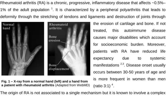
Rheumatoid arthritis (RA) is a chronic, progressive, inflammatory disease that affects ~0.5%– 1% of the adult population 1. It is characterized by a peripheral polyarthritis that leads to deformity through the stretching of tendons and ligaments and destruction of joints through the erosion of cartilage and bone. If not treated, this autoimmune disease causes major disabilities which account for socioeconomic burden. Moreover, patients with RA have reduced life expectancy due to systemic manifestations 2,3. Disease onset usually occurs between 30-50 years of age and is more frequent in women than men (ratio 3:1) 4.
The origin of RA is not associated to a single mechanism but it is known to involve a complex dysregulation in the immune system, genotype influence and environmental interactions. Thus, RA is presented as a heterogeneous condition mainly characterized by synovial inflammation and hyperplasia (swelling), cartilage and bone destruction, and systemic features, including cardiovascular, pulmonary, psychological, and skeletal disorders 5,6.
1.2 Diagnostic and clinical assessment
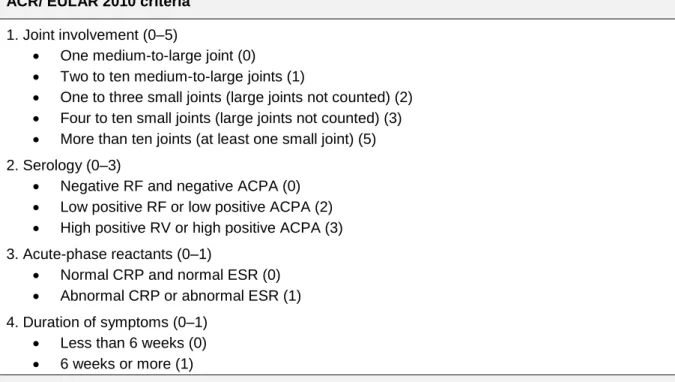
For years, RA diagnosis was established according to the 1987 American College of Rheumatology (ACR) criteria (Table 1) 7. In 2010, the ACR and the European League Against Rheumatism (EULAR) developed a new criteria that allowed more specificity when distinguishing RA from other joint diseases and more sensitivity to identify individuals in the early stages of RA (Table 2) 8–10.
The early stages of RA are mainly characterized by a symmetrical polyarthritis that affects the small joints of the hands and feet (metacarpophalangeal, proximal interphalangeal and metatarsophalangeal joints) before larger joints are affected 11. Due to synovial hypertrophy, the joints become swollen, promoting pain and stiffness. Moreover, the levels of inflammatory markers in the blood, such as erythrocyte sedimentation rate (ESR) and C-reactive protein (CRP) raise 12,13. Progression of joint damage in patients with RA is also highly correlated with overall levels of disease activity assessed by the disease activity score of 28 joints
Fig. 1 – X-ray from a normal hand (left) and a hand from a patient with rheumatoid arthritis (Adapted from WebMD)
(DAS28), which comprises the count of 28 tender or swollen joints in the hands, upper limbs and knees.14. The joints are also evaluated by imaging studies but these changes frequently take several years to develop 15.
Table 1 - The ACR 1987 criteria for classification of RA (adapted from 6)
The presence of autoantibodies such as rheumatoid factor (RF) and anti-citrullinated protein antibodies (ACPA) (or anti-cyclic citrullinated peptides (anti-CCP)) in serum is also a characteristic of this autoimmune condition, although patients seronegative for these antibodies also manifest the disease 16,17.
Table 2 - New classification criteria for rheumatoid arthritis (Adapted from 6) RF = rheumatoid factor; ACPA= antibodies against citrullinated antigens; CRP= C-reactive protein; ESR = erythrocyte sedimentation rate
RF are autoantibodies that directly bind to the Fc portion of normal human IgG and ACPA are autoantibodies that recognize peptides or proteins containing citrulline, a non-standard
ACR 1987 criteria
1. Morning stiffness (at least 1h) 2. Arthritis of three or more joint areas 3. Arthritis of hand joints (≥1 swollen joints) 4. Symmetrical arthritis
5. Rheumatoid nodules 6. Serum rheumatoid factor
7. Radiographic changes (erosions)
Four of these seven criteria must be present. Criteria 1–4 must have been present for at least 6 weeks
ACR/ EULAR 2010 criteria 1. Joint involvement (0–5)
One medium-to-large joint (0) Two to ten medium-to-large joints (1)
One to three small joints (large joints not counted) (2) Four to ten small joints (large joints not counted) (3) More than ten joints (at least one small joint) (5) 2. Serology (0–3)
Negative RF and negative ACPA (0) Low positive RF or low positive ACPA (2) High positive RV or high positive ACPA (3) 3. Acute-phase reactants (0–1)
Normal CRP and normal ESR (0) Abnormal CRP or abnormal ESR (1) 4. Duration of symptoms (0–1)
Less than 6 weeks (0) 6 weeks or more (1)
Points are shown in parenthesis. Cutpoint for rheumatoid arthritis 6 points or more. Patients can also be classified as having rheumatoid arthritis if they have: (a) typical erosions, (b) long-standing disease previously satisfying the classification criteria
aminoacid generated by the post-translational modification of arginine by peptidylarginine deiminase (PAD) enzymes, in a process known as citrullination 18. RF and ACPA can form immune complexes that deposit in the joints, activate complement and cause inflammation. The clinical usefulness of RF and ACPA has been acknowledged due to their good diagnostic sensitivity and prognostic value 17,19. Of note, RF and ACPA can develop several years before clinical onset 16.
1.3 Etiology and pathophysiology
The etiology of RA is unknown. However, the development of this autoimmune disease seems to be a multistage process in which a number of genetic and environmental factors trigger the development of undifferentiated arthritis and a subsequent amount of stimuli are required for progression towards joint inflammation.
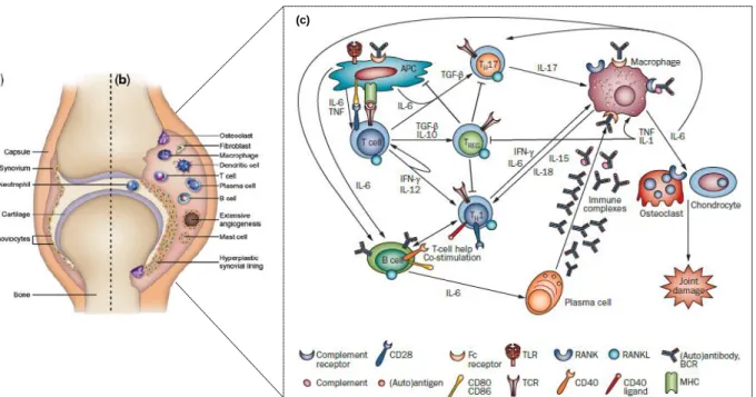
The synovial joint is composed of two adjacent bony ends each covered with a layer of cartilage, separated by a joint space and surrounded by the synovial membrane and joint capsule. The synovial lining (facing the cartilage and bone) consists of a thin layer of synoviocytes (macrophage and fibroblast-derived cells) that constitute the cellular part of the synovium. In healthy individuals, the synovium is almost an acellular structure (Fig. 2a) 20. Contrarily, in RA, a variety of mononuclear cells such as T cells, B cells, plasma cells, dendritic cells, macrophages and mast cells infiltrate the synovial membrane through the endothelia, forcing it to expand and become hyperplasic (Fig. 2b) 21. Hyperplasia also results
(c)
Fig. 2 - Pathogenesis of RA (Adapted from 158,159) a) represents the normal synovium; b) in RA the immune cells inflitrate the synovium making it hyperplastic with extensive angiogenisis; c) the adaptive and innate immune processes in the joint of RA
from a marked increase in macrophage-like and fibroblast-like synoviocytes as they produce cytokines and factors that maintain inflammation by activating either themselves or their neighboring cells 22.
RA pathogenesis involves a complex network between cells from both innate and adaptive immune system (Figure 2c). An initial trigger activates macrophages in the synovial membrane, mediated locally through the action of antigen-activated CD4+ T helper cells 23. The T cells invading the synovial membrane are primarily CD4+ memory cells, which produce interleukin-2 (IL-2) and interferon-gamma (IFN-γ), and are either already pre-activated or become further activated by antigen-presenting cells (APC) in conjunction with arthritogenic (auto)antigen(s) and appropriate MHC class-II molecules, co-stimulation (mainly through CD80/86 and CD28) and certain cytokines (IL-1, IL-15, IL-18) 24. Activated T cells stimulate monocytes, macrophages and synovial fibroblasts. This will lead to an overproduction of pro-inflammatory cytokines, mainly TNF 25, IL-1 26 and IL-6 27, which seem to constitute a key event leading to chronic inflammation 28–30. TNF and IL-1 also induce receptor activator of nuclear factor-кB (RANK) expression on macrophages that, after binding to RANK ligand (RANKL), stimulates macrophage differentiation into osteoclasts, which consequently resorb and destroy bone 31. Chondrocytes also become activated under the influence of 1 and IL-17A, leading to the release of metalloproteinases 5. Moreover, locally expressed degradative enzymes, serine proteases and aggrecanases, digest the extracellular matrix and destroy the articular structures 20. An extensive angiogenesis process also occurs in RA, promoted by vascular endothelium growth factor (VEGF) production by monocytes, endothelial cells and synovial fibroblast in response to all the inflammatory stimuli, hypoxia and reduced apoptosis
32. This supports the formation and maintenance of pannus, a membrane of fibrovascular
tissue that invades and destroys cartilage and bone 33.
Recent studies also implicate IL-17-producing T cells (TH17) as important effectors in RA
pathogenesis due to the overexpression of IL-17 in the synovial joints, which is associated with an aggravation of inflammation and joint damage 34,35. In fact, a neutrophil- and TH
17-driving cytokine pattern is present in the serum of untreated patients with very early RA 36. Regulatory T cells (TRegs) have also been detected in the synovium of RA patients with active
disease and particularly in synovial fluid, but they seem to have impaired regulatory function
37. The cytokine factors that sustain the expansion of T
Regs in the rheumatoid joint are not
defined but probably include the regulatory cytokines IL-10 and TGF-β 38
.
B cells also have a relevant role in RA pathogenesis 39, namely through autoantibody production (RF, ACPA), antigen presentation and cytokine release upon activation. Indeed, B cells produce RF and ACPA that form immune complexes that deposit in the joints. These immune complexes activate macrophages through Fc-gamma receptors (FcγR) that release
pro-inflammatory cytokines (TNF, IL-1, IL-6) 23 that exacerbate the inflammatory process as previously described. Furthermore, it has been recently demonstrated that a cytokine pattern directly related with B cell activation and survival is present since the first weeks of RA onset, which supports an early intervention of B cells in RA development 40.
1.4 Genetic factors and environment
RA comprises several genetic and environmental associations that are supposedly responsible for some alterations in post-transcriptional regulation 5. Twin studies have demonstrated a genetic role and showed that genetic factors contribute to 60% of risk of developing RA 41. The first established association was related with genes within the HLA (human leucocyte antigen) region, particularly the HLA-DRB1 allele that is associated with severe rheumatoid arthritis 42–44. This gene encodes major histocompatibility complex (MHC) class-II β-chain molecules whose function is the presentation of antigen to CD4+ T cells.
HLA-DRB1 alleles contain a common aminoacid motif termed “shared epitope” (SE) – a five aminoacid sequence motif in residues 70–74 of the HLA-DRβ chain 45. When antigens
undergo post-translational modifications by citrullination (conversion of the aminoacid arginine to citrulline) this change allows antigens to fit in the HLA alleles that harbor this shared epitope which might result in breaking of tolerance that allows antibody formation against these antigens 46,47. In fact, HLA class II alleles such as DR4 that form the ‘shared epitope’ are not primarily a risk factor for RA, but for the presence of ACPA 48. A second
association was established with the protein tyrosine phosphatase non-receptor 22 (PTPN22) gene, known to exert a negative feedback effect on the transmission of the signal generated by the T cell receptor (TCR) 5,32. These genetic associations might explain the emergence of autoreactive responses in antigen presentation, T cell repertoire selection and alterations in peptide affinity 6. Other genes linked to RA susceptibility were also associated with the NF-кB pathway (REL, TRAF1, and TNFAIP3) 49 and other pathways related to T cell
activation such as cytotoxic T-lymphocyte antigen 4 (CTLA-4) that suppresses the interaction between T cells and antigen-presenting cells (APC); and signal transducer and activator of transcription protein 4 (STAT-4), responsible for transducing cytokine signals that regulate proliferation, survival and differentiation of lymphocytes 50.
The environmental risk factors in RA include age and gender 18, a previous family history of RA 51, smoking 52–54, obesity 55 , cardiovascular events 56 and some infectious agents 57,58. In fact, it has been documented that changes in the female hormonal environment such as pregnancy, breastfeeding and the use of the oral contraceptive might affect the development of RA 59–61.
Some studies have also reported that the formation of immune complexes during exposure to some infectious agents might trigger the induction of RF autoantibodies, which have long been implicated in RA pathogenesis 58. Nonetheless, the most well-established environmental association with RA is smoking. Indeed, cigarette smoking has been associated with a consistently increased risk of developing RA, particularly in individuals that express the HLA-DRB1 SE alleles 53,54.
Overall, while RA susceptibility is genetically determined, disease onset may depend on non-genetic (i.e. environmental), epinon-genetic or post-translational events.
1.5 Treatment
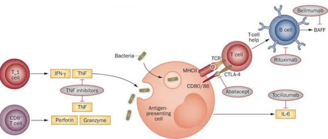
Although no cure has been found for RA, it is clear that establishing a diagnosis as early as possible and immediate treatment are the basis for a successful management of these patients 62. The main goals of RA treatment are the preservation of function and quality of life, remission of symptoms and control of systemic manifestations. Improvement in RA is defined by an outcome measure called ACR 20, ACR 50 or ACR 70, that is characterized as a reduction by 20, 50 or 70%, respectively, on the number of tender or swollen joints plus improvement in at least three of the five measures: pain, global assessment by the patient and levels of acute-phase reactants 6,63. Treatment options are divided in three main classes: 1) nonsteroidal anti-inflammatory drugs (NSAIDs); 2) corticosteroids; and 3) disease modifying anti-rheumatic drugs (DMARDS) (synthetic and biologic) 64. RA patients should begin their treatment with DMARDs, which not only relieve symptoms but also slow the progression of the disease. Often, DMARDs are prescribed along with NSAIDs and/or low-dose corticosteroids to decrease swelling and pain 6. DMARDs have greatly improved the symptoms, function and quality of life of nearly all patients with RA. Synthetic DMARDs include methotrexate, leflunomide, sulfasalazine (SSZ) and hidroxychloroquine (HCQ), which are widely used in RA and have proven to be highly beneficial in decreasing inflammation and joint damage 64. Gold used to be also administrated to RA patients, but nowadays is rarely prescribed since other drugs work better and/ or have fewer side effects 65. Nevertheless, patients still fail conventional DMARD therapy, maintaining a high disease activity and/ or suffering from severe adverse effects. In these cases, it is recommended that biologic DMARD therapy is initiated 64,66. Biologic DMARDs include TNF antagonists 67, which are sub-divided in neutralizing anti-TNF monoclonal antibodies (infliximab, adalimumab, golimumab, certolizumab) and a soluble TNF receptor-2–IgG-Fc fusion protein (etanercept)
68
. Other biologic therapies include abatacept, a fusion protein composed of the Fc region of IgG1 fused to the extracellular domain of cytotoxic T-lymphocyte antigen (CTLA)-4 that binds to the CD80/ CD86 molecule and inhibits T cell activation 69, anakinra, an IL-1 receptor
(IL-1R) antagonist 26; tocilizumab, an IL-6 receptor (IL-6R) blocking monoclonal antibody 70 and rituximab, an anti-CD20 B-cell-depleting monoclonal antibody 71.
1.5.1 Methotrexate
Methotrexate (MTX) is an anti-metabolite and a folate analog designed to compete for folate receptors, which affects the de novo synthesis of purine and pyrimidine precursors of DNA and RNA 72. MTX also has anti-inflammatory effects at low doses 73. MTX inhibits T-cell activation and proliferation, downregulates the expression of some activation and adhesion molecules, decreases immunoglobulin production, inhibits cyclooxygenases and lipooxygenases, and modulates the secretion of various cytokines by monocytes and macrophages. MTX, administrated alone or in combination with either synthetic or biologic DMARDs remains the first drug of choice for patients with RA 74.
1.5.2 TNF antagonists
TNF binds to two receptors, the type 1 TNF receptor (TNFR) (p55) and the type 2 TNFR (p75), that is expressed on many cell types, such as monocytes, T cells, B cells, NK cells, PMNs, mast cells, synovial fibroblasts and osteoblasts 22. TNF antagonists include neutralizing monoclonal antibodies such as infliximab, adalimumab or golimumab and a fusion protein, etanercept. Infliximab, is a chimeric IgG1 anti-TNF antibody containing the antigen-binding region of a mouse antibody and the constant region of a human antibody. It binds with high affinity to soluble and membrane-bound TNF, impairing the binding of TNF to its receptor. Infliximab also kills cells that express TNF through antibody-dependent and complement-dependent cytotoxicity (ADCC). Adalimumab and golimumab are recombinant human IgG1 monoclonal antibodies that bind to human TNF with high affinity, both impairing TNF binding to its receptors and lysing cells that express TNF on their surface. Etanercept, a soluble TNFR fusion protein composed of two dimers, each with an extracellular, ligand-binding portion of the higher-affinity type 2 TNFR (p75) linked to the Fc portion of human IgG1. This fusion protein binds to both TNF-α and TNF-β, thereby preventing each from interacting with its respective receptors. TNF antagonists are more effective when co-prescribed with MTX. Switching between anti-TNF treatments after secondary treatment failure can be associated with a sustained clinical improvement. Although usually effective, all anti-TNF treatments are associated with an enhanced risk of infections compared to non-biological DMARDs 69.
1.5.3 Tocilizumab
IL-6 forms a complex by binding to IL-6R on the cell membrane, which then combines with gp130 that also resides on the cell membrane, forming a homodimer and giving rise to
intracellular signal transduction 27. Tocilizumab (TCZ) is a recombinant monoclonal antibody that inhibits the induction of biological activity due to IL-6 in cells that express both membrane-bound IL-6R and gp130 molecules, and also inhibits the induction of biological activity due to IL-6/IL-6R complex formation in cells that express gp130 alone 75. Inhibition of IL-6 significantly has showed improved signs and symptoms of RA and normalized the acute-phase reactants 76.
1.5.4 Rituximab
CD20 is expressed almost exclusively by B cells at different stages of differentiation, but it is not expressed by stem cells or by earlier precursors (i.e. pro-B cells), or by terminally differentiated plasma cells 77. Rituximab (RTX) is a monoclonal antibody that selectively depletes B cells by directly binding to CD20 78. RTX depletes B cells by inducing cell lysis, which can be mediated by complement-dependent cytotoxicity, antibody-dependent cell-mediated cytotoxicity or apoptosis 79,80. The efficacy of B cell depletion therapy with RTX in RA patients has reinforced the relevance of B cells in RA pathogenesis 81–83. Importantly, RTX is generally well tolerated, with low risk of infections.
2.
B
CELLS2.1. B cell development
In adults, B cell development starts in the bone marrow. The B cell progenitor (pro-B cell) is the earliest distinctive B-lineage maturation stage, characterized by recombination activating gene (RAG)-mediated V, D and J region rearrangement of the immunoglobulin heavy-chain locus. Pro-B cells proliferate in the bone marrow and differentiate into precursor B cells (pre-B cells), characterized by the expression of the cell surface pre–(pre-B-cell receptor ((pre-BCR).The bone marrow stages of human B-cell development are characterized by the expression of CD19 by pro-B cells and the subsequent expression of CD20 by pre-B cells, the rearrangement of the heavy-chain and light-chain immunoglobulin (Ig) loci, and the expression of cell surface IgM. In bone marrow, autoreactive B cells are eliminated through apoptosis or undergo immunoglobulin receptor editing 85. Some evidence suggest that these tolerogenic mechanisms can be compromised during autoimmune processes 86. Once IgM and IgD surface expression are fully achieved, these immature B cells leave the bone marrow into circulation as naïve B cells. These naïve B cells, which have never encountered an antigen, circulate in the blood and lymphatic systems and are carried to secondary lymphoid organs (lymph nodes, spleen, Peyer’s Patches), where they further differentiate. In the presence of antigens, naïve B cells are stimulated via T-dependent and independent pathways in germinal centers (GC) and in the marginal zones (MZ) of lymphoid organs and differentiate into memory B cells or plasma cells 78,85,87.
2.2. B cells and rheumatoid arthritis
For years, RA was considered a T-cell driven disease 88,89. However, the disappointing results obtained with anti-CD4 therapy in humans reinforced the notion that perhaps other parts of the inflammatory process of RA should be better studied and understood 90,91.
B cell activity has become a subject of research not only because of the clinical success of B cell depletion therapy with rituximab 81,83, but also because the breakdown of tolerance mechanisms that normally regulate B cell development has been associated with autoimmunity 92,93. Importantly, recent studies have shown that very early RA patients (with less than 6 weeks of disease duration) have disturbances in circulating memory B cells 94 and increased levels of cytokines related with B cell activation and survival 36,40, which supports an active role of B cells in RA pathogenesis since early disease onset. Furthermore, histological studies have demonstrated that B cells highly infiltrate the synovial membrane, particularly memory B cells and plasma cells 95,96. Of note, the lymphocyte infiltrate in the synovium comprises various patterns of structural organization in which B cells are present: cells can be diffusely distributed, loosely aggregated or form ordered structures that contain
GCs (occurring in <20% of individuals affected with RA). The formation of ectopic GCs may contribute to the development of emerging self-reactive B cells as a result of local affinity maturation and receptor editing. Indeed, their presence may predispose to poorer outcome
97.
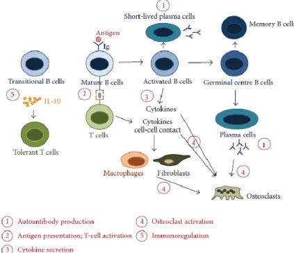
B cells can contribute to RA pathogenesis by several mechanisms (see figure 4):
1) Autoantibody production, such as RF and ACPA, to various synovium-derived antigens such as type II collagen, fibrinogen and filaggrin among others.
2) Antigen presentation and T cell activation. B cells can act as efficient APCs and stimulate T cells, allowing the development of memory T-cell subpopulations 98. Furthermore, T cell response in RA synovitis and ectopic lymphoid organization is B cell dependent 99. RF+ B cells are believed to play an important role in antigen presentation. In fact, it has been demonstrated that they can take up antigen–antibody immune complexes through their membrane Ig receptors, which have RF specificity 100.
3) Cytokine and chemokine secretion. B cells sequentially stimulated through the BCR and CD40 proliferate and secrete TNF, lymphotoxin-α and IL-6 that amplify the ongoing immune response 101.
Fig. 4 - The multiple roles of B cells in RA (Adapted from 102) Immune responses potentially orchestrated by B cells in rheumatoid arthritis. B-T cell interactions result in the activation and differentiation of plasma cells, responsible for the production of autoantibodies (1). In turn, activated B cells provide help to T cells and induce differentiation of effector T cells that produce proinflammatory cytokines (2). B cells can also impact on other immune and nonimmune cell functions through secretion of cytokines, such as interleukin (IL)-1, IL-6, TNF-α, and IL-17A (3). Proinflammatory cytokines and receptor activator of nuclear factor κB ligand (RANKL) produced by activated B cells, T cells,macrophages, and synovial fibroblasts promote the differentiation and activation of osteoclasts, leading to bone resorption (4). Further participation of B cells in bone homeostasis is suggested by the recognition that autoantibodies recognizing citrullinated vimentin are able to promote the differentiation ofmononuclear cells to osteoclasts (4). B cells can also be immunoregulatory through the provision of IL-10 and other mechanisms yet to be elucidated (5).
4) Osteoclast activation. B cell survival and activation signals are manly provided by TNF-superfamily members. B cells represent also a major source of RANKL in the rheumatoid environment, suggesting their direct involvement in the process of osteoclastogenesis and, consequently, bone erosion 103.
5) Immunoregulation. Recent studies indicate the presence of a subset of B cells – B regulatory cells (Breg) – specifically induced under inflammatory conditions that are capable of
suppressing inflammation and/or enhancing the recovery process 104. Breg can act through: a)
IL-10 production, which restores Th1/Th2 balance and directly inhibits inflammatory cascades; b) TGF-β1 production, which induces apoptosis of effector T cells; c) suppression of activated CD4+ T cells and d) interaction with other immune cells either directly or through secreted antibodies (reviewed in 38).
3.
A
IMSThe main goals of the present study were:
I. To analyze the expression of a group of genes directly related with B cell activation, maturation and survival in untreated early RA and established treated RA patients
II. To compare the effects of treatment options (methotrexate - MTX; TNF antagonists and Tocilizumab - TCZ) on B cell gene expression in established RA patients
4.
M
ATERIALS AND METHODSPatients
Blood samples were collected from 13 consecutive patients with untreated polyarthritis (Rheumatology Department, Hospital de Santa Maria, Lisbon) of ≤ 1 year disease duration. After a minimum follow-up of 3 months, the patients fulfilled the 2010 ACR/ EULAR criteria for RA and were classified as early RA (ERA). In addition, blood samples from 15 patients with established RA treated with methotrexate (MTX); 25 patients with established RA treated with MTX before starting biologic DMARD therapy (MTX pre-biologic, MTX pre-bio); 7 patients under treatment with TNF inhibitors and 9 patients under tocilizumab (TCZ) treatment were also collected for comparison (Rheumatology Department, Hospital de Santa Maria, Lisbon). Furthermore, blood samples from 15 healthy donors were also collected and processed for comparison.
This study was approved by the local ethics committee (Comissão de Ética do Hospital de Santa Maria), and all patients and healthy donors signed an informed consent form. Patient care was conducted in accordance with standard clinic practice, and the study was performed in accordance with the Declaration of Helsinki (2008).
Isolation of peripheral blood mononuclear cells and B cell separation
Peripheral blood mononuclear cells (PBMC) were isolated from 60 ml heparinized whole blood following density gradient centrifugation with Ficoll (Biowest, France). Cellular counts were estimated with 0.4% Trypan Blue (Sigma-Aldrich, USA) in a Neubauer’s chamber. B cells were isolated by positive MACS Separation using CD19 Microbeads and LS Columns (Miltenyi Biotec GmbH, Germany), according to the manufacturer’s instructions. Purity of isolated B cells was analysed by flow cytometry using fluorochrome-conjugated CD20 FITC (BD Biosciences, USA) and CD3 APC (eBioscience, USA) antibodies. A total of 20000 cells/ sample were acquired with LSR Fortessa (BD Biosciences, USA). The remaining B cells were stored at -80ºC in RLT Buffer (Qiagen, Germany) supplemented with RNase inhibitor β-mercaptoethanol (1:100) until further use.
RNA extraction and complementary DNA (cDNA) synthesis
Total RNA was extracted from B cells using the RNeasy Mini kit (Qiagen, Germany) according to the manufacturer’s instructions and treatment with RNase-free DNase Set (Qiagen, Germany) was performed to avoid contamination of genomic DNA. RNA
concentration and purity were determined with NanoDrop ND-1000 spectrophotometer (NanoDrop Technologies, USA). Total RNA was reverse-transcribed into cDNA using DyNAmoTM cDNA Synthesis Kit for qRT-PCR (Finnzymes, Finland) with Moloney murine leukemia virus (M-MuLV) reverse transcriptase, random hexamers (300 ng/l) and 2X RT Buffer, according to the manufacturer’s instructions, performed on Piko Thermal Cycler (Finnzymes, Finland). The cDNA samples were stored at -20ºC.
Real-time quantitative polymerase chain reaction
The expression of a group of genes directly related with B cell activation, maturation and survival was assessed by real-time quantitative polymerase chain reaction (qPCR) performed on Rotor-Gene 6000 (Corbett Life Science, USA) using SensiMix SYBR No-ROX Kit (Bioline, United Kingdom). The qPCR program consisted of an initial denaturation step at 95ºC for 10 min, followed by 40 cycles of 95ºC for 15 s, 60ºC for 15 s, and 72ºC for 15 s. Genes and primer sequences analyzed in this study are indicated in Table 3. Primers were designed using the National Center for Biotechnology Information (NCBI)/ Primer-BLAST. The 18S ribosomal RNA (18S rRNA) was used as endogenous controls in relative quantification using the standard curve method. All data were analyzed with Rotor-Gene 6000 Series Software.
Statistical analysis
Statistical differences were determined with GraphPad Prism (GraphPad, San Diego, USA). For populations that did not follow a Gaussian distribution, nonparametric tests were used. The Mann-Whitney test was used for comparisons between 2 independent groups. For comparisons between 3 or more groups, the Kruskal-Wallis and Dunn’s multiple comparison tests were used. The Wilcoxon matched pairs test was used for comparisons between 2 paired groups. Correlation analysis was performed using Spearman’s test. Differences were considered statistically significant for p < 0.05.
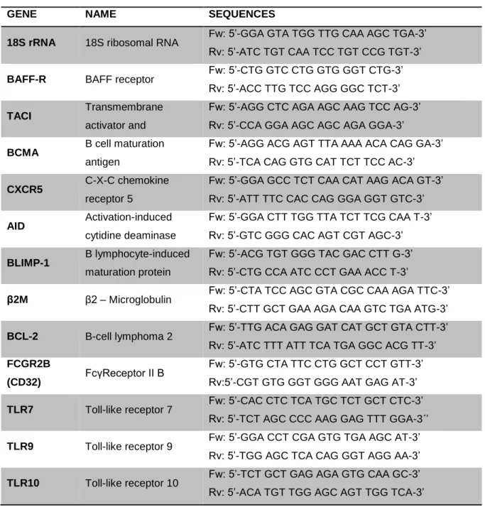
Table 3 - Primers used in real time PCR assay
GENE NAME SEQUENCES
18S rRNA 18S ribosomal RNA Fw: 5’-GGA GTA TGG TTG CAA AGC TGA-3’ Rv: 5’-ATC TGT CAA TCC TGT CCG TGT-3’
BAFF-R BAFF receptor Fw: 5’-CTG GTC CTG GTG GGT CTG-3’ Rv: 5’-ACC TTG TCC AGG GGC TCT-3’
TACI Transmembrane
activator and
Fw: 5’-AGG CTC AGA AGC AAG TCC AG-3’ Rv: 5’-CCA GGA AGC AGC AGA GGA-3’
BCMA B cell maturation antigen
Fw: 5’-AGG ACG AGT TTA AAA ACA CAG GA-3’ Rv: 5’-TCA CAG GTG CAT TCT TCC AC-3’
CXCR5 C-X-C chemokine receptor 5
Fw: 5’-GGA GCC TCT CAA CAT AAG ACA GT-3’ Rv: 5’-ATT TTC CAC CAG GGA GGT GTC-3’
AID Activation-induced cytidine deaminase
Fw: 5’-GGA CTT TGG TTA TCT TCG CAA T-3’ Rv: 5’-GTC GGG CAC AGT CGT AGC-3’
BLIMP-1 B lymphocyte-induced maturation protein
Fw: 5’-ACG TGT GGG TAC GAC CTT G-3’ Rv: 5’-CTG CCA ATC CCT GAA ACC T-3’
β2M β2 – Microglobulin Fw: 5’-CTA TCC AGC GTA CGC CAA AGA TTC-3’ Rv: 5’-CTT GCT GAA AGA CAA GTC TGA ATG-3’
BCL-2 B-cell lymphoma 2 Fw: 5’-TTG ACA GAG GAT CAT GCT GTA CTT-3’ Rv: 5’-ATC TTT ATT TCA TGA GGC ACG TT-3’ FCGR2B
(CD32) FcγReceptor II B
Fw: 5’-GTG CTA TTC CTG GCT CCT GTT-3’ Rv:5’-CGT GTG GGT GGG AAT GAG AT-3’
TLR7 Toll-like receptor 7 Fw: 5’-CAC CTC TCA TGC TCT GCT CTC-3’ Rv: 5’-TCT AGC CCC AAG GAG TTT GGA-3´’
TLR9 Toll-like receptor 9 Fw: 5’-GGA CCT CGA GTG TGA AGC AT-3’ Rv: 5’-TGG AGC TCA CAG GGT AGG AA-3’
TLR10 Toll-like receptor 10 Fw: 5’-TCT GCT GAG AGA GTG CAA GC-3’ Rv: 5’-ACA TGT TGG AGC AGT TGG TCA-3’
5.
R
ESULTS5.1. Clinical characterization of patients
A group of untreated polyarthritis patients (n=13) with less than 1 year of disease duration and classified as early RA (ERA) according to the 2010 ACR/ EULAR criteria after a minimum follow-up of 3 months was included in this study. ERA patients had a mean ± standard deviation age of 58±14 years old, 85% were female, 69% were RF positive, 62% were anti-CCP positive and DAS28 score was 4.1±2.0. A group of established RA patients under MTX treatment (n=15) with a mean age of 55±14 years old, 87% female and a DAS28 of 2.7±1.2 was also included. Furthermore, a group of established RA patients under MTX treatment pre-biological therapy (MTX pre-bio, n=25) with a mean age of 58±12 years old, 88% female and a DAS28 of 5.2±1.5 was also analyzed. From the MTX pre-bio group, a second blood collection was performed to RA patients that had either initiated treatment with TNF-inhibitors (n=7) or with tocilizumab (TCZ) (n=9), after an average follow-up of 8 and 6 months of treatment, respectively. In addition, blood samples were collected from age and sex-matched healthy donors (n=15). The clinical information and data from all patients and healthy controls included in this study is indicated in Table 4.
Table 4 - Clinical information of controls, ERA and established RA patients after treatment with MTX, MTX pre-bio, TNF inhibitors and TCZ. ERA - Early Rheumatoid Arthritis; RA - Rheumatoid Arthritis; MTX – Methotrexate; MTX pre-bio – Methotrexate pre-biologic; TNF – TNF inhibitors; TCZ – Tocilizumab; CRP - C-reactive protein; ESR - Erythrocyte Sedimentation Rate; VAS – Visual Analogue Scale; DAS28 – Disease Activity Score of 28 joints; RF – Rheumatoid Factor; ACPA– anti-cyclic citrullinated peptide; NA – not applicable; ND – not determined. Values are represented as mean ± standard deviation.( ** p < 0.05 in comparison with ERA; # p < 0.05 in comparison with MTX pre-bio; & p < 0.05 in comparison with TNF)
Controls (n=15) ERA (n=13) MTX (n=15) MTX pre-bio (n=25) TNF (n=7) TCZ (n=9) Age (years) 50±7 58±14 55±14 58±12 52±16 61±13 Sex (% female) 73 85 87 88 80 100 Disease duration (years) NA ≤ 1 7±6 11±8 6±3 12±7 CRP (mg/dl) ND 1.5±2.1 0.5±0.6 1.4±3.1 1.2±1.1 0.2±0.3# ESR (mm/1st hour) ND 42±25 23±23 29±16 44±47 4±2**#& VAS NA 40±33 42±33 69±18 66±27 67±10 DAS28 NA 4.1±2.0 2.7±1.2# 5.2±1.5 4.4±1.3 2.9±1.0# Swollen joints NA 3±5 1±2# 6±6 3±3 1±1 Tender joints NA 5±5 2±3# 12±8 7±4 2±3# RF (+) % ND 69 87 75 60 86 ACPA (+) % ND 62 60 78 40 86
5.2. Established RA, but not early RA patients have alterations in B cell gene expression levels in comparison with healthy controls
In this study, a group of genes related with B cell homeostasis and survival (BAFF-R, TACI, BCMA), class-switching (AID), chemotaxis (CXCR5), plasma cell differentiation (BLIMP-1), immune system activation (B2M), apoptosis (BCL-2), B cell inhibition (CD32) and activation through Toll-like receptors (TLR7, TLR9, TLR10) were analyzed in untreated ERA and established RA patients under treatment with MTX, MTX pre-bio, TNF inhibitors and TCZ. Results were compared with age and sex-matched healthy controls.
Since some of the studied genes are also expressed by monocytes and T cells, isolating B cells from PBMC samples was mandatory. Therefore, the purity of B cell isolation was tested for each sample by flow cytometry and samples were excluded if the purity was under 90%. Additionally, samples were also excluded if the RNA concentration obtained from isolated B cells was too low to perform a gene expression analysis of all genes of interest.
It was found that untreated ERA patients (≤ 1 year of disease duration) have similar B cell gene expression levels with no statistically significant differences when compared to healthy controls for all genes analyzed (Figures 5-8). However, established RA patients had alterations in B cell gene expression levels in comparison with controls.
The analysis of all BAFF receptors has revealed that BAFF-R gene expression was significantly increased in established RA patients after treatment with MTX (p=0.0212) and TNF inhibitors (p=0.0025) in comparison with the control group (Figure 5). Furthermore, BAFF-R gene expression was also significantly increased in established RA patients after treatment with TNF inhibitors when compared to ERA (p=0.0006). Nevertheless, no statistically significant differences were detected in TACI and BCMA gene expression levels in all groups studied.
Fig. 5 - BAFF-R gene expression is significantly increased in B cells from established RA patients after treatment with MTX and TNF inhibitors. A nonparametric statistical analysis was performed with Kruskall-Wallis and Mann-Whitney tests: bars represent median relative gene expression values. * p < 0.05 in comparison with Controls; # p < 0.05 in comparison with ERA.
Alterations in the B cell gene expression levels of TLR7, TLR9 and TLR10 were also observed in established RA patients (Figure 6). Indeed, it was found that TLR7 and TLR10 gene expressions were significantly increased in established RA patients after treatment with TNF inhibitors when compared to controls (p=0.0385 and p=0.0097, respectively). Furthermore, TLR10 gene expression levels were also significantly increased in established RA under anti-TNF therapy when compared to ERA patients (p=0.0801). Moreover, TLR9 gene expression was significantly increased in established RA patients under MTX treatment (p=0.0043) when compared to controls. No other statistically significant differences were detected in all groups analyzed.
Fig. 6 - Established RA patients have alterations in the gene expression levels of TLR7, TLR9 and TLR10 after treatment with MTX and/ or TNF inhibitors. A nonparametric statistical analysis was performed with Kruskall-Wallis and Mann-Whitney tests: bars represent median relative gene expression values. * p < 0.05 in comparison with Controls; # p < 0.05 in comparison with ERA.
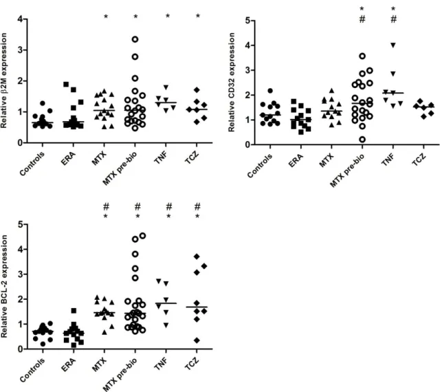
The analysis of the gene expression levels of B2M, an inflammatory marker, and CD32 (FCGR2B), an inhibitory B cell marker, also revealed disturbances in established RA patients (Figure 7). It was observed that B2M gene was overexpressed in B cells from established RA patients treated with MTX (p=0.0099), MTX pre-bio (p=0.0116), TNF inhibitors (p=0.0025) and TCZ (p=0.0112) when compared to controls, but no significant differences were detected in ERA patients in comparison with controls. The gene expression levels of CD32 were significantly elevated in established RA patients treated with TNF inhibitors in comparison not only with controls (p=0.0043), but also with ERA patients (p=0.0015). Furthermore, a tendency for increased CD32 gene expression levels was also observed in established RA patients treated with MTX pre-bio when compared to controls (p=0.0487). Nonetheless,
CD32 gene expression was significantly increased in established RA patients treated with MTX pre-bio when compared to ERA patients (p=0.0015).
Fig. 7 - Established RA patients have increased gene expression levels of B2M, CD32 and BCL-2 irrespective of the treatment. A nonparametric statistical analysis was performed with Kruskall-Wallis and Mann-Whitney tests: bars represent median relative gene expression values. * p < 0.05 in comparison with Controls; # p < 0.05 in comparison with ERA.
In addition, BCL-2 gene was overexpressed in B cells from established RA patients treated with MTX (p<0.0001), MTX pre-bio (p<0.0001), TNF inhibitors (p=0.0014) and TCZ (p=0.0042) in comparison with controls and also with ERA patients (p<0.0001) (Figure 7).
It was also observed that ERA and established RA patients (irrespective of treatment) had similar AID, BLIMP-1 and CXCR5 gene expression levels when compared to healthy controls and no significant differences were detected (Figure 8).
Fig. 8 - ERA and established RA patients have normal gene expression levels of AID, BLIMP-1 and CXCR5. No significant differences were observed in B cell gene expression levels of AID, BLIMP-1 and CXCR5 in all groups of patients when compared to healthy controls.
5.3. Treatment with TNF inhibitors and tocilizumab does not affect B cell gene expression
In order to understand the effect of treatment options before and after RA patients initiate treatment, a second blood collection was performed in established RA patients from MTX pre-bio group after a mean period of 8 months of treatment with TNF inhibitors (n=5) or after a mean of 6 months of treatment with TCZ (n=7). B cell gene expression levels were analyzed after treatment and compared to baseline values. Nevertheless, no statistically significant differences were observed in the gene expression levels of all genes studied in established RA patients after treatment with TNF inhibitors (Figure 9) or TCZ (Figure 10).
Fig. 9 - B cell gene expression is not affected by TNF inhibitors in established RA when compared to baseline. B cell gene expression levels of BAFF-R, TACI, BCMA, CXCR5, AID, BLIMP-1, B2M, BCL-2, CD32, TLR7, TLR9 and TLR10 were analyzed in established RA patients after 8 months of treatment with TNF inhibitors (n=5) and compared to baseline levels. No significant differences were observed in all B cell gene expression levels before and after anti-TNF therapy.
Fig. 10 - B cell gene expression is not affected by tocilizumab in established RA when compared to baseline. B cell gene expression levels of BAFF-R, TACI, BCMA, CXCR5, AID, BLIMP-1, B2M, BCL-2, CD32, TLR7, TLR9 and TLR10 were analyzed in established RA patients after 6 months of treatment with TCZ (n=7) and compared to baseline levels. No significant differences were observed in all B cell gene expression levels before and after treatment with TCZ.
3.4. Correlation analysis between B cell gene expression and clinical data
A correlation analysis was also performed between all the B cell gene expression levels of all groups included in this study with the clinical parameters previously indicated in Table 3. Nevertheless, no correlation was found between B cell gene expression with age, CRP, ESR, VAS, DAS28, swollen joints and tender joints in all patients’ groups (data not shown). Furthermore, no correlation could be detected between B cell gene expression levels and age in the control group (data not shown).