Abstract
Submitted: June 6, 2017
Modification: August 13, 2017 Accepted: September 14, 2017
The effect of periodontal therapy
on neopterin and vascular cell
adhesion molecule-1 levels in chronic
periodontitis patients with and without
acute myocardial infarction: a
case-control study
The presence of neopterin in gingival crevicular fluid (GCF) is a marker for local and acute immune activation, and the presence of vascular cell adhesion molecule (VCAM-1) in GCF is accepted as a marker for chronic vascular inflammation. Objectives: This study aimed to evaluate effects of periodontal treatment on GCF levels of neopterin and VCAM-1 in patients with chronic periodontitis (CP) with acute myocardial infarction (AMI) compared with systemically healthy CP patients. Material and methods: Sixty subjects (20 CP patients with AMI, 20 healthy CP patients, and 20 healthy controls) were included. GCF samples were analyzed at baseline and after 3 and 6 months, and the probing pocket depth (PD), clinical attachment level (CAL), bleeding on probing, gingival (GI) and plaque (PI) indices were recorded. We determined neopterin and VCAM-1 levels (concentration and total amount) using enzyme-linked immunosorbent assay (ELISA). No significant differences were seen between the AMI+CP and CP groups for PI, GI, GCF levels of neopterin and VCAM-1 at baseline. Results: The number of teeth with 5 mm≤CAL<7 mm and CAL≥7 mm were significantly increased in the AMI+CP group at baseline. There were no significant differences between the AMI+CP and CP for PI, CAL, GCF volumes, and the AMI+CP group had the highest clinical improvement in the number of teeth with 5 mm≤CAL<7 mm at the sixth month. There were significant positive correlations between clinical periodontal inflammation and the presence of neopterin and VCAM-1 in GCF prior to and following periodontal treatment, and between the GCF volume and clinical parameters. Conclusions: Data suggest that the total amount and concentration of neopterin and VCAM-1 in GCF seemed to be closely associated with periodontal disease severity in CP patients with AMI. Moreover, the results of our study demonstrate that the past periodontal status is potentially correlated between groups, with similar periodontal disease severity.
Keywords: Periodontitis. Gingival crevicular fluid. Neopterin. Vascular
cell adhesion molecule-1. Myocardial infarction. Zeynep TURGUT ÇANKAYA1
Ayşen BODUR1 Gülten TAÇOY2 İmge ERGÜDER3 Derya AKTUNA4 Atiye ÇENGEL2
1Gazi University, Faculty of Dentistry, Department of Periodontology, Ankara, Turkey. 2Gazi University, Faculty of Medicine, Department of Cardiology, Ankara, Turkey.
3Gazi University, Faculty of Medicine, Department of Biochemistry, Ankara, Turkey. 4Gazi University, Faculty of Medicine, Department of Biostatistics, Ankara, Turkey.
Introduction
Periodontitis is a bacterially-induced, localized and chronic inflammatory disease that destroys the connective tissue and bone that support the teeth. Periodontitis and acute myocardial infarction (AMI)
are two diseases that share common risk factors12. In
recent years, literature has paid attention to positive
correlation between periodontitis and coronary heart disease, acute coronary events, including AMI4. In
these studies, periodontitis is defined by clinical examination or radiologic criteria6,25. Such trials are
limited regarding gingival crevicular fluid (GCF)-based design and do not include periodontal treatment results. Site-directed measurements may allow for a more definitive identification of susceptible individuals
and evaluation of responsiveness to therapy10.
Neopterin (N) is produced by
interferon-gamma-stimulated monocytes. Elevated serum N concentrations predicted future adverse cardiac events
in patients with angina pectoris2 and one investigation
demonstrated a relationship with the extent of coronary
atherosclerosis30. The determination of N in GCF might
be useful for diagnosing and predicting periodontal
diseases21. Studies evaluated the N status in various
human biological fluids, including GCF21,22.
A number of cell adhesion molecules have
been detected as soluble circulating forms in human serum and other body fluids10. Vascular cell
adhesion molecule-1 (VCAM-1) is a member of the immunoglobulin superfamily of adhesion molecules and expressed on cytokine-activated endothelial cells. There are studies that investigated the levels of several adhesion molecules in different forms
of periodontitis24,26. However, there are no studies
specifically addressing the altered GCF profile concurrent with the onset of AMI (i.e. in the first 24 h) and no data are currently available about GCF N and VCAM-1 levels in AMI patients with chronic periodontitis (CP). Thus, in this study, we aimed at assessing whether GCF levels of CP patients with AMI have an alteration in GCF levels of N and VCAM-1. We also assessed whether these alterations might be related to treatment of existing periodontitis in AMI patients. We hypothesized that severe CP may play a role in initiating or exacerbating MI, and that there is an increased risk of AMI among systemically healthy people affected with severe CP. To test these hypotheses, we aimed to evaluate the effects of periodontal treatment on
GCF levels of neopterin and VCAM-1 in CP with AMI in comparison to systematically healthy CP patients. We also assessed whether AMI was associated with a local inflammatory response, as reflected in the levels of GCF N and VCAM-1. In addition, we tested the hypothesis that long-term exposure to severe AMI+CP in patients contributes to the development of myocardial infarction (MI), using a case-control model of MI to study the inflammatory response in GCF. To do this, we compared AMI patients with CP with those who were systemically healthy and had CP.
Material and Methods
The protocol of this clinical trial (ClinicalTrials.gov Identifier: NCT03005886) was approved by the Ethical Committee of the Faculty of Medicine. This study was executed between March 2006 and March 2010. Participants were recruited from individuals referred to the Department of Cardiology, Faculty of Medicine and Department of Periodontology, Faculty of Dentistry. Participants were classified as AMI+CP, CP and healthy control. Informed written consent was obtained from all subjects.
Inclusion/exclusion criteria
Patients, who were referred to the Department of Cardiology and met the AMI diagnostic criteria29,
with or without persistent ST-segment elevation, were screened for inclusion in the study. Patients with neoplasias, liver cirrhosis, HIV infection, chronic renal failure, hypo or hyperparathyroidism, diabetes mellitus and chronic inflammatory diseases were not included. None of the patients had received periodontal treatment during the past six months and none had received antibiotic medication during the past three months. AMI patients were regarded as suitable for the study if they were affected by CP and had at least 16 teeth, including at least four molars in different quadrants and at least two periodontal pockets at least 5 mm in depth, with a minimum of 2 mm attachment
loss. Data for 140 AMI patients were obtained from
examination in the coronary care unit. Of these, 53 met the inclusion criteria, but only 20 of them were approved to be included in this study. All patients with AMI were on medical therapy, and none of them used antiaggregant therapy other than salicylates. A total of 85 CP patients without history of systemic conditions were examined from individuals referred to the Department of Periodontology, Faculty of Dentistry. Of these, 65 returned for blood sample collection, medical assessment and cardiology consultation at the Department of Cardiology. Finally, a group of 20 systemically healthy volunteer CP patients participated in the study. In addition, to the abovementioned controls, 20 clinically healthy individuals who did not have any periodontal and systemic disease history were included in the scope of this study. All control subjects were referred to a cardiology specialist in the same hospital clinic, who conducted a comprehensive medical examination, including electrocardiogram, to confirm that they did not have any cardiovascular disease (CVD).
Clinical procedures
The periodontal examination included the assessment of plaque index (PI)27, gingival index
(GI)16, probing depth (PD), bleeding on probing
(BOP) and clinical attachment level (CAL). All subjects underwent a periodontal examination performed by the same periodontist (ZTÇ). Prior to the study, the examiner was calibrated for reproducibility of PD and CAL measurements. To determine repeatability of the
PD and CAL measurements, six sites per tooth in ten
patients, were measured twice. For PD 98% and for CAL 99% of the paired measurements were within ±1 mm. All periodontal parameters were measured with a Williams periodontal probe calibrated in millimeters (Nordent Manufacturing Inc., Elk Grove Village, IL, USA). Periodontal measurements were taken at six
sites per tooth (mesio-buccal, mid-buccal, disto-buccal,
mesio-palatal, mid-palatal and disto-palatal). The deepest six pockets found in the different segments of each subject were chosen for GCF sampling. Baseline periodontal examination of AMI patients and 24-48 h GCF collection were carried out in their hospital bed under sufficient illumination using artificial light. The examiners could not be “blinded” to the subject’s general condition, since they were examined in a hospital. Within a time period of two months after the proceeding infarction, none of the patients had received
periodontal treatment. AMI patients underwent periodontal therapy after the stabilization of their condition with the consent from same cardiologist. Periodontal disease was diagnosed based on the 1999 classification system developed by Armitage, and a preoperative periapical radiograph was taken, which provided baseline data in Faculty of Dentistry. All selected patients underwent a 2- to 4-week initial therapy, which included comprehensive proper plaque control program, scaling, subgingival curettage and root planning in Department of Periodontology. In all patients a periodontal reevaluation was performed four weeks after phase I therapy, to confirm the suitability of the sites for periodontal surgery. Mucoperiosteal flap operation was performed in cases when needed. Periodontal treatment including periodontal surgery was completed on the basis of 20 AMI and 20 CP patients, periodontal examination and GCF sampling repeated in baseline (T0), 3rd (T3) and 6th (T6) months.
GCF sampling and processing
We collected GCF samples using commercially available periopaper (Oraflow Inc., Box 219 Plainview,
NY), as described previously20. The sample site was
gently air-dried and all supragingival plaque was removed. The area was carefully isolated with cotton rolls and a saliva ejector was used to prevent samples from being contaminated. Periopaper strips were inserted into the pockets until slight resistance was felt and left in place for 30 s. We tried to avoid mechanical injury of the gingival tissues. Strips contaminated by bleeding or exudate were discarded. The amount of GCF on the strips was measured by weighing the accumulated fluid. Strips were placed into Eppendorf tubes. Weighing was then repeated immediately after collection to overcome any evaporation and then stored at -80°C until processed.
Laboratory analyses
Blood samples for serum were centrifuged for 10 min at 11.00 RPM separating serum from cells. Serum samples were then immediately divided into 0.2-0.5 mL aliquots and stored at -80°C until required for analysis. We assayed samples for N and VCAM-1 using quantitative enzyme immunoassays. Microcentrifuge tubes containing periopaper strips with absorbed GCF sample were allowed to reach room temperature and
eluted using a centrifugal method8. After centrifugation,
VCAM-1 in serum and GCF samples were measured using ELISA kit (Demeditec, Germany; Biosource, Invitrogen, Carlsbad, CA, USA). The ELISA plates were assessed spectrophotometrically at 450 nm. The concentrations of N and VCAM-1 in each sample were calculated by using the standards included with the kit. Results were expressed as ng/ml. Total amounts were also calculated by multiplying concentrations and GCF volumes31.
Statistical analysis
We performed data analysis using the SPSS statistical package program (Microsoft Corp., Chicago, IL, USA). For categorical variables, we used the chi-square test to compare two or more groups, and for metric variables, the Mann-Whitney U test for two groups, and for more than two groups, the Kruskal-Wallis analysis of variance (ANOVA). Two-way repeated measures ANOVA were used to evaluate both within and between group comparisons. Spearman correlation coefficient was calculated for assessing the
AMI+CP CP Healthy
n=20 n=20 n=20 p
Age(years) 56.5±8.02*(42-74) 47.25±7.34(39-73) 49.1±5.3(39-58) <0.001 BMI(kg/m2) 27.69±3.63**(23.29-35.16) 25.09±2.28(20.8-30.61) 24.1±2.15(20.31-29.41) <0.01
Smoking(n-%) 17§-%85 6-%30 3-%15 <0.001
Education status 2-2-12-2-2 2-2-10-3-3 1-1-8-6-4 >0.05
Level of income 6-14 7-13 6-14 >0.05
Number of teeth 22.05±5.05(7-28) 22.65±3.03(16-28) 23.55±1.84(20-26) >0.05 GCF volume(ml) 0.01±0.001(0.008-0.01) 0.01±0.001(0.008-0.01) 0.003±0.0006°(0.002-0.004) <0.001 Serum N v(mg/ml) 20.81±4.54(16.08-34.50) 19±1.21(17.49-21.86) 19.06±2.41(16.65-23.47) >0.05
GCF N c(ng/ml) 9.03±1.27(7.45-11.55) 8.64±0.94(6.84-11.31) 7.67±0.82°(5.67-8.84) <0.001 GCF N ta(ng/ml) 0.09±0.02(0.07-0.16) 0.09±0.01(0.06-0.14) 0.02±0.00(0.01-0.03) <0.001 Serum VCAM-1 (ng/ml) 1824.8±4597.9°(1230.9-3264) 1334.7±256.3(1017.0-2071) 1290.9±264.2(754.8-1938) <0.01
GCF VCAM-1 c(ng/ml) 10.21±6.60(5.79-36.19) 7.96±2.82(4.94-16.81) 7.23±2.34°(3.95-13.79) <0.05 GCF VCAM-1 ta(ng) 0.1±0.06°(0.06-0.33) 0.08±0.02°(0.04-0.14) 0.02±0.009°(0.01-0.04) <0.001
Data given as mean±standard deviation(min-max)values. n=Number of patients.The education status data given as the number of participants in primary school, secondary school, high school, university and graduate. The level of income describes number of participants with good or medium income.
*Statistically significant difference between groups; Anova. **Statistically significant difference between groups;Kruskal-Wallis test. §Statistically significant difference between groups; Chi-square test. °Statistically significant difference between groups; Kruskal-Wallis test. BMI=body mass ındex, N=Neopterin. GCF=Gingival Crevicular Fluid.VCAM-1=Vascular cell adhesion molecule. c=concentration, ta=total amount.
Table 1- Study subject characteristics
AMI+CP CP Healthy
n=20 n=20 n=20 p
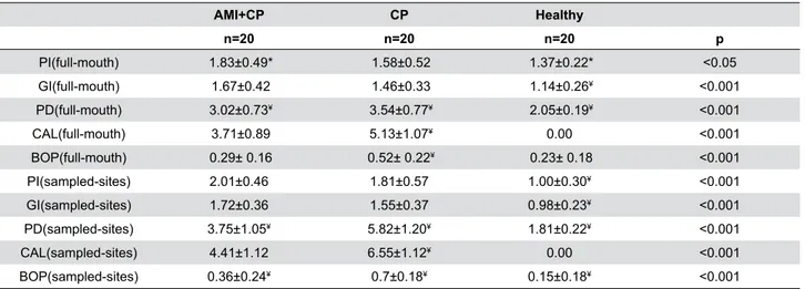
PI(full-mouth) 1.83±0.49* 1.58±0.52 1.37±0.22* <0.05 GI(full-mouth) 1.67±0.42 1.46±0.33 1.14±0.26¥ <0.001 PD(full-mouth) 3.02±0.73¥ 3.54±0.77¥ 2.05±0.19¥ <0.001
CAL(full-mouth) 3.71±0.89 5.13±1.07¥ 0.00 <0.001
BOP(full-mouth) 0.29± 0.16 0.52± 0.22¥ 0.23± 0.18 <0.001 PI(sampled-sites) 2.01±0.46 1.81±0.57 1.00±0.30¥ <0.001 GI(sampled-sites) 1.72±0.36 1.55±0.37 0.98±0.23¥ <0.001 PD(sampled-sites) 3.75±1.05¥ 5.82±1.20¥ 1.81±0.22¥ <0.001
CAL(sampled-sites) 4.41±1.12 6.55±1.12¥ 0.00 <0.001
BOP(sampled-sites) 0.36±0.24¥ 0.7±0.18¥ 0.15±0.18¥ <0.001
Data given as mean±SD; p value by Kruskal Wallis Test for PI=plaque index; GI=gingival index; PD=probing depth; BOP=bleeding on probing; p value by Mann-Whitney Test for CAL=clinical attachment level. *Statistically significant difference(p<0.05). ¥ Statistically significant difference(p<0.001)
association between variables. Frequency (percentage) for categorical variables, mean±standard deviation, median (minimum-maximum) for metric variables were given as descriptive statistics. We considered p<0.05 as statistically significant. When the power analysis is made for CAL, the power is sound 0.993 for 20 sample in each group.
Results
In total, sixty subjects were enrolled. In this scope, forty CP patients with and without AMI were examined in the third and sixth months following the periodontal treatment. No significant differences were found among the three groups with regarding gender, education
status, level of income, number of teeth, and serum N values at baseline (Table 1). The AMI+CP group had higher serum VCAM-1 levels compared with the other two groups. There were no significant differences between the CP and AMI+CP groups concerning PI
AMI+CP CP
Number of teeth with n=20 n=20 p
CAL<5 mm 22.30±4.94(7-28) 22.35±2.85(16-28) <0.001 5 mm≤CAL<7 mm 19.3±6.13*(6-17) 15.05±2.30(10-18) >0.001
CAL≥7 mm 15.6±5.9*(1-22) 10.2±1.93(7-14) <0.001
Data given as mean±SD(Min-Max)values; p value by Mann-Whitney Test.
Table 3- Number of teeth with a clinical attachment level (CAL) degree of 5 mm or more
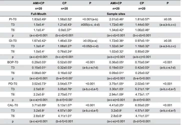
AMI+CP CP P AMI+CP CP P
n=20 n=20 n=20 n=20
Full-Mouth Sample sites
PI-T0 1.83±0.49a 1.58±0.52b <0.001(a-b) 2.01±0.46a 1.81±0.57b ≥0.05 T3 1.5±0.4c 1.21±0.43c ≥005(c-c, d-d) 1.72±0.46c 1.44±0.50c (a-a,b-b,c-c) T6 1.1±0.4d 0.9±0.37d 1.34±0.42d 1.06±0.46d
p (a-c-d)<0.001 (b-c-d)<0.001 (a-c-d)<0.001 (b-c-d)<0.001
GI-T0 1.67±0.42a 1.46±0.33a ≥0.05(a-a) 1.72±0.36a 0.97±0.15a ≥0.05 T3 1.3±0.4b 1.08±0.27c <0.05(b-c-d) 1.32±0.34b 1.18±0.32b (a-a,b-b,c-c) T6 1.0±0.4c 0.76±0.24d 1.02±0.32c 0.85±0.29c
p (a-c-d)<0.001 (b-c-d)<0.001 (a-b-c)<0.001 (a-b-c)<0.001
BOP-T0 0.29±0.03a 0.52±0.05b <0.001 0.36±0.05a 0.70±0.04b <0.001 T3 0.15±0.01c 0.32±0.03e (a-b,c-e,f-d) 0.19±0.03c 0.42±0.03e (a-b,c-e,f-d) T6 0.08±0.00d 0.16±0.02f 0.09±0.01d 0.25±0.02f
p (a-c-d)<0.001 (b-e-f)<0.001 (a-c-d)<0.001 (b-e-f)<0.001
PD-T0 3.02±0.73a 3.54±0.77b <0.001 3.75±1.05a 2.02±0.14b <0.001 T3 2.5±0.6c 3.05±0.76e (a-b,c-d,e-f) 3.30±1.03c 5.21±1.19d (a-b,c-d,e-f) T6 2.2±0.6e 2.75±0.71f 2.94±1.09e 4.73±1.17f
p (a-c-e)<0.001 (b-d-f)<0.001 (a-c-e)<0.001 (b-d-f)<0.001
CAL-T0 3.71±0.89a 5.13±1.07b <0.001 4.41±0.25a 6.55±0.25b <0.001 T3 3.2±0.8c 4.57±1.00d (a-b,c-d,e-f) 3.2±0.8c 4.57±1.00d (a-b,c-d,e-f) T6 2.8±0.8d 4.11±1.01f 2.8±0.8e 4.11±1.01f
p (a-c-e)<0.001 (b-d-f)<0.001 (a-c-e)<0.001 (b-d-f)<0.001
Data given as mean±SD; p value by two way ANOVA. Same superscripts indicate that there is no statistically significant difference (p≥0.05), different superscripts indicate that the difference is statistically significant (p˂0.001, p˂0.05) ;PI=Plaque Index; GI=Gingival Index; PD=Probing Depth; CAL=Clinical Attachment Level; BOP=Bleeding on Probing; SD=Standart Deviation. T0, theraphy initiation; T3, 3 months after T0; T6, 6 months after T0
and GI, which, in addition, were higher in both groups when compared with the healthy control group (Table 2). An analysis was carried out to investigate the relationship between periodontal disease severity and AMI using the number of teeth with a CAL degree of 5 mm or more (Table 3). In AMI patients, there were significantly fewer individuals with mild periodontitis and significantly more individuals with severe (and moderate-to-severe) periodontitis compared with patients in the CP group. At the third and sixth months,
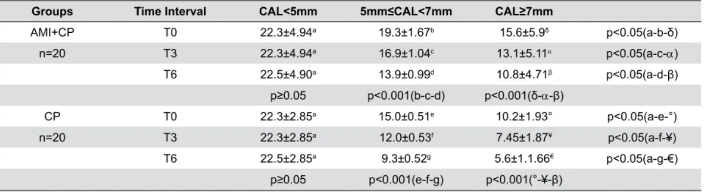
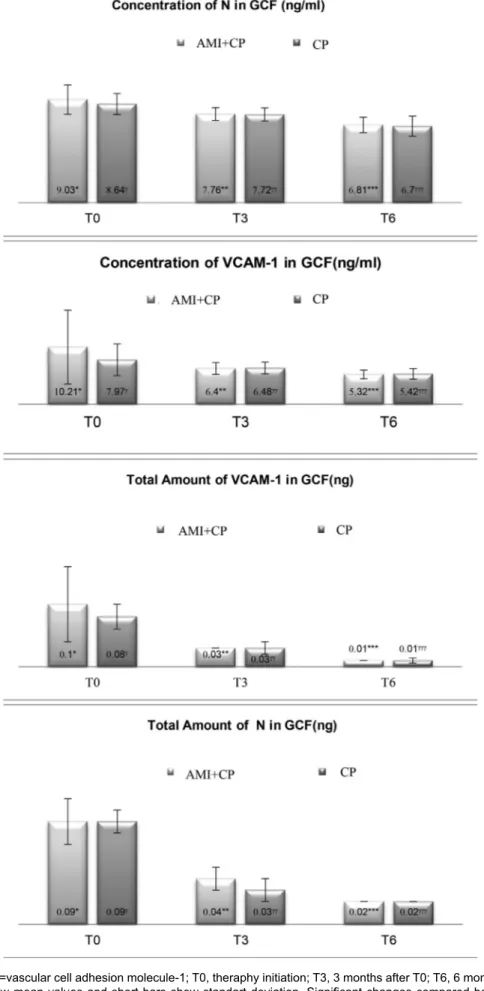
there were no significant differences between CP and CP+AMI groups for PI and CAL (Table 4). Within each group, all clinical parameters showed statistically significant decreases at the third and sixth months. The AMI+CP group showed statistically significant decreases in the number of teeth with 5≤CAL<7 and CAL≥7 mm after the periodontal treatment (Table 5). AMI+CP and CP groups showed significant decreases in total amounts and concentrations of N and VCAM-1 in the GCF during the post-treatment period (Figure 1).
Groups Time Interval CAL<5mm 5mm≤CAL<7mm CAL≥7mm
AMI+CP T0 22.3±4.94a 19.3±1.67b 15.6±5.9δ p<0.05(a-b-δ) n=20 T3 22.3±4.94a 16.9±1.04c 13.1±5.11α p<0.05(a-c-α)
T6 22.5±4.90a 13.9±0.99d 10.8±4.71β p<0.05(a-d-β) p≥0.05 p<0.001(b-c-d) p<0.001(δ-α-β)
CP T0 22.3±2.85a 15.0±0.51e 10.2±1.93° p<0.05(a-e-°)
n=20 T3 22.3±2.85a 12.0±0.53f 7.45±1.87¥ p<0.05(a-f-¥) T6 22.5±2.85a 9.3±0.52g 5.6±1.1.66€ p<0.05(a-g-€)
p≥0.05 p<0.001(e-f-g) p<0.001(°-¥-β)
Data given as mean±SD; p value by two way ANOVA. Same superscripts indicate that there is no statistically significant difference (p≥0.05), different superscripts indicate that the difference is statistically significant (p˂0.001, p˂0.05) CAL=Clinical Attachment Level; T0, therapy initiation; T3, 3 months after T0; T6, 6 months after T0.
Table 5- Number of teeth according to the clinical attachment level (CAL)
AMI+CP GCF N
Cont.(T6)
GCF VCAM-1 T.A(T6)
Full-mouth PI(T6) 0.557* Sample PI(T6) 0.480* Full-mouth CAL(T6) 0.580**
Full-mouth BOP(T6) -0.454*
CP GCF N
Cont.(T0)
GCF N T.A (T3)
GCF N Cont.(T6) Full-mouth CAL(T0) 0.452*
CAL≥7(T0) 0.467*
Sample PD(T3) 0.472*
Full-mouth BOP(T6) 0.543*
Full-mouth PI(T6) 0.536*
Full-mouth PD(T6) 0.543*
CP GCF VCAM-1 Cont.(T0) GCF VCAM-1 T.A(T0) GCF VCAM-1 Cont.(T3) GCF VCAM-1 T.A(T3)
Sample PD(T0) 0.563** 0.586**
Sample CAL(T0) 0.470* 0.481*
CAL≥7(T0) 0.461*
Full-mouth GI(T3) 0.467*
CAL<5(T3) 0.460*
5<CAL≤7(T3) 0.499*
CAL≥7(T3) 0.482* 0.529*
*p<0.05,**p<0.01 T.A:Total Amount (ng), Cont:Concentration (ng/ml), AMI:Acute Myocardial Infarction, CP:Chronic Periodontitis, N:Neopterin, VCAM-1:vascular cell adhesion molecule-1, PI:Plaque Index, CAL:Clinical attachment level, BOP:Bleeding on probing, PD:Probing Depth (r)= Spearman correlation coefficient.
N=Neopterin; VCAM-1=vascular cell adhesion molecule-1; T0, theraphy initiation; T3, 3 months after T0; T6, 6 months after T0. Numbers above the column show mean values and short bars show standart deviation. Significant changes compared between T0, T3 and T6 are indicated by*,**,***(p˂0.001) for AMI+CP and γ,γγ,γγγ (p˂0.001) for CP group. Different superscripts indicate statistically significant differences from baseline to 6 months between and within the groups
The decreases in the AMI+CP group were greater than those in the CP group, and this difference approached statistical significance (p<0.001). During pre- and post-treatment periods, there were no statistically
significant differences between the two groups regarding the concentration and total amount of N and VCAM-1. There were significant positive correlations between concentrations, the total amount of GCF N and VCAM-1 and the periodontal variables at the given time points in the AMI+CP and CP group (Table 6). The concentration of VCAM-1 in GCF was negatively correlated with BOP in the AMI+CP group.
Discussion
To our knowledge, there have been no studies evaluating the concentration and total amount of serum and GCF VCAM-1 and N in association with AMI patients with CP. This study provides the opportunity to examine the GCF profile of AMI patients with CP obtained in the first 24 to 48 h after MI that have not been modified by medical and periodontal treatment. This study achieved the following issues: (i) evaluated markers in GCF were simultaneously assessed; (ii) N and VCAM-1 levels in the GCF were assessed within the first 24-48 h after AMI, which allowed us to evaluate whether there was a local inflammatory burden in chronically infected patients; this was important because CP enhances the expression of cytokines that might contribute to an acute cardiac event; (iii) the levels of markers were assessed at three time points: before the initiation of periodontal therapy and twice after therapy (at 3 and 6 months), which provided data on sequential changes in the GCF with periodontal therapy; (iv) a correlation was established between the serological and biochemical markers of CVD with periodontal clinical variables before the initiation of and following the periodontal therapy; (v) standardization of the methods was used to evaluate the systemic
and periodontal health conditions of the enrolled individuals; (vi) matching of participants between the groups was possible based on gender, education status, level of income, number of teeth, and serum N values. Because of the selection criteria of the study, matching between groups was not possible for age, smoking and BMI variables. After adjusting for age, BMI and smoking, most of the studied parameters such as periodontal parameters, total amount of N,
concentration and total amount of VCAM-1 were found stable. Only the GCF N concentration was affected by BMI, PI was affected by smoking and the number of teeth with 5<CAL<7 was affected by BMI.
The measurement of clinical attachment loss and PD are indicators of previous periodontal disease rather
than of present activity21. In this study, periodontal
examination consisted of both clinical periodontal variables and a GCF assessment. This is the first study to profile N and VCAM-1 in the GCF of patients with recent history of AMI with CP and to investigate the effect of a comprehensive periodontal treatment on
within the levels of these molecules in the same patient group. The GCF samples were taken from patients with AMI within the first 24-48 h of their arrival to the hospital in the scope of this study. This relatively short time interval was chosen to examine the initial effects of the MI process on the GCF profile. The 24-48 h GCF collection reflected the actual N and VCAM-1 profile and the local presence of N and VCAM-1 within the gingival environment at the time of AMI. Patients with AMI and CP had worse PI scores than patients with CP alone. PI scores represent a measure of the
infectious burden associated with periodontal tissues9.
However, the increase in PI scores in patients with AMI may be due to their stay in the intensive care unit. GI and BOP represent measures of the severity of
the inflammatory burden within the gingival tissues9.
Although it may be expected that patients with AMI might have worse GI and BOP scores, because of the high rate of anti-platelet drug administration in these patients, carrying out a periodontal examination within 24-48 h of the infarction, eliminated this issue. The levels of N and VCAM-1 were similar in patients with AMI and those with CP only. In light of these results, it can be noted that the past periodontal history was not significantly different between the groups, indicating similar periodontal disease awareness, extent and severity. This similarity existed in both CP patients and patients with AMI, and it appears to be a reflection of the host response to periodontitis that is reflected in both N and VCAM-1 levels of patients. Determination of crevicular fluid levels of markers related to AMI is important to explain the exact role of periodontitis-associated systemic inflammation in AMI.
In a previous study,to define a threshold for
periodontitis at which the risk of AMI was greatest, we compared different cutoff levels of pathologic PDs
indicative of periodontal pathology28. The presence of>50% of sites with PDs≥4 mm showed the highest discrepancy between the groups, although this parameter lost its significance after an adjustment for known risk factors28. In the study mentioned,
radiographs were not available, and periodontitis was defined with clinical measurements. The clinical periodontal findings of our study indicate a positive association between moderate and severe periodontitis
and AMI, which was reported previously6,28. In studies
by Beck, et al.3 (2001) and Arbes, et al.1 (1999)
to evaluate the extent of periodontal disease, the percentage of tooth sites with different CAL levels were investigated. In this study, the association between CP and AMI varied by CAL levels, with a positive association between the number of teeth with CAL≥5 mm. We found that the number of teeth with a CAL of 5 mm or more was a significant predisposing factor for AMI in patients with CP when compared with healthy CP patients. We noted a possible similar modified effect with an association among patients with AMI+CP and, more importantly, among those with CP but not AMI. The AMI+CP patients demonstrated significantly increased of the disease severity as assessed by the number of teeth with a CAL of ≥5 mm. The interaction between CAL and AMI indicates that the cumulative destructive effects of CP on the periodontal tissues, and clinical periodontal infection with bleeding on probing
on the same tooth with attachment loss, may cause
long-term systemic side effects low-grade periodontal
infection as an independent or additional predisposing factor for future AMI after simultaneously considering several other recognized risk factors for AMI.
Periodontal disease is suggested to affect cytokine
levels13. Studies suggest that periodontitis exerts its
clinical effects via the systemic dissemination of locally produced mediators, such as CRP, IL-6, IL-1β and
TNF-α11,17.We chose to study N because changes in its
levels are systemic, unlike other inflammatory cytokines (the levels of which only change at local lesion sites), and peripheral serum level of N may help to determine
the severity of coronary atherosclerosis18. VCAM-1 is
observed in the remote myocardium of experimental
models of AMI15, while other adhesion molecules that
are constitutively expressed remain unchanged19. It
would be interesting to examine GCF versus serum N and VCAM-1 levels during the MI process. In this study, we found that serum and GCF N levels were similar in AMI+CP and CP patients. These similarities have clinical
importance for patients with apparently healthy non-MI+CP patients because determination of the N level may help when performing risk stratification of patients with coronary artery disease. Measuring N in GCF might be useful for diagnosing and predicting periodontal
disease since it has been well documented that T cells, in addition to other inflammatory infiltrates, mediate the immunopathologic events in the periodontal
disease22. Özmeriç, et al.21 (2002) found that N
increased in parallel with the severity of inflammatory disease. VCAM-1 is expressed on cytokine-activated endothelial cells and its induction occurs within 6 h10.
Evidence suggests that some of the cell trafficking to
the periodontium is probably fulfilled by VCAM-11,14.
VCAM-1 have been used for risk assessment for future
cardiovascular disease23.
Statistical significant reduction in pocket depth, PI, GI, BOP and gain in clinical attachment were found after 3 and 6 months of the follow-up concerning baseline in AMI patients. Total amounts and concentrations of N and VCAM-1 were markedly reduced following periodontal treatment in AMI patients. Periodontal treatment may improve efficiency of MI treatment by reducing the infectious load of the body associated with inflamed periodontium. Inflamed periodontium can contribute to the dynamics of inflammatory reaction in the body. Thus, the existence of inflamed periodontium is more important in patients with periodontitis after MI, compared to those without periodontal disease. We observed lower levels of N and VCAM-1 levels in both CP patients and the patients with AMI after periodontal treatment, which would represent an anti-inflammatory protective factor for future MI in these patients.
decreased. It has been suggested that total cytokine amounts in GCF might be more representative of
disease status than concentrations13. Reduced levels
of N and VCAM-1 in GCF after periodontal disease therapy could reduce the risk of myocardial infarction in systemically healthy patients with periodontal disease.
This study demonstrated a significant positive correlation between clinical periodontal parameters and the presence of N and VCAM-1 in GCF prior to and following periodontal treatment. Therefore, we might conclude that the severity of periodontal disease influenced the levels of N and VCAM-1. GCF levels of N and VCAM-1 might reflect chronic periodontal pathogenic burden and its contribution to the systemic inflammatory burden. In AMI+CP group, GCF N concentration was positively correlated with full-mouth PI, sample PI and full-mouth CAL, while the total amount of GCF VCAM-1 was negatively correlated with full-mouth BOP after treatment. The correlation between CAL and GCF N may reflect the potential significance of local inflammation for systemic inflammatory burden in patients with AMI and periodontitis. In CP patients, GCF N and VCAM-1 were correlated with number of teeth with CAL≥7 mm. These correlations indicate a relationship of N and VCAM-1 with periodontal breakdown and an association of these levels that relate to CAL degree and potentially reflect the level of periodontal health. These correlations in CP patients suggest that CF levels of N and VCAM-1 likely reflect a direct local contribution to the body regarding the severity of periodontal disease. Cumulative measures of past periodontal disease (CAL) and measures of ongoing inflammatory activity (PD, BOP) differed significantly in the AMI patients. These correlations may reflect the long-term exposure to severe periodontal disease in AMI+CP patients and appears to contribute to the development of MI. This study reported similar serological, clinical and immunological characteristics between CP patients with and without AMI. These findings might offer an etiological explanation for the causal relationship between periodontitis severity and acute cardiac events. We hypothesized that CVD could be triggered by systemic mechanisms in addition to local inflammatory factors, being chronic periodontal infection one of the possibilities to be
considered5. Periodontitis is regarded as an infection
in which putative periodontal pathogens trigger a chronic inflammatory and an immune response against
periodontal structures7. As a source of systemic
inflammatory burden, it is biologically plausible to consider periodontal disease severity as a putative risk/predisposing factor for AMI. Our findings suggest that reduced VCAM-1 levels following periodontal treatment may play a role in improving endothelial function. Determination of N levels helps to perform
risk stratification ofsystemically healthy CP patients.
Periodontal disease therapy is potentially beneficial in post-AMI+CP patients and might play a preventive role in non-AMI+CP patients.
Conclusion
In accordance with the outcomes of this study, we conclude that development of a protective model (including periodontal treatment needs) would provide greater public benefit than a single risk prediction regarding multiple behavioral risk factors. The findings of this study might be considered when determining the risk of AMI in individuals who have severe periodontal disease. The presence of CP in patients with AMI might form a vicious circle, mutually enhancing each other’s severity in susceptible hosts. Further comprehensive studies are needed to investigate the independent
role of severe periodontitis in triggering acute cardiac
events and the possibility of a relationship between periodontitis and recurrent episodes of MI. This study highlights the possibility regarding periodontal disease
therapy to be depended of changes in biological
potentials of traditional factors through increase of their risk effect.
Acknowledgments
This study was supported by the Scientific Research Foundation of Gazi University (Grant 03/2006-30). The authors report no conflicts of interest related to this study.
References
1-Arbes SJ Jr, Slade GD, Beck JD. Association between extent of periodontal attachment loss and self-reported history of heart attack: an analysis of NHANES III data. J Dent Res. 1999;78(12):1777-82. 2-Avanzas P, Arroyo-Espliguero R, Quiles J, Roy D, Kaski JC. Elevated serum
neopterin predicts future adverse cardiac events in patients with chronic
3-Beck JD, Elter JR, Heiss G, Couper D, Mauriello SM, Offenbacher S. Relationship of periodontal disease to carotid artery intima-media wall thickness: the atherosclerosis risk in communities (ARIC) study. Arterioscler Thromb Vasc Biol. 2001;21(11):1816-22.
4-Bochniak M, Sadlak-Nowicka J, Kedzia A, Sobiczewski W. [Bacteriological spectrum of periodontal pocket in patients with coronary heart disease and myocardial infarction]. Przegl Lek. 2009;66(7):373-9.
5-Bullon P, Morillo JM, Ramirez-Tortosa MC, Quiles JL, Newman HN, Battino M. Metabolic syndrome and periodontitis: is oxidative stress a common link? J Dent Res. 2009;88(6):503-18.
6-Cueto A, Mesa F, Bravo M, Ocaña-Riola R. Periodontitis as risk factor for acute myocardial infarction. A case control study of Spanish adults. J Periodontal Res. 2005;40(1):36-42.
7-Dutzan N, Rivas C, García-Sesnich J, Henríquez L, Rivera O, Dezerega A, et al. Levels of interleukin-21 in patients with untreated chronic periodontitis. J Periodontol. 2011;82(10):1483-9.
8-Griffiths GS, Curtis MA, Wilton JM. Selection of a filter paper with optimum properties for the collection of gingival crevicular fluid. J Periodontal Res. 1988;23(1):33-8.
9-Gotsman I, Lotan C, Soskolne WA, Rassovsky S, Pugatsch T, Lapidus L, et al. Periodontal destruction is associated with coronary artery disease and periodontal infection with acute coronary syndrome. J Periodontol. 2007;78(5):849-58.
10-Hannigan E, O’Connell DP, Hannigan A, Buckley LA. Soluble cell adhesion molecules in gingival crevicular fluid in periodontal health and disease. J Periodontol. 2004;75(4):546-50.
11-Ide M, McPartlin D, Coward PY, Crook M, Lumb P, Wilson RF. Effect of treatment of chronic periodontitis on levels of serum markers of acute-phase inflammatory and vascular responses. J Clin Periodontol. 2003;30(4):334-40.
12-Kodovazenitis G, Pitsavos C, Papadimitriou L, Vrotsos IA, Stefanadis C, Madianos PN. Association between periodontitis and acute myocardial infarction: a case-control study of a nondiabetic population. J Periodontal Res. 2014;49(2):246-52.
13-Lamster IB, Ahlo JK. Analysis of gingival crevicular fluid as applied to the diagnosis of oral and systemic diseases. Ann N Y Acad Sci. 2007;1098:216-29.
14-Lappin DF, McGregor AM, Kinane DF. The systemic immune
response is more prominent than the mucosal immune response
in the pathogenesis of periodontal disease. J Clin Periodontol. 2003;30(9):778-86.
15-Lee WW, Marinelli B, van der Laan AM, Sena BF, Gorbatov R, Leuschner F, et al. PET/MRI of inflammation in myocardial infarction. J Am Coll Cardiol. 2012;59(2):153-63.
16-Loe H, Silness J. Periodontal disease in pregnancy. I. Prevalence and severity. Acta Odontol Scand. 1963;21:533-51.
17-Loos BG, Craandijk J, Hoek FJ, Wertheim-van Dillen PM, van der Velden U. Elevation of systemic markers related to cardiovascular diseases in the peripheral blood of periodontitis patients. J Periodontol. 2000;71(10):1528-34.
18-Lyu Y, Jiang X, Dai W. The roles of a novel inflammatory neopterin in subjects with coronary atherosclerotic heart disease. Int Immunopharmacol. 2015;24(2):169-72.
19-Nakashima Y, Raines EW, Plump AS, Breslow JL, Ross R. Upregulation of VCAM-1 and ICAM-1 at atherosclerosis-prone sites on the endothelium in the ApoE-deficient mouse. Arterioscler Thromb Vasc Biol. 1998;18(5):842-51.
20-Ozmeric N, Bal B, Baloş K, Berker E, Bulut S. The correlation of gingival crevicular fluid interleukin-8 levels and periodontal status in localized juvenile periodontitis. J Periodontol. 1998;69(11):1299-304. 21-Ozmeric N, Baydar T, Bodur A, Engin AB, Uraz A, Eren K, et al. Level of neopterin, a marker of immune cell activation in gingival crevicular fluid, saliva, and urine in patients with aggressive periodontitis. J Periodontol. 2002;73(7):720-5.
22-Pradeep AR, Kumar MS, Ramachandraprasad MV, Shikha C. Gingival crevicular fluid levels of neopterin in healthy subjects and in patients with different periodontal diseases. J Periodontol. 2007;78(10):1962-7. 23-Pearson TA, Mensah GA, Alexander RW, Anderson JL, Cannon RO 3rd, Criqui M, et al. Markers of inflammation and cardiovascular
disease: application to clinical and public health practice: A statement
for healthcare professionals from the Centers for Disease Control and Prevention and the American Heart Association. Circulation. 2003;107(3):499-511.
24-Pischon N, Hägewald S, Kunze M, Heng N, Christan C, Kleber BM, et al. Influence of periodontal therapy on the regulation of soluble cell adhesion molecule expression in aggressive periodontitis patients. J Periodontol. 2007;78(4):683-90.
25-Renvert S, Ohlsson O, Persson S, Lang NP, Persson GR. Analysis of periodontal risk profiles in adults with or without a history of myocardial infarction. J Clin Periodontol. 2004;31(1):19-24.
26-Schenkein HA, Best AM, Brooks CN, Burmeister JA, Arrowood JA, Kontos MC, et al. Anti-cardiolipin and increased serum adhesion molecule levels in patients with aggressive periodontitis. J Periodontol. 2007;78(3):459-66.
27-Silness J, Loe H. Periodontal disease in pregnancy. II. Correlation between oral hygiene and periodontal condition. Acta Odontol Scand. 1964;22:121-35.
28-Stein JM, Kuch B, Conrads G, Fickl S, Chrobot J, Schulz S, et al. Clinical periodontal and microbiologic parameters in patients with acute myocardial infarction. J Periodontol. 2009;80(10):1581-9.
29- Thygesen K, Alpert JS, White HD, Joint ESC/ACCF/AHA/WHF Task Force for the Redefinition of Myocardial Infarction. Universal definition of myocardial infarction. Eur Heart J. 2007;28(20):2525-38. 30-Videm V, Wiseth R, Gunnes S, Madsen HO, Garred P. Multiple inflammatory markers in patients with significant coronary artery disease. Int J Cardiol. 2007;118(1):81-7.