UNIVERSIDADE DE LISBOA · FACULDADE DE CIÊNCIAS DEPARTAMENTO DE BIOLOGIA VEGETAL
CONTRIBUTION OF THE GJB2 GENE
TO NONSYNDROMIC SENSORINEURAL HEARING LOSS
IN THE PORTUGUESE POPULATION
Tiago Daniel Lopes Morim Pereira de Matos
DOUTORAMENTO EM BIOLOGIA (Especialidade Genética)
UNIVERSIDADE DE LISBOA · FACULDADE DE CIÊNCIAS DEPARTAMENTO DE BIOLOGIA VEGETAL
CONTRIBUTION OF THE GJB2 GENE
TO NONSYNDROMIC SENSORINEURAL HEARING LOSS
IN THE PORTUGUESE POPULATION
Tiago Daniel Lopes Morim Pereira de Matos
This thesis was supervised by Professor Doutora Maria da Graça Monteiro de Azevedo Fialho (Center for Biodiversity, Functional and Integrative Genomics, Faculty of Science, University of Lisbon) and by Professor David Kelsell PhD (Center for Cutaneous Research, Barts and The London School of Medicine and Dentistry, Queen Mary, University of London).
DOUTORAMENTO EM BIOLOGIA (Especialidade Genética)
DECLARAÇÃO
Para efeitos do disposto no n°2 do artigo 8 do Decreto-Lei 388/70, declaro que a presente dissertação inclui artigos científicos dos quais sou co-autor.
This work was supported by the POPH and the FSE, through the PhD grant SFRH/BD/19988/2004 from the Fundação para a Ciência e a Tecnologia (FCT).
Table of contents
Agradecimentos · Aknowledgements ... vii
List of abbreviations ... x
Statement of work... xiii
List of manuscripts ... xv
Resumo ... xvii
Abstract ... xix
CHAPTER 1 General Introduction ... 1
Section 1 - Physiology of Hearing ... 2
Ear Anatomy ... 2
Transduction of sound into nerve impulses ... 4
K+ recycling ... 6
Stria vascularis and the endocochlear potential (EP) ... 8
Cochlear gap junction systems ... 10
Section 2 - Hearing loss ... 11
Section 3 - Genetic hearing loss ... 12
Section 4 - The role of GJB2 (Cx26) and GJB6 (Cx30) genes in hearing loss ... 12
The Connexins and Gap Junctions ... 12
The GJB2 and GJB6 genes (DFNB1/DFNA3 loci) ... 14
Cx26 and Cx30 in the cochlea ... 15
GJB2 mutations ... 18
Gross deletions compromising GJB2/GJB6 ... 21
Unelucidated GJB2 or GJB6 heterozygotes ... 23
GJB2 mutations with unknown or controversial pathogenicity ... 23
DFNB1 mutations and human evolution ... 23
Section 5 - Functional Studies ... 24
Section 6 - Aims and outlines of the thesis ... 25
CHAPTER 2 GJB2 mutations and NSSHL in Portugal ... 43
CHAPTER 3 A novel mutation (c.-259C>T) impairs GJB2 basal promoter activity ... 65
CHAPTER 4 A dominant GJB2 mutation (p.Met163Leu) causes cell death ... 87
CHAPTER 5 The controversial p.Arg127His mutation in GJB2 ... 109
CHAPTER 6 Noncoding regions of GJB2 ... 121
Agradecimentos · Aknowledgements
À Professora Doutora Maria da Graça Monteiro de Azevedo Fialho, pela grande dedicação com que sempre me acompanhou desde que me acolheu no Grupo da Surdez para realizar o estágio de licenciatura, até a este momento em que termino a minha tese de Doutoramento, sob a sua supervisão. Entre uma etapa e outra, passaram-se mais de 10 anos, durante os quais me transmitiu conhecimentos e orientou, com uma preocupação constante pelo rigor, e sempre demonstrando confiança no meu trabalho e capacidades. Durante esta convivência, a sua orientação transcendeu várias vezes a esfera académica. Por tudo isto, agradeço-lhe, mas agradeço-lhe, acima de tudo, pela sua grande amizade. Estou-lhe também muito grato pelo tempo e esforço dispendidos na meticulosa revisão que fez desta tese.
To Professor David P. Kelsell PhD, for kindly accepting me as his PhD student, for receiving me in his lab, for his guidance, for the careful revision of some of the published manuscripts, for a first critical appreciation of this thesis, and for being so supportive. Thank you!
À Professora Doutora Maria Helena de Figueiredo Ramos Caria, pelos ensinamentos sobre os procedimentos laboratoriais, que constituíram importantes bases da minha formação, e pela sua orientação durante o estágio de licenciatura. Agradeço também o acompanhento que tem continuado desde então. Agradeço ainda a grande amizade, companheirismo e dedicação, os quais têm sido inestimáveis ao longo de todos estes anos.
To Trond Aasen, for his teachings on laboratory techniques and procedures, for his guidance, and for his friendship.
Ao Professor Doutor Rogério Tenreiro, por ter em grande medida ajudado a proporcionar as condições necessárias ao desenvolvimento do trabalho laboratorial conducente a esta tese.
À Professora Doutora Maria do Céu Correia, pelos seus ensinamentos, e pelos seus conselhos e sugestões, que sempre primaram pelo elevado sentido crítico.
À Helena Teixeira, pela ajuda e companhia no decorrer do trabalho laboratorial, e principalmente pelo grande companheirismo, pela grande amizade, acompanhados de muitos e bons momentos de humor! May the Force be with you!
Ao grupo das “Moscas”: Professor Doutor Rui Gomes, Isabel Salazar, Cristina Bastos, Paulo Dário e Sílvia Pimentel, pelo apoio, companhia e bom humor que caracterizaram o tempo vivido no Complexo Interdisciplinar.
À Liza Lima, pela sua amizade e boa disposição, sempre presentes, no período em que trabalhámos juntos no Complexo Interdisciplinar.
Ao Sr. João, pela companhia e pelos cafés, quando o trabalho no Complexo Interdisciplinar se prolongava pela noite dentro.
À Teresa Semedo-Lemsaddek e ao Abdou Lemsaddek, pela companhia e ajuda no laboratório do ICAT, e pela sua grande amizade. Agradeço-lhes também por terem acolhido o Ben no seu agregado familiar, e por lhe proporcionarem uma vida feliz. Obrigado por tudo!
Aos restantes amigos e colegas do ICAT.
À Rita Cascão Rodrigues, pela companhia e ajuda no laboratório, e pela sua amizade.
À Nádia Cavaleiro e à Linda Hamrol Pereira, pela sua amizade.
À Ana Cláudia Gonçalves, pela sua amizade, pela sua solicitude, e pela companhia nos cafézinhos.
To all the friends and colleagues at the Center for Cutaneous Research, who have so kindly received me and who were always ready to help me. A special thank you to Professor David P. Kelsell’s Group: Anna Thomas, Caroline Trolove, Claire Scott, Clair Sinclair, Dan Tattersall, Diana Blaydon, Harriett Unsworth, Rauda Al-Kuwaiti, Scott Edmunds, Stella Man and Trond Aasen. Agradeço profundamente a Vera Martins e Rita Cabral pela sua ajuda, companheirismo e amizade.
I would like to thank Christina Fleischmann at the Genome Center, for all the help and support in sequencing DNA samples.
To Fundação para a Ciência e a Tecnologia for my PhD grant.
To the patients and their families, as well as to the normal-hearing participants of this study.
Aos meus amigos, com um agradecimento especial ao Zé António, pela sua grande amizade, e ao Francisco Gonçalves pelo seu apoio durante um dos períodos importantes da elaboração desta tese.
Aos meus pais, avós, irmãos, tia João, tia Fernanda, prima Zaida, Carla, e António Vaz, por todo o apoio, incentivo e amizade.
List of abbreviations
ADP Adenosine diphosphateATP Adenosine triphosphate
BC Basal cell
bp Base pair
cAMP Cyclic adenosine monophosphate CDE Constitutive decay element CFP Cyan fluorescent protein
CMTX X-linked Charcot-Marie-Tooth disease
Cx26 Connexin-26 Cx30 Connexin-30 Cx31 Connexin-31 Cx32 Connexin-32 Cx46 Connexin-46 Cx47 Connexin-47 dB Decibel
E1/E2 Extracellular loop 1 or 2
EGFP Enhanced green fluorescent protein EtBr Ethidium bromide
FACS Fluorescence activated cell sorting
GAPDH Glyceraldehyde-3-phosphate dehydrogenase G-CSF Granulocyte colony-stimulating factor GJA3 Gap junction alpha-3
GJB1 Gap junction beta-1 GJB2 Gap junction beta-2 GJB3 Gap junction beta-3 GJB6 Gap junction beta-6 GJC2 Gap junction gamma-2
Hz Hertz
IC Intermediate cell
IHC Inner hair cell
IP3 Inositol 1,4,5-trisphosphate IRE Iron responsive element IRES Internal ribosome entry site IS Intrastrial space
ISO International Organization for Standardization
kbp Kilobase pair
KCNE1 Potassium voltage-gated channel, subfamily E, member 1 KCNJ10 Potassium inwardly-rectifying channel, subfamily J, member 10 KCNQ1 Potassium voltage-gated channel, subfamily Q, member 1
kHz Kilohertz LD Linkage disequilibrium LY Lucifer yellow M1-M4 Transmembrane domains 1 to 4 MC Marginal cell μg Microgram
MGF Mammary growth factor
μL Microliter
mM Millimolar
mRNA Messenger ribonucleic acid mtDNA Mitochondrial DNA
mV Millivolt
NBN Neurobiotin
NF-kB Nuclear factor of kappa light polypeptide gene enhancer in B-cells
ng Nanogram
NKCC Na-K-Cl cotransporter NSHL Nonsyndromic hearing loss
NSSHL Nonsyndromic sensorineural hearing loss OHC Outer hair cell
pb pares de base (portuguese for base pairs) PBS Phosphate buffered saline
PCR Polymerase chain reaction
PI Propidium iodide
RFLP Restriction fragment length polymorphism RT-PCR Reverse transcription polymerase chain reaction SDS Sodium dodecyl sulphate
SECIS Selenocysteine insertion sequence siRNA Small interfering RNA
SLDE Stem-loop destabilizing element SOX10 SRY (sex determining region Y)-box 10 Sp1/3 Specificity protein 1 or 3
SSCP Single-strand conformation polymorphism TFRC Transferrin receptor
TIA-1 T-cell-restricted intracellular antigen-1 TNF-alpha Tumor necrosis factor alpha
TSP Transcription start point
V Volt
Wt Wild-type
Statement of work
The experimental work described in this PhD thesis was developed under the supervision of Prof. Doutora Maria da Graça Monteiro de Azevedo Fialho and Prof. David P. Kelsell PhD, at the Centro de Genética e Biologia Molecular (presently Center for Biodiversity, Functional and Integrative Genomics), Faculty of Science, University of Lisbon, and at the Center for Cutaneous Research, Barts and The London School of Medicine and Dentistry, Queen Mary, University of London, between October 2004 and June 2009.
The patients who participated in this study originated from Portugal and were mainly referred from the following otolaryngology departments and genetic units, where blood samples were collected and where the patients were first clinically evaluated:
Serviço de ORL, Hospital Santa Maria (Centro Hospitalar de Lisboa Norte, E.P.E.), Lisboa; Serviço de ORL, Hospital Dona Estefânia (Centro Hospitalar Lisboa Central);
Department of Head and Neck, Hospital Egas Moniz (Centro Hospitalar de Lisboa Ocidental, E.P.E.), Lisboa;
Serviço de Genética, Hospital Santa Maria (Centro Hospitalar de Lisboa Norte, E.P.E.), Lisboa. The analysis of the coding region of the GJB2 gene has been performed by the author in collaboration with Helena Caria, Helena Simões-Teixeira and Joana Chora.
The screening for the two common GJB6 deletions was performed by Helena Simões-Teixeira and Ana Cláudia Gonçalves.
The dye transfer and capacitance studies performed in order to assess the functionality of p.Val84Leu-Cx26 intercellular channels, using a contruct mutated by the author, are credited to Dr Regina Nickel and Dr Daniel Jagger, from the Centre for Auditory Research, UCL Ear Institute, University College London.
Assessment of the presence of p.Met34Thr in controls, referred in the article included in Chapter 6, was done by Rita Cascão.
List of manuscripts
Published as first author:
Matos, T. D., Simões-Teixeira, H., Caria, H., Cascão, R., Rosa, H., O’Neill, A., Dias, Ó., et al. (2011). Assessing Noncoding Sequence Variants of GJB2 for Hearing Loss Association. Genetics Research International, 2011. doi:10.4061/2011/827469
Matos, T. D., Simões-Teixeira, H., Caria, H., Rosa, H., O’Neill, A., & Fialho, G. (2010). The controversial p.Arg127His mutation in GJB2: report on three Portuguese hearing loss family cases. Genetic Testing and Molecular Biomarkers, 14(1), 141-144.
Matos, T. D., Caria, H., Simões-Teixeira, H., Aasen, T., Dias, O., Andrea, M., Kelsell, D. P., et al. (2008). A novel M163L mutation in connexin 26 causing cell death and associated with autosomal dominant hearing loss. Hearing Research, 240(1-2), 87-92.
Matos, T. D., Caria, H., Simões-Teixeira, H., Aasen, T., Nickel, R., Jagger, D. J., O’Neill, A., et al. (2007). A novel hearing-loss-related mutation occurring in the GJB2 basal promoter. Journal of Medical Genetics, 44(11), 721-725.
Submitted:
Matos, T. D., Simões-Teixeira, H., Caria, H., Gonçalves, A. C., Chora, J., Rosa, H., Monteiro, L., et al. (2012). Contribution of GJB2 mutations to nonsyndromic sensorineural hearing loss in Portugal. (Submitted).
Published as co-author:
Simões-Teixeira, H., Matos, T. D., Marques, M. C., Dias, O., Andrea, M., Barreiros, E., Barreiros, L., et al. (2011). Novel splice-site mutation c.1615-2A>G (IVS14-2A>G) in the SLC26A4 gene causing Pendred syndrome in a consanguineous Portuguese family. American Journal of Medical Genetics Part A, 155A(4), 924-927.
Chora, J. R., Matos, T. D., Martins, J. H., Alves, M. C., Andrade, S. M., Silva, L. F., Ribeiro, C. A., et al. (2010). DFNB1-associated deafness in Portuguese cochlear implant users: prevalence and impact on oral outcome. International Journal of Pediatric Otorhinolaryngology, 74(10), 1135-1139.
Caria, H., Matos, T., Oliveira-Soares, R., Santos, A. R., Galhardo, I., Soares-Almeida, L., Dias, O., et al. (2005). A7445G mtDNA mutation present in a Portuguese family exhibiting hereditary deafness and palmoplantar keratoderma. Journal of the European Academy of Dermatology and Venereology, 19(4), 455-458.
Resumo
Mutações no gene GJB2 são responsáveis por uma percentagem significativa de casos de surdez neurossensorial não sindrómica em várias populações. Este gene pertence à família de genes que codificam as conexinas, as subunidades dos hemi-canais (conexões) que formam os canais intercelulares das gap junctions existentes nos vertebrados e nos tunicados. A conexina-26 (Cxconexina-26), codificada pelo gene GJB2, é expressa em vários tecidos, incluíndo tecidos específicos epiteliais e conjuntivos da cóclea, órgão auditivo localizado no ouvido interno. Na cóclea, a Cx26 é co-expressa com a conexina-30 (Cx30), codificada pelo gene GJB6, em vários tipos de células. Duas extensas deleções afectando o gene GJB6 são também responsáveis por vários casos de surdez, sendo a maior parte deles devido a heterozigotia composta com uma mutação em GJB2. Dada a relevância dos genes GJB2 e GJB6 na etiologia da surdez em várias populações, o diagnóstico molecular de casos de surdez neurossensorial não sindrómica é habitualmente iniciado pela análise da região codificante do gene GJB2 (a maior parte das mutações patogénicas foram encontradas nesta região), seguida da investigação da presença das deleções do gene GJB6 quando nenhuma ou apenas uma mutação é encontrada no gene GJB2. Em certos casos, a atribuição da causa da surdez ao genótipo GJB2/GJB6 é complicada pelo facto de a patogenicidade de algumas mutações do gene GJB2 ser indeterminada ou controversa, quer por falta de evidência genética (segregação evidente da mutação com a surdez), quer devido a dados genéticos contraditórios (indivíduos com audição normal e indivíduos com surdez que apresentam o mesmo genótipo). Nesses casos, os estudos funcionais constituem um meio adequado de investigar a eventual patogenicidade dessas mutações, tendo várias sido já desde modo analisadas.
Um dos primeiros objectivos deste trabalho foi o estudo da região codificante e local de splicing receptor do gene GJB2 em pacientes portugueses com surdez neurossensorial não sindrómica. Estendemos, seguidamente, a análise do gene a regiões não codificantes (promotor basal e os cerca de 700 pb imediatamente a montante, o exão 1, o local de splicing dador e toda a 3’UTR). Estas regiões não codificantes têm sido até agora raramente estudadas. No entanto, duas mutações patogénicas não codificantes tinham sido já identificadas, ambas no local dador de splicing. Inicialmente, analisámos apenas alguns indivíduos portadores de uma mutação
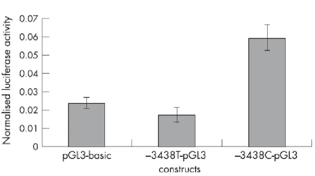
codificante, relativamente ao promotor basal, exão 1 e local dador de splicing. No decorrer desta análise inicial de regiões não codificantes, encontrámos uma nova mutação (c.-259 C>T) no promotor basal, num dos locais de ligação de factores de transcrição Sp1/Sp3. Esta mutação foi encontrada numa paciente com surdez profunda, em trans com p.Val84Met, por nós identificada anteriormente como uma nova mutação associada a surdez em GJB2.
Posteriormente, analisámos todas as regiões não codificantes atrás referidas, num conjunto maior de pacientes (n=89), com apenas uma ou nenhuma mutação codificante em GJB2, e em 91 controlos, considerados como tendo audição normal. Os resultados decorrentes deste análise revelaram que, nos pacientes, está sobre-representado o genótipo c.[*168A>G(+)*931C>T], um duplo heterozigótico respeitante aos SNPs rs55704559 e rs5030700 (ambos localizados na 3’UTR), respectivamente. A análise in silico prevê que a variante c.*168A>G (independentemente de estar uma citosina ou uma timina na posição c.*931) cause uma alteração da conformação do mRNA. Deste modo, os nossos dados sugerem que o alelo c.*168G poderá estar associado a surdez.
Outro dos objectivos deste trabalho foi a elucidação da patogenicidade das referidas mutações c.-259C>T e p.Val84Met, assim como da mutação p.Met163Leu, também encontrada pela primeira vez, num trabalho prévio, numa outra família Portuguesa. Os dados genéticos disponíveis sobre as três mutações (os nossos e os publicados por outros autores) eram insuficientes para a confirmação da sua patogenicidade, e assim efectuámos estudos funcionais para investigarmos os seus efeitos.
Os resultados obtidos sugerem que as três mutações são de facto patogénicas, exercendo o seu efeito de formas distintas. A mutação c.-259C>T reduz muito significativamente a actividade do promotor basal. Relativamente às mutações codificantes, enquanto a p.Val84Met altera as propriedades de permeabilidade iónica e molecular do canal intercelular, o que poderá in vivo traduzir-se num défice funcional, a p.Met163Leu causa a morte celular, possivelmente por outro mecanismo que não o funcionamento anómalo de conexões não emparelhados (já descrito na literatura), e apresenta um efeito dominante negativo parcial sobre a Cx26 e Cx30 do tipo selvagem.
Abstract
Mutations in the GJB2 gene are responsible for a considerable proportion of nonsyndromic sensorineural hearing loss (NSSHL), in several populations. This gene is a member of a gene family coding for connexins, the subunits of the hemichannels (connexons) which form the intercellular channels of the gap junctions existing in the vertebrates and tunicates. Connexin-26 (Cx26), encoded by the GJB2 gene, is expressed in several tissues, including specific epithelial and conjunctive tissues of the cochlea, the auditory organ which is localised to the inner ear. In the cochlea, Cx26 is co-expressed with connexin-30 (Cx30), encoded by the GJB6 gene, in several cell types. Two large GJB6 deletions are involved in several hearing loss (HL) cases, as well, being most of these cases due to compound heterozygosity with a GJB2 mutation.
Given the relevance of both GJB2 and GJB6 genes to the HL etiology in several populations, the molecular diagnosis of NSSHL cases with probable genetic cause is usually initiated by the analysis of the GJB2 coding region (in which most pathogenic mutations have been found), followed by the investigation of the presence of the GJB6 deletions in the cases where none or only one GJB2 mutation is found. In some cases, attributing the cause of the HL to the GJB2/GJB6 genotype is complicated because some GJB2 mutations are of unclear or controversial pathogenicity, due to either lack of genetic evidence (evident segregation of the mutation with the HL) or contradictory genetic data (same genotype occurring in both normal-hearing and normal-hearing-impaired individuals). In those cases, functional studies constitute an adequate approach for the investigation of the putative pathogenicity of such mutations, several of which have already been studied in this way.
One of the aims of this work consisted in the study of the coding region and acceptor splice site of the GJB2 gene in Portuguese individuals presenting with NSSHL. We have further extended the analysis of the gene to noncoding regions (basal promoter and about 700 bp immediately upstream, exon 1, donor splice site and the whole 3’UTR). These noncoding regions have rarely been studied. Nonetheless, two pathogenic noncoding mutations had previously been identified, both in the donor splice site. Initially, we analysed the basal promoter, exon 1 and donor splice site in some patients who only harboured one coding mutation. We then found
a novel mutation (c.-259C>T) in the basal promoter, in one of the binding sites for Sp1/Sp3 transcription factors. This mutation was found in a profoundly hearing-impaired patient, in trans with p.Val84Met, that we had identified as a novel mutation, in a previous work.
Later, we analised all the fore mentioned noncoding regions in a larger sample, including monoallelic patients and also those harbouring no mutation in the GJB2 coding region (n=89) as well as 91 controls who reported to have normal hearing. The obtained data revealed that the c.[*168A>G(+)*931C>T] double heterozygous genotype, regarding the rs55704559 and rs5030700 SNPs (both localised to the 3’UTR), respectively, is overrepresented in the patients. In silico analysis predicts that the c.*168A>G variant (regardless of the fact that in position c.*931 is a cytosine or a thymine) causes an alteration of mRNA folding. Thus, our data suggest that the c.*168G allele might be associated with HL.
Other aim of this work was to investigate the effects of the mutations c.-259C>T and p.Val84Met, mentioned above, and the mutation p.Met163Leu, also identified for the first time in a previous work, in other Portuguese family. The available genetic data regarding these mutations were insufficient to prove their pathogenicity, and thus we have performed functional studies on the three mutations. The obtained results suggest that the three mutations are indeed pathogenic, exerting their effect in distinct ways. The c.-259C>T mutation decreases very significantly the basal promoter activity. As regards the coding mutations, p.Val84Met alters the ionic and molecular permeability properties of the intercellular channel, which may compromise their function in vivo, while p.Met163Leu leads to cell death, possibly by a mechanism other than the malfunctioning of undocked hemichannels (already described in the literature), and has a partial dominant-negative effect on wild-type Cx26 (wtCx26) and Cx30 (wtCx30).
CHAPTER 1
Section 1 - Physiology of Hearing
The mammalian auditory system is capable of detecting and analyzing sounds over a wide spectrum of frequencies (humans can hear sounds between 20 Hz to 20 kHz) and over an intensity range of 12 orders of magnitude or 120 dB (Robles & Ruggero, 2001).
Ear Anatomy
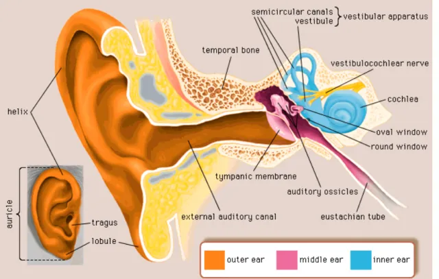
The human auditory system, responsible for the complex process of hearing, is composed of the outer ear, middle ear and inner ear (fig. 1).
Figure 1. Structure of the human ear. Reproduced from Encyclopædia Britannica Inc. (n.d.).
The outer ear is composed of the external ear (auricle) and the ear canal (external auditory canal). The middle ear includes the tympanic membrane, at the end of the ear canal, and three ossicles (maleus, incus and stapes). The middle ear cavity is connected by the
Eustachian tube to the pharynx. The inner ear consists of the vestibular apparatus, responsible for the balance function, and the cochlea, the organ of hearing (Møller, 2006).
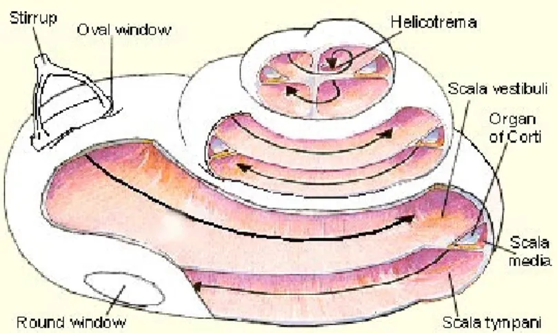
The human cochlea is a structure coiled in the form of a snail (fig. 2), which number of turns is generally considered to be 2 ½. However, a study conducted by (Biedron, Westhofen, & Ilgner, 2009) suggests that the majority of individuals (65% in their study) has more than 2 ½ cochlear turns, of which a small fraction has more than 2 ¾ turns. The cochlea is composed of three contiguous, fluid-filled, membranous tubes, scala vestibuli, scala tympani and scala media (fig. 2), being enclosed by a bony shell, the otic capsule.
The scala vestibuli and scala tympani, which communicate near the apex of the otic capsule, at helicotrema (fig. 2), are filled with perilymph, a fluid with an ionic composition similar to that of other extracellular fluids. The scala media is filled with endolymph, a fluid that, contrary to perilymph, has a high potassium concentration (Wangemann, 2006), which is necessary for the normal auditory function.
Figure 2. Cochlea opened from the side. The three compartments (scala vestibuli, scala media, scala tympani) are shown. The arrows indicate the direction of the sound wave traveling through the perilymph along scala vestibuli and scala tympani, which are connected at helicotrema. The stirrup is also called the stapes. Figure reproduced from Hearing Central LLC (n.d.).
Transduction of sound into nerve impulses
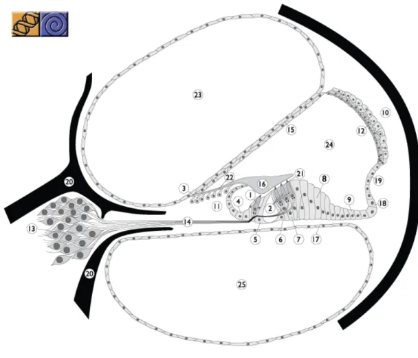
The sound is conducted through the outer ear to the tympanic membrane, which transmits vibration to the three ossicles of the middle ear. These ossicles impart the vibration to the oval window of the cochlea (fig. 2), creating a mechanical wave in the cochlear fluid and in the basilar membrane (fig. 3). The mechanical vibration is transduced into electric signals in the organ of Corti (fig. 3), which contains several cell types, including inner and outer hair cells (IHCs and OHCs, respectively) as well as supporting cells (e.g. inner and outer pillar cells, Claudius’, Hensen’s and Deiters’ cells) (fig. 3).
Figure 3. Representation of a cross-section of one turn of the cochlea, showing the three compartments, scala vestibuli, scala media and scala tympani, and the organ of Corti, resting on the basilar membrane. 1. Inner hair cell; 2. Outer hair cell; 3. Interdental cell (IDC); 4. Inner sulcus cells; 5. Inner pillar cells; 6. Outer pillar cells; 7. Deiters’ cells; 8. Hensen’s cells; 9. Claudius’ cells; 10. Spiral ligament; 11. Spiral limbus; 12. Stria vascularis; 13. Spiral ganglion; 14. Auditory nerve; 15. Reissner’s membrane; 16. Tectorial membrane (TM); 17. Basilar membrane; 18. External sulcus cells; 19. Spiral prominence; 20. Bony spiral lamina; 21. Reticular lamina; 22. Between IDC and TM; 23. Scala vestibuli; 24. Scala media; 25. Scala tympani. Reproduced from Van Camp & Smith (2011a).
The OHCs play an active role in increasing sensitivity to low intensity acoustic stimuli, and in frequency selectivity (especially for weak sounds) while the IHCs are responsible for the transduction of mechanical vibration (caused by sound) into nerve impulses (Møller, 2006). The movement of the basilar membrane leads to the deflection of the stereocilia at the apical
surface of IHCs and OHCs, which elicits the opening of the transducer channels (Beurg, Evans, Hackney, & Fettiplace, 2006; Ricci, Kennedy, Crawford, & Fettiplace, 2005; Stauffer & Holt, 2007). The current that then passes through these non-selective cation channels, carried by Ca2+ and mainly by K+ (LeMasurier & Gillespie, 2005), leads to depolarization of the cell’s membrane. Influx of calcium ions through IHCs’ voltage-gated Ca2+ channels elicits exocytosis (Brandt, Striessnig, & Moser, 2003; Moser & Beutner, 2000) and consequently the release of the neurotransmitter which stimulates type I afferent fibers via glutamatergic synapses, involving the α-amino-3-hydroxy-5-methyl-4-isoxazolepropionic acid (AMPA) receptor and the glutamate-aspartate transporter (GLAST) (Fuchs, Glowatzki, & Moser, 2003; Glowatzki et al., 2006). This primary acoustic input is then transmitted to the brain.
K+ recycling
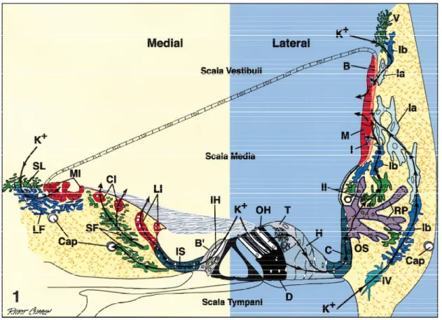
The process of transduction requires K+ to be returned to the endolymph. It is believed that K+ ions that exit hair cells through K+ channels at their basolateral membrane into the perilymph may be taken up by supporting cells (Hibino & Kurachi, 2006; Zdebik, Wangemann, & Jentsch, 2009), and move either through supporting cells, epithelial cells, and fibrocytes of the spiral limbus back into the endolymph, or through supporting and epithelial cells to the spiral ligament, where they are taken up by fibrocytes and conducted via the fibrocyte network to the stria vascularis where they are then secreted back into the endolymph (Spicer & Schulte, 1998) (fig. 4).
Figure 4. A schematic representation of the proposed transcellular pathways of K+ ions, effluxed from hair cells during auditory transduction, back to the endolymph. B=basal cell; BP=border cell; Cap=capillary; C=Claudius’ cell; CI=central interdental cell; D=Deiters’ cell; H=Hensen’s cell; I=intermediate cell; IH=inner hair cell; IS=inner sulcus cell; M=marginal cell; MI=medial interdental cell; LF =light fybrocyte; LI=lateral interdental cell; OH=outer hair cell; OS=outer sulcus cell; RP=root process; SF =stellate fybrocyte; SL=supralimbal fybrocyte; T=tectal cell; Ia, Ib, II, IV, and V=types of lateral wall fybrocytes. Reproduced from Spicer and Schulte (1998).
In addition to K+ circulation induced by acoustic stimulation, evidence supports the existence of standing currents in the cochlea (fig. 5), not depending on acoustic stimulation but being modulated by it (Zidanic & Brownell, 1990).
Figure 5. Model for standing currents in the cochlea in terms of current density field lines. The current leaks from scala media through the mechanically-sensitive transduction channels in the stereocilia of hair cells. Current leakage from scala media is thought to occur also through Reissner's membrane and through (or between) the supporting cells lateral to the organ of Corti. Reproduced from Zidanic and Brownell (1990).
Stria vascularis and the endocochlear potential (EP)
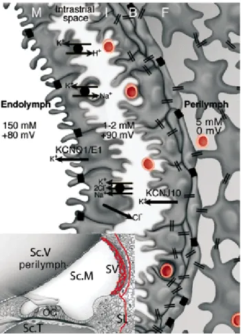
The stria vascularis, located in the cochlear lateral wall, is a tissue composed of two epithelial layers, with one composed of basal cells (BCs) and intermediate cells (ICs), and the other composed of marginal cells (MCs). The space that exists between the MCs and ICs is designated by intrastrial space (IS), and contains a dense capillary network (fig. 6).
Figure 6. Schematic diagrams illustrating the structure and location in the cochlea of the stria vascularis and the electrogenic machinery involved in the endocochlear potential generation. Inset: cross section of a cochlea’s turn. OC = organ of Corti; Sc.V = scala vestibuli; Sc.M = scala media; Sc.T = scala tympani; SL = spiral ligament; SV = stria vascularis. Red lines represent the vascular system of the cochlea lateral wall. Large diagram: stria vascularis in detail. B = basal cells; I = intermediate cells; M = marginal cells. Black boxes indicate tight junctions (TJs) between marginal cells and between basal cells. Endothelial cells of the SV capillaries are also linked by TJs (not represented). Intermediate and basal cells of the SV and fibrocytes (F) of the SL are connected by gap junctions (GJs) (double bars) composed of Cx26 and Cx30 (Forge et al., 2003; Lautermann et al., 1998; W. Liu, Boström, Kinnefors, & Rask-Andersen, 2009; Y.-P. Liu & Zhao, 2008). The K+ concentration (in mM) and the electric potential (in mV) are indicated in each of the three extracellular fluid spaces, namely the perilymph, intrastrial fluid, and endolymph. Each electric potential is indicated relative to that of the perilymph, which is taken as reference (0 mV). Figure reproduced from Cohen-Salmon et al. (2007).
As mentioned previously, stria vascularis secretes K+ ions into the endolymph. Additionally, stria vascularis generates an EP of approximately +80 mV which constitutes the main driving force for sensory transduction (Wangemann, 2002), thus being critical for the normal auditory function. The EP comprises two different K+-diffusion potentials, one across the apical membrane of the ICs and the other across the apical side of the MCs (Nin et al., 2008). This EP is dependent on KCNJ10 (Kir4.1) (Marcus, Wu, Wangemann, & Kofuji, 2002; Nin et al., 2008) and KCNQ1/KCNE1 (Nin et al., 2008) K+ channels, localised to the apical surfaces of ICs and MCs, respectively. The Na+,K+-ATPase and NKCC K+ transporters, that exist in the basolateral membrane of MCs, are also important for the generation of the EP (Nin et al., 2008). The gastric H+,K+-ATPase is expressed at the basolateral membrane of MCs as well, and may participate in EP formation (Shibata et al., 2006), but its role in the regulation of the electrochemical milieu of the IS remains unclear (Nin et al., 2008).
Cochlear gap junction systems
Two gap junction systems, the epithelial cell and the connective cell gap junction systems, exist in the cochlea and are thought to play a role in the recirculation of K+ by providing a mechanism for the uptake of the K+ released by the hair cells, its transcellular transport laterally, and its delivery to the stria vascularis (Kikuchi, Adams, Miyabe, So, & Kobayashi, 2000; Kikuchi, Kimura, Paul, & Adams, 1995; Kikuchi, Kimura, Paul, Takasaka, & Adams, 2000). Spicer and Schulte (1998) suggested the existence of a medial K+ recycling pathway from IHCs, in parallel with the formerly hypothesized lateral pathway (fig. 4), implicating the participation of gap junctions in both transcellular routes. Jagger and Forge (2006) presented evidence for the existence of two distinct compartments (medial and lateral) in the organ of Corti, which are isolated from each other, in terms of gap junctional intercellular communication. This finding supports Spicer and Schulte’s model of two distinct transcellular routes (medial and lateral) for the recirculation of K+, in which gap junctions would play a role, but it also demonstrated the existence of gap junction channels with different permeability properties, suggesting that the gap junction intercellular communication may play additional roles in the organ of Corti beyond its putative contribution for K+ recycling.
Section 2 - Hearing loss
Hearing loss is the most common sensorial impairment being a major public health concern. In 2005, about 278 million people had moderate to profound hearing impairment, of whom 80% live in low- and middle-income countries (World Health Organization, 2011a). HL affects 6 to 8% of the population in developed nations (Schrijver, 2004). This condition can occur at any age. In children before speech, HL (prelingual HL) occurs in 1 in 800-1000 while in early childhood HL occurs in 1 in 400-500. The probability of impairment increases with age (phenomenon designated by age-related HL or presbyacusis), affecting 2.3 % of the population aged 40-50 years, and over 30% of the population above 70 years of age (Petit et al., n.d.).The auditory defect can be located in the external or middle ear (conductive HL), or in the inner ear (sensorineural HL), affecting the cochlea or the auditory nervous system. Mixed HL results from anomalies in both external/middle ear and inner ear. HL may present itself with variable grades, which are described in table 1.
Table 1. Grades of hearing impairment, adapted from World Health Organization (2011b). Grades 2, 3 and 4 are classified as disabling hearing impairment. The audiometric ISO values are averages of values at 500, 1000, 2000, 4000 Hz. Grade of impairment Corresponding audiometric ISO value Performance 0 - None 25 dB or better (better ear)
No or very slight hearing problems. Able to hear whispers.
1 - Slight (Mild) 26-40 dB (better ear)
Able to hear and repeat words spoken in normal voice at 1 metre.
2 - Moderate 41-60 dB (better ear)
Able to hear and repeat words spoken in raised voice at 1 metre.
3 - Severe 61-80 dB (better ear)
Able to hear some words when shouted into better ear.
4 - Profound 81 dB or greater (better ear)
Unable to hear and understand even a shouted voice.
HL can be caused by environmental factors (infections, prematurity, trauma and exposition to ototoxic medications), however, in developed countries, most cases have a genetic cause.
Section 3 - Genetic hearing loss
Genetic hearing loss can be syndromic (~30%) or nonsyndromic (~70%). About 80% of nonsyndromic hearing loss (NSHL) cases are autosomal recessive, 15-20% are autosomal dominant, about 1% are X-linked and at least 1% are due to mutations in the mitochondrial DNA (mtDNA) (Schrijver, 2004).
To date 60 nuclear genes associated with NSHL have been cloned (Van Camp & Smith, 2011b). While eight of them are associated with both autosomal recessive and dominant HL, 17 genes have been strictly associated with autosomal dominant HL, and 32 other genes are so far related with autosomal recessive HL only. Three genes are implicated in X-linked NSHL. Two mitochondrial genes are also associated with NSHL.
Mutations in one particular gene, GJB2, coding for the Cx26 protein, are a major cause of NSHL/NSSHL in several populations (Kenneson, Van Naarden Braun, & Boyle, 2002). As such, screening for mutations in this gene is generally the first step undertaken in the molecular diagnosis of this pathology. Two gross deletions involving one other connexin gene, GJB6 (Cx30) (F. J. del Castillo et al., 2005; I. del Castillo et al., 2002; Lerer et al., 2001; Pallares-Ruiz, Blanchet, Mondain, Claustres, & Roux, 2002) have also been associated with NSHL in several cases.
Section 4 - The role of GJB2 (Cx26) and GJB6 (Cx30) genes in hearing loss
The Connexins and Gap Junctions
Connexins are transmembrane proteins, with four transmembrane domains (M1-M4), two extracellular loops (E1, E2), a cytoplasmic loop and cytoplasmic amino (N)- and carboxy (C)- termini (Kumar & Gilula, 1996). The N-terminus, transmembrane domains and extracellular loops are highly conserved among connexins, while the cytoplasmic loop and C-terminus are quite variable in both length and sequence (Haefliger et al., 1992).
Connexins oligomerize into hemichannels termed connexons, which are assembled from six connexin subunits (Baker, Caspar, Hollingshead, & Goodenough, 1983; Makowski, Caspar,
Phillips, & Goodenough, 1977; Perkins, Goodenough, & Sosinsky, 1997; Unwin & Zampighi, 1980; Yeager, 1998). Two connexons in adjacent plasma membrane
intercellular channel (fig. 7).
Figure 7. Tri-dimensional representation of a connexon (A), and of two connexons docked to form a intercellular channel (B). Figure adapted from
Several of these intercellular channels cluster into structures termed gap ju Channels formed by connexins have been shown to be permeable to atomic
glutathione, glutamate (Harris, 2001) and even siRNAs (Valiunas et al
nervous system, respiratory epithelium, bone, lens, heart and vasculature, reproductive system, inner ear) (Harris & Locke, 2009)
proteins are of importance in kerati
wound healing (Aasen & Kelsell, 2009; Langlois Becker, 2007). Connexins are also involved in Saffitz, 2001), and in the electric signaling between tissue homeostasis (Mathias, White, & Gong, 2010)
subunits of the same connexin (homomeric connexons) or from different connexins (heteromeric connexons). The intercellular channels are homotypic if both connexons are identical, or heterotypic if they are composed of two different connexons. Heteromeric Phillips, & Goodenough, 1977; Perkins, Goodenough, & Sosinsky, 1997; Unwin & Zampighi,
. Two connexons in adjacent plasma membrane
dimensional representation of a connexon (A), and of two connexons docked to form Figure adapted from Perkins, Goodenough, and Sosinsky (
Several of these intercellular channels cluster into structures termed gap ju Channels formed by connexins have been shown to be permeable to atomic
(Harris, 2001), second messengers such as cAMP and IP3
et al., 2005). Connexins are expressed in several tissues (e.g. skin, nervous system, respiratory epithelium, bone, lens, heart and vasculature, reproductive system, (Harris & Locke, 2009), playing key roles in tissue physiology. For example, these proteins are of importance in keratinocyte biology, being involved in skin differentiation and in (Aasen & Kelsell, 2009; Langlois et al., 2007; C. M. Wang, Lincoln, Cook, & . Connexins are also involved in the electric conduction in myocardium
the electric signaling between neurons (Connors, 2009)
(Mathias, White, & Gong, 2010). Connexons can be assembled from of the same connexin (homomeric connexons) or from different connexins heteromeric connexons). The intercellular channels are homotypic if both connexons are identical, or heterotypic if they are composed of two different connexons. Heteromeric Phillips, & Goodenough, 1977; Perkins, Goodenough, & Sosinsky, 1997; Unwin & Zampighi, . Two connexons in adjacent plasma membranes dock forming an
dimensional representation of a connexon (A), and of two connexons docked to form and Sosinsky (1998).
Several of these intercellular channels cluster into structures termed gap junctions. Channels formed by connexins have been shown to be permeable to atomic ions, ADP, ATP, , second messengers such as cAMP and IP3 (Harris, 2007), n several tissues (e.g. skin, nervous system, respiratory epithelium, bone, lens, heart and vasculature, reproductive system, , playing key roles in tissue physiology. For example, these nocyte biology, being involved in skin differentiation and in ., 2007; C. M. Wang, Lincoln, Cook, & the electric conduction in myocardium (Kanno & (Connors, 2009), and contribute to . Connexons can be assembled from six of the same connexin (homomeric connexons) or from different connexins heteromeric connexons). The intercellular channels are homotypic if both connexons are identical, or heterotypic if they are composed of two different connexons. Heteromeric
connexons have been found in several tissues (Diez, Ahmad, & Evans, 1999; He, Jiang, Taffet, & Burt, 1999; Jiang & Goodenough, 1996; Locke et al., 2007, 2000), including cochlear tissues (Shoab Ahmad, Chen, Sun, & Lin, 2003; Forge et al., 2003; X. Z. Liu, Yuan, et al., 2009).
Genomic data has provided evidence of the generalised presence of connexins in vertebrates (Cruciani & Mikalsen, 2006). Connexins have also been identified in tunicates (Sasakura et al., 2003; Seo et al., 2001; White, Wang, Mui, Litteral, & Brink, 2004). These findings, together with additional genomic data from other taxonomic groups obtained so far, suggest that this protein family may be specific to vertebrates and tunicates (Panchin, 2005; Shestopalov & Panchin, 2008; Sodergren et al., 2006).
The GJB2 and GJB6 genes (DFNB1/DFNA3 loci)
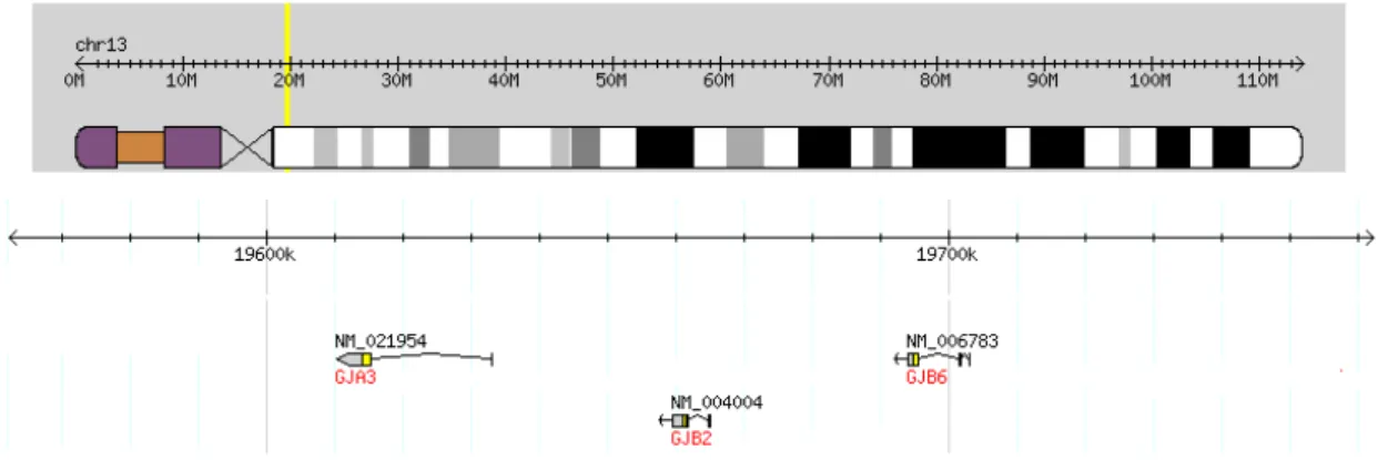
Twenty connexin genes have been identified in humans (Cruciani & Mikalsen, 2005). The genes GJB2 and GJB6, together with gene GJA3, which codes for connexin-46 (Cx46), form a connexin gene cluster which localise to chromosome 13q12.11 (fig. 8).
Figure 8. GJA3, GJB2 and GJB6 connexin gene cluster in human chromosome 13q12.11. Adapted from The International HapMap Consortium (n.d.).
The genomic region comprising the GJB2 and GJB6 genes is commonly known as the DFNB1 locus or the DFNA3 locus, when referring to mutations in these connexins causing
recessive or dominant HL, respectively 1998).
The GJB2 gene comprises a first nonc
5’ capped cDNA sequences), a 3179 kb intron, and a second exon containing the whole coding region (fig. 9a). There is also supporting evidence for an unspliced transcript, containing a shorter ORF initiating at an AUG upstream of the main ORF (fig. 9b), which is likely to impair the efficacy of the translation of the Cx26 protein. Additionally, there are
polyadenylation sites (figs. 9a, b). The possibility of alternative splicing, out of a 2357 pb intron (fig. 9c), is supported by one cDNA sequence Mieg, 2010).
Figure 9. Scheme of human b represents an unspliced transcript. polyadenylation sites (three different sites) 2010).
In the GJB6 gene the coding region is contained within a single exon, prece upstream noncoding exons (Essenfelder, Larderet, Waksman, & Lamartine, 2005)
Cx26 and Cx30 in the cochlea Cx26 and Cx30 are expressed
et al., 2009) cochleae, in the supporting cells of the organ of Corti and in the lateral wall, where they are part of the two independent gap junction systems, the epithelial cell gap junction , respectively (Smith & Van Camp, 1998; Smith, Sheffield, & Van Camp,
gene comprises a first noncoding exon (different TSPs are suggested by distinct 5’ capped cDNA sequences), a 3179 kb intron, and a second exon containing the whole coding region (fig. 9a). There is also supporting evidence for an unspliced transcript, containing a ting at an AUG upstream of the main ORF (fig. 9b), which is likely to impair the efficacy of the translation of the Cx26 protein. Additionally, there are
(figs. 9a, b). The possibility of alternative splicing, by means of (fig. 9c), is supported by one cDNA sequence (D. Thierry
Scheme of human GJB2 alternative transcripts. a and c represent spliced transcripts while b represents an unspliced transcript. Filled flags represent 5’ cap sites; empty flags represent validated
(three different sites). Adapted from AceView (D. Thierry
gene the coding region is contained within a single exon, prece (Essenfelder, Larderet, Waksman, & Lamartine, 2005)
Cx26 and Cx30 in the cochlea
30 are expressed in rodent (Forge et al., 2003) and human
in the supporting cells of the organ of Corti and in the lateral wall, where they are part of the two independent gap junction systems, the epithelial cell gap junction (Smith & Van Camp, 1998; Smith, Sheffield, & Van Camp,
oding exon (different TSPs are suggested by distinct 5’ capped cDNA sequences), a 3179 kb intron, and a second exon containing the whole coding region (fig. 9a). There is also supporting evidence for an unspliced transcript, containing a ting at an AUG upstream of the main ORF (fig. 9b), which is likely to impair the efficacy of the translation of the Cx26 protein. Additionally, there are alternative by means of the splicing (D. Mieg &
Thierry-a Thierry-and c represent spliced trThierry-anscripts while Filled flags represent 5’ cap sites; empty flags represent validated (D. Thierry-Mieg & Thierry-Mieg,
gene the coding region is contained within a single exon, preceded of five (Essenfelder, Larderet, Waksman, & Lamartine, 2005).
human (W. Liu, Boström, in the supporting cells of the organ of Corti and in the lateral wall, where they are part of the two independent gap junction systems, the epithelial cell gap junction
system and the connective tissue cell gap junction system (Kikuchi et al., 1995). W. Liu, Boström, et al. (2009) also observed expression of the two connexins in human spiral ganglion neurons.
Co-localisation of Cx26 and Cx30 at the same gap junction plaques was observed in supporting cells of the organ of Corti, in basal and intermediate cells of stria vascularis and in fibrocytes of the spiral ligament, in rodents (Forge et al., 2003). In the same study, an immunoprecipitation assay using mature mouse cochleae demonstrated the existence of heteromeric connexons comprising both Cx26 and Cx30. In humans, co-localisation was detected in the basal cell layer of the stria vascularis and in Deiters’ cells of the organ of Corti (W. Liu, Boström, et al., 2009). Therefore, it is very likely that heteromeric connexons, and/or heterotypic channels, comprising both Cx26 and Cx30 also exist in the human cochlea.
Despite of being co-localised in certain cochlear regions, Cx26 and Cx30 display distinct localisation patterns in the cochlear sensory epithelium (W. Liu, Boström, et al., 2009; Zhao & Yu, 2006) and in the cochlear lateral wall (W. Liu, Boström, et al., 2009; Y.-P. Liu & Zhao, 2008).
Since Cx26 and Cx30 are constituents of the cochlear gap junction systems, mutations in these genes have been considered to cause HL by interfering with the recirculation of K+ in the cochlea (Kikuchi, Adams, et al., 2000). However, a more complex role of both connexins in cochlear physiology has been emerging in the recent years.
Loss of Cx30 in mice impairs the development of the EP (Teubner et al., 2003), through disruption of the endothelial barrier in the stria vascularis (Cohen-Salmon et al., 2007). Cx30-null mice also present degeneration of the organ of Corti (Teubner et al., 2003). Shoeb Ahmad et al. (2007) observed that Cx30-null mice express a reduced Cx26 protein level in the cochlea and, interestingly, the consequent hearing impairment is prevented in Cx30-null mice overexpressing Cx26, which present levels of the protein close to the wild-type mice. A recent study has detected a reduction of Cx26 mRNA and protein levels in Cx30-null mice cochlea, particularly significant in cells from the region of the outer sulcus when compared to the whole cochlea. The authors presented evidence of co-regulation of Cx26 and Cx30 through NF-kB pathway as a feature of supporting cells in the outer sulcus region (Ortolano et al., 2008). Chang, Tang, Ahmad, Zhou, and Lin (2008) found that Cx30-null mice presented near normal ionic coupling but impaired metabolic coupling in outer sulcus and Claudius’ supporting cells,
which normally co-express Cx26 and Cx30, as assessed by Zhao and Yu (2006), in guinea-pig Claudius’ cells, and by Y.-P. Liu and Zhao (2008), in rat outer sulcus cells. The results of the fore mentioned studies suggest that ablation of Cx30 and the subsequent decrease of the Cx26 protein levels in supporting cells, results in impaired metabolic coupling, which could explain cell death in the organ of Corti.
Loss of expression of Cx26 in the mice epithelial gap junction network specifically does not seem to impair the EP development, but results in the degeneration of the organ of Corti soon after onset of hearing. It was proposed that the degeneration could be due to local extracellular accumulation of K+ near the basolateral region of IHCs (Cohen-Salmon et al., 2002). However, the fore mentioned finding of the co-regulation of Cx26 and Cx30 could suggest that a downregulation of Cx30 occurs in these conditional Cx26 knocked-out mice, leading to a defective epithelial gap junction system, and consequently to metabolic coupling impairment and cell death.
Additional evidence suggesting the participation of Cx26 and Cx30 in cochlear homeostasis, beyond their putative role in the recirculation of K+, arises from functional studies of some Cx26 and Cx30 mutant proteins. It was found that some mutations in these connexins (p.Val84Leu, p.Ala88Ser in Cx26, and p.Thr5Met in Cx30) do not impair ionic permeability of intercellular channels but impair their permeability to the larger cationic molecule propidium iodide (PI), as well as to the anionic second messenger IP3, which initiated propagation of Ca2+ waves in organotypic cochlear cultures (Beltramello, Piazza, Bukauskas, Pozzan, & Mammano, 2005; Y. Zhang et al., 2005). Also, the study of other Cx26 mutants (p.Thr8Met and p.Asn206Ser) showed that mutant and wild-type channels had similar unitary conductance, and that permeability to the anionic molecules cAMP and Lucifer yellow (LY) were retained by the mutant channels whereas their permeability to the cationic dye ethydium bromide (EtBr) was greatly reduced compared with that of the wild-type channel. This finding indicated differential selectivity to large molecules which suggested that altered permeability to large molecules of mutant channels between the supporting cells might be associated to HL (Meşe, Valiunas, Brink, & White, 2008). Moreover, faster intercellular Ca2+ signaling was observed in cells coupled by
heteromeric channels containing both Cx26 and Cx30 (Sun et al., 2005), suggesting that such channels may mediate and be important to Ca2+ signaling in cochlear tissue.
Although HL in Cx30 null mice had been prevented by overexpression of Cx26 (Shoeb Ahmad et al., 2007), the distinct localisation of the two connexins in the cochlea (W. Liu, Boström, et al., 2009; Y.-P. Liu & Zhao, 2008; Zhao & Yu, 2006) might be a strong indication that the two connexins have non-redundant functions in the normal physiology of the cochlea, further supporting their function beyond the putative role in K+ recycling.
Therefore, it is very likely that gap junctions play a crucial role in the cochlea by mediating the intercellular diffusion of large biological molecules, as part of biochemical and signaling pathways in vivo.
GJB2 mutations
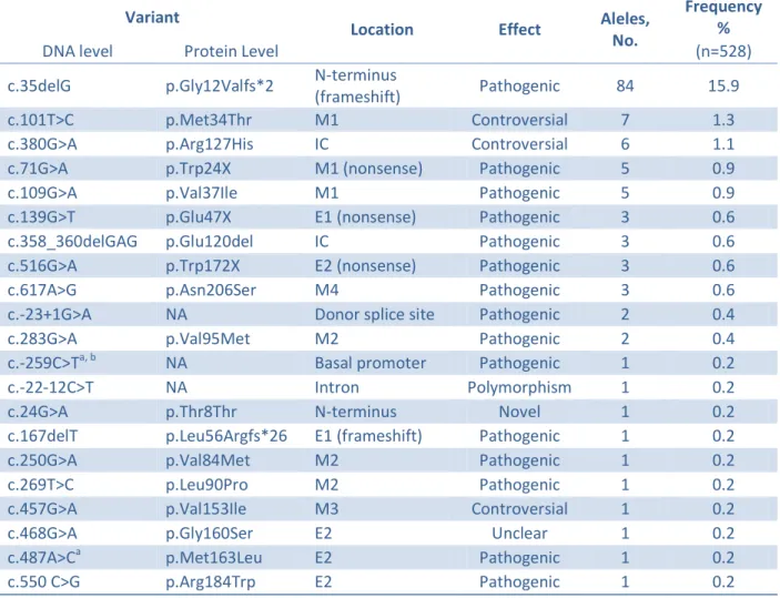
There are over 200 pathogenic GJB2 mutations described (Stenson et al., 2009). Most of them are responsible for recessive NSHL, but a few others are dominant causing either NSHL or HL associated with dermatological disorders (syndromic HL).
The spectrum and the prevalence of GJB2 mutations vary among different populations. The recessive mutation c.35delG is the most frequent GJB2 pathogenic mutation identified in NSHL European patients (Antoniadi et al., 2000; Gabriel et al., 2001; Janecke et al., 2002; T. Löppönen et al., 2003; Minárik, Feráková, Ficek, Poláková, & Kádasi, 2005; Roux et al., 2004; Santos et al., 2005; Seeman et al., 2004; Šterna et al., 2009), in other populations of predominantly European ancestry (Batissoco et al., 2009; Dalamón et al., 2005; Kelley et al., 1998) and in the Mediterranean populations from Algeria (Ammar-Khodja et al., 2009), Egypt (Snoeckx, Hassan, Kamal, Van Den Bogaert, & Van Camp, 2005), Lebanon (Mustapha et al., 2001), Morocco (Abidi et al., 2007) and Tunisia (Masmoudi et al., 2000). Two other recessive mutations, c.167delT and c.235delC, are the most frequent GJB2 pathogenic mutations found in NSHL affected individuals among Ashkenazi Jews (Lerer et al., 2000), and in Asian populations from Japan (Abe, Usami, Shinkawa, Kelley, & Kimberling, 2000; Fuse et al., 1999), Korea (H. J. Park, Hahn, Chun, Park, & Kim, 2000) and China (Dai et al., 2009), respectively.
The c.35delG mutation has a high carrier frequency in Europe in general (1.96%), but is higher (2.86%) in southern Europe (Gasparini et al., 2000). This deletion has also a high carrier frequency (2.08%) among Americans of European ancestry (Kelley et al., 1998). Regarding the c.167delT mutation, carrier frequencies of 2.78% (Sobe et al., 1999), 4.03% (Morell et al., 1998) and as high as 7.46% (Lerer et al., 2000) have been found among Ashkenazi Jewish individuals. Carrier frequencies for c.235delC mutation range from 0.5% to 2.08% in Eastern Asia (Abe et al., 2000; S.-H. Han et al., 2008; Hwa et al., 2003; Kudo et al., 2000; Y. Liu, Ke, Qi, Li, & Zhu, 2002; H. J. Park et al., 2000; Shi et al., 2004).
One other recessive mutation, p.Arg143Trp, firstly identified in Ghana (Brobby, Müller-Myhsok, & Horstmann, 1998), is the prevalent mutation in congenital, profound NSSHL Ghanese cases (Hamelmann et al., 2001). Interestingly, this mutation has a high prevalence among NSHL Japanese cases, compatible with recessive inheritance (Abe et al., 2000; Hayashi et al., 2011). To a lesser extent, the p.Arg143Trp mutation has been identified in NSHL cases from other Asian countries, such as India (Maheshwari et al., 2003; Mani et al., 2009), Iran (Hashemzadeh Chaleshtori et al., 2005; Najmabadi et al., 2005), and Korea (H. J. Park et al., 2000). A significant frequency of p.Arg143Trp among moderate to profound NSSHL cases, compatible with recessive inheritance, was also found in Argentina, a country which has received major immigration fluxes from Italy and Spain (Gravina et al., 2010). Noteworthy, the p.Arg143Trp was among the most frequent GJB2 variants detected in North American HL patients of Hispanic ethnicity (Putcha et al., 2007).
A different pathogenic mutation, p.Trp24X, was the predominant GJB2 mutation identified in NSHL cases in India (RamShankar et al., 2003), and in Gypsies (an ethnic group that traces back to the Indian subcontinent) from Eastern Slovakia (Minárik et al., 2003) and Spain (Álvarez et al., 2005). This mutation was also found to have a high carrier frequency (2.4%) in the Indian population (RamShankar et al., 2003), and in some subpopulations of Gypsies (Minárik et al., 2003; Álvarez et al., 2005).
As previously mentioned, due to the high frequency of GJB2 mutations among NSHL/NSSHL cases in several populations worldwide, this gene is routinely screened for mutations in patients with this pathology. However, only the coding region has been
systematically analysed. Nonetheless, several studies have also searched for mutations in exon 1 (Al-Qahtani et al., 2010; Denoyelle et al., 1999; Godbole et al., 2010; Green et al., 1999; Hamelmann et al., 2001; Hashemzadeh Chaleshtori et al., 2002; Janecke et al., 2002; X. Z. Liu, Xia, et al., 2002; Mani et al., 2009; Marlin et al., 2005; Matos et al., 2008, 2007, 2011, 2010; Najmabadi et al., 2002, 2005; Pollak et al., 2008; Prasad, Cucci, Green, & Smith, 2000; RamShankar et al., 2003; Riazalhosseini et al., 2005; Roux et al., 2004; Seeman & Sakmaryová, 2006; Sirmaci, Akcayoz-Duman, & Tekin, 2006; H.-Y. Tang et al., 2006; Y. Yuan et al., 2009, 2010). The promoter region has also been screened, but in fewer studies (Hashemzadeh Chaleshtori et al., 2002; Houseman et al., 2001; Matos et al., 2008, 2007, 2011, 2010; Pollak et al., 2008; Sirmaci et al., 2006; Y. Yuan et al., 2009, 2010). As a result, a few noncoding pathogenic mutations have been described. Their identification contributed to the elucidation of the genetic etiology of the HL in some patients who harboured only one recessive mutation in the GJB2 coding region.
The donor splice site c.-23+1G>A (commonly known as IVS1+1G>A) mutation has explained a high proportion of such unelucidated cases in some populations. This mutation, first identified by (Denoyelle et al., 1999), and later proven to prevent the transcription of the gene (Shahin et al., 2002), has been identified in 45% (9/20) of Czech and 23% (11/47) of Hungarian hearing-impaired individuals with only one GJB2 coding mutation (Seeman & Sakmaryová, 2006; Tóth et al., 2007). In one other study, 50% (8/16) of Turkish HL patients heterozygous for an exon 2 mutation, were also heterozygous for the c.-23+1G>A mutation (Sirmaci et al., 2006). Compound heterozygosity between the c.-23+1G>A mutation and a coding GJB2 mutation has also been detected in patients from other populations (Janecke et al., 2002; Medica, Rudolf, Balaban, & Peterlin, 2005; Najmabadi et al., 2005; Prasad et al., 2000; Y. Yuan et al., 2010).
One other noncoding mutation, c.-23G>T, located at the position -1 in respect to the donor splice site, has been recently identified (Mani et al., 2009). This mutation, predicted to impair the splice site, was present in trans with p.Trp24X, in a severe to profound HL patient from India.
Y. Yuan et al. (2010) reported a patient harbouring a novel exon 1 variant, c.-3175C>T, in compound heterozygosity with c.235delC. These variants were most likely in trans since the
patient’s father was simple heterozygous for the exon 1 variant. The pathogenicity of c.-3175C>T, which was not present in 105 normal-hearing controls, is possible, but still unclear.
A pathogenic mutation within the basal promoter has been found in the context of the present work (Matos et al., 2007).
Gross deletions compromising GJB2/GJB6
Pathogenic point mutations in the GJB2 gene are numerous and are a frequent cause of NSHL. In contrast, GJB6 point mutations are seemingly a rare cause of this pathology (Bhalla, Sharma, Khandelwal, Panda, & Khullar, 2011; Gürtler, Egenter, Bösch, & Plasilova, 2008; Nahili et al., 2008). Nonetheless, a few GJB6 variants have been identified in some NSHL cases. The GJB6 mutations p.Thr5Met (Grifa et al., 1999), and c.63delG (Pandya, personal communication in Ballana, Ventayol, Rabionet, Gasparini, & Estivill, 2011) have been associated with dominant NSHL, while two other GJB6 variants, c.631T>G (p.Cys211Gly) and c.689insA, were identified in compound heterozygosity in two NSSHL cases (Putcha et al., 2007). The first one was accompanied by the rare c.110T>C (p.Val37Ala) GJB2 variant [firstly identified by Azaiez et al. (2004), in heterozygosity, in a NSHL patient born to normal-hearing parents; classified as pathogenic (Husami et al., 2011)] and the second one by the GJB2 c.35delG deletion. Y. Yuan et al. (2010) found a NSHL patient, with no GJB2 mutations identified, carrying one other GJB6 variant, c.404C>A (p.Thr135Lys). While GJB6 point mutations are rare, two gross deletions, del(GJB6-D13S1830) (I. del Castillo et al., 2002; Lerer et al., 2001; Pallares-Ruiz et al., 2002) and del(GJB6-D13S1854) (F. J. del Castillo et al., 2005), localised upstream of the GJB2 gene, and disrupting the GJB6 gene, have been identified and shown to be associated with HL in several populations, most often due to compound heterozygosity with GJB2 mutations (Angeli, 2008; Cama et al., 2009; Chora et al., 2010; Cordeiro-Silva et al., 2011; Dalamón et al., 2005; F. J. del Castillo et al., 2005; I. del Castillo et al., 2003; Gravina et al., 2010; Marlin et al., 2005; Pandya et al., 2003; Piatto, Bertollo, Sartorato, & Maniglia, 2004; Roux et al., 2004; Santos et al., 2005; Seeman et al., 2005; Taitelbaum-Swead et al., 2006). Less frequently, homozygosity for del(GJB6-D13S1830), or compound heterozygosity for both deletions, have also been identified in HL patients (Angeli, 2008; F. J. del Castillo et al., 2005; I. del Castillo et al., 2003; Pandya et al.,
2003; Roux et al., 2004). Interestingly, (Erbe, Harris, Runge-Samuelson, Flanary, & Wackym, 2004) reported one hearing-impaired child, not harbouring any GJB2 coding mutations, who was compound heterozygous for del(GJB6-D13S1830) and for a GJB6 coding mutation (p.His124Gln).
Common et al. (2005) observed loss of Cx26 expression from the allele bearing the del(GJB6-D13S1830) deletion, in ductal sweat gland epithelium, and suggested the existence of a regulatory element of Cx26 expression. Rodriguez-Paris and Schrijver (2009) demonstrated, by qualitative allele-specific RT-PCR analyses using total RNA isolated from buccal ephitelium, that del(GJB6-D13S1830) causes loss of expression of GJB2 gene in cis, providing further support for the hypothesis of existence of a cis-regulatory element regulating GJB2. Later, Rodriguez-Paris, Tamayo, Gelvez, and Schrijver (2011) showed that the del(GJB6-D13S1854) GJB6 deletion, which is smaller than, and included within, the del(GJB6-D13S1830) also causes loss of GJB2 expression in cis, as previously suspected.
A third deletion, of 131.4 kbp, del(chr13:19,837,344-19,968,698), localised upstream of GJB2 and GJB6 has been identified segregating with HL and in trans with c.35delG, in one large American family of German descent. The GJB2 and GJB6 expression from alleles bearing the deletion was found to be reduced (Wilch et al., 2010, 2006). Wilch et al. (2010) hypothesized a similar mechanism for the pathogenic effect of this deletion, and the two deletions truncating GJB6, which would involve, in addition to loss or reduction in expression of GJB6, the loss of a GJB2 cis-regulatory element located within the deleted 95.4 kbp genomic interval shared by the three deletions. The expression of GJB6 gene might also be regulated by this element, or, instead, a distinct cis-regulatory element of this gene might exist and be deleted by del(chr13:19,837,344-19,968,698). Alternatively, the authors suggest that this third deletion could be in linkage disequilibrium (LD) with a pathogenic variant closer to both GJB2 and GJB6 that disrupts cis-regulatory function of these genes.
Feldmann et al. (2009) identified a large deletion, encompassing at least 920 kbp, which completely deletes the three connexin genes GJA3, GJB2 and GJB6, as well as at least four other genes. This deletion was identified in trans with the c.250G>A (p.Val84Met) GJB2 mutation in an individual presenting prelingual, profound HL.
Unelucidated GJB2 or GJB6 heterozygotes
In some patients, after searching for mutations in the GJB2 coding region, splice sites and exon 1, and screening for the del(GJB6-D13S1830) and del(GJB6-D13S1854) GJB6 deletions, only one recessive GJB2 coding mutation is identified (F. J. del Castillo et al., 2005; I. del Castillo et al., 2003). Some of these individuals may indeed just be carriers of a GJB2 mutant allele but in some other patients a second mutation may exist in other regions of GJB2 gene, such as the promoter, as shown in this work (Matos et al., 2007), the intron or the 3’ UTR, or in the GJB6 gene. The fore mentioned 131.4 kbp or the ~920 kbp GJB2/GJB6 gross deletions, other deletions truncating GJB2 or GJB6, or mutations disrupting a putative upstream regulatory element of these genes, may account for some of these unresolved cases.
GJB2 mutations with unknown or controversial pathogenicity
The pathogenicity of several GJB2 mutations is not always clear, due to the lack of evidence for or to existing contradictory data regarding the segregation of the mutation with the HL. Examples of such mutations are c.-684_-675del (Houseman et al., 2001; Zoll et al., 2003), p.Met34Thr (Bicego et al., 2006; Cama et al., 2009; Feldmann et al., 2004; Griffith et al., 2000; Houseman et al., 2001; Kenna, Wu, Cotanche, Korf, & Rehm, 2001; Pollak et al., 2007; Snoeckx, Huygen, et al., 2005; B.-L. Wu et al., 2002), p.Arg127His (Matos et al., 2010), p.Val153Ile (Cryns et al., 2004; Dalamón et al., 2005; Hashemzadeh Chaleshtori et al., 2005; Kenna et al., 2001; Marlin et al., 2001; RamShankar et al., 2003; Snoeckx, Hassan, et al., 2005; Snoeckx, Huygen, et al., 2005) and p.Gly160Ser (X. Cheng et al., 2005; Hashemzadeh Chaleshtori, Farhud, & Patton, 2007; Janecke et al., 2002; Löffler et al., 2001; Santos et al., 2005; Snoeckx, Huygen, et al., 2005; H.-Y. Tang et al., 2006).
DFNB1 mutations and human evolution
The high frequency of DFNB1-related HL, observed in several populations, has been hypothesized to be due to the combined effects of relaxed selection and assortative mating (Nance & Kearsey, 2004). Furthermore, it has been suggested that a carrier status for Cx26