UMinho | 2014
Universidade do Minho
Escola de Ciencias da Saúde
Maio de 2014
Vânia Rita de Faria Cardoso
Impact of differential Toll-like receptor
recognition of
Mycobacterium tuberculosis
strains on the adaptive immune response
Mycobacterium tuberculosis
Vânia Rita de Faria Cardoso
Impact of dif ferential T oll-lik e recep tor recognition of My cobacterium tuberculosis s trains on t he adap
tive immune response
My
cobacterium
Vânia Rita de Faria Cardoso
Impact of differential Toll-like receptor
recognition of
Mycobacterium tuberculosis
strains on the adaptive immune response
Maio de 2014
Dissertação de Mestrado
Mestrado em Ciências da Saúde
Trabalho Efetuado sob a orientação da
Doutora Margarida Saraiva
e da
Doutora Lúcia Moreira Teixeira
Universidade do Minho
Escola de Ciencias da Saúde
ii
DECLARAÇÃO
Nome: Vânia Rita de Faria Cardoso
Endereço eletrónico: [email protected] Telefone: +351 916 262 967
Número do Bilhete de Identidade: 13892195
Título da dissertação: Impact of differential Toll-like receptor recognition of Mycobacterium tuberculosis strains on the adaptive immune response
Orientadora:
Doutora Margarida Saraiva Coorientadora:
Doutora Lúcia Moreira Teixeira
Ano de conclusão: 2014
Designação Ramo de Conhecimento do Mestrado: Ciências da Saúde
DE ACORDO COM A LEGISLAÇÃO EM VIGOR, NÃO É PERMITIDA A REPRODUÇÃO
DE QUALQUER PARTE DESTA TESE
Universidade do Minho, 21 de Maio de 2014
iii
The work presented in this thesis was done in Microbiology and Infection Research Domain of the Life and Health Sciences Research Institute (ICVS), School of Health Sciences, University of Minho, Braga, Portugal (ICVS/3B’s – PT Government Associate Laboratory, Braga/Guimarães, Portugal).
v
“The real voyage of discovery consists not in seeking new landscapes but in having new eyes.” Marcel Proust
vii ACKNOWLEDGEMENTS
Gostaria de agradecer a todas as pessoas que contribuíram para a realização desta tese e que me encorajaram, motivaram e acreditaram em mim especialmente durante este último ano. Dessas pessoas, gostaria de salientar:
A Doutora Margarida Saraiva, minha orientadora, com quem muito aprendi nestes últimos dois anos. Quero agradecer pela oportunidade de trabalhar no seu laboratório, pela transmissão de conhecimentos pela sua constante disponibilidade, empenho, entusiasmo, pelo rigor científico e pela capacidade de organização.
A Lúcia, minha coorientadora, pelos conhecimentos sobretudo ao nível experimental pela tua constante disponibilidade e sobretudo pela AMIZADE, companheirismo e motivação mesmo estando longe continuas perto, as palavras são poucas para agradecer tudo o que fizeste por mim.
O Professor Gil Castro pela oportunidade de trabalhar no seu laboratório, conhecimentos transmitidos e pela disponibilidade e entusiasmo.
O Egídio pelos conhecimentos transmitidos, pela disponibilidade, pela ajuda, pela motivação, pela nova forma de encarar o BSL-3 e pelas interessantes discussões.
A todos eles queria agradecer por lerem criticamente o meu trabalho e pelas sugestões e críticas que ajudaram a melhorá-lo e mais importante por fomentarem o gosto pela ciência e por me ajudarem a crescer enquanto aluna de investigação. Espero que reconheçam todo o respeito e admiração intelectual que tenho por vós.
A Flávia e a Ritinha pela AMIZADE, pelos bons momentos passados, pelo apoio e motivação nos bons e maus momentos, vocês estão cá dentro. Obrigada!
A Joana Gaifem pela boa disposição e pelo seu particular sentido de humor e a Joana Gouveia às duas obrigadas pela amizade, apoio, motivação e ajuda nos bons e maus momentos.
viii
A Joana Guedes pela iniciação no laboratório ainda durante a licenciatura e amizade e por ter sempre um sorriso amigo na cara.
O Jeremy pelo amizade e por ser sempre tão prestável e preocupado.
A Alex, a Isabel, a Carine, o Bruno, o Bernardo, a Palmira, a Cláudia, o Diogo, a Gabriela, a Ana, a Filipa, o Henrique e a Alice pela amizade e disponibilidade e por tornarem o ambiente no laboratório bom.
Todos os meus amigos pela amizade, apoio, confiança e cumplicidade mesmo nas ausências.
Os meus pais, irmãos e sobrinhos porque são a minha vida, pelo apoio e amor incondicional por tudo o que sempre fizeram e fazem por mim, por me motivarem e por me mostrarem sempre o orgulho que tem em mim. E ainda ao novo membro da família que apesar de ainda não ter chegado já me trouxe muita felicidade. Amo-vos.
O Barreto, o meu amor há quase seis anos, quero agradecer por tudo, pelo amor, partilha e apoio incondicional nos bons e em especial nos maus momentos. E por sempre tentar compreender que a minha vida científica é também importante para mim. Tu sabes o quanto és fundamental na minha vida.
ix ABSTRACT
Mycobacterium tuberculosis (Mtb) infection, remains one of the major worldwide threats, affecting about one third of the human population. The vaccine Bacillus Calmette-Guérin (BCG), although effective against childhood disseminated tuberculosis (TB), has variable and limited efficacy against adulthood pulmonary TB. Co-infection with the human immunodeficiency virus (HIV) and the emergence of multidrug resistant strains of Mtb has further complicated the TB control and eradication. It is becoming increasingly recognized that the Mtb complex (MTBC) is more genetically diverse than previously believed. In our laboratory, we have been interested in the study of the differential host-pathogen interactions among Mtb strains, particularly those from the Beijing family. Among this Mtb lineage, we found that strain 02-171, is recognized by both toll-like receptor 2 (TLR2) and TLR4, resulting in a distinct outcome of infection in vitro and in vivo.
In the first part of this work we investigated the impact of TLR4 triggering on T cell responses during a primary infection by 02-171 Mtb strain via the aerosol route. We found that TLR4 triggering increased the frequency of lung CD4+ T cells, which appears to be associated with enhanced T cell recruitment. Furthermore, TLR4 activation did not impact interferon (IFN)- response neither nitric oxide synthase 2 (Nos2) expression. In accordance, TLR4 triggering did not impact bacterial growth after aerosol 02-171 infection. Interestingly, TLR4 absence increased lung interleukin (IL)-17 CD4+ T cell responses. Overall, our data suggest no apparent function for TLR4 triggering during a primary infection with a low dose of 02-171 Mtb strain inoculated via the aerosol route. In the second part of this work, we focused on the role of TLR4 activation by 02-171 Mtb strain during a recall response after BCG vaccination. We found that BCG vaccination is effective at controlling the progression of infection and lung pathology in 02-171 infection. This finding does not support the idea that Beijing strains are resistant to protection evoked by BCG vaccination. In vaccinated mice, we observed an overall anticipation of protective responses, particularly the presence of multifunctional and granulocyte macrophage colony-stimulating factor (
GM-CSF)-producing CD4+ T cells. Furthermore, while wild-type (WT) vaccinated mice upregulated IL-17 responses, in TLR4 deficient mice such induction was not observed, without impairment in BCG-induced protection. Overall, TLR4 triggering seems to be not essential for BCG-mediated immunity against 02-171 infection. Understanding, the mechanisms that dictate the development of adaptive immunity will certainly contribute for the identification of T cell subsets and other immune factors important for protection against Mtb infection, as well as, novel correlates of protection, particularly relevant to evaluate the effectiveness of new TB vaccine candidates.
xi RESUMO
A infeção por Mycobacterium tuberculosis (Mtb) constitui um grave problema em todo o mundo, afetando cerca de um terço da população humana. A vacinação com o Bacilo Calmette-Guérin (BCG), embora eficaz contra tuberculose (TB) disseminada na infância, tem uma eficácia variável e limitada contra a TB pulmonar em idade adulta. A co-infeção com o vírus da imunodeficiência humana (HIV) e o aparecimento de estirpes multirresistentes de Mtb contribuiu para o deficiente controlo e erradicação da TB no mundo.
Mtb é geneticamente mais heterogéneo do que inicialmente se acreditava. O nosso laboratório interessa-se pelo estudo da heterogeneidade das interações patogénio-hospedeiro com estirpes de Mtb, nomeadamente as da família Beijing. Recentemente, publicámos que a estirpe 02-171 desta linhagem ativa o recetor do tipo Toll 2 (TLR2), como também o TLR4, com consequências na resposta do hospedeiro in vitro e in vivo.
Na primeira parte do trabalho nós investigamos o papel da ativação do TLR4 no desenvolvimento de respostas T durante uma infeção primária pela estirpe 02-171. Nós observamos que a ativação do TLR4 induz um aumento na frequência de células T CD4+ do pulmão, o que parece estar associado com um aumento do recrutamento dessas mesmas células. Apesar da ativação do TLR4 não ter afetado a resposta do interferão (IFN)- nem a expressão da óxido nítrico sintase 2 (Nos2), a ausência deste recetor causou o aumento da resposta IL-17 pelas células T CD4+ no pulmão. Em suma, os dados obtidos neste trabalho sugerem que a ativação do TLR4 durante uma infeção primária por 02-171 não confere proteção contra a infeção.
Na segunda parte do trabalho focámo-nos no papel da ativação do TLR4 pela estirpe 02-171 durante a resposta de memória após a vacinação com BCG. Observamos que a vacinação é eficaz no controlo da progressão da infeção e inflamação pulmonar induzida pela estirpe 02-171, não suportando a ideia de que estirpes Beijing são resistentes à proteção conferida pela vacinação com BCG. Em ratinhos vacinados, observamos uma antecipação global da resposta protetora, em particular a presença de células T CD4+ multifuncionais e produtoras de GM-CSF. Além disso, nos ratinhos wild-type (WT) vacinados observamos um aumento da resposta IL-17, enquanto em ratinhos vacinados deficientes para TLR4 o aumento desta resposta induzida pela vacinação não foi observada, sem qualquer prejuízo para a proteção induzida pela imunização por BCG. Em suma, a ativação do TLR4 não parece ser essencial para a imunidade mediada por BCG após a infeção com 02-171. Perceber os mecanismos que determinam o desenvolvimento da imunidade adaptativa contribuirá para a identificação de subpopulações de células T e outros fatores imunológicos importantes para a proteção contra TB, bem como, para a identificação de novos biomarcadores de proteção, particularmente relevantes para a avaliação da eficácia de novas vacinas.
xiii TABLE OF CONTENTS
ABSTRACT ... ix
RESUMO ... xi
TABLE OF CONTENTS ... xiii
FIGURE INDEX... xv
LIST OF ABBREVIATIONS ... xvii
INTRODUCTION... 1
1.1 Tuberculosis: a problem with major proportions ... 3
1.2 Mycobacterium tuberculosis... 4
1.3 Mycobacterium tuberculosis recognition by Toll-like receptors ... 5
1.4 Innate immune response to Mycobacterium tuberculosis ... 9
1.5 Adaptive immune response to Mycobacterium tuberculosis ... 10
1.5.1 General ... 10
1.5.2 CD4+ T cell responses during Mycobacterium tuberculosis infection ... 11
1.6 TB prevention and treatment ... 15
1.6.1 BCG vaccination ... 15
1.6.2 The immune response underlying BCG vaccination ... 16
1.6.3 Novel vaccination approaches ... 18
1.6.4 TB treatment ... 19
AIMS ... 21
MATERIAL AND METHODS ... 25
RESULTS – CHAPTER I ... 33
RESULTS – CHAPTER II ... 51
DISCUSSION ... 71
xv FIGURE INDEX
Estimated worldwide TB incidence rates in 2012. ... 4 TLR signaling pathway.. ... 6 Subsets of effector Th cells. ... 14 TLR4 triggering by 02-171 Mtb strain impacts the frequency of CD4+ T cells at day 25 post-infection. ... 36 Cell proliferation was not responsible for the increased frequency of CD4+ T cells in the lungs of WT 02-171-infected mice. ... 37 Chemokines responsible for attracting T cells were differentially expressed in the lungs of WT and TLR4 -/- mice during 02-171 Mtb infection. ... 39 Absence of TLR4 signaling during 02-171 Mtb infection does not impact the phenotype of CD4+ T cells.. ... 40 TLR4 absence does not impact IFN- response in lung CD4+ T cells during 02-171 infection. .. 42 TLR4 deficiency influences the frequency of lung IL-17 CD4+ T cell responses ... 44 TLR4 triggering by 02-171 Mtb strain does not impact lung neutrophil accumulation neither lung inflammation. ... 45 TLR4 triggering by 02-171 Mtb strain does not impact Nos2 mRNA neither NOS2 protein expression. ... 47 TLR4 deficiency does not impact bacterial growth after aerosol infection with 02-171 Mtb strain ... 48 Higher infectious dose results in higher susceptibility of NOS2 -/- mice to the 02-171 Mtb infection. ... 49 BCG immunization induces protective immunity against a Beijing TLR4-activating Mtb strain in WT mice ... 54 Increased frequency and number of lung CD4+ T cells at day 14 of infection in BCG vaccinated WT mice. ... 55 BCG vaccination leads to an early accumulation of lung IFN-+TNF+IL-2+ multifunctional CD4+ T cells after Mtb infection. ... 58 BCG vaccination leads to an early accumulation of lung IL-17, GM-CSF and IL-17+TNF+IL-2+ multifunctional CD4+ T cells after Mtb infection. ... 60 Reduced lung inflammation in WT BCG vaccinated mice following aerosol infection with 02-171 Mtb strain.. ... 62
xvi
TLR4 absence does not influence the effectiveness of BCG vaccination in protecting mice against a Beijing TLR4-activating Mtb strain infection. ... 63 BCG-induced protection associates with an increased frequency of lung CD4+ T cell response at day 20 of infection in TLR4 -/- mice ... 64 BCG vaccination leads to an early accumulation of lung IFN-+TNF+IL-2+ multifunctional CD4+ T cells in the lungs of TLR4 -/- mice. ... 66 BCG vaccination leads to an early accumulation of lung GM-CSF but not IL-17 and IL-17+TNF+IL-2+ multifunctional CD4+ T cells after Mtb infection of TLR4 -/- mice. ... 69 BCG immunization reduces lung inflammation in TLR4 -/- mice infected with 02-171 Mtb strain.. ... 70
xvii LIST OF ABBREVIATIONS
ANOVA Analysis of variance APC Antigen presenting cell AraLAM Arabinofuranosyl lipoarabinomannan BCG Bacillus Calmette-Guérin BMDM Bone marrow-derived macrophages BrdU Bromodeoxyuridine BSA Bovine serum albumin CCR C-C chemokine receptor CD Cluster of differentiation
cDMEM Complete Dulbecco's Modified
Eagle Medium
CFU Colony forming units CO2 Carbon dioxide CpG Cytosine-phosphate-guanine CXCL C-X-C motif chemokine CXCR CXC chemokine receptor DAPI 49,6-diamino-2-phenylindole hydrochloride DC Dendritic cell
DC-SIGN Dendritic cell-specific intercellular adhesion molecule-3-grabbing nonintegrin
DEPC Diethylpyrocarbonate
DMEM Dulbecco's Modified Eagle
Medium
ELISA Enzyme-linked immunosorbent
assay
FACS Fluorescence-activated cell sorting
FBS Fetal bovine serum
G-CSF Granulocyte-colony stimulating
factor
GLA Glucopyranosyl Lipid Adjuvant GM-CSF Granulocyte macrophage colony
stimulating factor
H&E Hematoxylin and eosin H2O2 Hydrogen peroxide
HIV Human immunodeficiency virus HPRT Hypoxanthine
phosphoribosyltransferase
IFN Interferon IL Interleukin
iNOS Inducible nitric oxide synthase IP IntraperitoneaI
IRF IFN regulatory factor
KLRG1 Killer-cell lectin like receptor G1 LAM Lipoarabinomannan
LM Lipomannan LN Lymph node
LSP Large sequence polymorphisms Ly6G Lymphocyte antigen 6G Man-LAM Mannose-capped
lipoarabinomannan
MAPK Mitogen-activated protein kinase MCP Monocyte chemoattractant protein MDR Multi-drug resistant
MHC Major histocompatibility complex MPL Monophosphoryl lipid A
xviii
MTBC Mycobacterium tuberculosis complex
MVA85A Modified Vaccinia Ankara virus
expressing antigen 85A
MyD88 Myeloid differentiation factor 88 NF-κB Nuclear factor kappa-B
NH4Cl Ammonium chloride
NK Natural Killer
NKG2D NK group 2, member D NKp46 NK cell p46-related protein NLR Nucleotide-binding oligomerization
domain-like receptor
NO Nitric oxide
NOD Nucleotide-binding oligomerization
domain
NOS2 Nitric oxide synthase 2 OADC Oleic acid/ albumin/
dextrose/catalase
ON Overnight
PAMP Pathogen associated molecular
pattern
PB Proskauer Beck
PBS Phosphate-buffered saline PD-1 Programmed cell death 1
PIM Phosphatidyl-myo-inositol mannoside PMA Phorbol myristate acetate
PRR Pattern recognition receptor R Receptor
Rag Recombination-activating gene RFLP Restriction fragment length
polymorphism
RIG Retinoic acid-induced gene
RLR Retinoic acid-induced gene-I-like
receptor
RNI Reactive nitrogen intermediate ROI Reactive oxygen intermediate
ROR-t Retinoic acid receptor related
orphan receptor-t
RT Room temperature
RT-PCR Real-time polymerase chain
reaction
SCID Severe combined immunodeficiency SE Stable oil-in-water emulsion
SEM Standard error of the mean
STAT Signaling transducer and activator
of transcription
TAP Transporter associated with antigen
processing
TB Tuberculosis
T-bet T-cell-specific T-box transcription
factor
TCR T cell receptor
TGF Transforming growth factor Th T helper
TIR Toll-interleukin 1 receptor
TIRAP TIR domain-containing adaptor
protein
TLR Toll-like receptor TNF Tumour necrosis factor
TRAM TRIF-related adaptor molecule Treg cell Regulatory T cell
TRIF TIR domain-containing adaptor
xix WHO World health organization
WT Wild-type
1
3 1.1 Tuberculosis: a problem with major proportions
Tuberculosis (TB) is an infectious respiratory disease that essentially occurs in the lung, although it can also affect other organs (as lymph nodes (LN), meninges and bone) – extrapulmonary TB [1, 2]. Despite the increasing understanding of the disease pathogenesis, TB still has a significant impact on public health worldwide (Figure 1), being the second leading cause of death from an infectious disease, second only to the human immunodeficiency virus (HIV) [1]. In 2013, the World Health Organization (WHO) Global Tuberculosis Report estimates that there were approximately 8.6 million new TB cases and around 1.3 million TB deaths (1.0 million among HIV-negative people and 0.3 million HIV-associated TB deaths) [1]. The number of deaths resulting from TB is still incongruously high, given that most of the cases are avoidable if patients had access to primary health care for diagnosis and if the right treatment was provided and accomplished [1]. Nowadays, despite the descending trends in the incidence rates of the disease, the situation in Portugal is considered one of the most severe among Western European countries (Figure 1) [3].
TB is a highly transmittable disease, due to the fact that it is disseminated through the air [1]. When a patient with active disease sneezes, coughs or spits, the bacilli that cause the disease spread into the air and can then be inhaled by surrounding people [1]. It is estimated that one-third of the world’s population is infected with Mycobacterium tuberculosis (Mtb), the causative agent of most cases of TB [1, 2, 4], with only 5 to 10% of those people developing active disease with symptoms (fever, fatigue, cough with bloody sputum, night sweats and weight loss) and culturable bacilli in sputum [2, 5, 6]. In the remaining TB cases, the infected individuals develop an effective immune response that culminates in latent infection without clinical symptoms [7]. In those cases mycobacteria rests in the host inside the granuloma – a hallmark of TB disease [8, 9]. However, those individuals are susceptible to disease reactivation, which normally happens in situations associated with immunosuppression [7]. Just a small percentage of individuals remain uninfected upon exposure to Mtb possibly owing to the expression of high innate immunity [8, 10]. In the past years, TB renewed a new significance mainly due to diabetes, co-infection with HIV and the emergence of multi-drug resistant (MDR) and extensively drug-resistant (XDR) strains of Mtb [2, 6, 7, 11].
4
Figure 1 – Estimated worldwide TB incidence rates in 2012, as determined by the WHO. From [1].
1.2 Mycobacterium tuberculosis
TB is caused by a group of phylogenetically related species collectively denominated as Mtb complex (MTBC) [9, 12]. Besides Mtb, other Mycobacterium species can cause disease like Mycobacterium africanum, Mycobacterium canettii, Mycobacterium microti, Mycobacterium bovis and Mycobacterium avium in immunocompromised individuals [9]. Mtb is considered a Gram-positive and an acid-fast bacteria, characterized by slow growth, dormancy and genetic variability among Mtb strains [5, 12]. Mtb is a facultative intracellular bacteria that grows inside phagocytic cells, particularly macrophages and monocytes. One of the most strictly characteristics of this pathogen is the complex cell wall, rich in lipids. The cell wall core comprises peptidoglycan bound to arabinogalactan, which in turn is attached to mycolic acids. The most external layer, also denominated capsule, consists in mannose-capped lipoarabinomannan (Man-LAM), lipomannan (LM), and manno-glycoproteins [13, 14].
Genomic studies revealed that Mtb consists in 7 main lineages and 15 sublineages based on the analysis of large sequence polymorphisms (LSPs) and other phylogenetic markers [12, 15, 16]. Each lineage is linked to specific geographical regions. For instance, in Europe, the Euro-American lineage of Mtb prevails, which suggest that lineages of Mtb are more adapted to particular human populations [15].
5
The Beijing genotype (East-Asia lineage) has been described as one of the most successful lineages of Mtb [17, 18]. Strains are classified as belonging to the Beijing family based on their distinct spoligotype and IS6100 restriction fragment length polymorphism (RFLP) patterns [17, 18]. Strains of this clade were firstly found in China and Mongolia, but their frequency has emerged worldwide, representing nowadays about 13% of the isolates [17, 18]. Strains of the Beijing genotype are a major concern, because of their global association with worldwide TB epidemics, high virulence and mortality, increased drug resistance, non-protective immune responses and possible inefficacy of Bacillus Calmette–Guérin (BCG) vaccination [17-21].
1.3 Mycobacterium tuberculosis recognition by Toll-like receptors
The innate immune response is initiated by the activation of germline-encoded receptors, globally designated as pattern recognition receptors (PRRs), which recognize highly conserved microbial features, in a wide range of pathogens [22, 23]. These microbial structures, known as pathogen-associated molecular patterns (PAMPs), are essential biomolecules of the microorganisms, that therefore do not suffer changes easily [22]. During infection, several host cell receptors are involved in the pathogen recognition [22]. For example, in the case of Mtb, the mannose receptor, complement receptors, dectin-1, dendritic cell-specific intercellular adhesion molecule-3-grabbing nonintegrin (DC-SIGN), mincle, nucleotide-binding oligomerization domain (NOD-) like receptor (NLR), retinoic acid-induced gene (RIG)-I-like receptor (RLR), Fc receptors, scavenger receptors and toll-like receptors (TLRs) are involved in pathogen recognition by innate immune cells [8, 13, 24, 25]. From here on, we focus on TLRs, the scope of this thesis.
TLRs are expressed on several immune cells, including macrophages, dendritic cells (DCs), some T cell subsets, B cells and also non-immune cells such as epithelial cells and fibroblasts [26] [22]. To date, 12 TLRs have been characterized in mice, while the human TLR family comprises 10 known members [23, 24]. TLRs differ from each other essentially in ligand specificity, expression patterns and signaling pathways triggered downstream TLR activation [22]. Moreover, TLRs can be expressed either extra- or intracellularly. In that sense, TLR1, -2, -4, -5 and -6 are expressed on the cell surface and therefore recognize mainly pathogen membrane structures [23, 24]. In contrast, TLR3, -7, -8 and -9 are localized in intracellular organelles and recognize mostly nucleic acids [23, 24]. The TLR triggering by PAMPs activates intracellular signaling cascades that induce the expression of genes involved in antimicrobicidal mechanisms [23, 24]. Upon ligand binding the interaction among TLRs and adaptor proteins is initiated. Myeloid differentiation factor 88 (MyD88)
6
adaptor molecule is critical for all TLR signaling with exception of TLR3 (Figure 2). TLR3 signals exclusively via toll-interleukin 1 receptor (TIR) domain-containing adaptor protein inducing interferon (IFN)- (TRIF) adaptor molecule, while TLR4 utilizes both MyD88 and TRIF (Figure 2). In the plasma membrane, TLR4 interacts with TIR domain-containing adaptor protein (TIRAP) and MyD88 triggering signaling cascades that culminates in the activation of nuclear factor kappa-B (NF-κB) or in the phosphorylation of mitogen-activated protein kinase (MAPK), leading to the production of proinflammatory cytokines and chemokines that intervene in the immune response (Figure 2). After internalization, TLR4 form a complex with TRIF-related adaptor molecule (TRAM) and TRIF (Figure 2). In addition to NF-κB and MAPK activation, the TRIF-dependent pathway leads to the activation of IFN regulatory factors (IRFs) with consequent transcription of type I IFN (Figure 2) [23, 24].
Figure 2 – TLR signaling pathway. From [23].
TLR2 in association either with TLR1 or TLR6, TLR4 and TLR9 have been described as mediating in vitro and in vivo recognition of Mtb [8, 27-34]. TLR2 is involved in the recognition of 38-kDa
7
protein, 19-kD glycoprotein, LAM, LM, arabinofuranosyl LAM (AraLAM), phosphatidyl-myo-inositol mannoside (PIM), triacylated (TRL2/TLR1) and dyacylated (TLR2/TLR6) lipoproteins of Mtb. TLR4 also recognizes PIM and heat-shock protein 60/65, while TLR9 is activated by unmethylated cytosine-phosphate-guanine (CpG) motifs in mycobacterial DNA [8, 13, 30].
As anteriorly referred, TLR2 is known to recognize several mycobacteria ligands, being accepted as the main TLR for the in vitro activation upon Mtb infection [8]. In vitro studies of TLR activation showed that TLR2 triggering by Mtb ligands causes the inhibition of macrophage antigen presentation via major histocompatibility complex (MHC) class II and also blocks the response of macrophages to IFN-, which suggest that TLR2 signaling could negatively impact macrophage functions [8]. Additionally, TLR2 and TLR9 are important receptors for driving interleukin (IL)-12 production in response to Mtb [30].
Data from our laboratory showed that in a panel of Mtb strains, including the reference Mtb strain H37Rv, and strains from the Beijing lineage, most of the Mtb strains were recognized by TLR2 by bone marrow-derived macrophages (BMDM) [34]. However, we also found that Mtb Beijing strain 02-171 was also recognized by TLR4 [34]. This Mtb strain induced a different profile of cytokines in BMDM, with high pro- and anti-inflammatory responses [34]. Interestingly, only 02-171-infected BMDM produced IFN- , a type I IFN [34].
In vivo studies showing a fatal infection of mice deficient for MyD88, evidence the role for TLR activation in the control of Mtb infection [35, 36]. Nevertheless, the involvement of individual TLR signaling for the protection against Mtb infection remains to be elucidated. In vivo experimental Mtb infection using mice deficient for specific TLRs suggests a differential role of TLRs in Mtb infection. Mice deficient for TLR9 display an increased susceptibility to high dose of Mtb infection than wild-type (WT) animals [30]. Also upon a high dose of Mtb infection TLR2 defective mice showed a reduced bacterial clearance compared to WT mice [28, 33]. However, in response to a low dose of Mtb infection, TLR2 and -9 deficient mice presented similar protection compared to WT mice [30, 33]. Furthermore, TLR2 triggering has a key role on the regulation of p19 (Il-23a) expression in response to Mtb and therefore impact the maintenance of T helper 17 (Th17) cells in the lung of Mtb-infected animals [27].
Regarding the role of TLR4 triggering during Mtb there are inconsistent results. Some studies showed that TLR4-defective C3H/HeJ mice were as resistant to aerosol Mtb infection as C3HHeN control mice [32, 33]. In contrast, other reports showed that TLR4 mutant mice were more susceptible to Mtb infection when compared to WT mice [29, 31]. Recently, our group showed that
8
TLR4 deficient mice were more susceptible to an intranasal infection by a TLR4-activating Mtb strain, while upon infection by a TLR2-activating Mtb strain TLR4 deficient mice were as resistant as WT mice, supporting the involvement of TLR4 during a protective response to at least some strains of Mtb [34].
Human genetic variations have been reported within the TLR genes, which might affect their functionality and increase TB susceptibility. Indeed, some human TLR4 polymorphisms, mainly TLR4 Asp299Gly and Thr399Ile polymorphisms, have been associated with TB susceptibility in Asian Indian and in a cohort of HIV-infected Tanzanian patients [37, 38]. However, in Southeastern Iran, Gambia, South Indian and Southeastern Chinese population the same association was not found [39-43]. Different polymorphisms in TLR2 in cohorts from Turkey, Vietnam, Pakistan, Southeast Iran and Korea associated with increased susceptibility to TB [44-47], whereas such association was not observed in cohorts from Southeastern China and South India [41, 43, 48]. Finally, TLR9 polymorphisms have also been shown to associate with TB in cohorts from Mexican, China, Indonesia and Vietnam [49-51]. However, the same association was not found in Southeastern Iran and India [39, 41]. These discrepant data might be explained on the basis of a dynamic host-pathogen genetic and pathogen phenotypic interplay. Indeed, further studies are needed to clarify the specific contribution of TLRs for the outcome of Mtb infection.
TLR triggering can also play a role during adaptive immunity [52, 53]. In the absence of T cell receptor (TCR) signaling, murine Th1 effector T cells can be activated by TLR2 ligands with consequent IFN- production. In those conditions, TLR2 agonists can also stimulate proliferation and survival of Th1 cells [53, 54]. Moreover, TLRs induce T cell differentiation through the activation antigen presenting cells (APCs) that in response to TLR binding produce polarizing cytokines, as IL-12 and IL-23, and chemokines, necessary for cell recruitment to the site of infection [53]. Thus, T cell activation can be induced either through T cell/APC interactions or directly via TLR activation in the absence of APCs. TLR2 deficiency by compromising p19 (IL-23a) expression limits Th17 cell responses to Mtb infection [27]. Memory T cells are also responsive to TLR ligands. In this case, TLR triggering could help in T cell survival and in the mounting of a fast memory response [53]. Furthermore, TLR agonists can temporally suppress regulatory T cell (Treg) function, allowing the activation of pathogen-specific T cells [53].
9 1.4 Innate immune response to Mycobacterium tuberculosis
Upon aerosol exposure, Mtb spreads into the lungs, firstly infecting the alveolar resident macrophages [5, 6]. After that, other cells take part in this process. APCs, such DCs and macrophages, are the main innate immune cells involved in protection against TB [6, 55]. Following Mtb recognition, TLR triggering in APCs promotes the phagocytic process and activates their microbicidal mechanisms in order to eliminate or reduce Mtb growth [8]. Upon phagocytosis and inside the phagolysosome (formed by phagosome fusion with lysosomes), Mtb is eradicated through several mechanisms, such as decrease of the phagosomal pH, production of reactive oxygen intermediates (ROIs) such as hydrogen peroxide (H2O2) and reactive nitrogen intermediates
(RNIs) like nitric oxide (NO), and release of hydrolytic enzymes or other antimicrobicidal components into the phagosome [56]. However, Mtb can resist to macrophage microbicidal mechanisms and survive due to modulation of the host immune response, for instance preventing the phagolysosome fusion [6, 57]. Upon activation by TLRs, and in the presence of IFN- and tumor necrosis factor (TNF), activated macrophages produce NO via inducible nitric oxide synthase (iNOS) enzyme using L-arginine and molecular oxygen as substrates [5, 56, 58]. In murine models of infection, Nos2 (the gene encoding for iNOS) deficient (NOS2-/-) mice have an impaired clearance of Mtb infection compared to WT-infected mice, corroborating the protective role of NO against Mtb infection in mice [5, 56, 58]. However, whether NO production is relevant to human macrophage microbicidal mechanisms remains unclear [6].
Despite the important function of macrophages in eliminating Mtb infection, these cells are poor activators of naïve T cells [59]. In contrast, DCs are the main APCs able to sense, uptake, process and present mycobacterial antigens [8]. Upon recognition, immature DCs phagocyte Mtb or Mtb-infected apoptotic cells at the site of infection. Upon maturation, DCs migrate to draining LN to present mycobacterium antigens coupled with MHC molecules to naïve T lymphocytes initiating an antigen-specific adaptive immune response [4, 55]. During this maturation process, costimulatory molecules as cluster of differentiation (CD)40, CD80, CD86 and MHC-II molecules are upregulated and polarizing cytokines, as IL-12, IL-23, IL-1β, TNF and IL-6 involved in T cell differentiation, and chemokines are secreted (Figure 3) [4, 22, 55, 60, 61]. All these processes are controlled by PPR activation on DCs.
Despite the central role of DCs and macrophages in innate immunity against Mtb, other innate immune cells as neutrophils, natural killer (NK) cells and δ T cells are also involved in Mtb challenge [5, 56]. For instance, after Mtb infection neutrophils are rapidly recruited to the site of
10
infection, where they phagocytize bacteria [62]. However, the role of neutrophils is still unclear. Despite conflicting, studies where neutrophils were depleted and mice infected with Mtb suggest that these cells might contribute to early defense against Mtb infection. Nevertheless, in the chronic phase of the disease neutrophil accumulation is most likely to contribute to pathology [62]. NK cells are granular lymphocytes with both cytotoxicity and cytokine-producing effector functions [63]. NK cells are able to lysis Mtb-infected cells, such monocytes, through the cytotoxicity receptors as NK cell p46-related protein (NKp46) and NK group 2, member D (NKG2D) [64, 65]. In addition, NK cells also produce IFN-, which contributes for the initiation of macrophage microbicidal mechanisms, resulting in early resistance to many infections, such as Listeria monocytogenes [66]. In a mouse model with intact T cell function, antibody depletion of NK cells during in vivo Mtb infection, does not cause an impairment in Mtb control [67]. In contrast, studies in severe combined immunodeficiency (SCID) mice have suggested a putative protective role for NK cells in the immune response to Mtb [66]. Indeed, IFN- secreted by NK cells does not seem to be essential for normal control of Mtb infection, but this response may become important in situations in which T cell function is compromised, such as in HIV patients or SCID mice [66]. T cells, are a subtype of T cells that classically belongs to the innate arm of the immune system [68]. T cells, differ from classical T cells, in the range of antigens that they can recognize, as non-peptide antigens, independently of MHC-II [68]. T lymphocytes are able to efficiently kill extracellular and intracellular Mtb by releasing the cytotoxic molecule granulysin [68]. T cells are also able to secrete IFN- [68]. In addition to IFN-, it has been reported that δ T cells are the main source of IL-17 production in response to Mtb infection [69]. In the mouse model, the absence of δ T cells results in defective granuloma formation, however, the bacterial burden in the lungs was similar to the exhibited by WT mice [70]. The precise role of these cells in Mtb infection remains unclear [68].
1.5 Adaptive immune response to Mycobacterium tuberculosis
1.5.1 General
In contrast to innate immunity, adaptive immunity relies on highly specialized cells that react specifically to pathogen antigens and have the ability to generate immunologic memory that leads to an enhanced and fast response to following encounters. B (humoral immunity) and T (cell mediated immunity) cells are the main effector cells of the adaptive immune arm [71].
11
The role of B cells during Mtb infection is still unclear. Murine models of B cell deficiency showed that after high, but not low inoculum dose of Mtb, mice were more susceptible to infection compared to control mice [4, 72, 73].
T cells play a fundamental protective role in immunity to TB [2, 4, 5, 56]. CD8+ T cells or cytotoxic T cells recognize the cognate antigen in the context of MHC-I molecules and besides cytokine production, such as IFN-, that activates macrophage activity, CD8+ T cells can directly eliminate Mtb-infected cells by the release of perforin, granzymes, and granulysin [56, 74]. Studies with 2-microglobulin (a component essential for MHC-I expression), transporter associated with antigen processing 1 (TAP-1) (involved in the transport of cytosolic peptides to the endoplasmic reticulum, where they bind to MHC-I molecules) and CD8 gene disrupted mice suggest a protective role of CD8+ T cells in Mtb infection [5, 56, 74].
CD4+ T cells or Th cells recognize peptides presented by MHC-II molecules and act essentially by cytokine production [56]. Since the study of CD4+ T cell response is the scope of this thesis, we now focus mainly in CD4+ T cell responses.
1.5.2 CD4+ T cell responses during Mycobacterium tuberculosis infection
T cells mainly CD4+ T cells have extreme importance in resolution of Mtb infection in human and in mice [4, 5, 56]. This fact was confirmed by the observation that individuals infected with HIV, who present low numbers of CD4+ T cells, are very susceptible to Mtb [2, 4, 75]. Also murine studies have shown, by antibody depletion of CD4+ T cells or by the use of genetically manipulated mice, that the CD4+ T cells are required for Mtb control [4, 5, 56, 76].
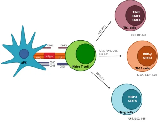
Upon Mtb infection, DCs phagocyte Mtb at the site of infection. Mature DCs migrate to the LN to present processed mycobacterium antigens to naïve T lymphocytes [4]. After TCR engagement and depending of cytokine milieu and co-stimulation provided by APCs, naïve CD4+ T cells are activated, proliferate and become committed to one of several lineages of Th cells, including Th1, Th17 and Treg cells among others (Figure 3) [4, 77, 78]. Effector Th cells migrate to the lungs in response to specific inflammatory mediators and develop an immune response that further promotes activation of macrophage microbicidal mechanisms [4].
Following Mtb challenge, the induction of T cell polarizing cytokines is dependent of PRRs that are expressed and activated in APCs [4]. In the absence of IFN- and after Mtb-induced TLR2-dependent DCs stimulation, IL-23 is higher expressed than IL-12 [79], whereas in the presence of IFN- Mtb-infected DCs secrete both IL-12 and IL-23 which potentiate both Th1 and Th17
12
polarization [79, 80]. In vitro and after Mtb infection both TLR2 and TLR9 activation induce the production of IL-12p40 by DCs [30, 80]. IL-12p40 subunit is shared by IL-12 and IL-23, key innate cytokines involved in Th1 and Th17 responses. Consequently the innate stimulus that leads to the production of these cytokines has an important impact in the type of Th response and has to be different and highly regulated [80]. Recently, we reported that certain Beijing strains of Mtb preferentially activate TLR2, whereas others also activate TLR4 resulting in a distinct cytokine pattern in vitro and in vivo [34]. Thus, it is possible that distinct Mtb strains by differentially stimulating DCs, lead to secretion of distinct polarizing cytokines which may impact the Th cell response generated. Indeed, the work developed in this thesis focus on the impact of TLR4 triggering by a TLR4-activating Mtb strain on the development of adaptive immunity.
Each T cell lineage is characterized by their cytokine production profile, function and different patterns of cell surface molecules (Figure 3) [33, 77, 78]. The signaling pathways triggered in CD4+ T cells by polarizing cytokines leads to the activation of signaling transducer and activator of transcription (STAT) proteins which are involved in induction and binding of the main transcription factors to genes that are under their influence [77, 81]. The hallmark cytokine of Th1 cells is IFN-, but these cells also produce IL-2, TNF and lymphotoxin (Figure 3) [77, 78, 82]. The main cytokine involved in Th1 polarization is IL-12 (Figure 3) [77, 78, 81]. IL-12 is a heterodimer formed by two subunits namely IL-12p35 and IL-12p40 that signal through the IL-12 receptor (IL-12R) composed by IL12R 1 and IL12R 2 chains [83]. Mtb infected-DCs secrete IL-12 that potentiates IFN- production by those cells [84]. T box expressed in T cells (T-bet) is the master transcription factor associated with IFN- production and Th1 differentiation (Figure 3) [77, 78, 81]. After TCR engagement, the binding of IFN- to IFN-R triggers the activation of STAT1 and T-bet [77, 78]. Following, T-bet further induces the production of IFN- and IL-12R, which potentiates the action of IL-12 and subsequently the Th1 expansion and maintenance (Figure 3) [77, 78, 85]. STAT4 is another protein that is involved in Th1 differentiation and intervenes in IL-12 signaling pathway (Figure 3) [77, 78, 85].
In Mtb infection, Th1 cells are fundamental for protective immunity [2, 4, 5, 56]. The main functions of IFN- secretion in Mtb infection are to enhance macrophage phagocytosis and increase the induction of NO and ROIs, which are mechanisms involved in pathogen clearance [5, 56]. Humans with defects in the production or signaling of IL-12 and IFN- cytokines are highly susceptible to TB [4, 86]. Furthermore, the importance of IL-12 and IFN- was demonstrated in mice deficient in those molecules that succumbed early in response to Mtb infection [56]. Studies
13
revealed that IL-12p40 deficient mice are more susceptible to Mtb infection than IL-12p35 knockout mice, suggesting a protective role for IL-12p40 [2]. Indeed, upon Mtb infection IL-12p40 homodimers are involved in DC migration and T cell activation [2, 84]. Additionally, the arrival and accumulation of IFN--producing T cells in the lungs is temporally associated with the stop of bacterial growth [4, 87, 88].However, a recent report using a model of adoptive transfer of Mtb specific effector CD4+ T cells, propose a mycobactericidal effector function of CD4+ T cells that is independent of both IFN- and TNF production [89]. These findings show that, despite the fact that CD4+T cells and IFN- are important for Mtb control, CD4+ T cells can mediate protection by other IFN--independent mechanisms during primary and secondary challenge [89, 90].
Upon Mtb infection, and in addition to Th1 cells, Th17 cells are also induced [80]. Th17 cells secrete IL-17A (IL-17), IL-17F and IL-22 as their signature cytokines (Figure 3) [80, 91]. The cytokines that drive Th17 differentiation are IL-6, IL-21 and transforming growth factor (TGF)- in low quantities (Figure 3) [80, 91]. Others cytokines such IL-1 and TNF act as cofactors during Th17 development [80, 91]. The main transcription factor involved in Th17 polarization is retinoic acid receptor related orphan receptor-t (ROR-t) [80, 91]. IL-6, IL-23 and TFG- intervene through activation of STAT3 pathway, which in turn induces ROR-t and IL-23R expression that allows Th17 differentiation and maintenance (Figure 3) [80, 91, 92]. IL-23 is a heterodimeric cytokine formed by IL-12p40 and IL-23p19 subunits [91]. IL-23 plays an important function not in initial differentiation but in the later maintenance of Th17 phenotype, since that IL-23R is not expressed in naïve CD4+ T cells [77, 80, 91, 92]. During intracellular bacterial infections as Mtb infection the function of IL-17 is not well understood [71, 80, 93]. Recent reports propose that IL-17 confers protective immunity against some intracellular pathogens like Salmonella enterica and Listeria monocytogenes [76, 80]. In low dose of Mtb infection, the lack of IL-17 and IL-23 does not have a significant impact in the capacity of mice to limit Mtb infection. Although following a high dose challenge, IL-17 is required to the resolution of Mtb infection [80]. Th17 cells have an important role in induction of tissue inflammation and in development of immunopathology [80, 91, 93]. IL-17 is a pro-inflammatory cytokine that induces the expression of several cytokines like IL-6, IL-8, IL-1 , TNF, granulocyte-colony stimulating factor (G-CSF) and granulocyte macrophage–colony stimulating factor (GM-CSF); chemokines, as monocyte chemoattractant protein-1 (MCP-1), C-X-C motif chemokines 1 (CXCL1), CXCL9, CXCL10 and CXCL11 and antimicrobial proteins as defensins [80, 91]. Due to the induction of these chemokines by IL-17, this cytokine has an impact in neutrophil recruitment and inflammation. Indeed, after BCG infection and in absence of IL-17
14
the number of neutrophils recruited upon Mtb infection decreases as compared with WT mice [80]. Thus, upon Mtb infection, the IL-23/Th17 axis might be preponderant for granuloma organization, because it promotes chemokine secretion that potentiates an early neutrophil recruitment. Furthermore, the accumulation of neutrophils can potentiate IL-12 production and stimulate Th1 differentiation [80]. However, if excessive IL-17 production occurs, continuous neutrophil recruitment leads to an exacerbate immune response and tissue damage [80, 94]. This exacerbated immune response can be inhibited by many regulators such as IFN-, IL-27 and IL-10 whose signaling pathways inhibit Th17 differentiation preventing immunopathology [91, 92]. Thus, Th17 cells may have contrasting functions during Mtb infection either in fighting infection or in promoting tissue damage [80].
In sum, a fine balance between Th1 and Th17 responses is needed to stop bacterial growth and restrict immunopathology [80]. In this sense, Treg cells downregulate the immune response generated to avoid exacerbated pathology by secreting anti-inflammatory cytokines such as IL-10 and TGF- (Figure 3). However, during Mtb infection Treg cells seem to play a detrimental role since they prevent pathogen clearance by suppressing protective CD4+ T cell responses [95].
Figure 3 – Subsets of effector Th cells. Depending on the cytokine milieu present at the time of antigenic
stimulation, naïve CD4+ T cells can differentiate into various subsets of Th cells (Th1, Th17, Treg and others). Specific transcription factors have been identified as master regulators of each subset. Differentiated Th cells are characterized by the secretion of a combination of effector cytokines. Adapted from [77, 78, 85, 91, 95].
15
One particular feature of TB is the slow induction of the adaptive immunity, which allows an uncontrolled growth of mycobacteria and the establishment of a chronic infection, which is detrimental for the outcome of infection [4]. The mechanisms responsible for the delayed T cell priming remain elusive, but some assumptions have been proposed [4]. It was demonstrated that dissemination of Mtb-infected DCs to LN is essential to priming T cell responses [4, 55, 96, 97]. One hypothesis is that Mtb suppresses APC function, thereby delaying the dissemination of antigens from the lungs to the LN. The slow bacterial growth of Mtb may also contribute for the delay in the initiation of adaptive immunity, since a long time is needed to reach an adequate antigen concentration to stimulate the immune system [4, 10, 98]. Effective levels of adaptive immune responses are only achieved after 18-20 days post-infection and consequently the bacterial growth and dissemination become controlled [4]. Between 2 to 6 weeks post-infection, the influx of lymphocytes and activated macrophages to the local of infection results in granuloma formation [4]. This structure consists in infected phagocytes delimited by activated giant cells and epitheloid macrophages and an external layer of lymphocytes and fibrosis. The granuloma main function is to encircle Mtb in immune cells and impede dissemination and transmission of infection into the rest of the uninfected lung [99, 100].
The dynamics and characteristics of the T cell response induced upon Mtb infection needs further investigation in order to understand which T cell subpopulations are important for protection and how do they differentiate. This knowledge will certainly contribute for the development of new and more effective vaccines against TB.
1.6 TB prevention and treatment
1.6.1 BCG vaccination
BCG is currently the only available vaccine against TB. BCG is a live strain of Mycobacterium bovis developed in 1921 by Calmette and Guérin through consequent passages of a virulent strain of M. bovis that suffered attenuation [101, 102]. This vaccine is able to interact with different components of the innate and adaptive immune systems [101, 102]. Currently, BCG is administrated intradermally soon after birth. However, the efficiency of this vaccine is variable, ranging from 0 to 80%, and it only prevents the dissemination of disease in childhood, not avoiding the development of pulmonary TB in adulthood [101, 102]. The observed variation in BCG efficiency can be due to several factors like: early exposure to environmental mycobacteria,
16
differences in BCG sub-strains or in subsequent Mtb strains infecting the individual, inadequate stimulation of immune system by BCG, age of administration and host genetic variation [101, 102]. In addition, the protection conferred by BCG is limited and has the duration of 10 to 20 years [101, 102]. However, a second immunization with BCG or other mycobacterial preparations may not be a good approach because repeated exposure of mice with previous latent Mtb infection, to mycobacterium antigens was shown to induce an exacerbated immune response leading to severe lung damage [94].
1.6.2 The immune response underlying BCG vaccination
Although BCG has been used for many years, being one of the most widely used vaccines in the world, our knowledge on the cellular and molecular mechanisms involved in the protection conferred by BCG is still limited [101].
Achievement of BCG mediated protection, requires antigen-specific effector T cells to be recruited quickly to the site of infection and to activate the infected phagocytes [4, 103]. Furthermore, these cells should be able to persist within the infection site [104, 105].
As discussed before, IFN--producing CD4+ T cells are essential for Mtb control and for the host survival [106, 107]. For this reason, IFN--producing T cells have been widely used as a correlate of protection upon Mtb infection [108, 109]. However, the level of IFN- production does not necessarily associate with vaccine mediated protection [108, 110], suggesting that other immune factors are also important for T cell-mediated protection. Indeed, it has been demonstrated that BCG vaccinated IFN--deficient mice, although limited, exhibit protection against a subsequent Mtb aerosol infection, in a mechanism dependent of CD4+ T cells [90, 110].
Th17 cells have also been associated with vaccine-induced protection by anticipating Th1 arrival to the infected lung [83]. As explained before, Th17 cells increase the expression levels of chemokines responsible for attracting T cells, such as CXCL9, 10, and 11, which in turn enhance the recruitment of IFN--producing T cells to the site of infection [103]. Nevertheless, repeated exposure of mice, with a stabilized Mtb-infection, to BCG vaccination leads to an uncontrolled IL-17 cellular response that induce tissue damage related with increased neutrophil recruitment to the lung [94].
Clearly, CD4+ effector T cells comprise several T cell subsets, going from early activated T cells only producing IL-2, single cytokine secretors such as IFN-- or IL-17-producing T cells, to CD4+ T cells that might have several effector functions by the simultaneous secretion of various immune
17
factors such as IFN-, TNF and IL-2 [4]. The presence of these multifunctional T cells has been associated with protection in mouse models of Leishmania major infection [111]. Given that IFN- by itself was not associated with vaccine-induced protection, it is expected that CD4+ T cells secreting multiple cytokines might play an important function in the control of Mtb infection [108]. However, the protective role of these cells in TB still unclear [112]. Mtb-infected mice primed with BCG and boosted with Modified Vaccinia Ankara virus expressing antigen 85A (MVA85A) showed a higher control of bacterial burden in the lungs that was associated with the presence of multifunctional T cells producing IFN-, TNF and IL-2 compared with BCG alone [108]. Also, studies of BCG vaccination with infants demonstrated the presence of T cells that secrete several combinations of IFN-, TNF and IL-2 [113]. However, another study reported that adults with active Mtb infection expressed higher multifunctional CD4+ T cells than individuals with latent disease [114]. This observation suggests that multifunctional CD4+ T cells might associate with active disease, instead of protective immunity. Moreover, it seems that the presence of CD4+ T cells secreting multiple cytokines correlates with bacterial load, as indicated by the reduction of multifunctional T cells in TB subjects after the end of TB therapy [114].
It has been reported that BCG immunization is less protective against Beijing Mtb strains when compared with H37Rv infection, a reference laboratory strain [19, 21]. In a study performed by Ordway et al. until day 30 post-aerosol infection, mice infected with Beijing strains were equally protected comparative to H37Rv-infected ones. However, at day 60 post-infection the protection conferred by BCG was lost in Beijing-infected mice. This observation suggests that BCG protection against the Beijing strains tested was only transient and fails in long term-protection. This raises the question that BCG immunization or other BCG-like vaccines could be unsuccessful in regions of the world with high prevalence of Beijing genotype strains [20]. In contrast, other study demonstrated no strain-specific differences in BCG-induced protection [115]. Also, in humans, some studies tried to correlate BCG inefficacy with Beijing endemic areas, while others failed in demonstrating such association [19]. In our laboratory, in a panel of Beijing Mtb strains, we found a Mtb strain that in addition to TLR2 also triggers TLR4, with an impact for the outcome of Mtb infection. Thus, in this thesis, we investigated whether BCG vaccination is equally effective in inducing protection against this Beijing TLR4-activating Mtb strain [34]. In addition, taking into account the particular feature of this Mtb strain we want to understand the role of TLR4 triggering on the development of CD4+ T cell responses evoked by BCG vaccination.
18
1.6.3 Novel vaccination approaches
The impairment of BCG vaccination in protecting the host against TB in addition to the emergence of drug resistant strains of Mtb highlights the urgency of alternative strategies for prevention and therapy. Therefore, it is of extreme importance understand the underlying mechanisms that mediate both natural and vaccine-induced immunity [116]. Several strategies have been implemented to develop a better vaccine against TB. However most of them are based on boosting the immune response to BCG, in order to improve their effectiveness. As explained before, BCG is not an ideal vaccine and the protection conferred is not life-long, although it can prevent the most severe childhood manifestations of TB [101, 102]. Thus, the achievement of a most effective vaccine against TB may require the development of a booster vaccine [101, 102]. For instance, BCG vaccination early in life followed by an adjuvant to increase the strength and the durability of an effective immune response [117].
The most advanced of these improved vaccines is MVA85A that enhanced the protective efficacy of BCG in animal models by delivering the immunodominant antigen 85A as a booster using a replication-deficient poxvirus [118]. Recently, the phase IIb of clinical trials was completed but no significant protection against Mtb was obtained after MVA85A administration in infants [119]. Adjuvants can be added to vaccines such as in Hepatitis A virus vaccine in order to enhance effective T cell responses. PAMPs, specifically those binding the TLRs, are the basis of many adjuvants [11]. Therefore, the new generation of vaccines often integrates agonists of TLRs to enhance T helper cell responses [120]. The glycolipid monophosphoryl lipid A (MPL ®), which activates TLR4, was the first TLR ligand used as adjuvant in an approved human vaccine (the Hepatitis B vaccine Fendrix ®) [120]. M72 consisting of Mtb72F polyprotein formulated with the adjuvant AS02A (a MPL® based adjuvant) has been tested as a TB candidate vaccine [117, 118]. M72 was administrated as a boost of BCG in the cynomolgus monkey model demonstrating superior protection than that afforded by using BCG alone [121]. Furthermore this vaccine was well tolerated by healthy adult subjects in a phase I clinical trial [118]. Another study showed that boosting of BCG with the recombinant fusion protein ID93, adjuvanted with the TLR4 agonist Glucopyranosyl Lipid Adjuvant (GLA), formulated in a stable oil-in-water emulsion (SE) (ID93+GLA-SE), protects mice and guinea pigs against Mtb compared to BCG alone [122]. ID93+GLA-SE is presently under Phase I clinical trials [123].
19 1.6.4 TB treatment
Since BCG is not effective in preventing TB, treatment with anti-TB drugs is mandatory. Generally the treatment of drug-sensitive Mtb stains can be performed with a combination of first line drugs which comprise isoniazid, rifampin, pyrazinamide and ethambutol for at least 6 months. However, if the therapy is not successful, for instance with MDR and XDR strains, a second line of anti-TB drugs such as kanamycin, amikacin, capreomycin, para-aminosalicyclic acid, cycloserine, prothionamide and thiacetazone has to be implemented for at least 2 years [124, 125].
Therefore it is clear that alternative strategies for prevention and therapy are urgently needed to tackle the TB burden in the world.
21
A
IMS
23
Previous data from our laboratory demonstrated that different Mtb strains trigger different TLRs, with impact in both in vitro and in vivo immune responses. Using in vitro and in in vivo models of infection we showed that 02-171 Mtb strain from the Beijing lineage is recognized not only by TLR2, but can also trigger TLR4. In contrast, the reference strain H37Rv mainly activates TLR2. Interestingly, we observed that TLR4 deficiency led to a higher susceptibility of mice upon intranasal infection with 02-171 Mtb strain, whereas the same did not occur in H37Rv-infected mice, thus suggesting a protective role for TLR4 triggering [34]. Given that TLR activation could influence the differentiation of T cells [52, 53], which are fundamental for protection against TB [2, 4, 5, 56], in the Chapter I of this thesis we focus on the consequences, at the adaptive immune level, of TLR4 activation during a primary mycobacterial infection by 02-171. Therefore the main goals of this chapter were to investigate:
i) the role of TLR4 activation on T cell priming, activation and recruitment;
ii) the consequence of TLR4 activation on the lung pathology and granuloma organization
upon infection by 02-171 Mtb strain.
Recent studies suggest that BCG is less effective to protect mice against infection with Beijing strains comparative to the strain H37Rv [19, 21]. Given that BCG is mainly recognized by TLR2 [126], we speculated that the type of acquired immunity induced by BCG immunization may be protective against TLR2-activating Mtb strains, but defective towards a TLR4-activating Mtb strain challenge. Thus, we hypothesized that the differential TLR recognition of Mtb strains could also account for the variability of BCG protection. In this line, Chapter II focuses on the role of TLR4 activation by 02-171 Mtb strain during a recall response after BCG vaccination. The main goals of this part of the work were to evaluate:
i) the effectiveness of BCG vaccination in protecting WT mice against Mtb infection with
02-171 Mtb strain;
ii) the impact of TLR4 triggering in the effectiveness of BCG vaccination against
TLR4-activating Mtb strain infection;
iii) how TLR4 activation during Mtb infection influences BCG-induced CD4+ T cell responses.
The knowledge of how natural and vaccine induced immunity generated in response to Mtb infection could have a huge impact on the development of new strategies of vaccination and therapy.
25
27 Animals
WT C57BL/6 mice were purchased from Charles River (Barcelona, Spain). TLR4 deficient (TLR4−/−) mice were breed at the ICVS animal facility. NOS2 deficient (NOS2−/−) mice were kindly provided by Dr. Rui Appelberg, Institute for Molecular and Cell Biology (IBMC), University of Porto, Porto, Portugal. All mice were kept at the ICVS animal facility under conventional conditions, with food and water given ad libitum. Both male and female mice between the ages of 8 and 12 weeks were used. All procedures involving animals were carried out in accordance with the European Union Directive 86/609/EEC, and previously approved by the Portuguese National authority Direcção Geral de Veterinária.
Bacterial strains and growth conditions
Mtb clinical isolate 02-171 was kindly provided by Dr. Gunilla Källenus Karolinska Institutet, Sweden and Mycobacterium bovis BCG Pasteur (hereafter simply referred as BCG) was originally from the Trudeau Institute Mycobacterial Collection. Bacteria were first grown at 37ºC in Middlebrook 7H9 Broth (BD Bioscience) for 7–10 days and then diluted into Proskauer Beck (PB) medium supplemented with 30% glycerol and 0.05% Tween 80. At mid-log phase bacterial stocks were collected and frozen in 1 mL aliquots at -80ºC.
Experimental aerosol infection and BCG vaccination
Mice were infected via the aerosol route by using an inhalation exposure system (Glas-Col) calibrated to deliver a dose of 100 to 200 colony forming units (CFUs) of Mtb 02-171. In some experiments a lower aerosol infection dose of nearly 70 CFUs was used. The mycobacterial inoculum was prepared from the frozen stock, firstly by passing it through a 26G needle 6 times to disrupt bacterial clumps and then diluted in water (Aqua B. Braun) to the desirable concentration. Then, 10 mL of this suspension were placed into the nebulizer and the mice were exposed to the aerosol cloud for 40 minutes. The infection dose was confirmed for every experiment by plating the entire lung 3 days after the aerosol infection, as described below.
In some experiments, mice were subcutaneously vaccinated in the chest with 106 CFUs of BCG
28
Preparation of single cell suspensions
At day 3 and selected time-points post-infection mice were killed by CO2 asphyxiation and the organs
were aseptically excised. Lungs were first perfused with cold phosphate-buffered saline (PBS) through the right ventricle of the heart until the lungs appeared white, minced and incubated for 30 minutes at 37ºC with collagenase IX (0.7mg/mL, from Sigma-Aldrich). Digested lungs, LN or livers were disrupted by passage through a 40-μm-pore-size nylon cell strainer (BD Biosciences). LN cells were resuspended in complete Dulbecco's Modified Eagle Medium (cDMEM, DMEM supplemented with 10% of heat-inactivated fetal bovine serum (FBS), 1% of HEPES, 1% L-glutamine and 1% sodium pyruvate (all from GIBCO) and used for bacterial burden determination. Liver cells were used for bacterial burden determination and lung suspensions were treated with a red blood cells lysis buffer (0.87% of NH4Cl solution and 5% of PBS in water) to remove red blood cells and
resuspended in cDMEM. Lung single cell suspensions were enumerated with a Countess Automatic Cell Counter (Life Technologies).
Bacterial Burden determination
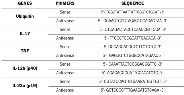
Bacterial burdens were determined by incubating single cell suspensions with 0.1% saponin (Sigma-Aldrich) for 10 minutes at room temperature (RT) to disrupt the cells that harbor Mtb. CFUs were determined by plating 10-fold serial dilutions of the disrupted cell suspensions in Middlebrook 7H11 (BD Biosciences) agar plates supplemented with 10% oleic acid/albumin/dextrose/catalase (OADC) and 0.5% glycerol. BBL™ MGIT™ PANTA™ antibiotic mixture (BD Bioscience) was used to prevent contaminations. Viable mycobacteria colonies were enumerated after 3 weeks of incubation at 37ºC. Values were transformed in Log10 before plotting and statistically analyzed.
Flow cytometry analysis of single cell suspensions
For cell surface antigen staining antibodies against CD8-FITC (clone 5H10-1 from BioLegend); CD69-PerCP-Cy5.5 (clone H1.2F3 from BD Biosciences); CD4-APC-Cy7 (clone GK1.5 from BioLegend); CD3-PerCP-Cy5.5 (clone 145-2C11 from BioLegend); killer-cell lectin-like receptor G1 (KLRG1)-APC (clone 2F1 from eBioscience); programmed cell death 1 (PD-1)-PECy7 (RMP1-30
from BioLegend ) CD11b-PE (clone M1/70 from BioLegend); lymphocyte antigen 6G (Ly6G)-APC (clone 1A8 from BioLegend) were used. Single cell suspensions were first incubated with Fc block (BioLegend) for 10 minutes on ice and then stained in fluorescence-activated cell sorting (FACS) buffer (PBS containing 2% of FBS plus 0.01% of azide) for surface markers at 4ºC for 30 minutes.
29
Cells were then washed with FACS buffer, resuspended in PBS containing 2% of formol and kept overnight (ON) at 4ºC in the dark before analyzed.
For intracellular cytokine staining, cell suspensions were restimulated with a mixture of phorbol myristate acetate (PMA) (50ng/mL) and ionomycin calcium salt (4μg/mL) for 4 hours, in the presence of brefeldin A (10μg/mL) (all from Sigma-Aldrich). For the detection of cytokine production, antibodies against TNF-FITC (clone MP6-XT22 from BioLegend); GM-CSF-PE (clone MP1-22E9 from eBioscience); INF--PE-Cy7 (clone XMG1.2 from eBioscience); IL-2-PB (clone JESG-5H4 from BioLegend) and IL-17-APC (clone TC11-18H10.1 from BioLegend) were used. Upon surface staining, cells were fixed and permeabilized with the Fixation/Permeabilization buffer (eBioscience) in the dark for 30 minutes at 4ºC. Cells were washed with the Permeabilization/Wash buffer (eBioscience) and incubated with a cocktail of fluorochrome-labeled antibodies at 4ºC for 30 minutes, in the dark. Finally, cells were resuspended in PBS containing 2% of formol and kept ON at 4ºC in the dark.
For Bromodeoxyuridine (BrdU) staining mice were subjected to an intraperitoneaI injection (IP) of 0.8 mg of BrdU (Sigma-Aldrich) 24 hours before harvest. BrdU staining was performed with APC BrdU Flow Kit (BD Biosciences) according to the manufacturer's instructions. Briefly, after surface staining, cells were fixed and permeabilized using the BD Cytofix/Cytoperm buffer for 20 minutes at 4ºC in the dark. Cells were then washed with BD Perm/Wash buffer and resuspended in BD Cytofix/Cytoperm PLUS and incubated 10 minutes at 4ºC in the dark. After washing, cells were fixed again with the BD Cytofix/Cytoperm Buffer for 5 minutes at 4ºC in the dark, washed and incubated with DNAse (300µg/mL in PBS), to expose incorporated BrdU, for 1 hour at 37ºC in the dark. Cells were then washed and stained with anti-BrdU antibody (APC) for 30 minutes at RT in the dark. Finally, cells were resuspended in PBS containing 2% of formol and kept ON at 4ºC in the dark.
Unstained cells were used to access autofluorescence and single stained controls were performed for fluorescence compensation. Samples were acquired on a LSRII flow cytometer (BD Bioscience). All data were analyzed using FlowJo software (TreeStar). The frequency of responding cells was determined and applied to the number of cells per sample to generate the total number of specific cell populations per organ.
![Figure 1 – Estimated worldwide TB incidence rates in 2012, as determined by the WHO. From [1]](https://thumb-eu.123doks.com/thumbv2/123dok_br/17683931.826775/26.892.135.768.112.478/figure-estimated-worldwide-tb-incidence-rates-determined.webp)
![Figure 2 – TLR signaling pathway. From [23].](https://thumb-eu.123doks.com/thumbv2/123dok_br/17683931.826775/28.892.133.768.481.972/figure-tlr-signaling-pathway.webp)