Correlation between GDF-15 gene polymorphism and the formation
of collateral circulation in acute ST-elevation myocardial infarction
XIAO-PING CHEN1*, XIAO-SEN SHANG1, YAN-BIN WANG1, ZHI-HUA FU1, YU GAO1, TAO FENG1
1Department of Cardiology, Taiyuan Central Hospital, Taiyuan, Shanxi, China
S
UMMARYStudy conducted at Taiyuan Central Hospital, Taiyuan,
Shanxi, China
Article received: 4/1/2017 Accepted for publication: 4/3/2017
*Correspondence:
Protomedicus, Department of Cardiology, Taiyuan Central Hospital
Address: 5E 3rd Alley
Taiyuan, Shanxi – China
Postal code: 030009 [email protected]
http://dx.doi.org/10.1590/1806-9282.63.12.1049
Objective: To explore the correlation between growth differentiation factor 15 (GDF-15) -3148C/G polymorphism and the formation of collateral circulation in acute ST-elevation myocardial infarction (STEMI) in Han population of Taiyuan area.
Method: The present study included 92 STEMI patients and 56 normal controls based on coronary angiography; STEMI group was divided into collateral group and non-collateral group according to Rentrop’s grading method. Polymerase chain reaction (PCR) and DNA sequencing methods were used to detect and analyze the GDF-15 -3148C/G polymorphism in all participants.
Results: There was significant difference in GDF-15 -3148C/G CC and GC distri-bution between STEMI group and control group (p=0.009); the allele frequencies between these two groups were also significant different (p=0.016); and the risk genotype for STEMI was CC with increased OR=2.660. For STEMI group, GDF-15 -3148C/G CC and GC distribution was also significantly different between patients with and without collateral (p=0.048), and CC genotype significantly promote the formation of collateral circulation. However, there were no significant dif-ferences in allele frequencies between these two subgroups of STEMI.
Conclusion: There was correlation between GDF-15-3148C/G polymorphism and the formation of collateral circulation in patients with acute STEMI.
Keywords: collateral circulation, polymorphism, genetic, growth differentiation factor 15, ST elevation myocardial infarction.
I
NTRODUCTIONWhen the coronary artery is stenotic or occluded, the formation of collateral circulation is an alternative source of blood supply to the myocardium, which may play an important role in reducing sudden cardiac death and infarct size. Therefore, the presence of coronary collater-als is crucial during acute myocardial infarction. How-ever, significant differences exist in the degree of collat-eral development among different patients.1 It is very necessary to find out why some patients can develop suf-ficient collateral circulation while others do not.
Growth differentiation factor 15 (GDF-15) is a mem-ber of the transforming growth factor β (TGF-β) super-family, which primarily regulates multiple cellular func-tions as well as the growth of multiple organs and the differentiation and renovation of tissues.2 The role of GDF-15 in suppressing the progress, invasiveness and metastasis of tumors has been verified by researchers.3
Besides, it is also related to cardiovascular diseases, in-cluding cardiac hypertrophy4 and heart failure.5 Re-cently, GDF-15 +157 A/T polymorphism was found to be associated with coronary collateral formation in acute non-ST segment elevation myocardial infarction.6 Since GDF-15 -3148C/G polymorphism is closely related to left ventricular remodeling,7 whether it also plays a role in the formation of collateral vessels in the cardiovascular system remains unclear. The present study aimed to investigate the correlation between GDF-15 -3148C/G polymorphism and the formation of collateral circula-tion in acute ST-elevacircula-tion myocardial infarccircula-tion (STEMI) in the Han population of the Taiyuan area.
M
ETHOD ParticipantsHospital, who were allocated into a collateral group and a non-collateral group according to Rentrop’s grading method. Fifty-six (56) patients with chest pain but with normal coronary angiography were enrolled in the control group. We excluded patients with the following condi-tions: Non-ST-elevation myocardial infarction (NSTEMI), acute and chronic inflammatory disease, neoplastic disease, valvular disease, cardiomyopathy, angina pectoris without fixed coronary artery stenosis, X syndrome, coronary artery expansion, severe kidney disease (blood creatinine > 2.5 mg/dL) and severe liver disease (ALT or AST two times higher than normal). Standardized forms were used to collect baseline data of all participants. All participants provided written informed consent.
Coronary angiography and collateral vessels evaluation
Selective coronary angiography was used according to the Judkins method. Any major coronary artery (left main coronary artery and right anterior descending coronary artery, cyclotron branch, the main diagonal branch or blunt edge branch) with pipe cavity diameter stenosis 50% and above was defined as significant coronary artery stenosis. Using Rentrop’s classification system to evaluate the collateral circulation. Level 0: no visible collateral vessel perfusion; Level 1: visible collateral blood vessels, but did not reach the contrast infusion; Level 2: visible collateral blood vessels, epicardial artery perfusion; Level 3: visible collateral blood vessels, epicardial artery was completely perfused. Rentrop’s grade zero level is non-collateral group whereas Rentrop’s grade 1 ~ 3 level for collateral group.
DNA extraction
DNA was extracted from 3 mL of fasting venous blood in anticoagulation tube using the whole blood genomic DNA extraction kit (Beijing Bao Lai technology co, LTD).
Polymerase chain reaction (PCR) and product identification
Primers were synthesized by Shanghai biological technol-ogy co., LTD. 3148C/G site: Forward 5 ´ - AGT-GAGTCCTTGTGTCTCTTAC - 3 ´; Reverse : 5 ´ - GCAG-GCTGGTGTAGAGTC - 3 ´. Amplification system: a total of 40 µL volume, 10×Buffer 4 μL, MgCl2 3 μL, dNTP (2.5 mM) 4 μL, forward (10 μM) 2 μL, reverse (10 μM) 2 μL, Taq enzyme (1 U) 2 μL, DNA 2 μL, ddH2O 21 μL. Poly-merase chain reaction (PCR) conditions: 94C° 5 min; 94°C 30 s, 55°C 30 s, 72°C 30 s, repeat 35 cycle; 72°C 7 min. Take 5 µL PCR amplification products with load sample buffer, add sample to 1.5% agarose gel electropho-resis; the gel imaging electrophoresis products were used for analyzing.
Enzyme digestion and product identification
The PCR products were digested for 12 hours (65°C). Reaction system: a total of 20 μL volume, PCR products 10 μL, 10×Buffer 2 μL, enzyme (BsrI enzyme) 0.5 μL. Take 15 µL enzyme-digested products with load sample buffer,
add sample to 2.5% agarose gel electrophoresis, the gel imaging electrophoresis products were used for analyzing.
DNA sequence alignment
DNAMAN and Chromas biological software were used to analyze sequencing results.
Statistical methods
SPSS 19.0 was used for statistical analysis. Measurement data were presented as mean ± standard deviation (Mean±SD) and compared by t test; use X2 test to compare genotype distribution in compliance with Hardy Wein-berg genetic balance law and inter-group gene frequen-cies and alleles. p-value < 0.05 was considered statisti-cally significant.
R
ESULTSThere was no any statistical significance between three groups in the baseline data (age, gender, smoking history, hyperlipidemia, hypertension, diabetes, family history) (all p>0.05) (Table 1).
As shown in Figure 1, both the size of GDF-15 gene PCR amplification product and CC genotype enzyme diges-tion product was 251 bp, while GC genotype enzyme di-gestion product contained three components, whose siz-es were 251 bp, 191 bp and 60 bp, rsiz-espectively. Both CC genotype and GC genotype were observed in all three groups.
400bp
300bp
200bp
150bp
100bp
50bp
M 1 2 3
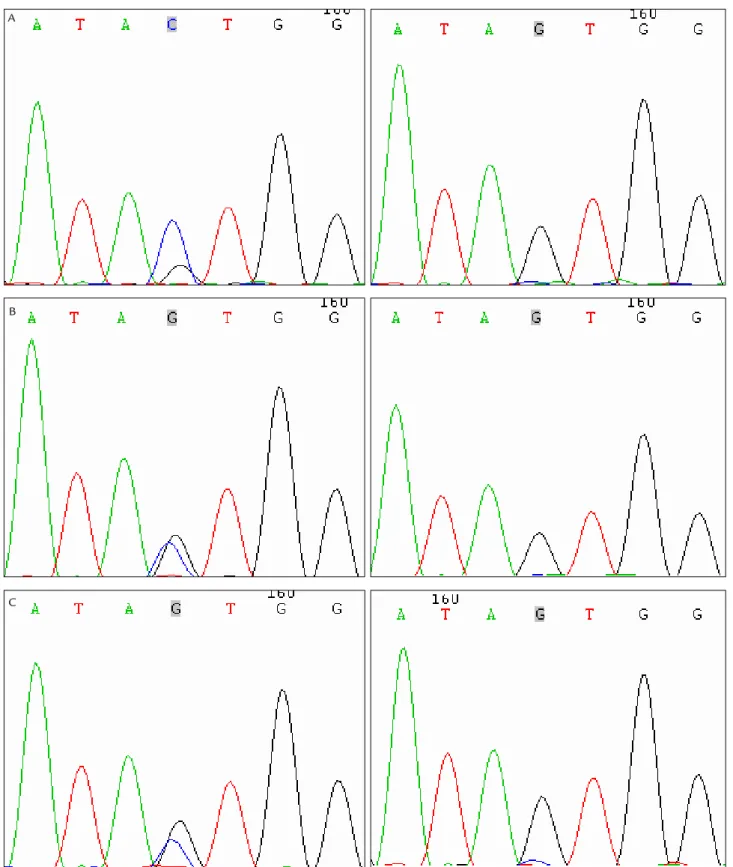
The results of compared sequences were the same as the enzyme digestion results. Both CC genotype and GC genotype were observed in the STEMI collateral circula-tion group (Figure 2A), STEMI non-collateral circulacircula-tion group (Figure 2B) and normal control group (Figure 2C).
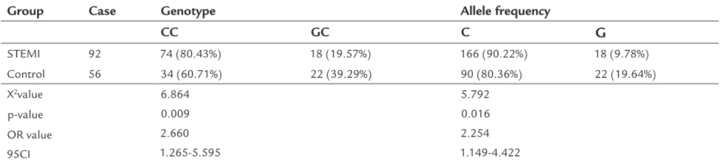
The comparison of genotype frequency and allele frequency of GDF-15 -3148C/G site between the STEMI and control groups were presented in Table 2. There was statistically significant difference in -3148C/G site geno-type distribution (X2=6.864, p=0.009) between the two groups. Allele frequency between the two groups also had statistical significance (p=0.016). The risk genotype for STEMI was CC with increased OR=2.660.
The comparison of genotype frequency and allele frequency of GDF-15 -3148C/G locus between the col-lateral circulation and non-colcol-lateral circulation groups was shown in Table 3. There was statistically significant difference in -3148C/G site genotype distribution (p=0.048) between the two groups. However, the allele frequency (p=0.062) between the two groups had no sta-tistical significance. GDF-15 -3148C/G site CC genotype might promote the formation of collateral circulation in patients with STEMI (OR=2.900).
D
ISCUSSIONWhen the original coronary artery cannot provide enough blood flow, the collateral circulation has the potential to become the main blood supply, which can reduce sudden cardiac death and infarct size.8 As we all known, many factors may be associated with collateral development, including severity of coronary stenosis, history of myocar-dial infarction, use of angiotensin-converting enzyme inhibitors.9 However, none of the above reasons seems to explain an interesting phenomenon: some patients can develop sufficient collateral circulation while others do not. Therefore, we need to find new influencing factors.
GDF-15 is a member of the transforming growth factor β (TGF-β) superfamily. The role of GDF-15 in car-diovascular disease has been explored by many research-ers in recent years. In 2002, Brown et al. reported that serum GDF-15 protein level was an independent risk factor for women’s atherosclerosis and other cardiovascu-lar events. It was the first report that connected GDF-15 and cardiovascular disease.10 Then, the correlations of GDF-15 with cardiac hypertrophy,4 heart failure5 and coronary heart disease (CHD)11 were found gradually. A recently published article reviewed the association of
TABLE 1 Baseline data comparison (means±SD or absolute numbers).
Survey index STEMI group (n=92) Control group
(n=56) p Collateral circulation group (n=68) Non-collateral circulation group (n=24)
Age (years) 59.15±1.58 59.73±1.63 60.73±1.71 0.453
Gender (n) Male Female 50 18 15 9 34 22 0.283
Smoking history (n) Yes No 36 32 16 8 35 21 0.388 Hypertension (n) Yes No 38 30 18 6 29 27 0.148 Hyperlipidemia (n) Yes No 39 29 13 11 25 31 0.361 Diabetes (n) Yes No 32 36 10 14 17 39 0.164
Family history of CHD (n) Yes No 28 40 7 17 17 39 0.363
FIGURE 2 A. -3148 site genotype in STEMI collateral circulation group. B. -3148 site genotype in STEMI non-collateral circulation group. C. -3148 site genotype in normal control group (a: GC genotype; b: CC genotype).
A
GDF-15 with the prognosis of acute coronary syndrome (ACS), finding that high plasma GDF-15 levels were as-sociated with an increased risk of mortality and recurrent myocardial infarction in patients with ACS.12 All of the studies above indicated the vital role of GDF-15 in car-diovascular disease. Interestingly, in 2010, Sun et al. found that GDF-15 levels increased with the extent of collat-eral formation;13 however, the underlying mechanism was not clarified. Recently, Jing et al. observed that there was correlation between GDF-15 + 157 A/T polymorphism and the formation of collateral circulation in patients with non-ST segment elevation myocardial infarction,6 firstly connecting GDF-15 gene polymorphism with the formation of coronary collateral circulation. Apart from the + 157 A/T site, the relation between -3148C/G (rs4808793) polymorphism and cardiovascular disease was also investigated. One study failed to prove an asso-ciation of - 3148C/G polymorphism with CAD or its se-verity in a Chinese population.14 Conversely, the other research suggested that GDF-15 -3148C/G polymorphism was closely related to left ventricular remodeling.7 There-fore, whether GDF-15 -3148C/G polymorphism also plays a role in the formation of coronary collateral vessels or not remains unclear and needs to be further clarified.
In the present study, we included 92 STEMI patients and 56 normal controls based on coronary angiography;
the STEMI group was divided into a collateral group and a non-collateral group. Two genotypes of -3148C/G sites, CC and GC, were found both in the STEMI group and the control group, meaning that GDF-15 -3148C/G poly-morphism existed in the Han population of the Taiyuan area. There was significantly difference in the distribution of these two genotypes between the STEMI group and the control group (p=0.009). Allele frequencies between these two groups were also significantly different (p=0.016). Moreover, CC genotype significantly increased the risk of STEMI occurrence (OR=2.660). At the same time, the possibility of the existence of collateral circulation in patients with STEMI carrying CC genotype could be in-creased by 2.9 times. However, there were no significant differences in allele frequencies between the two subgroups of STEMI; this may be due to the small sample size of this study. We need larger samples in the future researches.
Most importantly, we found that GDF-15 -3148C/G polymorphism (CC genotype) might have a correlation with STEMI occurrence and the formation of collateral circulation. Even though one study suggested that GDF-15 might predict more severe coronary stenosis, which had a higher probability to develop collaterals,13 the spe-cific mechanism of action is still unknown, further func-tional studies are thus needed to clarify it. Of course, our study has some limitations: first, this is a cross-sectional
TABLE 2 Comparison of genotype frequency and allele frequency of GDF-15 -3148C/G site between the STEMI and control groups (X2 analysis).
Group Case Genotype Allele frequency
CC GC C G
STEMI 92 74 (80.43%) 18 (19.57%) 166 (90.22%) 18 (9.78%)
Control 56 34 (60.71%) 22 (39.29%) 90 (80.36%) 22 (19.64%)
X2value 6.864
0.009 2.660 1.265-5.595
5.792 0.016 2.254 1.149-4.422 p-value
OR value 95CI
TABLE 3 Comparison of genotype frequency and allele frequency of GDF-15 -3148C/G site between the collateral circulation and non-collateral circulation groups (X2 analysis).
Group Case Genotype Allele frequency
CC GC C G
Collateral circulation 68 58 (85.29%) 10 (14.71%) 126 (92.65%) 10 (7.35%)
Non-collateral circulation 24 16 (66.67%) 8 (33.33%) 40 (83.33%) 8 (16.67%)
X2 value 3.911
0.048 2.900 0.983-8.556
3.487 0.062 2.520 0.931-6.819 p-value
study and does not provide a mechanism explaining the results; second, the study sample might be considered small, which limits the reliability of our results; third, our results may not be applicable to the Chinese population as a whole. More longitudinal studies and functional studies in different ethnicities are needed to further in-vestigate the pathophysiological effects of GDF-15.
C
ONFLICT OF INTERESTThe authors declare no conflict of interest.
R
EFERENCES1. Pohl T, Seiler C, Billinger M, Herren E, Wustmann K, Mehta H, et al. Frequency distribution of collateral flow and factors influencing collateral channel development. Functional collateral channel measurement in 450 patients with coronary artery disease. J Am Coll Cardiol. 2001; 38(7):1872-8. 2. Ago T, Sadoshima J. GDF15, a cardioprotective TGF-beta superfamily protein.
Circ Res. 2006; 98(3):294-7.
3. Mimeault M, Batra SK. Divergent molecular mechanisms underlying the pleiotropic functions of macrophage inhibitory cytokine-1 in cancer. J Cell Physiol. 2010; 224(3):626-35.
4. Dominguez-Rodriguez A, Abreu-Gonzalez P, Avanzas P. Relation of growth-differentiation factor 15 to left ventricular remodeling in ST-segment elevation myocardial infarction. Am J Cardiol. 2011; 108(7):955-8.
5. Anand IS, Kempf T, Rector TS, Tapken H, Allhoff T, Jantzen F, et al. Serial measurement of growth-differentiation factor-15 in heart failure: relation to disease severity and prognosis in the Valsartan Heart Failure Trial. Circulation. 2010; 122(14):1387-95.
6. Jing R, Liu Q, Xie Q, Qian Z. Correlation between GDF 15 gene polymorphism and the collateral circulation in acute non-ST segment elevated myocardial infarction. Int J Clin Exp Med. 2015; 8(8):14383-7.
7. Wang X, Yang X, Sun K, Chen J, Song X, Wang H, et al. The haplotype of the growth-differentiation factor 15 gene is associated with left ventricular hyper-trophy in human essential hypertension. Clin Sci (Lond). 2009; 118(2):137-45. 8. Traupe T, Gloekler S, Marchi SF, Werner GS, Seiler C. Assessment of the human coronary collateral circulation. Circulation. 2010; 122(12):1210-20. 9. Altin T, Kilickap M, Tutar E, Turhan S, Atmaca Y, Gulec S, et al. The relationship of chronic angiotensin converting enzyme inhibitor use and coronary collateral vessel development. Int Heart J. 2007; 48(4):435-42. 10. Brown DA, Bauskin AR, Fairlie WD, Smith MD, Liu T, Xu N, et al.
Antibody-based approach to high-volume genotyping for MIC-1 polymorphism. Biotechniques. 2002; 33(1):118-20.
11. Kempf T, Sinning JM, Quint A, Bickel C, Sinning C, Wild PS, et al. Growth-differentiation factor-15 for risk stratification in patients with stable and unstable coronary heart disease: results from the AtheroGene study. Circ Cardiovasc Genet. 2009; 2(3):286-92.
12. Zhang S, Dai D, Wang X, Zhu H, Jin H, Zhao R, et al. Growth differentiation factor-15 predicts the prognoses of patients with acute coronary syndrome: a meta-analysis. BMC Cardiovasc Disord. 2016; 16:82.
13. Sun T, Huang Y, Phillips MI, Luo X, Zhu J, Shi H, et al. Growth differentiation factor 15 and coronary collateral formation. Clin Cardiol. 2010; 33(1):E1-5. 14. Chen Z, Xie F, Ma G, Feng Y, Qian Q, Liu N. Study of the association between