2351-9789 © 2015 Published by Elsevier B.V. This is an open access article under the CC BY-NC-ND license (http://creativecommons.org/licenses/by-nc-nd/4.0/).
Peer-review under responsibility of AHFE Conference doi: 10.1016/j.promfg.2015.07.337
Procedia Manufacturing 3 ( 2015 ) 823 – 828
ScienceDirect
6th International Conference on Applied Human Factors and Ergonomics (AHFE 2015) and the
Affiliated Conferences, AHFE 2015
A real time biofeedback system using visual user interface for
physical rehabilitation
M.Barandas
a, H. Gamboa
b, J.M. Fonseca
aa
Centre of Technology and Systems, Uninova, 2829-516 Caparica, Portugal
b
Physics Department, FCT-UNL, 2829-516 Caparica, Portugal
Abstract
This study was undertaken to investigate the effectiveness of biofeedback training when compared to conventional physical rehabilitation. Real time biofeedback plays an important role for patients and therapists to assessthe success and performance of the training process. Visual user interface allows new forms of therapy simplifying the process and increasing patients’ motivation. An experimental investigation was conducted to study the effect of using a visual interface as an add-on therapy to standard exercises for Range of Motion (ROM) measurements in glenohumeral movements. Human movement was collected by a Kinect sensor and the ROM measurements were computed using spatial coordinates provided by the official Microsoft Kinect SDK. The design allows patients to train therapeutic exercises, while receiving different types of real time feedback indicating measures of performance evaluation through the course of the training. Moreover, both the environment and the training task are customizable to the patient needs. In order to evaluate the biofeedback effectiveness, subjects that participated in the study were required to do a therapeutic exercise twice: firstly following the instructions of the therapist and secondly adding the visual biofeedback to the therapist instructions. The results obtained suggest that the proposed visual interface was an effective tool to achieve a significant improvement in the performance of the exercise. The exercises correctness when performed with visual feedback was significantly higher than the same exercises performed without this visual stimulus. In trials where subjects received visual feedback, it was observed a greater effort to achieve the proposed objective and superior movement control. Given the potential benefits of biofeedback, the proposed interface can become a helpful tool to patients that can confirm the correctness of their exercises and therapiststhat can adapt the prescribed exercises considering patients’ evolution and performance.
© 2015 The Authors. Published by Elsevier B.V. Peer-review under responsibility of AHFE Conference.
Keywords:Biofeedback; Kinect; Range of Motion; Rehabilitation; Therapy effectiveness
© 2015 Published by Elsevier B.V. This is an open access article under the CC BY-NC-ND license (http://creativecommons.org/licenses/by-nc-nd/4.0/).
1. Introduction
In motor rehabilitation, the use of biofeedback has been shown to be a potent variable affecting motor skill learning and an effective way of providing motivating tasks, facilitating the rehabilitation process [1-3]. One of the most important features of practice is the information patients receive about their attempts to produce an action [3]. This information can be received through two general types of feedback to consciously and voluntarily correct movement or posture. One type is called intrinsic feedback and refers to the natural part of performing a skill (sensory perceptual information). The other type is called extrinsic or augmented feedback and refers to adding to or enhancing intrinsic feedback with an outside source [4]. The external source may be a therapist or a device such as a biofeedback system. The recent development of technology has dramatically increased the tools available to augment movement-related feedback, encouraging patients to practice active functional tasks and providing relevant information to their therapists. These technologies may be used to complement direct efforts by therapists and, occasionally, may even act as surrogates [5]. During the exercises, movement quantification (kinematic analyses),movement quality evaluation and instructions to improve patient performance can be provided in real time. In this regard, patients can monitor their own movements and be directly involved in the evaluation of their progress.
People with motor disabilities experience limitations in fine motor control, strength and range of motion. To overcome these limitations, repetitive exercises are usually applied within the rehabilitation setting. However, the number of exercises in a therapy session is typically insufficient and one of the reasons for not performing the exercises regularly out of the therapy sessions is lack of motivation [6]. Knowledge about performance of the training session and progress are reasons to keep patients motivated. Moreover, a growth in motivation may be translated into a greater effort during task practice [7]. Therefore, identifying effective methods of encouraging patients to correctly perform exercises can play a vital role for helping them retaining or enhancing their motor control.Within the rehabilitation setting, therapeutic interventions are often aimed at improving motor function of the upper extremity [4]. This study aims to develop a biofeedback system that is able tomotivate people with motor disabilities of the upper extremity by evaluating their performance during task practice based on Range of Motion (ROM) measurements and giving positive feedback when they achieve good results.
ROM measurements can be obtained using different sensing principles such as optical, magnetic or inertial sensors. Although these systems usually have high accuracy and sensitivity, they also present limitations related with complexity, space requirements and cumbersome cables carried by the patient. Therefore, these limitations make their adoption difficult in the field, especially on patient’s home environment. In the last years, Microsoft KinectTM gained the attention of several researches due to its low cost, portability and markerless anatomical measurements. Due to the potential of the KinectTMsensor, its applications have been quite diverse, such as human body movement detection [8-9], tracking and action recognition [10], clinic assessment [11] and rehabilitation training [12-14]. In the rehabilitation field, studies performed with the KinectTMsensor typically take advantage of the specific software developed by PrimeSense or Microsoft [13-15] for 3D kinematic data measuring.
In order to assist therapists in the rehabilitation process and to motivate patients to practice daily exercises independently, a biofeedback system based on Microsoft KinectTM was developed. Furthermore, this study was undertaken to investigate the effectiveness of biofeedback training when compared to conventional physical rehabilitation.
2. Material and methods
In this section, technical aspects of capture sessions and description of the developed application are presented. The data analysis is also shown.
2.1. Data collection
The angular field of view is 57º on the horizontal and 43º on the vertical. Themotion data was recorded at a rate of 30 Hz and the depth image resolution was640x480 pixels. The Kinect sensor was used to record joints' spatial coordinates provided by the official Microsoft Kinect SDK.
2.2. Developedapplication
In the tests carried out, the KinectTMsensor was positioned in the front of the subjects at anapproximate distance of 2 meters.In order to evaluate the biofeedback effectiveness, subjects that participated in the study were required to do a therapeutic exercise twice: firstly following the instructions of the therapist and secondly adding the visual biofeedback to the therapist instructions. Both exercises were recorded with the KinectTMsensor. The captured data include joints’ spatial coordinates of poses estimated by the Microsoft Kinect, acquisition time, ROM measurements and patient information.Shoulder abduction movement, due to its frequent use in rehabilitation exercises, was used as case study to evaluate the effectiveness of biofeedback training.Global Coordinate System (GCS)was used to compute ROM measurements. The GCS can be considered as the thoraxcoordinate system provided that there are no motions of the thorax during theacquisitions [17]. A physiotherapist was present in all acquisitions ensuring that the subject was not compensating the motion with the thorax.
The developed applicationconsists of a method for movement recognition, focused on visual feedback in real time. The main features of this system are the visualization of the angle executed in real time (value updated every second), intuitive animations that help subjects executing the required movement and training data storage for posterioranalysis of the patient evolution and re-adaptation to the training. The system design allows patients to train shoulder abduction using three different types of exercises: repetitions, holding on a defined angle and controlling the movement. For repetitions exercise, the therapist defines the number of repetitions and the target angle adequate to the patient needs. In order to help the subject, a count of the number of repetitions executed was added to the visual interface. In the second type of exercise - holding on a defined angle - the therapist defines the holding time and the target angle. Besides the previous features, the extra feedback for this exercise is a countdown that starts when the patient reachesthe target angle.Finally, the control movement exercise trains the patient to do the exercise by following the animation at a certain speed, i.e., the patient cannot reach abruptly the target angle.
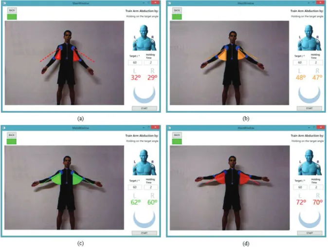
In Figure 1, a representation of a holding on a target angle exercise is presented. At the beginning of the exercise, Fig.1(a), the abduction angle is shown in the red zone. Here, the patient must continue increasing the angle. As soon as the ROMstarts to come closer to the target angle, the animation turns into orange, Fig1.(b), and changes to green when the ROM is equal, or very close, to the target angle, Fig.1(c). If the patient continues to increase the abduction angle, above the upper specification limit, the animation will turn red again, Fig.1(d), until the patient returns to the specified target zone. Therefore, thesoftware helps patients reaching the desired goals, thru interactive color information animation.
2.3. Data analysis
In order to study the KinectTMSkeleton Tracking (KST) for rehabilitation purposes, an application to receive the information from the KinectTMsensor was developed in C# using Visual Studio 2010. To access the data streams and the coordinates of anatomical landmarks, the official KinectTMSDK drivers were used. The ROM measurements were computed using the following equation (equation 1),
ߠ=ܽݎܿܿݏ ൬ԡܣԡԡܤԡ൰ܣ×ܤ (1)
whereߠis the angle performed in degrees, A is the vector composed by spatial coordinates of the joints of interest (shoulder-elbow) and B is vector fixed to the thorax and coincident with the Y-axis of the global coordinate system (reference vector)[17].
ܧݎݎݎ =หߠݐܽݎ݃݁ݐߠ െ ߠห ݐܽݎ݃݁ݐ
(2)
whereߠݐܽݎ݃݁ݐ is the target angle defined by the therapist and ߠis the measured angle in real time.
For the repetitions exercise, patient arm should perform a movement from the position attached to the body to the position of the target angle. Therefore, the error was calculated for each repetition at the maximum angle performed. On the other hand, for holding on a target angle, error was calculated for each second, analysing deviations along the holding time. Lastly, for the control movement exercise, instead of calculating the error in the final position (corresponding to the target angle), the error was calculated in several intervals before reaching the target angle. Therefore, it is possible to analyse patient performance during all movement.
3. Results
On this study ten healthy subjects,four females and six males, participated. During the acquisitions, subjects wore tightclothing to ensure data integrity. The lower body clothes were not relevant, but clotheswith shiny surfaces, like belts, were avoided.
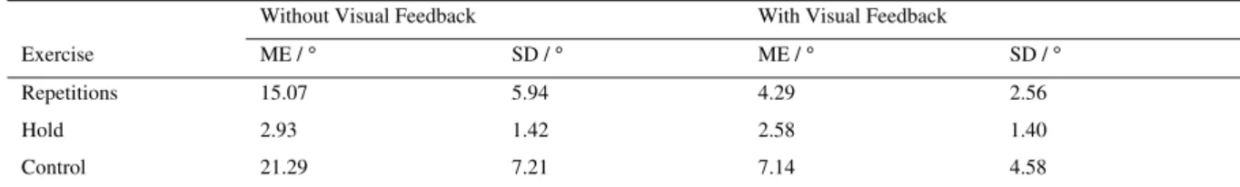
All subjects did each abduction exercise twice, firstly following the instructions of the therapist and secondly adding visual biofeedback to the therapist instructions. Although the number of repetitions and the holding time are customizable in the interface, it was chosen always 10 repetitions and 10 seconds for all subjects that participated in this study. In order to compare ROM measurements obtained with feedback with those obtained without feedback, Mean Error (ME) and Standard Deviation (SD) were computed. Results are presented in Table 1.
Table 1.Errors comparison between exercises performed without visual feedback with those performed with visual feedback.
Without Visual Feedback With Visual Feedback
Exercise ME / ° SD / ° ME / ° SD /°
Repetitions 15.07 5.94 4.29 2.56
Hold 2.93 1.42 2.58 1.40
Control 21.29 7.21 7.14 4.58
As it can be seen in Table 1, the exercises realized without feedback present a ME that is always larger than the ones realized with visual feedback. In fact, the control movement is the one that presents a larger variation between the two repetitions. In this exercise, the high errors are essentially due to the fact that the exercise evaluation is continuously done during all range of motion. Therefore,it is the exercise that requires more attention and control during the execution.
Regarding the repetition exercise, the error obtained without using feedback was also significantly higher compared with the same exercise usingvisual feedback. In this exercise it was observed that the last repetitions were the ones with higher error, probability due to the fact that subjects start to get fatigued at end of the exercise. Therefore, the biofeedback system acts as an incentive for patients to support the physical effort and perform correctly the prescribed exercises up to the end.
Although there are differences between MEs of the hold exercise, the ME obtained without using feedback is very similar to the one obtained using visual feedback. Therefore, in this exercise the visual feedback did not make a significant difference. One of the reasons may be related to the chosen holding time.Since subjects that participated in this study did not have any shoulder pain and the holding time was not enough to fatigue themselves it is natural that they performed correctly even without any feedback. Thus, we can conclude that in this exercise the biofeedback system is helpful, at least,for alerting and correcting the patients when they start to get fatigued, preventingthe decrease of patients’ performance.
4. Conclusions
Patients reported that visual feedback promoted a better execution of the exercise and the guidance provided by the system increased their motivation. Although the positive results, the system should be improved in order to add more feedback thatcanincreasesubjectsmotivation during the entire training programme. Further work is necessary to prove these preliminary results, and to extend the same exercises to other body parts, reaching a larger number of cases.
References
[1] Ma, M., McNeill, M., Charles, D., McDonough, S., Crosbie, J., Oliver, L., & McGoldrick, C. (2007). Adaptive virtual reality games for rehabilitation of motor disorders. In Universal Access in Human-Computer Interaction. Ambient Interaction (pp. 681-690). Springer Berlin Heidelberg.
[2] Piron, L., Tonin, P., Piccione, F., Iaia, V., Trivello, E., & Dam, M. (2005). Virtual environment training therapy for arm motor rehabilitation. Presence: Teleoperators and Virtual Environments, 14(6), 732-740.
[3] Schmidt RA, Lee TD. Motor control and learning: a behavioral emphasis. 3rd edn. Champaign, IL: Human Kinetics; 1999. [4] Dijk, H., Jannink, M. J., & Hermens, H. J. (2005). Effect of augmented feedback on motor function of the affected upper extremity in
rehabilitation patients: a systematic review of randomized controlled trials. Journal of Rehabilitation Medicine, 37(4), 202-211. [5] Liebermann, D. G., Buchman, A. S., & Franks, I. M. (2006). Enhancement of motor rehabilitation through the use of information
technologies. Clinical biomechanics, 21(1), 8-20.
[6] Chang, Y. J., Chen, S. F., & Huang, J. D. (2011). A Kinect-based system for physical rehabilitation: A pilot study for young adults with motor disabilities.Research in developmental disabilities, 32(6), 2566-2570.
[7] Lünenburger, L., Colombo, G., & Riener, R. (2007). Biofeedback for robotic gait rehabilitation. Journal of NeuroEngineering and Rehabilitation, 4(1), 1.
[8] Xia, L., Chen, C. C., & Aggarwal, J. K. (2011, June). Human detection using depth information by kinect. In Computer Vision and Pattern Recognition Workshops (CVPRW), 2011 IEEE Computer Society Conference on (pp. 15-22). IEEE.
[9] Galna, B., Barry, G., Jackson, D., Mhiripiri, D., Olivier, P., & Rochester, L. (2014). Accuracy of the Microsoft Kinect sensor for measuring movement in people with Parkinson's disease. Gait & posture, 39(4), 1062-1068.
[10] Oikonomidis, I., Kyriazis, N., & Argyros, A. A. (2011, August). Efficient model-based 3D tracking of hand articulations using Kinect. In BMVC (Vol. 1, No. 2, p. 3).
[11] Ning, X., & Guo, G. (2013). Assessing Spinal Loading Using the Kinect Depth Sensor: A Feasibility Study. Sensors Journal, IEEE,13(4), 1139-1140.
[12] Metsis, V., Jangyodsuk, P., Athitsos, V., Iversen, M., and Makedon, F. (2013). Computer aided rehabilitation for patients with rheumatoid arthritis. In Computing, Networking and Communications (ICNC), 2013 International Conference on (pp. 97–102). IEEE.
[13] Fernandez-Baena, A., Susin, A., and Lligadas, X. (2012). Biomechanical validation of upper-body and lower body joint movements of kinect motion capture data for rehabilitation treatments. In Intelligent Networking and Collaborative Systems (INCoS), 2012 4th International Conference on (pp. 656–661). IEEE.
[14] Kitsunezaki, N., Adachi, E., Masuda, T., and Mizusawa, J. I. (2013). Kinect applications for the physical rehabilitation. In Medical Measurements and Applications Proceedings (MeMeA), 2013 IEEE International Symposium on (pp. 294–299). IEEE.
[15] Obdrzalek, S., Kurillo, G., Ofli, F., Bajcsy, R., Seto, E., Jimison, H., & Pavel, M. (2012). Accuracy and robustness of Kinect pose estimation in the context of coaching of elderly population. In Engineering in Medicine and Biology Society (EMBC), 2012 Annual International Conference of the IEEE (pp. 1188-1193). IEEE.
[16] Khoshelham, K. and Elberink, S. O. (2012). Accuracy and resolution of kinect depth data for indoor mapping applications. Sensors, 12(2), 1437–1454.