Escola de Ciências
António Miguel Araújo Rego
Characterization of the role of
sphingolipids in the modulation of
acetic acid-induced apoptosis
Tese de Mestrado em Genética Molecular
Trabalho efectuado sob a orientação da
Professora Doutora Manuela Côrte-Real
e co-orientação do
Professor Doutor Vítor Costa
Escola de Ciências
António Miguel Araújo Rego
Characterization of the role of
sphingolipids in the modulation of
acetic acid-induced apoptosis
Tese de Mestrado em Genética Molecular
Trabalho efectuado sob a orientação da
Professora Doutora Manuela Côrte-Real
e co-orientação do
Nome: António Miguel Araújo Rego
Endereço electrónico: [email protected] Telefone: +351 252 916 567 / +351 918 837 301 Nº do Bilhete de Identidade: 12730015
Título da Tese de Mestrado:
Characterization of the role of sphingolipids in the modulation of acetic acid-induced apoptosis
Orientadores:
Professora Doutora Manuela Côrte-Real Professor Doutor Vítor Costa
Instituições de Acolhimento:
Centro de Biologia Molecular Ambiental (CBMA) Instituto de Biologia Molecular e Celular (IBMC)
Ano de Conclusão: 2011
Designação do Mestrado:
Mestrado em Genética Molecular
1. É AUTORIZADA A REPRODUÇÃO INTEGRAL DESTA TESE, APENAS PARA EFEITOS DE INVESTIGAÇÃO, MEDIANTE DECLARAÇÃO ESCRITA DO INTERESSADO, QUE A TAL SE COMPROMETE.
Universidade do Minho, 31 de Outubro de 2011 _____________________________________________ António Miguel Araújo Rego
Agradecimentos
É com a escrita dos agradecimentos, que consciencializo que o presente trabalho não é apenas meu mas também de uma série de pessoas com quem tenho convivido e que, direta ou indiretamente, são responsáveis por este resultado final. Assim sendo, gostaria de aqui registar o meu público agradecimento:
Aos meus orientadores, Professora Manuela Côrte-Real e Professor Vítor Costa pela excecional orientação, dedicação, disponibilidade e partilha do rigor científico ao longo da minha passagem pelos seus laboratórios. Obrigado a ambos pela oportunidade e confiança.
À Susana, por toda a disponibilidade, por todas as sugestões diárias na realização do trabalho experimental e escrita da tese. Obrigado por tudo.
À Fundação Portuguesa para a Ciência e Tecnologia pela bolsa de investigação que me foi atribuída, no âmbito do projeto financiado PTDC/BIA-BCM/69448/2006.
A todos os meus colegas dos laboratórios de Microbiologia Celular e Aplicada do IBMC e de Microbiologia I do CBMA, por me terem proporcionado um excelente ambiente de trabalho, pelo companheirismo e bons momentos passados diariamente. Um obrigado especial para a Margarida, Rita, Daniel, Catarina Pacheco, Sílvia, Rodrigo, Maria João, Catarina Santos, Tiago, Vanda, Liliana, D. Amélia e D. Helena do MCA e para a Andreia Pacheco, Dulce, Helena, Dário, Sara, Flávio, Andreia Afonso, Marlene, Rui e Gabriela do Micro I.
A todos os amigos que me acompanharam não apenas nesta etapa, mas também ao longos deste últimos anos. Obrigado Pedro, Juliana, Catherine, Bruno Freitas, Carla, André Charrua, Bruno Pacheco, Bruno Panta e Joana pelo companheirismo, incentivo e amizade constantes.
À Rita simplesmente porque sem ti nada faria sentido. Obrigado por me ajudares a sorrir, por confortar nos momentos de desânimo e por estares sempre comigo.
A toda a minha família por serem quem são! Em especial, muito obrigado Mãe, Pai, Filipe, Carina e Eduardo pelo amor, alegria, confiança e apoio.
Characterization of the role of sphingolipids in the modulation of
acetic acid-induced apoptosis
Abstract
The yeast Saccharomyces cerevisiae can undergo programmed cell death in response to different stimuli. Exposure of yeast cells to acetic acid has been shown to trigger a mitochondrial pathway displaying, as in mammalian cells, typical apoptotic markers such as externalization of phosphatidylserine, DNA fragmentation, chromatin condensation, mitochondrial dysfunction with cytochrome c release and production of reactive oxygen species (ROS).
Sphingolipids are lipid second messengers generated in response to different physiological signals and stress stimuli. They affect multiple aspects of cellular function, including apoptosis. Changes in sphingolipid metabolism have been linked to apoptosis and oxidative stress in both yeast and mammalian cells. The increase of ceramide and sphingosine levels leads to cell growth arrest and apoptosis whereas the increase of sphingosine-1-phosphate levels promotes proliferation and inhibits apoptosis. Moreover, ceramides have been detected in mitochondria and accumulate upon stress treatments, increasing the permeability of the mitochondria to cytochrome c and leading to the generation of ROS.
Our working hypothesis was that acetic acid may elicit ceramide production and, therefore, may trigger apoptosis by a signal transduction pathway modulated by ceramide. For that reason, we aimed to characterize the relative contribution of biosynthesis versus catabolism of ceramides to the apoptotic cell death induced by acetic acid in yeast. For our studies, yeast cells lacking Lag1p, Lac1p (unable to generate ceramide by de novo synthesis), Ydc1p and Ypc1p (unable to breakdown ceramide) and Isc1p (unable to generate ceramide by degradation of inositolphosphosphingolipids), were generated by homologous recombination.
Our results showed that lag1∆ and isc1∆ mutant cells exhibited a higher resistance to acetic acid that was correlated with lower levels of ROS production and reduced mitochondrial alterations. In comparison with the wild-type strain, lag1∆ and isc1∆ mutant cells display, under acetic acid stress, lower levels of mitochondrial fragmentation and degradation, and reduced alterations of the mitochondrial membrane potential. Associated with these events, there was also less translocation of cytochrome c to the cytosol in response to acetic acid than in the wild-type strain.
In conclusion, our results suggest that ceramide production contributes to cell death induced by acetic acid, especially through the hydrolysis of complex lipids catalyzed by Isc1p and through de novo synthesis catalyzed by Lag1p.
Caracterização do papel dos esfingolípidos na modulação da
apoptose induzida por ácido acético
Resumo
A levedura Saccharomyces cerevisiae pode sofrer morte celular programada em resposta a diferentes estímulos. O tratamento de células de levedura com ácido acético tem sido descrito como capaz de ativar a via mitocondrial apresentando, exposição de marcadores celulares típicos de apoptose de mamíferos tais como externalização de fosfatidilserina, fragmentação de DNA, condensação da cromatina, disfunção mitocondrial, incluindo a libertação de citocromo c, e produção de espécies reativas de oxigénio (ROS).
Os esfingolípidos são lipídos mensageiros gerados em resposta a diferentes sinais fisiológicos e estímulos de stress. Alterações no metabolismo de esfingolípidos têm sido associadas a apoptose e stress oxidativo tanto em leveduras como em células de mamíferos. O aumento dos níveis de ceramidas e esfingosina promove a paragem do crescimento celular e a apoptose, enquanto o aumento dos níveis de esfingosina-1-fosfato promove a proliferação e inibe a apoptose. Adicionalmente, tem-se verificado a deteção e acumulação de ceramidas nas mitocôndrias após tratamentos de stress, aumentando a permeabilidade da mitocôndria ao citocromo c e levando à produção de ROS.
A hipótese de trabalho deste projeto consistiu na suposição de que o ácido acético pode levar à produção de ceramidas e portanto, desencadear a apoptose por via de transdução de sinal modulado por ceramida. Por essa razão, foi nosso objetivo caracterizar a contribuição relativa da biossíntese e do catabolismo de ceramidas para a morte celular por apoptose induzida por ácido acético na levedura. Mutantes de levedura deficientes nas enzimas Lag1p e Lac1p (incapazes de gerar ceramidas pela síntese de
novo), Ydc1p e Ypc1p (incapazes de catabolizar ceramidas) e Isc1p (incapaz de gerar
ceramidas pela degradação da inositolfosfoesfingolípidos), foram gerados por recombinação homóloga.
Os resultados mostraram que os mutantes lag1Δ e isc1Δ exibem uma maior resistência ao ácido acético que se correlaciona com níveis baixos de produção de ROS e alterações mitocondriais menos intensas. Em comparação com a estirpe selvagem, lag1Δ e isc1Δ exibem, sob stress de ácido acético, menores níveis de fragmentação e degradação mitocondrial e reduzidas alterações do potencial de membrana mitocondrial. Associados a estes eventos, observou-se igualmente uma menor translocação de citocromo c para o citosol do que na estirpe selvagem.
Em conclusão, os resultados sugerem que a produção de ceramidas contribui para a morte celular induzida por ácido acético, especialmente através da hidrólise de lípidos complexos catalisada por Isc1p e através de síntese de novo catalisada por Lag1p.
Index
Agradecimentos ... iii Abstract ... iv Resumo ... v Index ... vi Abbreviations ... viii 1. Introduction ... 1 1.1. Cell death ... 3 1.2. Apoptosis ... 3 1.2.1. Caspases ... 41.2.2. The Bcl-2 family members ... 5
1.2.3. Apoptotic pathway ... 6
1.2.3.1. Extrinsic pathway ... 6
1.2.3.2. Intrinsic pathway ... 8
1.3. Yeast apoptosis ... 10
1.4. Acetic acid as an inducer of apoptosis ... 11
1.5. Sphingolipids ... 12
1.5.1. Yeast sphingolipid metabolism ... 13
1.5.2. De novo synthesis ... 14
1.5.3. Sphingolipid turnover ... 16
1.6. Sphingolipids and cell fate ... 17
1.6.1. Ceramides ... 17
1.6.2. Ceramides and its metabolites: Sphingosine and sphingosine-1-phosphate .. 19
1.7. Sphingolipids and yeast apoptosis ... 20
2. Objectives ... 21
3. Materials and Methods ... 24
3.1. Yeast strains ... 26
3.2. Plasmids ... 28
3.3. Genomic DNA isolation ... 29
3.4. Yeast electroporation ... 30
3.6. Analysis of oxidative stress and apoptotic markers ... 31
3.6.1. Assessment of plasma membrane integrity/PI staining ... 31
3.6.2. ROS ... 31
3.6.3. Protein carbonylation ... 31
3.6.1.1. Preparation of protein extracts ... 32
3.6.1.2. Derivatization ... 32
3.6.1.3. SDS gel electrophoresis/Western blot ... 32
3.6.1.4. Silver staining ... 33
3.6.4. Mitochondrial fragmentation and degradation ... 33
3.6.5. Mitochondrial membrane potential ... 34
3.6.6. Cytochrome c detection ... 34
3.6.6.1. Subcellular fractionation/preparation of yeast mitochondria ... 34
3.6.6.2. Mitochondrial integrity ... 34
3.6.6.3. SDS gel electrophoresis/Western blot ... 35
3.7. Flow cytometric assays ... 35
3.8. Reproducibility and statistic analysis of the results ... 35
4. Results ... 36
4.1. Acetic acid stress response ... 38
4.2. Oxidative markers ... 41 4.2.1. Intracellular ROS ... 41 4.2.2. Protein oxidation ... 42 4.3. Mitochondrial dynamics ... 43 4.3.1. Mitochondrial fragmentation ... 44 4.3.2. Mitochondrial degradation ... 45
4.3.3. Mitochondrial membrane potential ... 46
4.3.4. Cytochrome c release ... 47
5. Discussion and Future perspectives ... 48
Abbreviations
AIF - Apoptosis-inducing Factor ANT - Adenine Nucleotide Translocator Apaf-1 - Apoptotic Protease Activating Factor-1 ATP - Adenosine Triphosphate
BSA - Bovine Serum Albumin c.f.u. - Colony forming units
CAPK - Ceramide-activated Protein Kinase CAPPs - Ceramide-activated Protein
Phosphatase
CARD - Caspase Recruitment Domain c-FLIP - Cellular-FLICE (FADD-like
IL-1β-converting enzyme)-inhibitory Protein
CK - Creatine Kinase Cyp D - Cyclophilin D
DAPI - 4,6-Diamino-2-phenyl-indole
dihydrochlorid
DD - Death Domain
DED - Death Effector Domain DHE - Dihydroethidium DHS - Dihydrosphingosine
DiOC6 -3,3′-Dihexyloxacarbocyanine iodide
DISC - Death Inducing Signaling Complex DNA - Deoxyribonucleic Acid
DNP - Dinitrophenyl
DNPH - 2,4-Dinitrophenylhydrazine DR - Death Receptors
DTNB - 5,5′-Dithiobis (2-nitrobenzoic acid) DTT - Dithiothreitol
EDTA - Ethylenediaminetetraacetic acid Endo G - Endonuclease G
ER - Endoplasmatic Reticulum FADD - Fas-Associated Death Domain GFP - Green Fluorescent Protein H2O2– Hydrogen Peroxide
HtrA2/Omi - High Temperature Requirement
Protein A2
IAPs - Inhibitors of Apoptosis Proteins IPC - Inositolphosphorylceramide
KSR - Kinase Suppressor of Ras LCB – Long Chain Base
M(IP)2C - Mannosyldiinositolphosphorylceramide
MAC - Mitochondrial Apoptosis-induced Channel MAPK - Mitogen-activated Protein Kinases MIPC – Mannosylinositolphosphorylceramide MOMP - Mitochondrial Outer Membrane
Permeabilization
NADH - Nicotinamide Adenine Dinucleotide NADPH - Nicotinamide Adenine Dinucleotide
Phosphate
NO - Nitric Oxide
PARP-1 – polyADP-ribose Polymerase PBR - Peripheral Benzodiazepine Receptor PCD – Programmed Cell Death
PCR - Polymerase Chain Reaction PHS - Phytosphingosine
PI - Propidium Iodide PKC - Protein Kinase C
PTP - Permeability Transient Pore Rb - Retinoblastoma Product Gene ROS - Reactive Oxygen Species SAPK - Stress-activated Protein Kinase
SC Gal - Synthetic Complete Galactose medium SDK1 - Sphingosine-dependent Protein Kinase SDS - Sodium Dodecyl Sulfate
SK1 - Sphingosine Kinase
Smac/Diablo - Second Mitochondria-derived
Activator of Caspases/Direct Inhibitor of Apoptosis Protein (IAP)-Binding Protein With Low Pi
TNF-R - Tumor-Necrosis Factor Receptor TRADD - TNF-R-Associated Death Domain TRAIL-R - TNF-related Apoptosis-Inducing
Ligand Receptor
TUNEL - Terminal dUTP Nick-end Labeling VDAC - Voltage-Dependent Anion Channel
1.1. Cell death
In the last decades, the understanding of cell death has attracted a lot of attention, mostly because of its crucial role in tissue homeostasis and development of multicellular organisms (Baehrecke, 2002). Cell death may occur through different processes, some of which may have a physiological role. Classification of the types of cell death and associated terminology has evolved since the 19th century. Initially, isolated studies recognized that cell death occurs during metamorphosis and embryogenesis, but studies on dying cells led Lockshin to introduce the concept of programmed cell death (PCD) in 1965. PCD was categorized as a type of cell death that was not accidental (Necrosis), but a genetically controlled sequence of steps that lead to morphological and biochemical changes (Lockshin and Zakeri, 2001). Later, in 1972, Kerr and coworkers coined the term apoptosis to define a new pattern of cell death genetically controlled, with specific morphological features (Kerr et al., 1972). Apoptosis was later considered synonym of PCD and cell death classified into apoptosis and necrosis. However, since it was later found that necrosis may also be highly regulated (Ellis and Horvitz, 1986) and that exist other forms of cell death, including autophagic cell death, this classification was abandoned (Debnath et al., 2005).
Recently, the Nomenclature Committee on Cell Death proposed a unified criterion for the definition of cell death, according to different morphological characteristics. Cell death is now classified into three major pathways: apoptotic, necrotic and autophagic (Kroemer et al., 2009).
1.2. Apoptosis
The term "apoptosis" derived from an ancient Greek word meaning "falling petals of flowers" or "fall of leaves in autumn". It was the term chosen by John Kerr and his coworkers Andrew Wyllie and Alastair Currie in 1972 to define a new type of death (Kerr et al., 1972). Apoptosis is a type of cell death with morphological characteristics distinct from those found in necrosis and autophagy (Figure 1).
In necrosis, the cell content is released in an uncontrolled manner, resulting in damage to neighboring cells and a strong inflammatory response in the corresponding tissue (Proskuryakov et al., 2003). In autophagy, the cells recycle their own damaged intracellular components via the lysosome when the nutrients are scarce (Codogno and Meijer, 2005). In apoptosis, cells undergo a series of morphological changes such as exposure of phosphatidylserine from the inner leaflet to the external leaflet of the plasma membrane, chromatin condensation, internucleosomal DNA fragmentation, cell
volume decrease and finally formation of apoptotic bodies which are subsequently removed by phagocytes without causing an inflammatory response (Saraste and Pulkki, 2000; Lawen, 2003).
Figure 1. Molecular and morphological events associated with the different types of cell
death: apoptosis, autophagy and necrosis (Hotchkiss et al., 2009)
1.2.1. Caspases
Most of the alterations observed during apoptosis are caused by proteases called caspases (Cysteine-dependent aspartate-specific proteases). Conserved through evolution, caspases can be found in humans, insects, nematodes and hydra. Caspases contain a cysteine residue in the active site that is critical for their proteolytic activity and exhibit a high affinity for aspartate (Asp), cleaving their substrates after these residues (Cohen, 1997). Close to one hundred caspase substrates have already been identified, ranging from complex macromolecular complexes (e.g. actin network) to single enzymes [e.g. polyADP-ribose polymerase (PARP-1)] (Fischer et al., 2003).
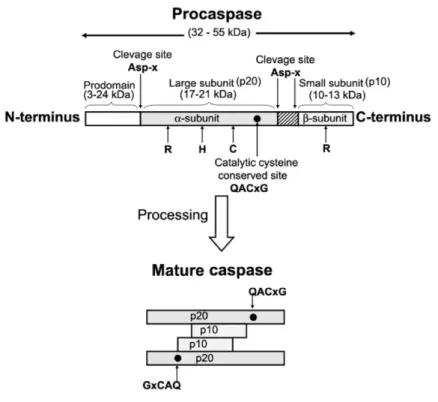
Caspases are synthesized as inactive zymogens, named procaspases. Procaspases can be proteolytically cleaved between the large and small subunit by an upstream caspase, resulting in the separation of these subunits and activation of volume decrease and finally formation of apoptotic bodies which are subsequently removed by phagocytes without causing an inflammatory response (Saraste and Pulkki, 2000; Lawen, 2003).
Figure 1. Molecular and morphological events associated with the different types of cell
death: apoptosis, autophagy and necrosis (Hotchkiss et al., 2009)
1.2.1. Caspases
Most of the alterations observed during apoptosis are caused by proteases called caspases (Cysteine-dependent aspartate-specific proteases). Conserved through evolution, caspases can be found in humans, insects, nematodes and hydra. Caspases contain a cysteine residue in the active site that is critical for their proteolytic activity and exhibit a high affinity for aspartate (Asp), cleaving their substrates after these residues (Cohen, 1997). Close to one hundred caspase substrates have already been identified, ranging from complex macromolecular complexes (e.g. actin network) to single enzymes [e.g. polyADP-ribose polymerase (PARP-1)] (Fischer et al., 2003).
Caspases are synthesized as inactive zymogens, named procaspases. Procaspases can be proteolytically cleaved between the large and small subunit by an upstream caspase, resulting in the separation of these subunits and activation of
procaspases, now known only as caspases (Figure 2). Caspases can also be activated by induced proximity, where the low intrinsic protease activity of procaspases is sufficient to allow them to mutually cleave and activate each other (Chowdhury et al., 2008).
Figure 2. Schematic representation of caspase structure and processing. The active
site residues are represented by R, H and C (Chowdhury et al., 2008).
Caspases can be divided into initiators (caspases-2, -8, -9 and -10) and executioners (caspases-3, -6, and -7). Executioner caspases have a small pro-domain, while initiator caspases have a long pro-domain, and a Death Effector Domain (DED), in the case of caspases -8 and -10, or a Caspase Recruitment Domain (CARD), in the case of caspases -2 and -9 (Budihardjo et al., 1999).
1.2.2. The Bcl-2 family members
The gene encoding the protein Bcl-2 (B-cell lymphoma 2) was the first proto-oncogene to be related to the regulation of cell cycle progression. However, its oncogenic characteristic stems from its ability to prevent apoptosis rather than promoting proliferation. Since then, several homologues of Bcl-2 have been identified that can be defined by the presence of conserved domains (BH1-BH4) necessary for their anti- or pro-apoptotic functions (Tsujimoto, 1998). Based on structural and
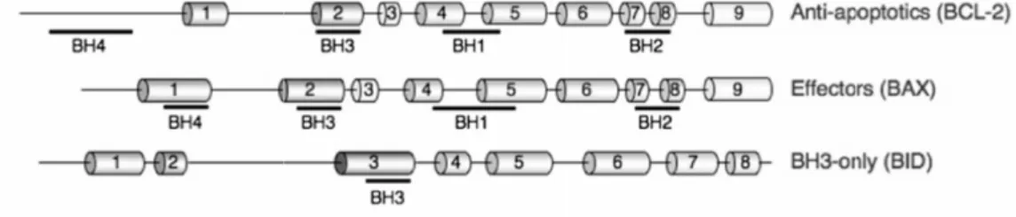
functional similarities, the Bcl-2 family of proteins can be divided into four sub-families: anti-apoptotic (A1, Bcl-2, Bcl-w, Bcl-xL and Mcl-1), effector proteins (Bax and Bak), direct activator BH3-only proteins (Bid and Bim) and de-repressor/sensitizer BH3-only proteins (Bad, Bik, Bmf, Hrk, Noxa and Puma) (Chipuk et al., 2010) (Figure 3). In viable cells, pro-apoptotic proteins are antagonists of anti-apoptotic proteins, and the levels of pro- and anti-apoptotic proteins determine susceptibility to apoptosis (Korsmeyer, 1995).
Figure 3. Schematic representation of a member of each Bcl-2 sub-family. The
conserved domains are underlined. Cylinders represent α helices (Chipuk et al., 2010).
1.2.3. Apoptotic pathways
Similarly to other signaling pathways, the events responsible for the apoptotic death process are mediated by two classical pathways: the extrinsic and intrinsic pathways, which are activated by the binding of ligands to death receptors and by stress triggered by oncogenes, irradiation, reactive oxygen species (ROS) and exposure to several drugs, respectively (Figure 4).
1.2.3.1.
Extrinsic pathway
The extrinsic pathway is one of the best characterized apoptotic signaling pathways. This pathway involves binding of specific extracellular ligands to their cognate cell surface death receptors (DR) such as Tumor-Necrosis Factor Receptor (TNF-R1), CD95 (also called as Apo-1 or Fas), TNF-related Apoptosis-Inducing Ligand Receptor (TRAIL-R1/2) and DR3/6 (Sartorius et al., 2001). Subsequent signaling is mediated by the cytoplasmic domain of the DR, named Death Domain (DD). Adapter proteins such as Fas-Associated Death Domain (FADD) or TNF-R-Associated Death Domain (TRADD) have their own DD and are recruited to the DD of the activated death functional similarities, the Bcl-2 family of proteins can be divided into four sub-families: anti-apoptotic (A1, Bcl-2, Bcl-w, Bcl-xL and Mcl-1), effector proteins (Bax and Bak), direct activator BH3-only proteins (Bid and Bim) and de-repressor/sensitizer BH3-only proteins (Bad, Bik, Bmf, Hrk, Noxa and Puma) (Chipuk et al., 2010) (Figure 3). In viable cells, pro-apoptotic proteins are antagonists of anti-apoptotic proteins, and the levels of pro- and anti-apoptotic proteins determine susceptibility to apoptosis (Korsmeyer, 1995).
Figure 3. Schematic representation of a member of each Bcl-2 sub-family. The
conserved domains are underlined. Cylinders represent α helices (Chipuk et al., 2010).
1.2.3. Apoptotic pathways
Similarly to other signaling pathways, the events responsible for the apoptotic death process are mediated by two classical pathways: the extrinsic and intrinsic pathways, which are activated by the binding of ligands to death receptors and by stress triggered by oncogenes, irradiation, reactive oxygen species (ROS) and exposure to several drugs, respectively (Figure 4).
1.2.3.1.
Extrinsic pathway
The extrinsic pathway is one of the best characterized apoptotic signaling pathways. This pathway involves binding of specific extracellular ligands to their cognate cell surface death receptors (DR) such as Tumor-Necrosis Factor Receptor (TNF-R1), CD95 (also called as Apo-1 or Fas), TNF-related Apoptosis-Inducing Ligand Receptor (TRAIL-R1/2) and DR3/6 (Sartorius et al., 2001). Subsequent signaling is mediated by the cytoplasmic domain of the DR, named Death Domain (DD). Adapter proteins such as Fas-Associated Death Domain (FADD) or TNF-R-Associated Death Domain (TRADD) have their own DD and are recruited to the DD of the activated death
receptors. These adapter proteins also have a death-effector domain (DED), with which the DED of procaspase-8 can interact with to form the Death Inducing Signaling Complex (DISC). Concentration of several pro-caspase-8 molecules within DISC leads to autocatalytic cleavage, activation and release of active caspase-8, which initiates a cascade of caspases by processing caspases -3, -6 and -7, which then cleave certain substrates. This eventually leads to the morphological and biochemical features of apoptosis. DISC signaling can be inhibited by expression of cellular-FLICE (FADD-like IL-1β-converting enzyme)-inhibitory protein (c-FLIP), a caspase-8 inhibitor, leading to inactivation of DISC (Hengartner, 2000; Lawen, 2003).
Figure 4. Schematic representation of the two major apoptotic pathways in mammalian
1.2.3.2.
Intrinsic pathway
In the intrinsic pathway, mitochondria have a central role in the induction of apoptosis. Mitochondria are able to propagate the death signals generated in the cells and/or amplify the apoptotic signal from the extrinsic pathway through mitochondrial outer membrane permeabilization (MOMP) and release of pro-apoptotic proteins. The connection between the extrinsic and intrinsic pathways and amplification of death signal is mediated by Bid, a pro-apoptotic Bcl-2 family member. Bid is cleaved by caspase-8 and when the truncated form (tBid) is translocated into the mitochondria it acts to induce MOMP and release of pro-apoptotic proteins (Luo et al., 1998).
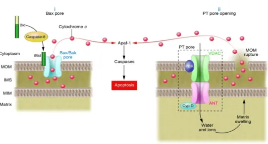
MOMP occurs at the early stages of the intrinsic pathway, though the precise mechanism has not been determined (Figure 5). According to one model, MOMP is mediated by members of the pro-apoptotic Bcl-2 family, Bax and Bak, which interact and form the mitochondrial apoptosis-induced channel (MAC), a pore that allows the release of pro-apoptotic proteins. In fact, Mcdonnell proposed that cleavage of Bid changes the conformation of the protein, leading to exposure of hydrophobic residues that allows the insertion of tBid into the membrane and binding of its BH3 domain to Bax and Bak to form pores (Mcdonnell et al., 1999). Moreover, it was observed that Bcl-2 and Bcl-xL can directly induce changes in conformation of the proteins Bax and Bak, preventing their activation and polymerization, and blocking the release of pro-apoptotic mitochondrial factors that lead to apoptosis (Letai et al., 2002). Another model suggests a permeability transition pore (PTP) is formed, which allows the passage of solutes and water into the mitochondrial matrix, causing mitochondrial depolarization, uncoupling of oxidative phosphorylation and osmotic swelling. This leads to the rupture of the outer membrane and subsequently to the release of pro-apoptotic proteins that circulate freely in the intermembrane space. The precise localization and composition of PTP has not been fully determined, but it appears to be localized at the site of contact between the inner and outer mitochondrial membranes and to contain as main components the voltage-dependent anion channel (VDAC), the adenine nucleotide translocator (ANT), hexokinase, creatine kinase (CK), the peripheral benzodiazepine receptor (PBR), and the mitochondrial matrix cyclophilin D (Cyp D) (Garrido et al., 2006).
Figure 5. Molecular mechanisms of MOMP: (i) Bax/Bak pore formation and (ii) PTP
opening (Bouchier-Hayes et al., 2005).
The permeabilization of mitochondria results in the release of pro-apoptotic proteins into the cytoplasm, such as cytochrome c, Apoptosis-inducing factor (AIF), Endonuclease G (Endo G), Second Mitochondria-derived Activator of Caspases/Direct Inhibitor of Apoptosis Protein (IAP)-Binding Protein With Low Pi (Smac/Diablo) and High Temperature Requirement protein A2 (HtrA2/Omi) (Shimizu et al., 2001; Gulbins
et al., 2003).
The cytochrome c released from the intermembrane space of mitochondria contributes to the formation of the apoptosome, together with Apoptotic Protease Activating Factor-1 (Apaf-1) and deoxyadenosine triphosphate (dATP). The apoptosome activates caspase-9, which further mediates the activation of the caspase cascade and execution of apoptosis (Acehan et al., 2002). Smac/Diablo inhibits the Inhibitors of Apoptosis Proteins (IAPs), which inhibit the activity of executing caspases. When translocated into the nucleus, AIF induces deoxyribonucleic acid (DNA) fragmentation and chromatin condensation, whereas Endo G induces internucleosomal DNA fragmentation (van Loo et al., 2001). In addition to the mitochondrial factors released, dissipation of the membrane potential also causes the loss of cell homeostasis via generation of ROS that quickly saturate the antioxidant systems and, consequently, led to cessation of ATP synthesis, Ca2+ release, oxidation of redox
molecules such as nicotinamide adenine dinucleotide reduced form (NADH), nicotinamide adenine dinucleotide phosphate reduced form (NADPH) and glutathione, and activation of stress response genes (Kroemer et al., 2007).
1.3. Yeast apoptosis
It has become clear that the apoptotic program is not restricted to multicellular organisms, but also occurs in unicellular organisms such as the budding yeast S.
cerevisiae. The physiological role of apoptosis in single-cell organisms was initially
questioned because there was no obvious reason for a unicellular organism to commit suicide. However, yeast tends to cluster and form communities. It was hypothesized that apoptosis may occur in yeast during chronological and replicative aging, unsuccessful mating processes, and to remove virus-infected and damaged cells from colonies. This altruistic cell death spares nutrients for younger cells and releases nutrients that can be metabolized by younger cells, contributing to the viability and reproductive success of healthier members of the community (Figure 6) (Büttner et al., 2006).
Figure 6. Scenarios of yeast apoptosis (Büttner et al., 2006).
Several studies have shown the occurrence of cell death in yeast can display with some characteristics of apoptosis similar to that of mammalian cells. The first observation of apoptosis in yeast was made in a temperature-sensitive mutant of S.
cerevisiae (mutant Cdc48S565G). CDC48 is an essential gene that encodes an
AAA-ATPase localized in the endoplasmic reticulum and necessary for vesicle trafficking/ translocation of ubiquitinated proteins from the endoplasmic reticulum to the
proteasome for degradation. Surprisingly, when incubated above the restrictive temperature, Cdc48S565G cells showed an apoptotic phenotype characterized by
phosphatidylserine exposure, DNA damage, chromatin condensation and fragmentation, release of cytochrome c and ROS production (Madeo et al., 1997; Braun et al., 2006). Since then, several studies also identified yeast orthologs of several members of the mammalian apoptotic machinery, including caspases (Madeo
et al., 2002), AIF (Wissing et al., 2004), Omi/HtrA2 (Fahrenkrog et al., 2004), IAP
(Walter et al., 2006) and Endo G (Büttner et al., 2007). In addition, apoptosis has often been associated with scenarios involving apoptotic mitochondrial fragmentation (Fannjiang et al., 2004) and cytochrome c release (Ludovico et al., 2002). Regulators such as Apaf-1 and of most members of the Bcl-2 family proteins seem to be absent in yeast. Until now, only a yeast BH3-only protein was identified. Ybh3p translocates to the mitochondria and is capable of mediating the mitochondrial pathway of apoptosis (Büttner et al., 2011). However, heterologous expression of Bax in yeast leads to apoptotic cell death that can be prevented by heterologous expression of anti-apoptotic Bcl-2 and Bcl-xL, suggesting the function of Bcl-2 family proteins is potentially conserved in yeast (Hanada et al., 1995).
Several assays for apoptosis detection are routinely used in yeast. They include determination of viability, ROS accumulation, DNA fragmentation [TUNEL (Terminal dUTP nick-end labeling) assay], exposure of phosphatidylserine (Annexin-V staining), chromatin condensation [DAPI (4,6-diamino-2-phenyl-indole dihydrochloride) staining] and cell integrity [Propidium iodide (PI) staining] (Carmona-Gutierrez et al., 2010). Using these assays, it was found that exposure of yeast cells to a variety of stimuli such as acetic acid, sodium chloride, ethanol, hypochlorite, amiodarone, gallium arsenide, pheromones, valproic acid, edelfosine, jasplakinolide, glucose or sorbitol, or high concentrations of glucose in the absence of other nutrients can trigger the apoptotic process (for a review see Pereira et al., 2008).
1.4. Acetic acid as an inducer of apoptosis
Acetic acid is a weak acid that can be formed as an end sub-product of alcoholic fermentation by S. cerevisiae. This compound is mainly produced by yeast strains in order to equilibrate the intracellular redox balance. In response to hyperosmotic stress caused by high sugar concentrations, yeasts increase glycerol production, oxidizing NADH to NAD+. In order to regenerate reducing equivalents,
yeasts increase the oxidation of ethanol to acetate, thus increasing the production of acetic acid (Nissen et al., 2000).
As all weak carboxylic acids, acetic acid is partially ionized in solution. It has a pKa value of 4.75, and the extracellular pH determines the proportion between the undissociated and anionic form (acetate) and the main mechanism of acetic acid cellular uptake. Acetate can be transported by two different carriers: an acetate-propionate-formate permease (Paiva et al., 1999) or an acetate-proton symport enconded by JEN1 (Casal et al., 1999). In glucose-repressed yeast cells, acetic acid enters the cell in the undissociated form by simple diffusion (Casal et al., 1996), but also potentially by a Fps1p channel. Mollapour and Piper demonstrated that deletion of
FPS1, a gene that encodes an aquaglyceroporin channel, abolishes the accumulation
of undissociated acetic acid in the cell. Moreover, they correlated loss of Fps1p with resistance to acetic acid and found that Fps1p is regulated by Hog1p signaling. Hog1p directly phosphorylates Fps1p, targeting the channel for endocytosis and degradation in the vacuole (Mollapour and Piper, 2007). After acetic acid entry, it dissociates (when the intracellular pH is higher than the extracellular pH), compromising cell viability (Pinto et al., 1989), leading to the intracellular acidification (Casal et al., 1996) and induction of apoptosis (Ludovico et al., 2001).
Exposure of S. cerevisiae to low doses to acetic acid at pH 3.0 results in cell death with features of mammalian apoptosis. Cells exposed to low doses of acetic acid exhibit chromatin condensation, exposure of phosphatidylserine and DNA strand breaks (Ludovico et al., 2001). Like in mammalian cells, yeast apoptosis induced by acetic acid was linked to mitochondria. It was shown that acetic acid can lead to the release of cytochrome c, ROS production, transient hyperpolarization of mitochondria followed by depolarization, decrease of mitochondrial respiration associated with decrease in cytochrome oxidase activity (Ludovico et al., 2002) and mitochondrial ultrastructural changes, namely decrease of cristae number, formation of myelinic bodies, and swelling (Ludovico et al., 2003). Acetic acid has been extensively used as an inducer of apoptosis in namely in the study of the involvement of the yeast AIF1 (Wissing et al., 2004), mitochondrial fragmentation (Fannjiang et al., 2004), modulation of mammalian protein kinase C (PKC) (Saraiva et al., 2006), involvement of metacaspase YCA1 (Guaragnella et al., 2006), MOMP (Pereira et al., 2007) and Pep4p involvement (Pereira et al., 2010).
1.5. Sphingolipids
Sphingolipids were considered for a long time simply structural molecules residing in membranes. They are now known to be important in cell stress responses and act as messengers in a variety of signaling pathways such as senescence,
differentiation, apoptosis, cell-cycle arrest, proliferation, mitogenesis, inflammation, migration, and angiogenesis (Hannun and Obeid, 2008), and are associated with several human diseases such as sphingolipidoses, cancer, neurodegenerative diseases, and cardiovascular pathologies (Ozbayraktar and Ulgen, 2009). Consequently, understanding how sphingolipid metabolism regulates these signaling pathways and the mechanisms underlying these diseases is of utmost importance.
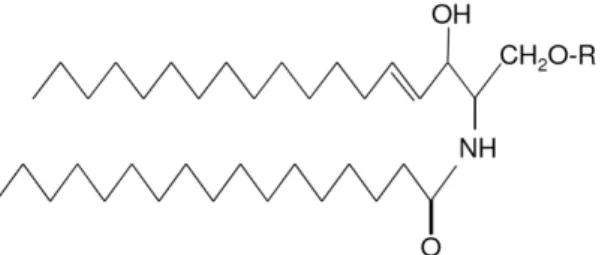
Sphingolipids were first named by Johann Thudichum in 1884 to describe the enigmatic nature and properties of complex lipids present in brain tissue. Several species of sphingolipids have been identified since then, and nowadays sphingolipids are one of the major classes of membrane lipids with a structural role in the eukaryotic lipid bilayer (Futerman and Hannun, 2004). As all membrane lipids, sphingolipids are amphipathic molecules, i.e. molecules with both hydrophilic and hydrophobic properties. The hydrophobic region is constituted by a sphingoid long-chain base (LCB), which forms the backbone of sphingolipids, to which a fatty acid is attached by an amide bond. The hydrophilic region is constituted by a polar head group (Figure 7). The type of LCB, fatty acid, and polar head group determine the type of sphingolipid (Dickson, 1998; Ozbayraktar and Ulgen, 2009).
Figure 7. General structure of sphingolipids. The LCB is linked to a fatty acid by an
amide bound and a polar head group, R (Fuller, 2010).
1.5.1. Yeast sphingolipid metabolism
The study of sphingolipids is a recent field. Simple model organisms such as the yeast S. cerevisiae have been used to uncover the pathways involved in sphingolipid metabolism and function. All the genes that encode the enzymes involved in yeast sphingolipid metabolism are known, the steps involved take place in the same organelles as in mammals and most of the enzymes have orthologs in mammalian cells. S. cerevisiae sphingolipid metabolism has ceramides as central molecules and comprises a de novo biosynthesis pathway as well as sphingolipid turnover (Figure 8).
Figure 8. Schematic overview of yeast sphingolipid metabolism displaying the
metabolic intermediates, genes involved and cellular locations of the enzymatic reactions.
1.5.2. De novo synthesis
Phytoceramide and dihydroceramide are assumed to be the yeast counterparts of mammalian ceramides, and were found to mediate regulation of cell growth and stress responses. As in all organisms, sphingolipid synthesis in yeast begins in the endoplasmatic reticulum (ER) with the condensation of serine and palmitoyl-CoA by serine palmitoyltransferase to yield 3–ketodihydrosphingosine and release of carbon dioxide and Coenzime A (CoA) (Dickson and Lester, 1999). This membrane-bound enzyme is composed of two homologous subunits encoded by LCB1 and LCB2, required for its activity (Nagiec et al., 1994), and a third small subunit, encoded by
TSC3 (temperature-sensitive suppressor of calcium sensitivity), a post-translational
activator that is essential at high temperatures (Gable et al., 2000).
In the next step of the sphingolipid metabolism, 3–ketodihydrosphingosine is reduced and converted to dihydrosphingosine (DHS) by the NADPH-dependent 3– ketoreductase encoded by TSC10. Deletion of the TSC10 gene confers an unviable
phenotype that can be rescued only when the medium is supplemented with DHS or phytosphingosine (PHS) (Beeler, 1998). DHS can be hydroxylated at C-4 by Sur2p/Syr2p hydroxylase to form PHS and both long chain bases, DHS and PHS, can suffer phosphorylation and N-acylation. The Sur2p/Syr2p hydroxylase is not essential for cell growth; however, deletion results in a mutant cell that has only DHS, whereas wild-type cells have mostly PHS (Haak et al., 1997).
DHS and PHS can be phosphorylated by two LCB kinases, encoded by LCB4 and LCB5, forming DHS-1-phosphate and PHS-1-phosphate, respectively. Finally, these phosphorylated products can either be dephosphorylated back to DHS and PHS by the phosphatases Lcb3p/Ysr2p and Ysr3p or catabolized by dihydrosphingosine-1-phosphate lyase (Dpl1p) to release palmitaldehyde and phosphoethanolamine (Sims et
al., 2004).
In the N-acylation step, C26fatty acyl-CoA is added via an amide bond to DHS
and PHS to yield dihydroceramide and phytoceramide, respectively. N-acylation of both long chain bases requires two ceramide synthases, encoded by LAG1 (longevity assurance gene 1) and LAC1 (longevity assurance gene 1 cognate) (Guillas et al., 2001). LAG1 was first identified by D’mello in 1994 as a gene involved in cell aging whose expression is decreased in aged yeast cells and its deletion results in an increased lifespan (D’mello et al., 1994). LAC1 was later identified as a homologue of
LAG1 (Jiang et al., 1998), and since then both genes have been implicated in
acyl-CoA-dependent ceramide synthesis (Schorling et al., 2001) and shown to play a role in the transport of glycosylphosphatidylinositol-anchored proteins from the ER to the Golgi (Barz and Walter, 1999). In addition, Lip1p forms a heteromeric complex with Lac1p and Lag1p and is essential for ceramide synthase activity in vivo and in vitro (Vallée and Riezman, 2005).
After generation of ceramides, they are transported to the Golgi for incorporation into complex sphingolipids. Ceramides are first converted into inositol phosphorylceramide (IPC) by transferring of a phosphorylinositol group from phosphatidylinositol to ceramide with release of diacylglycerol. This step is catalyzed by the IPC synthase encoded by AUR1, a essential gene whose deletion is lethal (Nagiec et al., 1997). More recently, Kei1p was identified as a novel component of IPC synthase. It was observed that Kei1p interacts with Aur1p and is essential for its enzymatic activity and localization (Sato et al., 2009). The complex sphingolipid IPC can further be mannosylated to form mannosylinositolphosphorylceramide (MIPC) via three enzymes, encoded by CSG1, CSG2 and CSH1. The enzymes can form two complexes, composed of Csg1p-Csg2p and Csh1p-Csg2p, that function as two different IPC mannosyltransferases, which transfer the mannose from the nucleotide sugar GDP-mannose to the inositol group in IPC (Uemura et al., 2003). The Ca2+
-binding protein Csg2p functions as a regulatory subunit in the complex because it regulates the transport and protein levels of the Csg1p and Csh1p (Uemura et al., 2007).
The final step in sphingolipid synthesis is the synthesis of the most abundant complex lipid in yeast, mannosyldiinositolphosphorylceramide (M(IP)2C). M(IP)2C is
synthesized by the addition of another inositol phosphate group to MIPC by inositolphosphotransferase (Ipt1p) (Dickson et al., 1997).
1.5.3. Sphingolipid turnover
Yeast ceramides can be catabolized by two homologous alkaline ceramidases. Dihydroceramidase (Ydc1p) and phytoceramidase (Ypc1p) are associated with the deacylation of dihydroceramide and phytoceramide, respectively (Mao et al., 2000a; Mao et al., 2000b).
Ceramides can also be produced through the turnover of complex sphingolipids. This reaction is performed by inositol phosphosphingolipid phospholipase C (Isc1p), which has phospholipase-C type activity and hydrolyses the polar head groups from complex sphingolipids, releasing dihydroceramide and phytoceramide. Isc1p is activated by phosphatidylserine, phosphatidylglycerol, and cardiolipin, and is dependent on the presence of Mg2+and inhibited by Mn2+. Isc1p overexpression results
in an increase of ceramide levels, whereas Isc1p knockout results in an accumulation of complex lipids (Sawai et al., 2000). In the pre-diauxic phase, i.e. fermentation phase, in which the cells preferentially metabolize the sugar, Isc1p is located in the ER whereas in the post-diauxic phase, i.e. respiration phase, in which cells utilize the ethanol produced during the fermentative phase, Isc1p is located in the mitochondria (Vaena de Avalos et al., 2004). ISC1 deleted strains grow very slowly in media with nonfermentable carbon sources such as glycerol, lactate, ethanol, or acetate (Vaena de Avalos et al., 2005), and have an altered mitochondrial lipid profile (lower content of α-hydroxylated phytoceramide) (Kitagaki et al., 2007). This suggests Isc1p has a critical role in mitochondrial function and/or in the regulation of pre/post-diauxic shift, because respiration and utilization of nonfermentable carbon sources require intact mitochondrial function. In addition, ISC1 deletion has been associated with premature aging and decreased cellular resistance to hydrogen peroxide (H2O2) (Almeida et al.,
2008), ethidium bromide (Kitagaki et al., 2007), genotoxic agents (methyl methanesulfonate and hydroxyurea) (Matmati et al., 2009), and increased cellular resistance to high concentrations of NaCl and LiCl (Betz et al., 2002).
1.6. Sphingolipids and cell fate
Over the past years, sphingolipids have generated considerable interest, not only due to their structural role but also as secondary signal effector molecules that can control vital biological functions.
Ceramide, a central molecule in the sphingolipid pathway, can be generated by
de novo synthesis and by catabolism of complex lipids and has a number of metabolic
fates, including catabolism to sphingosine and sphingosine-1-phosphate. Cells maintain a dynamic equilibrium of the levels of ceramide, sphingosine and sphingosine-1-phosphate. The relative amounts of these different sphingolipids and lipid–protein interactions determine cell fate. In mammalian cells, the increase of ceramide and sphingosine levels leads to cell growth arrest and apoptosis, whereas the increase of sphingosine-1-phosphate levels promotes proliferation and inhibits apoptosis (Futerman and Hannun, 2004).
1.6.1. Ceramides
Cellular ceramide levels increase in response to a variety of stimuli either by de
novo synthesis, breakdown of complex sphingolipids, or inhibition of ceramidases. The
established idea that ceramide has a role as a bioactive lipid in apoptosis is based on the identification of putative and direct targets for ceramide action (Pettus et al., 2002). The effects of ceramide appear to be mediated by activation of protein kinases and phosphatases, such as ceramide-activated protein kinase (CAPK), PKC, mitogen-activated protein kinases (MAPK) and ceramide-mitogen-activated protein phosphatases (CAPPs), or through the interaction with caspases and mitochondria (Mathias et al., 1998).
Kinase suppressor of Ras (KSR) is a direct target of ceramide. It was first identified as a CAPK and required for both inflammatory responses and ceramide-inducing stress (Zhang et al., 1997). Through phosphorylation, KSR activates Raf-1, which phosphorylates MEK, activating MAPK (Yan and Polk, 2001) and promoting apoptosis in cells expressing small amounts of the pro-apoptotic protein BAD (Basu et
al., 1998). Ceramide has also been directly associated with PKC ζ. Cells treated with
ceramide exhibit a high activation of PKC ζ, which promotes the activation of the stress-activated protein kinase (SAPK) pathway and suppression of cell growth (Bourbon et al., 2000). Moreover, activation of PKC ζ by ceramide seems to be essential in the formation of a pro-apoptotic complex in differentiating stem cells (Wang
Another target of ceramide is the CAPP: PP2A and PP1. Ceramide-activated PP2A is able to mediate the apoptotic process by dephosphorylation and consequent inactivation of pro-growth kinases such as PKC α (Lee et al., 1996) and Akt (Schubert
et al., 2000), and anti-apoptotic proteins such as Bcl-2 (Ruvolo et al., 1999) or
activation of pro-apoptotic proteins such as Bad (Xin and Deng, 2006). On the other hand, PP1 was shown to be involved in ceramide-induced dephosphorylation of the retinoblastoma product gene (Rb), interfering with cell cycle regulation (Kishikawa et
al., 1999).
Cathepsin D (endosomal acidic aspartate protease) was identified by Heinrich in 1999 as a novel ceramide-binding protein. This interaction induces the autocatalytic proteolysis of the pro-enzyme to the active form of cathepsin D (Heinrich et al., 1999). Since then, cathepsin D has been implicated in apoptosis due its role in cleavage and activation of Bid, Bax activation and translocation to the mitochondria, destabilization of mitochondria, cytochrome c release and caspase activation (Guicciardi et al., 2004).
Several studies have demonstrated that ceramide has a role in mitochondria-involving apoptosis. First, cellular ceramide levels increase prior to the activation of the mitochondrial pathway of apoptosis (Rodriguez-Lafrasse et al., 2001). Second, ceramide has been shown to interact with and inhibit components of the mitochondrial respiratory chain in isolated mitochondria (Gudz et al., 1997), induce ROS production in cells (France-Lanord et al., 1997) and isolated mitochondria (García-Ruiz et al., 1997), mitochondrial depolarization and dysfunction (Hearps et al., 2002), and release of pro-apoptotic proteins, such as cytochrome c and AIF (Di Paola et al., 2004; Zhang
et al., 2008). Third, the discovery that mitochondria contain the enzymes involved in
ceramide synthesis and the observation that several agents such as TNF, UV radiation and Fas increase the levels of ceramide in isolated mitochondria confirmed that apoptosis occurs via an increase in mitochondrial ceramide levels (Siskind, 2005).
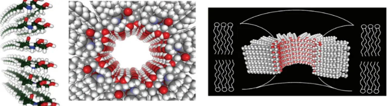
Siskind and Colombini showed that ceramide can form large and stable channels with an estimated diameter of about 10 nm (Samanta et al., 2011) (Figure 9) that allows the release of proteins in the intermembrane space with a molecular weight up to 60 kDa (Siskind et al., 2002) such as cytochrome c (12 kDa), AIF (57 kDa), Endo G (28 kDa) and Smac/DIABLO (42 kDa). In addition, ceramide channels can be disassembled by binding of anti-apoptotic proteins, such as Bcl-xL (Ganesan and Colombini, 2010), and that Bax seems to be responsible for enlargement of the ceramide channels, improving mitochondrial membrane permeability (Ganesan et al., 2010). Furthermore, ceramide channels are specifically formed in mitochondrial membranes at physiologically concentrations of ceramide. Indeed, at concentrations 20 times higher than those required for channel formation in mitochondrial, ceramide
channel formation does not occur in the plasma membrane of erythrocytes (Siskind et
al., 2006).
Figure 9. Structure of ceramide pores. They are composed of a ring of a variable
number of columns each consisting of six ceramides interconnected by hydrogen bonds between the amide linkage (Siskind and Colombini, 2000).
1.6.2. Ceramides and its metabolites: Sphingosine and Sphingosine-1-phosphate
Sphingosine and sphingosine-1-phosphate are sphingolipids derived from ceramides that can also function as signaling molecules. Sphingosine has been shown to play a role in the induction of apoptosis in several types of cells, such as HL-60 cells (Sakakura et al., 1996), human neutrophils (Ohta et al., 1994), cardiac myocytes (Krown et al., 1996), neurons and astrocytes (Kanno and Nishizaki, 2011), among others. Sphingosine interacts with several signaling pathways, including activation of JNK, p38 MAPK, sphingosine-dependent protein kinase (SDK1) and caspases, stimulation of PARP-1 cleavage, induction of BID and BAX truncation and cytochrome
c release, inhibition of PKCα, Akt kinase, ERK1/ERK2 kinases, 14-3-3 chaperone
protein, β1 integrin and Ca2+/calmodulin-dependent protein kinase and reduction of the
expression of Bcl-2 and Bcl-xL (Figure 10). In addition, sphingosine is able to stimulate the activation of Rb and promotes cell cycle arrest (Cuvillier, 2002, Taha et al., 2006). On the other hand, sphingosine-1-phosphate functions as an antagonist of ceramide and sphingosine and plays a crucial role in the promotion of survival, proliferation and inhibition of apoptosis. Moreover, the fact that sphingolipids are interconvertible led to the proposal of the so-called “sphingolipid rheostat” model, which postulates that the relative levels of these lipids determine the cell fate (Spiegel and Milstien, 2003). Indeed, it has been proposed that the sphingolipid signaling pathway is a target of interest for cancer therapy (Cuvillier et al., 2010). Many external stimuli, namely growth factors, cytokines and mitogens, were shown to activate the sphingosine kinase (SK1),
leading to increased sphingosine-1-phosphate levels and decreased ceramide levels (Alvarez et al., 2007). Once generated, sphingosine-1-phosphate can act either extracellularly, by binding to G-protein-coupled receptors present on the cells, initiating downstream G-protein-mediated signaling pathways, or intracellularly, by regulating the calcium levels, activating pro-survival mediators like nitric oxide (NO), ERK, Akt, or by inhibiting the mitochondrial pathway by blocking JNK or activating several transcriptional factors such as AP-1 and NF-ĸB (Spiegel and Milstien, 2003; Pitson, 2011).
Figure 10. Sphingosine (Sph) and sphingosine-1-phosphate (S1P) targets (Taha et al., 2006).
1.7. Sphingolipids and yeast apoptosis
Until now, few studies were performed in yeast to address the involvement of sphingolipids in apoptosis. Siskind observed that expression of either recombinant Bcl-xL or CED-9, homologues of Bcl-2 proteins, disassemble ceramide channels in isolated mitochondria of yeast cells (Siskind et al., 2008). In another study, overexpression of Ydc1p ceramidase triggered vacuolar and mitochondrial fragmentation and dysfunction, shortened chronological lifespan and increased apoptosis (Aerts et al., 2008). Moreover, Isc1p deletion is associated with up-regulation of the iron regulon and leads to an overload of iron, which catalyzes the production of the highly reactive hydroxyl radicals via the Fenton reaction, and increases apoptotic cell death caused by exposure to hydrogen peroxide (Almeida et al., 2008). Recently, it was described that Isc1p is an upstream regulator of Sit4p, the catalytic subunit of PP2A in yeast. Deletion of the SIT4 gene in isc1∆ abolishes the premature ageing and oxidative stress sensitivity of this strain by reversing the mitochondrial dysfunction of isc1∆ cells (Barbosa et al., 2011).
Nowadays, it is consensual that the yeast S. cerevisiae undergoes apoptosis in a manner similar to that of mammalian cells. Several members of the mammalian apoptotic machinery have already been identified, and implicated in scenarios of yeast apoptosis. However, there are still numerous apoptotic regulators that remain to be discovered, and others whose function and hierarchy in apoptotic cell death has not yet been determined.
This work aimed to understand the role of ceramides in the mitochondrial apoptotic pathway induced by acetic acid, by taking advantage of bakers´s yeast as a powerful genetic system, and using the following approaches:
1. Construction of several mutants involved in sphingolipid metabolism;
2. Identification of the enzymes associated with sphingolipid metabolism that involved in acetic acid-induced apoptosis;
3. Characterization of the involvement of these enzymes in oxidative stress and mitochondrial apoptotic markers induced by acetic acid.
3. MATERIALS AND
METHODS
3.1. Yeast strains
All Saccharomyces cerevisiae strains used in this study are listed in Table I. S.
cerevisiae strain CG379 was used as the wild-type. The lac1Δ, lag1Δ, ydc1Δ, ypc1Δ
and isc1Δ mutants were constructed in CG379 by homologous recombination with disruption cassettes (KanMX4) amplified by Polymerase Chain Reaction (PCR), using the oligonucleotides listed in Table II (numbers 1-10) and genomic DNA isolated (as described below in 3.3) from the respective mutants of the yeast strain BY4741 (Euroscarf collection, Germany).
CG379 cells were transformed by electroporation (as described below in 3.4) and transformants selected on rich medium [YPD; 1% (w/v) yeast extract, 2% (w/v) bactopeptone, 2% (w/v) glucose] containing 200 μg/mL geneticin. The correct integration of the disruption cassettes was confirmed by PCR using oligonucleotides (numbers 11-20) that bind upstream and downstream of the insertion, plus an additional oligonucleotide (number 21) binding within the kanamycin gene (Figure 11). In addition, lag1∆ and isc1∆ were transformed by electroporation with pYES2-LAG1 and pYES2-ISC1 vectors, respectively. For mitochondrial studies, wild-type, lag1∆ and
isc1∆ strains were transformed with pYES2-mtGFP.
Table I. List of S. cerevisiae strains used in this study.
Strain Genotype Reference/Source
CG379 Matα, ade5, his2, leu2-112, trp1-289,ura3-52 Yeast Genetic StockCenter, University of California, USA
CG379 pYES2 CG379 harboring pYES2 This study
CG379 pYES2-mtGFP CG379 harboring pYES2-mtGFP This study
lac1Δ CG379 lac1Δ :: KanMX4 This study lag1Δ CG379 lag1Δ :: KanMX4 This study lag1Δ pYES2 lag1Δ harboring pYES2 This study lag1Δ pYES2-LAG1 lag1Δ harboring pYES2-LAG1 This study lag1Δ pYES2-asLAC1 lag1Δ harboring pYES2-asLAC1 This study lag1Δ pYES2-mtGFP lag1Δ harboring pYES2-mtGFP This study ydc1Δ CG379 ydc1Δ :: KanMX4 This study ypc1Δ CG379 ypc1Δ :: KanMX4 This study ypc1Δ pYES2 ypc1Δ harboring pYES2 This study ypc1Δ pYES2-asYDC1 ypc1Δ harboring pYES2-asYDC1 This study isc1Δ CG379 isc1Δ :: KanMX4 This study isc1Δ pYES2 isc1Δ harboring pYES2 This study isc1Δ pYES2-ISC1 isc1Δ harboring pYES2-ISC1 This study isc1Δ pYES2-mtGFP isc1Δ harboring pYES2-mtGFP This study
Table II. List of oligonucleotides used in this study for the construction of yeast mutants.
Restriction sites are marked in bold in respective oligonucleotide sequence.
Number Name Oligonucleotide Sequence
1 Lac1Fw 5´- GGAGGGAGAAAGTATTGGAATCT – 3´ 2 Lac1Rv 5´- GAAAGCACTAACATCAACATGGA – 3´ 3 Lag1Fw 5´- CGTCATCTTCCATTTGAAATCC – 3´ 4 Lag1Rv 5´- TCTTACTAGGAGTCTTGGCGAGA – 3´ 5 Ydc1Fw 5´- TGTCCGATAGCGTACGCCA – 3´ 6 Ydc1Rv 5´- GCCGGTTTTCCAAGCAG – 3´ 7 Ypc1Fw 5´- CGCGAGACATCGGAAAATA – 3´ 8 Ypc1Rv 5´- CATGTCCCGAATTAGCTAACAA – 3´ 9 Isc1Fw 5´- AGGTCGACTGCCGTCTAGAT – 3´ 10 Isc1Rv 5´- GCGGACTTCATTTTACTCCAGAC – 3´ 11 Lac1KanFw 5´- TGGGCATTGTACCTGATCATG – 3´ 12 Lac1KanRv 5´- GGCCTACTATGACAACGATAGCT – 3´ 13 Lag1KanFw 5´- CCAGTCCGTCAAGACTAATATCG – 3´ 14 Lag1KanRv 5´- CGATGATTCATTGAGATCTGTCA – 3´ 15 Ydc1KanFw 5´- AAATCCCTCGTTCCCGG – 3´ 16 Ydc1KanRv 5´- TATGTGCCGCCGACATG – 3´ 17 Ypc1KanFw 5´- GGACGGATTATCACGCAAGT – 3´ 18 Ypc1KanRv 5´- CAGAAGCCAAAATAGCATTCAA – 3´ 19 Isc1KanFw 5´- TTGCAGCAGCGAGTCCA – 3´ 20 Isc1KanRv 5´- CGAACGAGGCAGTAGTCATGTT – 3´ 21 KanRv 5´- AATCGAATGCAACCGGC – 3´
22 LAG1_HindIII_Fw 5´- ACGACAAGCTTAACATGACATCAGCTACGGACAAAT - 3´
23 LAG1_XhoI_Rv 5´- AGATACTCGAGCGTTTATTCACACTTTTCCTTAGAT - 3´
24 asLAC1Fw 5´- TAAAAGCTTGCTTCATCGACAATAAGCCAAG - 3´
25 asLAC1Rv 5´- CACCTCGAGCCTATGAATATCCTTTTTCGTTGGAGTA - 3´
26 asYDC1Fw 5´- GAAAAGCTTCAATTACTGTTCAGCTGGCCTTATCCA - 3´
27 asYDC1Rv 5´- CAACTCGAGTCCATGGTTATTCTTTTTTGTTTCATCATC - 3´
Figure 11. General scheme of the strategy used for construction of yeast mutants. Step
1 represents the procedure used for generation of KanMX4 cassettes from the respective mutants in strain BY4741, step 2 the homologous recombination mechanism, and step 3 the confirmation of the correct integration of disruption cassettes in proper position of genome of CG379 strain with the primers represented in step 2.
The construction of double mutants was performed by silencing the YDC1 gene in ypc1∆ mutants and the LAC1 gene in lag1∆ mutants using an antisense gene expression vector. The ypc1∆ and lag1∆ mutants were transformed by electroporation with pYES2-asYDC1 and pYES2-asLAC1, respectively (Figure 12).
Figure 12. General scheme of the strategy used for construction of double mutants.
When antisense mRNA from the plasmid is expressed in yeast cells, it hybridizes with the sense mRNA from the nucleus, blocking synthesis of the protein.
3.2. Plasmids
All the plasmids used in this study are listed in Table III. For expression of
LAG1, pYES2-LAG1 was constructed. The LAG1 gene was amplified by PCR from
genomic DNA isolated from the CG379 strain using the oligonucleotides LAG1_HindIII_Fw (number 22) and LAG1_XhoI_Rv (number 23), which introduce
HindIII and XhoI restriction sites in the flanks, and cloned into pYES2 using these
enzymes (Figure 13).
Table III. List of plasmids used in this study.
Plasmid Description Reference/Source
pYES2 URA3; AmpR Invitrogen
pYES2-LAG1 LAG1 inserted in pYES2 This study
pYES2-ISC1 ISC1 inserted in pYES2 Y. Hannun, Medical University ofSouth Carolina, Charleston, USA
pYES2-asLAC1 Antisense - LAC1 inserted in pYES2 This study
pYES2-asYDC1 Antisense - YDC1 inserted in pYES2 This study
pYES2-mtGFP mtGFP inserted in pYES2 Westermann and Neupert, 2000 The construction of double mutants was performed by silencing the YDC1 gene in ypc1∆ mutants and the LAC1 gene in lag1∆ mutants using an antisense gene expression vector. The ypc1∆ and lag1∆ mutants were transformed by electroporation with pYES2-asYDC1 and pYES2-asLAC1, respectively (Figure 12).
Figure 12. General scheme of the strategy used for construction of double mutants.
When antisense mRNA from the plasmid is expressed in yeast cells, it hybridizes with the sense mRNA from the nucleus, blocking synthesis of the protein.
3.2. Plasmids
All the plasmids used in this study are listed in Table III. For expression of
LAG1, pYES2-LAG1 was constructed. The LAG1 gene was amplified by PCR from
genomic DNA isolated from the CG379 strain using the oligonucleotides LAG1_HindIII_Fw (number 22) and LAG1_XhoI_Rv (number 23), which introduce
HindIII and XhoI restriction sites in the flanks, and cloned into pYES2 using these
enzymes (Figure 13).
Table III. List of plasmids used in this study.
Plasmid Description Reference/Source
pYES2 URA3; AmpR Invitrogen
pYES2-LAG1 LAG1 inserted in pYES2 This study
pYES2-ISC1 ISC1 inserted in pYES2 Y. Hannun, Medical University ofSouth Carolina, Charleston, USA
pYES2-asLAC1 Antisense - LAC1 inserted in pYES2 This study
pYES2-asYDC1 Antisense - YDC1 inserted in pYES2 This study