DECLARAÇÃO
Nome: Sónia Maria de Sousa Borges
Endereço electrónico: sonia.borges25@gmail.com Telefone: 917937056
Número do Bilhete de Identidade: 13612893 Título dissertação:
Searching for the neurobiological targets through which prenatal glucocorticoids program adult social and affective behaviors
Orientador(es):
Doutora Ana João Rodrigues, Professor Doutor Nuno Sousa, Professora Doutora Dorit Schuller Ano de conclusão: 2012
Designação do Mestrado ou do Ramo de Conhecimento do Doutoramento: Mestrado em Genética Molecular
É AUTORIZADA A REPRODUÇÃO INTEGRAL DESTA TESE/TRABALHO APENAS PARA EFEITOS DE INVESTIGAÇÃO, MEDIANTE DECLARAÇÃO ESCRITA DO INTERESSADO, QUE A TAL SE COMPROMETE;
Universidade do Minho, 31/10/2012
iii Para ser grande, sê inteiro: nada Teu exagera ou exclui. Sê todo em cada coisa. Põe quanto és No mínimo que fazes. Assim em cada lago a lua toda Brilha, porque alta vive. Fernando Pessoa, Poemas de Fernando Pessoa
v
Agradecimentos
Apesar de esta tese de mestrado ter o meu nome como a pessoa responsável pela sua realização, na realidade, este projecto não seria possível sem a ajuda de muitas outras pessoas. Primeiro e principalmente, à Doutora Ana João Rodrigues por me ter ajudado ao longo deste ano e dado a oportunidade de ser a “psicóloga” dos meus ratos, pela sua disponibilidade, e, principalmente, por ter acreditado em mim, pois qualquer grau de sucesso que eu alcancei enquanto estive no laboratório devo-o a ela. Sinto-me honrada por ter trabalhado com ela neste projecto. Um sincero obrigado!
Ao Professor Doutor Nuno Sousa pela sua disponibilidade, pela sua boa disposição e pelo seu interesse no meu trabalho e como estava a decorrer. À Professora Doutora Dorit Schuller pela sua disponibilidade e interesse neste projecto.
Um obrigado às minhas “team colleagues”, Bárbara e Carina. À Carina pela sua disponibilidade e pela sua grande ajuda na realização dos western blots e nos testes comportamentais deste projecto. À Bárbara pela sua boa disposição e pela parceria nas inúmeras horas passadas na escrita da tese e por tudo que teve de aturar.
À Adriana por ter me aturado muitas vezes e pelas inúmeras vezes que me elevou o espírito. À Joana Silva, Margarida, Sofia Esteves, Filipa, Miguel Oliveira, Cristina, Ana Ribeiro, Rita, André, Silvina, Mafalda pelo apoio moral demonstrado e pela boa disposição.
Ao grupo de Neurociências pelas discussões científicas e pelo apoio demonstrado durante a realização deste projecto.
Aos meus amigos, principalmente aos meus melhores amigos, Fábio “Furbs” e Susana “Su”, por me aturarem desde pequena, por serem meus amigos, por me fazerem rir e pelo apoio demonstrado.
E ao meu irmão pelo apoio demonstrado no meu trabalho.
Por último, às principais pessoas responsáveis por tudo, os meus pais. Obrigado, por sempre me terem ajudado a atingir os meus objectivos, por apoiarem sempre que precisei e por sempre acreditarem em mim, não só ao longo deste ano, mas ao longo da minha vida.
vii “Searching for the neurobiological targets through which prenatal
glucocorticoids program adult social and affective behaviors”
Abstract
Stress activates the hypothalamic-pituitary-adrenal axis and leads to a controlled release of glucocorticoids (GCs) in the blood stream. Due to their numerous effects, synthetic GCs are often prescribed in clinics, as for example in 10% of preterm risk pregnancies in order to accelerate fetal lung maturation. Previous studies have shown that exposure to stress, or administration of GCs during pregnancy can contribute for the development of neuropsychiatric disorders later in life such as anxiety, depression and addiction.
In this work we studied a rat model prenatally exposed to synthetic GC, dexamethasone, at embryonic days 18 and 19. Animals were subjected to standard behavioral tests to assess emotional and social behaviors, and, to further complement these results we measured the emission of ultrasonic vocalization (USVs). iuGC animals are hyperanxious, display enhanced fear response and present depressive-like behavior. In parallel, we hypothesized that emotional impairment observed in iuGC animals could influence social interaction. Indeed, prenatal GC exposure impairs social behavior at different ages, since iuGC animals present low motivation to engage in social play and seem to present less pleasure during the interaction. Considering the cross-talk between opioid-endocannabinoid-dopaminergic circuits in both emotional and social behaviors, we further analysed levels of specific receptors in several regions of mesolimbic circuitry. We found prominent changes in Drd2, MOR1 and KOR1 in the nucleus accumbens and amygdala, which may underlie the abovementioned impaired behaviors. As these animals present a significant hypodopaminergic status in the mesolimbic circuitry, together with Drd2 molecular changes, in the last part of the project we decided to treat these animals with L-DOPA, a dopamine precursor. Administration of a therapeutic dose of L-DOPA reverted most of the emotional and social deficits observed in iuGC animals, suggesting a crucial role of dopaminergic transmission in this type of behaviors.
Our results show that prenatal GC administration potentiates emotional and social deficits, probably as a consequence of a deficient dopaminergic signalling in the mesolimbic circuitry. Importantly, we were able to restrain these behaviors by a simple pharmacological approach.
ix
“Explorar os alvos neurobiológicos através dos quais glucocorticoides pré-natais programam comportamentos sociais e afetivos”
Resumo
O stress ativa o eixo hipotálamo-pituitária-adrenal e conduz à libertação de glucocorticoides (GCs) na corrente sanguínea. Devido aos seus efeitos diversos, os GCs sintéticos são usados frequentemente na prática clínica, como por exemplo em gravidezes com risco de parto pré-termo, para promover a maturação pulmonar fetal. Estudos anteriores demonstraram que exposição ao stress, ou a administração de GCs durante a gravidez, pode contribuir para o desenvolvimento de doenças neuropsiquiátricas em adultos, tais como ansiedade, depressão e adição.
Neste trabalho estudamos um modelo em rato de exposição ao GC sintético, dexametasona, nos dias embrionários 18 e 19 (iuGC). Os animais foram submetidos a uma bateria de testes comportamentais para avaliar o comportamento emocional e social, e, de forma a completar estes estudos, também medimos a emissão de vocalizações ultrassónicas (USVs). Os animais iuGC são mais ansiosos, apresentam comportamento do tipo depressivo, e uma resposta ao medo exacerbada.
Em paralelo, avaliamos o comportamento social e vimos que estes animais apresentam uma menor antecipação para iniciar uma interação social e menos prazer na mesma. Considerando a interação entre os sistemas opioide-endocanabinoide-dopaminérgico neste tipo de comportamento, decidimos analisar os níveis de determinadas moléculas destes circuitos nestes animais. Identificamos alterações proeminentes no receptor Drd2, no MOR1 e KOR1 no nucleus accumbens e na amigdala no grupo iuGC. Uma vez que os iuGC apresentam um estádio hipodopaminergico substancial no circuito mesolimbico juntamente com alterações significativas de Drd2, decidimos tratar os animais com um precursor da dopamina, a L-DOPA. Administração de uma dose terapêutica de L-DOPA reverteu quase todas as alterações comportamentais dos animais iuGC.
Estes resultados mostram que a administração pré-natal de GCs potencia o aparecimento de déficits emocionais e sociais, e que uma simples abordagem farmacológica com o objectivo de aumentar a dopamina nestes animais, foi suficiente para reverter a maioria das alterações comportamentais.
xi Contents Agradecimentos...v Abstract...vii Resumo...ix Abbreviations...xiv 1.Introduction ... 1
1.1. Stress and the Hypothalamic-Pituitary-Adrenal (HPA) axis ... 3
1.2. The physiological role of GCs ... 4
1.3. GCs early in life: molecular, structural and behavioral outcomes ... 6
1.4. Social Play Behavior ... 8
1.5. Modulation of social (and affective) behavior: neurobiological correlates ... 9
1.5.1. Opioids signalling ... 10
1.5.2. Cannabinoid signalling ... 11
1.5.3. Dopaminergic signalling ... 13
1.6. Ultrasonic vocalizations – a measure of emotional and social behavior in rodents... ... 15
2. Aims ... 19
3. Materials and Methods ... 23
3.1. Animals ... 25
3.2. Experimental design ... 25
3.3. Behavioral characterization ... 26
3.3.1. Emotional Status ... 26
3.3.1.1. Forced swimming test ... 26
3.3.1.2. Physical Enrichment ... 26
3.3.1.3. Anticipatory behavior to food (feeding sessions) ... 26
3.3.1.4. Light/dark box test ... 27
3.3.1.5. Confined cage test ... 27
3.3.1.6. Fear conditioning... 28
xii
3.3.2.1. Maternal Behavior ... 28
3.3.2.2. Tumble and play ... 28
3.3.2.3. Communicative function ... 29
3.3. Western Blot ... 29
3.4. Pharmacological reversal of behavioral alterations ... 30
3.5. USVs analysis ... 30
3.6. Statistical analysis ... 31
4. Results ... 33
4.1. Effects of iuGC treatment on emotional behavior of offspring ... 35
Depressive-like-behavior... 35
Anxiety ... 37
Fear ... 38
4.2. Effects of iuGC treatment on social behavior of offspring ... 40
4.3. Effects of iuGC treatment on adult social behavior ... 43
4.4. Effects of iuGC treatment on endocannabinoid, opioid and dopaminergic circuits…………. ... 45
4.5. Dopaminergic modulation of the emotional condition in iuGC animals ... 49
Depressive-like-behavior... 49
Anxiety ... 50
Fear ... 52
4.6. Dopaminergic manipulation of social behavior in iuGC animals ... 55
5. Discussion ... 59
5.1. Stress effects in affective state ... 61
Depressive-like behavior ... 62
Anxiety ... 63
Fear ... 63
5.2. Impact of prenatal stress on social/affective behaviors ... 64
5.3. Endocannabionid,opioid and dopaminergic correlates ... 66
5.4. Impact of dopaminergic manipulation ... 68
xiii
xv
Abbreviations
11β-HSD – 11-Beta hydroxysteroid dehydrogenase 5-HT - Serotonin
ACTH - Adrenocorticotropic hormone AH - Anterior hypothalamus
AMY - Amygdala
BDNF - Brain-derived neurotrophic factor BNST – Bed nucleus of the stria terminalis CAH - Congenital adrenal hyperplasia CORT – Corticosteroids
CP - Cerebral peduncle CPu - Caudate putamen
CRH - Corticotrophin-releasing hormone DA - Dopamine DAergic - Dopaminergic DEX - Dexamethasone Drd1 - Dopamine receptor 1 Drd2 - Dopamine receptor 2 GC - Glucocorticoid GR - Glucocorticoid receptor
GREs - Glucocorticoid response elements HPA - Hypothalamic-pituitary-adrenal
xvi
HPT – Hypothalamus
iuGC - in utero glucocorticoid KOR1 - k opioid receptor 1 LC - Locus coeruleus
LDT - Laterodorsal tegmental nucleus LS - Lateral septum
MOR1 - µ opioid receptor 1 MR - Mineralocorticoid receptor NAcc - Nucleus accumbens NEC - Necrotizing enterocolitis
NGFI-A - Nerve growth factor-inducible protein A NTS - Nucleus tractus solitarius
OXY – Oxytocin
PBN - Parabranchial nucleus PFC - Prefrontal cortex PO - Preoptic area
PVN - Paraventricular nucleus
RDS - Respiratory distress syndrome TH - Tyrosine hydrolase
USV - Ultrasound vocalization VLM - Ventrolateral medulla VTA - Ventral tegmental area
1
3
1. Introduction
1.1. Stress and the Hypothalamic-Pituitary-Adrenal (HPA) axis
Stress is defined as any perturbation of homeostasis of the organism, both psychological or physiological (Goldstein and Kopin, 2007). The stress response is coordinated by the brain; in the presence of a stressor, adrenaline and noradrenaline are released, and this constitutes the foundation of the classic reaction of the “fight-or-flight” response (Juruena et al., 2004). Simultaneously, stress activates the hypothalamic-pituitary-adrenal (HPA) axis. This endocrine response starts with the release of corticotrophin-releasing hormone (CRH) that is secreted by neurons from the paraventricular nucleus (PVN) of the hypothalamus (HPT). The CRH acts on the anterior pituitary, where it stimulates the synthesis and release of adrenocorticotropic hormone (ACTH) into the blood stream. In turn, ACTH induces the synthesis and secretion of corticosteroids in the adrenal cortex (Fig.1). The main corticosteroid is cortisol in humans and corticosterone in rodents (Mesquita et al., 2009).
Figure 1 - Regulation and negative feedback of glucocorticoids (GCs) in the hypothalamus-pituitary-adrenal (HPA)
axis. Under stressful stimuli the cells of the paraventricular nucleus (PVN) in the hypothalamus release corticotrophin-releasing hormone (CRH). The CRH activates the pituitary, which in turn, releases adrenocorticotropic hormone (ACTH), then it act on the adrenal cortex resulting in an increase in the levels of glucocorticoids in the blood stream. GCs have a negative feedback in order to inhibit further production of GCs.
4
There are two types of corticosteroids receptors, the high-affinity mineralocorticoid receptor (MR) and the low-affinity glucocorticoid (GC) receptor (GR) (Sanchez et al., 2000). Ligand binding results in the translocation of the receptor to the nucleus, where it regulates the expression of target genes due to the association of the complex receptor-ligand with the glucocorticoid response elements (GREs) in the promoter region of targets genes (Fig.2). Importantly, the GRs can regulate positive or negatively the genes to which they are coupled (Juruena et al., 2004).
The cessation of the HPA response to stress is coordinated by a glucocorticoid negative-feedback process (Fig.1)(Spencer et al., 1998, De Kloet et al., 1998), which is tightly regulated to prevent deleterious effects of both hyper and hypo-cortisolemia.
1.2. The physiological role of GCs
Endogenous GCs (CORT) increase the concentration of glucose and other metabolic fuels; they are also involved in the immune response. GCs have inhibitory effects on both innate and acquired immune response mediated by the T and B cells, and can suppress the effector functions of phagocytes. In short, they are extraordinary efficacious in managing numerous acute disease manifestations of inflammatory and autoimmune disorders (Chatham and Kimberly, 2001). During pregnancy, they are involved in lung maturation (regulation of surfactant production by the lungs), and for the regulation of immune and metabolic functions, and for neuro- and behavioral development (Mesquita et al., 2009).
5
Figure 2 - Scheme of the activation of glucocorticoid receptor (GR). GR in their inactive state are present in the
cytoplasm with a multimeric complex of chaperone proteins, including many heat sock proteins. Endogenous or synthetic glucocorticoids act as a ligand; GR-ligand undergoes a conformational change, and becomes active, then dissociates from the complex of chaperone proteins and translocates to the nucleus, where it regulates gene expression by binding to glucocorticoid response elements (GRE). The diagram is adapted from Strachan T., 1999 (Strachan T. and A.P., 1999).
The placenta as well as most foetal tissues expresses GRs, which can bind both endogenous and exogenous GC, from mid-gestation onwards (Johnson et al., 2008). Endogenous GCs have a higher affinity to MR and the expression of these receptors is more confined to specific brain areas and, in rodents, is only present in late gestation (Funder, 1996).
The foetus has limited capacity to degrade synthetic GCs, so, even low levels of GC can have a deleterious impact. The endogenous GCs are much lower in the foetus than in mother circulation due to the existence of the enzyme 11-Beta hydroxysteroid dehydrogenase (11β-HSD), which is located in the syncytiotrophoblast, the site of maternal-foetus exchange (Mesquita et al., 2009).This enzyme converts the GC in 11-keto metabolites, its inactive form. Nonetherless, 11β-HSD is not active throughout the pregnancy, in fact, it was show by McTernan and collaborators, variations of 11β-HSD activity in the end of pregnancy, in rats and
6
humans (McTernan et al., 2001), that may indicate that increased maternal GCs during this stage can affect the foetus. Moreover, exogenous GCs, such as dexamethasone (DEX) or betamethasone, are poor substrates for 11β-HSD, facilitating the passage through the placenta (White et al., 1997).
Due to their pleiotropic action and ability to cross the placenta, exogenous GCs are widely prescribed, in clinics, to accelerate lung maturation in preterm labour and perinatal period (Crane et al., 2003). Frequently, DEX, a synthetic glucocorticoid that has 25 times more potency than endogenous cortisol (Oliveira et al., 2006), is used in clinics. This seems to reduce neonatal death, respiratory distress syndrome (RDS), intraventricular hemorrhage, necrotizing enterocolitis (NEC) and respiratory support, intensive care admissions and systemic infections in the first 48 hours of life (Miracle et al., 2008). Less frequently, it is used during antenatal period in the foetuses at risk of congenital adrenal hyperplasia (CAH) (New et al., 2001). Although the positive effects of such treatment are undeniable, less is known about the deleterious effects that it can have in the developing brain.
1.3. GCs early in life: molecular, structural and behavioral outcomes
Adversity during early life, including, abuse or neglect, can induce effects on physical and mental health. For example, it increases the risk of development of conduct disorders, personality disorders, major depression, schizophrenia, anxiety and addictive disorders (Agid et al., 1999, Bernet and Stein, 1999, Dube et al., 2003, Heim and Nemeroff, 2001, Young et al., 1997). In contrast, the development of psychiatric disorders is very low in women abused during adulthood (Brown and Moran, 1994), (McCauley et al., 1997). Thus, studies demonstrate that the mechanisms that underlie programming of the adult HPA axis function are dependent on time of exposure, but also, dependent of the dose of GCs.
Some studies report that children exposed prenatally to GC have a reduction in their birth weight, and at the age of 3 present significant behavioural changes (Newnham, 2001). Oliveira and colleagues showed that prenatal administration of endogenous or synthetic (DEX) glucocorticoids can act permissively to accelerate some developmental processes, like eye opening (a measure of neurological development), while it delays others, like body growth (Oliveira et al., 2006), in rats. However, such treatments lead to a long-lasting impairment in
7 the HPA axis (Matthews et al., 2002). In utero exposure to GCs/stress, and consequent impairment in the HPA axis, was found to be associated with long-lasting deficits in cognitive, mood and addictive behaviors (Oliveira et al., 2006, Rodrigues et al., 2011b, Roque et al., 2011, Miracle et al., 2008). Indeed, in human pregnancies, evidence suggests that fetal exposure to synthetic GCs has detrimental effects on birth outcome, childhood cognition and long-term behavior (Seckl, 2004, Crowley, 1995, Miracle et al., 2008). Furthermore, previous work from our lab revealed that prenatal administration of DEX to rats induces a hyperanxious status in the progeny and increases the propensity for developing depression (Roque et al., 2011), together with reward-associated behaviors (Rodrigues et al., 2011b).
Several brain regions seem to be affected by early exposure to GC, however, the hippocampus (Sousa and Almeida, 2002), the prefrontal cortex (Cerqueira et al., 2007) and the bed nucleus of the stria terminalis (BNST) seem to be specially affected (Pego et al., 2008). Previous work showed that rhesus monkeys treated with prenatal DEX present a reduction in hippocampal neuronal number and volumes, until de age of 20 months old (Uno et al., 1990). Indeed, in humans and rats it has been demonstrated that alterations in hippocampus caused by elevated GCs during prenatal period, can be responsible for the development of depression in adulthood (Sheline et al., 1996, Stein et al., 1997). In addition, prenatal GCs administration leads to an increase in the levels of CRH in the amygdala (AMY), which in turn leads to the development of anxiety in adult life (Cratty et al., 1995).
Interestingly, the mesolimbic circuit also known as reward circuit, seems particularly sensitive to early life stress (Leao et al., 2007, Rodrigues et al., 2011a). This pathway involves dopaminergic inputs from the ventral tegmental area (VTA) to the AMY and nucleus accumbens (NAcc), and is activated in consumatory behaviors such as sex, food and drug consumption, and it is generally characterized by increased dopamine (DA) release from the VTA to NAcc (Baier et al., 2012). In this context, it is important to quote that mesolimbic neurons are strongly activated upon stress or GC exposure (Rodrigues et al., 2011a), increasing the strength of excitatory synapses on DA neurons and inducing similar patterns of dendritic organization in the NAcc. This suggests that dopaminergic transmission in the NAcc is GC-dependent (Barrot et al., 2000). McArthur et al showed that rats exposed to GC prenatally, significantly increases adult dopamine neuronal numbers in the ventral tegmental area (VTA) (McArthur et al., 2005). These data confirm that mesencephalic dopaminergic neurons are
8
sensitive to perturbations in circulating GC levels during development (McArthur et al., 2005). On the contrary, previous work from our lab, show that GC treatment induced a reduction in proliferating cells and reduced number of TH-positive cells in the VTA, leading to a decrease in the dopaminergic (DAergic) innervation to the NAcc (Rodrigues et al., 2011a). Such changes may underlie the addictive profile of these animals and partially explain their anxious profile (Oliveira et al., 2006, Leao et al., 2007, Rodrigues et al., 2011a, Rodrigues et al., 2011b).
Though several studies suggest that prenatal GCs affect emotional the status of individuals, less is known about its effects in terms of social behavior, which is the main focus of this thesis.
1.4. Social Play Behavior
Nothing is more familiar to mammalian species than their ordinary, everyday social interactions. Increasing evidence suggests that social support in humans, and affiliative behaviors in animals, can lead to a positive impact on health and decrease mortality from many different causes (DeVries et al., 2003). There are many forms of social behavior, but for the context of this thesis we will only focus on social play behavior. Social play behavior is one of the earliest forms of non-mother-directed social behavior appearing in animals, presenting a significant rewarding effect (Vanderschuren et al., 1997).
In rats, social play behavior exhibits an inverted U-shaped curve, being the highest level around day 30 of life and declining following maturation (Trezza and Vanderschuren, 2008b). Thus, social play is considered the most characteristic social behavior of adolescent animals (Panksepp et al., 1984). In juvenile rats, social interaction with a play partner displays various forms (Vanderschuren et al., 1997). Social play does not have the highest priority and is not displayed unconditionally, only appearing after the primary needs of animals have been satisfied. For example, food deprivation or exposure to intense light conditions suppres social play behavior (Siviy and Panksepp, 1985, Vanderschuren et al., 1995a). Previous studies have identified many behavioral acts during social play, which include pouncing, pinning and social exploration (Baenninger, 1967, Panksepp and Beatty, 1980). Pinning and pouncing are considered to be related with play behavior, while social exploration is not directly related with play (Vanderschuren et al., 1997). The social play behavior starts with one animal approaching
9 and soliciting another (pouncing), i.e., the soliciting rat attempts to nose or rub the nap of neck of the play partner. After this, pinning may follow. Pinning is defined as one of the animals lying with its dorsal surface on the floor with other animal standing over it. Thus, when a rat attempt to nose the nape of a conspecific, the pounced animal fully rotates to a supine position, pinning is the result (Vanderschuren et al., 1997).
Social interaction is not only rewarding for the animals, but also seems to modulate emotionality, in both rats and humans (Burgdorf et al., 2011). Social interaction profoundly influences the HPA axis activity in humans and other animals. Severe psychosocial stress, or the lack of certainty and control over one’s social environment, can lead to chronically elevated HPA axis activity and a deterioration of health (DeVries et al., 2003, DeVries, 2002). This is supported by earlier work that had shown that rats were very susceptible to effects of social isolation during the period of weaning and maturation (Einon and Morgan, 1977, Einon et al., 1978). Moreover, social defeat is one of the most powerful stress paradigms in rodents (McLaughlin et al., 2006). Animals that are deprived of play present abnormal patterns of social, sexual and aggressive behaviors (Gerall et al., 1967, Lore and Flannelly, 1977, Vanderschuren et al., 1997). The offspring of stress exposed pregnant female rats, during the final week of pregnancy, have a diminished quality the social interaction behavior indicative of a reduced social drive (Lee et al., 2007). Another study has shown that administration of DEX to pregnant rats on gestation days 6-8, leads to a decrease in juvenile social play (Kleinhaus et al., 2010).
In summary, stress/GCs may affect the development and/or manifestation of correct social behavior. On the other hand, social deprivation may be a powerful stressor at crucial life stages.
1.5. Modulation of social (and affective) behavior: neurobiological correlates
Social play is the most characteristic expression of social activity, and it is modulated by neural systems involved in reward and motivation, such as the opioid and endocannabinoid sistems, and this seems to rely on a strong interplay with DAergic circuit (Trezza and Vanderschuren, 2008b).
10
1.5.1. Opioids signalling
The opioid receptors are a group of G-protein-coupled receptors, which has the opiods as ligands. The opioids are found principally in the central and peripheral nervous system and in the gastrointestinal tract (Mansour et al., 1995). Until now, three types of receptors have been identified, referred as µ, k and δ (MOR, KOR and DOR). With the use of selective agonists or antagonists, it became clear that each type of opioid receptor has unique pharmacological properties, is differentially distributed in the central nervous system and has been implicated in a broad range of behaviors and functions, such as, regulation of pain, reinforcement and reward, release of neurotransmitters, and neuroendocrine modulation (Leadem and Yagenova, 1987, Schoffelmeer et al., 1988, Bozarth and Wise, 1984). Each opioid receptor type demonstrates a distinct anatomical distribution. The µ opioid receptor (MOR) is distributed in regions such as the septum, the BNST, the hippocampus, the medial preoptic area, the AMY, the locus coeruleus (LC), the parabranchial nucleus (PBN), the nucleus tractus solitarius (NTS) and the dorsal motor nucleus of the vaugus. The k opioid receptor (KOR) is present in regions such as the NAcc, the BNST, the medial preoptic area, the hypothalamus and the AMY. The δ opioid receptor (DOR) is located in the hippocampus, the AMY and the ventrolateral medulla (VLM) (Mansour et al., 1995).
With respect to the HPA axis, it is known that opioid receptors are able to modify the release of specific hormones, such as, increase the release of corticosteroids or decreasethe release of oxytocin (OXY) (Mansour et al., 1995). There is strong evidence showing that administration of an opioid receptor antagonist induces a greater increment in the HPA axis function in chronically stressed animals when compared with control animals (Drolet et al., 2001), suggesting that opioids have the effect of attenuating stress responses (defensive action of the organism).
Apart from this role in the HPA axis activity, the opioids are important in the modulation of affective and social behaviors (Vanderschuren et al., 1995b). The opioids are released during episodes of social contact and rewarding situations, and can induce odor and place preference (Nelson and Panksepp, 1998, Trezza and Vanderschuren, 2008b). It was also shown by many studies that opioid release results in a powerful attenuation of the reaction to social separation (Nelson and Panksepp, 1998). On the other hand, low basal levels of
11 opioids induce motivation to seek out social contact (Panksepp et al., 1985, Vanderschuren et al., 1995b).
Therefore, if the rat is treated with morphine it will increase social play. In contrast, an opioid antagonist decreases social play behavior (Trezza and Vanderschuren, 2008b). Actually, depending on the dose of morphine, it can have different effects on social behavior. Morphine treatment powerfully increases pinning, which indicates an increase in the rewarding properties of social play (Vanderschuren et al., 1997). When rats are tested in an unfamiliar environment, pinning and pouncing are initially suppressed, while the rats explore before engaging in social play, although the total levels of play are not affected. A dose of morphine 10 times lower than the dose used to increase social play, is capable to abolish the initial suppression of social play (Vanderschuren et al., 1997).
It has been shown that all types of opioid receptors are involved in reward-related processes. The rewarding effects of opioid drugs are generally mediated by µ-opioid receptors along with evidences that had shown also that the δ-opioid receptor is important in reward processes (Depue and Morrone-Strupinsky, 2005, Le Merrer et al., 2009). Regarding k-opioid receptors stimulation, some reports have shown that it results in dysphoric effects (Mucha and Herz, 1985).
Interestingly, it was found that the three types of opioid receptors have different effects on social behavior. The stimulation of µ- and k-opioid receptors has antagonistic effects. While the stimulation of µ-opioid receptor enhances social behavior, the opposite pattern was observed with k-opioid stimulation (Vanderschuren et al., 1995b). To what concerns the δ-opioid receptor, neither δ-δ-opioid receptor agonist nor δ-δ-opioid receptor antagonists are involved in the regulation of social behavior. Some reports have shown that this is only observed in juvenile rats (Vanderschuren et al., 1995b). In adult rats, δ-opioid receptor agonists have been reported to increase social behavior (Negri et al., 1991).
1.5.2. Cannabinoid signalling
There are currently two subtypes of known cannabinoid receptors, cannabinoid receptor 1 (CB1) and cannabinoid receptor 2 (CB2). The CB1receptor is expressed principally
12
in the central nervous system, but also in the lungs, liver and kidneys (Herkenham et al., 1991). Within the brain, there are at least two endogenous ligands that activate the CB1 receptor, namely N-arachidonoylethalamine (anandamine, AEA) and 2-arachidonoyglycerol (2-AG) (Hill and Tasker, 2012). The CB2receptor is expressed predominantly in the immune system and in the hematopoietic cells, but recent reports also show their expression in the brain (Gong et al., 2006).
With respect to the HPA axis, it is know that the endocannabinoid system is well distributed throughout cortico-limbic and hypothalamic circuitries, which regulates the activation of HPA axis (Gorzalka et al., 2008). The endocannabinoid system has been found to regulate both excitatory and inhibitory neurotransmitter release in the circuitry (Hill et al., 2010). Mice deficient in CB1 receptors or animals treated with a CB1 receptor antagonist present an increase in the basal activity of the HPA axis (Hill and Tasker, 2012). After a period of acute stress, CB1 knock out (KO) mice exhibit a larger peak of ACTH and corticosterone response, suggesting that the loss of CB1 receptor reduces the fast feedback inhibition and increases the magnitude and duration of the HPA axis response to acute stress (Barna et al., 2004). In addition, endocannabinoids (eCBs) play an important role in glucocorticoid-mediated negative feedback (Hill and Tasker, 2012). Interestingly, the endocannabinoid system appears to contribute differently to the feedback loops of GC feedback inhibition of the HPA axis and does so through distinct mechanism. Within the PVN, glucocorticoids cause a rapid release of endocannabinoids, which contribute to a fast-feedback inhibition of the HPA axis. Within the prefrontal cortex (PFC), GCs cause a delayed increase in endocannabinoid mobilization, which contribute to a delayed GC negative feedback of the HPA axis. Combined, this different recruitment of the endocannabinoid system leads to a both short (PVN) and long (PFC), loops of the negative feedback pathway in the brain (Hill and Tasker, 2012).
Besides the modulation of the HPA axis activity, cannabinoid neurotransmission is important in reward processes, and interacts with the opioid system in the modulation of drug, food and social reward (Trezza and Vanderschuren, 2008a, Solinas and Goldberg, 2005, Fattore et al., 2005). Tezza and Vanderschuren have shown that the cannabinoid system has an important role in the modulation of social play behavior, with opposite behavioral results depending on how the endocannabinoid system is stimulated (Trezza and Vanderschuren, 2008a). The direct CB1 cannabinoid receptor agonist and the indirect cannabinoid receptor
13 agonist exert opposite effects on social behavior (Trezza and Vanderschuren, 2008a). Preclinical reports revealed that both acute and chronic treatment with cannabinoid receptor agonist suppress social behavior (Schneider et al., 2008). In addition, the indirect cannabinoid receptor agonist, which inhibits fatty acid amide hydrolase (FAAH), the enzyme that degrades the endocannabinoid, anandamide, enhances social behavior. Reports have also shown that this indirect cannabinoid receptor agonist is dependent on opioid and dopaminergic neurotransmission, and it is blocked by antagonists of both systems (Trezza and Vanderschuren, 2008a).
Previous reports have exposed that the endocannabinoid and opioid systems interact with each other (Trezza and Vanderschuren, 2008b). Actually, it has been shown that the endocannabinoid system is involved in the reinforcing and dependence-producing properties of opioid drugs. Vanderschuren was the first to show an interaction between these two circuits in the positive modulation of social behavior (Trezza and Vanderschuren, 2008a, Trezza et al., 2012). It was also observed that the increase in social play induced by the indirect cannabinoid receptor agonist was completely block by opioid receptor antagonist, and surprisingly, the increase effect of morphine on social play behavior was reduced by CB1 cannabinoid receptor antagonist (Trezza and Vanderschuren, 2008a).
1.5.3. Dopaminergic signalling
The dopamine (DA) receptors are G-protein-coupled receptors. There are 5 types of DA receptors (Drd 1-5). The Drd1 is the most widespread of the DA receptors and is expressed at higher levels than the other types of DA receptors (Baier et al., 2012). It has been found in the NAcc, AMY, caudate putamen (CPu) and in the prefrontal cortex (PFC). On the other hand, D5
receptor, also called Drd1-like receptor, is less expressed in the rat brain in comparison to the Drd1 subtype, and it is expressed in the frontal cortex, hippocampus and CPu. The three Drd2-like type of receptors Drd2, Drd3, and Drd4 are all expressed in the NAcc, and olfactory tubercle, amongst other areas (Missale et al., 1998, Xu and Zhang, 2004)
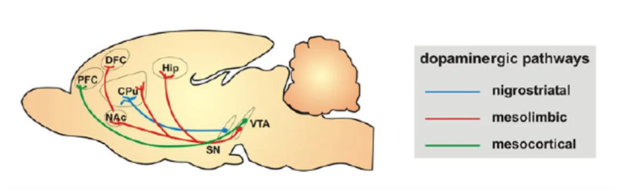
Dopaminergic circuitries can be divided into four main groups: nigrostriatal, mesolimbic, mesocortical, and tuberohypophyseal circuits (Fig.3). The first is the major dopaminergic tract in the brain, and plays an essential role in the control of voluntary motor
14
movement (Baier et al., 2012). The mesolimbic dopaminergic circuit originates in the cell bodies localized in the VTA and projects to NAcc, AMY and hippocampus. Other group of cell bodies in the VTA project to the prefrontal and perirhinal cortex, forming the mesocortical dopaminergic system. Previous reports have shown that these last two pathways overlap substantially, and are collectively referred as the mesocorticolimbic system, being involved in emotional behaviors including motivation and reward (Chinta and Andersen, 2005). In addition, these neurons have been hypothesized to play a critical role for the action of antipsychotic, antihyperactivity and psychostimulant drugs (Baier et al., 2012). Lastly, the tuberohypophyseal system, which is originated in the hypothalamus and innervates the pituitary and the median eminence, is important in the regulation of the release of pituitary hormones (Baier et al., 2012).
Figure 3 - Schematic representation of dopaminergic pathways. Dopaminergic transmition can be divide in four
pathways nigrostriatal, mesolimbic, mesocortical, and tuberohypophyseal systems. The diagram is adapted from Baier CJ, 2012 (Baier et al., 2012).
Work from Kuhn et al showed that Drd1 and Drd2 subtypes of DA receptors contribute for the dopaminergic regulation of the HPA axis function, and stimulated increase the HPA axis response (Borowsky and Kuhn, 1992). Besides modulation of the HPA axis, dopamine is also important for the regulation of emotional, affective and social behaviors (Rodrigues et al., 2011b, Vanderschuren et al., 1997), although, the role of the dopaminergic neurotransmission in the modulation of social play is not yet clear. It has been reported an increase and decrease on social play behaviour, after treatment with dopamine receptor agonists(Siviy et al., 1996, Vanderschuren et al., 1997). Part of the rewarding properties of both opioid and cannabinoid drugs involve dopamine-dependent processes, through direct interaction with dopaminoreceptive neurons in the NAcc (Pierce and Kumaresan, 2006). Complementing this,
15 it was shown by Vanderschuren and colleagues that the effects of the indirect endocannabinoid receptor agonist were blocked by the dopamine receptor antagonist, while those of morphine were not (Trezza and Vanderschuren, 2008a). These results indicate that although the endocannabinoid and opioid systems jointly facilitate social play, they do so through partly dissociable mechanism. The opioid and dopaminergic circuits also seem to interact and modulate the reward behavior (Berridge, 2007, Depue and Morrone-Strupinsky, 2005). Whereas, the mesolimbic dopaminergic system mediates the motivation to a reward, opioids modulate the pleasurable proprieties of a rewarding situation (Berridge, 2003, Kelley et al., 2005, Berridge, 2007). Indeed, there are significant studies showing that the rewarding aspects of social interaction depend on opioid activity (Depue and Morrone-Strupinsky, 2005). Actually, there are many reports evidencing the interaction between opioid and dopaminergic system in social bonding and sexual behavior (Balfour et al., 2004, Young and Wang, 2004).
1.6. Ultrasonic vocalizations – a measure of emotional and social behavior in rodents
Emotional and social behaviors are complex and dynamic and its analysis presents some limitations when using animal models. In the context of this thesis, we decided to complement the behavioral studies with the recording of ultrasonic vocalizations (USVs).
Rats use USVs as a regular feature of their social interactions. Social interactions include aversive and appetitive encounters and the measurement of such USVs can be used an indicative of their emotional status and social capacities (Knutson et al., 1998).
Rat vocalizations are produced through the larynx. Rats use their larynx in two modes: the first, to produce audible sounds of 2-4kHz, by causing vibrations of vocal folds (Nitschke, 1982); And the second, generates USVs, in this case, the larynx is stabilized and used as whistle with a very small orifice created by the vocal cords, which cannot vibrate (I. et al., 2001). This produces the ultrasonic sound by the same principle as a human whistle does, except that the small size of the respiratory tract of the rat produces sound in the ultrasonic range.
16
It is possible to identify three classes of USVs that the rat uses in different situations. Rat pups show evidence of ultrasonic vocalizations around the 40 kHz in response to situations like separation from their litters and mother, or even when the temperature of the environment drops (Hofer, 1996). Adolescent and adult rats emanate two different kinds of USVs, classified according to their frequency, as low (22 kHz) and high (50 kHz) frequency ultrasonic vocalizations.
In situations of danger, circumstances that originate an increase of anxiety (for example, in the presence of an aggressive opponent, predator, large mammal or when the noise is loud), or even when the rat is expecting an unpleasant stimulus, it emits 22 kHz alarm calls, also called as low frequency vocalizations (Brudzynski, 2009). These kinds of sounds are significant advantages in the present of some predators that do not have the capacity to hear ultrasonic vocalizations. Furthermore, these ultrasounds are propagated more directionally than audible sound, they are difficult to detect and localize, and they dissipate easily in environmental obstacles and weather conditions. And if these sounds are emitted in underground burrows the predators in the ground or in the air do not hear it (Brudzynski, 2009). Such vocalizations are not only emitted during the actual aversive event and in response to stimuli associated with these experiences but also in situations of sexual contact (Knutson et al., 1999). The intensity of an aversive emotional state can be revealed by latencies to pronounce vocalizations, their length and loudness or by the number of calls emitted (Wohr et al., 2005).
High frequency 50 kHz USVs occur in situations associated with a positive status and are representative of a state similar to joy (Knutson et al., 1999).They are expressed in rough-and-tumble play, tickling, exploratory activity, meeting rats after a period of separation or in the initial meeting of resident-intruder pair and in rewarding situations (Brudzynski, 2009, Brudzynski and Chiu, 1995). It was observed that these sounds are also emitted in the presence or anticipation of artificial rewarding situations, like pharmacological stimulus (Maier et al., 2010, Burgdorf et al., 2000).
Besides appetitive situations, 50 kHz USVs also occur after separation from conspecifics during short periods of social isolation. Rats taken out from their home cage and individually exposed to a clean cage, as well as the rat that stays alone in the home cage after the conspecifics has been removed, emit 50 kHz USV (Wohr et al., 2008). Actually, the fact
17 that the separation from conspecifics elicits 50 kHz USV is an indicative of an affiliative communicative function of positive USV, namely to (re)establish or to maintain social contact.
It is important to mention, that is possible for the rats to emit with a brief difference of time both types of call, but they cannot acoustically mix the 22 kHz and 50 kHz ultrasounds. These mutually exclusive types of vocalization are regulated by two ascending systems that originate in the brainstem tegmentum: the cholinergic system and the mesolimbic dopaminergic system (Fig.4) (Brudzynski, 2009).
The activation of the cholinergic system, which originates in the laterodorsal tegmental nucleus and extends to the basal forebrain and limbic areas (Fig.4, black arrows), induces defensive behavior and increases the number of 22 kHz ultrasonic vocalizations. It was shown that pharmacologic treatments, that activate this system, induce 22 kHz USVs and the use of antagonists inhibits or decreases these USVs, resulting in a negative affective state (Brudzynski, 2001).
The ascending dopaminergic system, that begins in the VTA and ends in the NAcc and other basal forebrain structures (Fig.4, white arrows), when activated induces behavioral activation (increase in locomotor activity and exploration) and increases 50 kHz vocalizations, resulting in a positive affective state. This state includes relevant changes in the somatic, autonomic, and endocrines systems (Brudzynski, 2009).
18
Figure 4 - Two ascending systems are responsible to initiate the ultrasonic vocalizations. The cholinergic system
has the origin in the laterodorsal tegmental nucleus (LDT), and release acetylcholine in the anterior hypothalamus (AH), preoptic area (PO), bed nucleus of the stria terminalis (BNST), and lateral septum (LS), and has been involved in 22 kHZ USVs emission. The ventral tegmental area (VTA) is the zone of origin of the dopaminergic system. Release dopamine in the nucleus accumbens (NAcc) and neighboring areas, inducing 50 kHz vocalization. The diagram is adapted from Brudzynski SM. 2009 (Brudzynski, 2009).
Due to the potential of the USVs to ascertain the emotional and motivational status of the animals, in this thesis we decided to complement the behavior studies by measuring this tool.
19
21
2. Aims
Accumulating evidence suggests that high levels of GCs during prenatal period increase the propensity to develop a multiplicity of psychiatric disorders in adulthood, such as anxiety and depression.
This project aims to characterize in detail the emotional, social and affective behaviors of adult animals exposed to GCs at gestation days 18 and 19. To further complement the standard behavioral tests, we will also evaluate the number and type of ultrasonic vocalizations (USVs) to ascertain emotional/effective condition in specific behavioral paradigms. As a last part of the project, we intend to identify the neurobiological correlates of such deficits.
The present thesis had the following objectives: 1. Establish and optimize USV measurement at ICVS;
2. Evaluate emotional and social behaviors of iuGC animals in conjunction with USVs recordings;
3. Ascertain the levels of specific neurotransmitter receptors (opioids, endocannabinoids and dopamine) in the mesolimbic circuits of iuGC animals; 4. Revert the behavioral deficits by pharmacological manipulate of affected circuits. We expect that this integrative and multimodal analysis will give insight to the mechanisms underlying stress-induced social and emotional dysfunction and will shed some light about the neurotransmitter system(s) affected.
23
25
3. Materials and methods
3.1. Animals
All manipulations were conducted in accordance with local regulations (European Union Directive 2010/63/EU) and National Institute of Health guidelines on animal care and experimentation. Pregnant Wistar rats were injected with the synthetic GC, dexamethasone (DEX; iuGC animals), at 1mg kg-1 or with saline (CONT; control animals), on days 18 and19 of
gestation.
Male offspring were pair housed, according with antenatal treatment, under standard laboratory conditions: artificial 12h light/dark cycle (lights on from 08:00 a.m. to 08:00 p.m.); room temperature 22ºC; food and water were provided ad libitum.
3.2. Experimental design
Different sets of animals were used during the experimental approach, represented in fig.5.All procedures were performed during the dark period (08:00 p.m.- 02:30 a.m.), except the protocol to assess maternal behavior, the forced swimming test and the light/dark box test.
Animals were assessed for social behavior at different ages (Fig.5). In adulthood, we analysed their emotional status using different behavioral paradigms to assess anxiety, depressive-like behavior and fear. A set of animals was sacrificed and brain areas (AMY, NAcc and HPT) were collected to be used for western blot analysis.
Finally, we tried to reverse the phenotype by pharmacological manipulation.
Figure 5 - Schematic representation of the experimental approach. Dexamethasone
(1mg/kg) or saline
injection on pregnant rats
PND1 PND21 3 months Sacrifice Pups: -Social Behavior Maternal Behavior. Juveniles: -Social Behavior: Tumble and play;
Adults: -Emotional Status: FST; Physical enrichment; Anticipatory behavior to f ood; L/D box test; Fear conditioning; -Communicative f unction. Western blot
26
3.3. Behavioral characterization
3.3.1. Emotional Status
3.3.1.1. Forced swimming test
The forced swimming test (FST) apparatus consists in a glass cylindrical tank, which was filled with water (24 ± 1ºC) to the depth enough so the animals could not support themselves by placing the paws or tail on the base of the cylinders. The water was cleaned between trials.
On the first day (pre-test day) and second day (test day), rats were placed inside the cylinder for 5 min (Roque et al., 2011). On both days, UVS and behavior was recorded but only the second day was scored. The characteristics assessed were climbing time, immobility time (when the rat is floating in the water), and latency to be immobile.
3.3.1.2. Physical Enrichment
The apparatus consisted in an arena with 43.2x43.2 cm, with transparent acrylic walls and white floor (MedAssociates). Adult rats were individually habituated to the arena for 10 min with toys for two consecutive days. On the testing day, the animals were social isolated for 3.5 h before testing (Trezza and Vanderschuren, 2008b). After this period, the animals were individually placed in the arena with toys for 10 min. Immediately after, the ultrasonic vocalizations were recorded. Before each trial, the apparatus was cleaned with Ethanol 10%, in order to remove odor from the previous animal.
3.3.1.3. Anticipatory behavior to food (feeding sessions)
The feeding sessions protocol was carried out for seven days as described previously (Burgdorf et al., 2000). Testing occurred in cages similar to their home cages. The testing box measured 30x20x30 cm, with a steel grid and approximately 3 cm of wood shavings covering the floor.During 6 consecutive days, the subjects were allowed to access food for 1hr per day. In the beginning of the test, the subjects were placed individually in the testing cage. After 3 min (basal situation), a white light located 10cm above the floor was turned on and it stayed on
27 (‘cue exposure’) during 2 min, until the beginning of the feeding session, when they received a recipient full of food. On the seventh day, after the 2 min with a light on, instead of receiving food, an empty recipient was placed inside the testing box (‘extinction’). USVs were recorded throughout the days, during the cue exposure and extinction periods. After the seventh day, the subjects were allowed to access food ad libitum.
3.3.1.4. Light/dark box test
The light/dark box test (L/D test) was performed inside the open field arena (43.2x43.2 cm, transparent acrylic walls and white floor) (MedAssociates Inc., St.Albans, Vermont). The animals were individually placed in the center of the illuminated part. A dark compartment was attached to one side with an opening facing the center of the open field. The distance travelled and time spent in each compartment were recorded in a single-trial of 10 min.
3.3.1.5. Confined cage test
The confined cage test was performed in a non-restrictive Plexiglas cylinder (inner diameter 8.8 cm, length 22.2 cm), mounted on a Plexiglas platform and placed in a ventilated, sound-attenuated chamber (SR-LAB, San Diego Instruments, San Diego, CA,USA). Inside of the cylinder was placed a stainless steel grid, through which an electric current could be passed (shock chamber). The microphone and a video camera were placed inside the sound attenuated chamber. The protocol was realized in two consecutive days, where the animals were placed inside of the shock chamber for 11 min. The USVs and percentage of total time freezing (total time which the animal didn’t move) were measured. Percentage of total time freezing was calculated as the probability of the time freeze of an animal by the total time in the chamber. Chambers were cleaned between tests (ethanol 10 %) in order to remove olfactory cues.
28
3.3.1.6. Fear conditioning
The fear conditioning test was performed in the same apparatus as the confined cage test. The protocol was realized in three consecutive days (Borta et al., 2006). During the first day (habituation), each animal was placed in the sock chamber for 11 min. On conditioning day (second day), each subject was positioned again inside of the shock chamber. After the first 3 min, where no light or shock was given, the animal was exposed to six lights on/shock pairings, each followed by an inter-stimulus interval (ISI) of 60s. This shock (0,4mA ± 0,1) was administered during 500ms, after the period (20s) with the light on. Following conditioning, animals return to their home cages.
In the following day (test day), the animals were again placed in the shock chamber for 11 min. After the initial phase of 3 min, the light (3 W, incandescent light bulb) was presented six times for 20s each but not shock was given, again with an ISI of 60s. During all procedures USVs and behavior (freezing) were recorded. Percentage of total time freezing was calculated as the probability of the time freeze of an animal by the total time in the chamber. Chambers were cleaned between tests, as mentioned before.
3.3.2. Social behavior
3.3.2.1. Maternal Behavior
Maternal behavior was assessed on postnatal days (PND) 3, 7 and 15. The mothers and pups (litters) were measured together in their home cage (basal situation), during 5 min (Hofer and Shair, 1978). The procedure was performed during the light phase (08:00 a.m.) and during dark phase (08:00 p.m.).
3.3.2.2. Tumble and play
Tumble and play test (Trezza and Vanderschuren, 2008b) was performed in cages similar to their home cages, which measured 30x20x30 cm, with approximately 2 cm of wood shavings covering the floor. For behavior recordings a camera with zoom lens was used. The characteristics of the social behavior scored were: pouncing (this is an index of play solicitation, i.e., when one of the animals is attempting to nose or rub the nape of the neck of the test
29 partner), pinning (when one of the animals is lying with is dorsal surface on the floor with the one animal on top of him) and social exploration (when animals are sniffing any part of the body, including the anogenital area, of the test partner). Play response to solicitation was calculated as the probability of an animal of being pinned in response to pouncing by the test partner.
Juvenile rats were socially isolated for 3.5 h before testing. Each animal was then placed into the test cage with their sibling pair for 15 min. This protocol was performed three times. On the first trial, two familiar animals living in the same cage (from the same exposure group) were tested (familiar pairs). On the second trial, two unfamiliar animals (from the same exposure group) were tested (unfamiliar pairs). Lastly, we tested social interaction between one CONT animal and one iuGC animal (CONT-iuGC pairs). Besides behavior, we also measured the positive USVs during 15 min of the test, immediately after the animals were placed in the test cage.
3.3.2.3. Communicative function
In order to assess the social interaction in adult animals, we isolated one animal from the home cage and individually exposed it to a clean new cage for 15 min. During this time we recorded the USVS from the two animals, i.e., from the one isolated and the remaining animal in the home cage. After this period, animals were reunited and, once again, USVs were recorded, along with their social behavior (pinning, pouncing and social exploration) throughout the 15 min of the test.
3.3. Western Blot
Specific areas (AMY, NAcc and HPT) were collected by macrodissection, and subsequently frozen at -80ºC. For western blot, lysis buffer (50 mM Tris pH 7.4, 50 mM EDTA, 50 mM NaCl, 100 mM phenylmethylsulfonyl fluoride, complete protease inhibitors) was added to each frozen area. After, samples were manually homogenized and centrifuged at 13 000 r.p.m. for 10 min at 4ºC. The supernatant was collected and quantified using the Bradford method. Fifty µg of protein was loaded into a SDS-polyacrylamide gel, and gel was then
30
transferred to a nitrocellulose membrane. After, membranes were incubated overnight at 4ºC with the primary antibodies: rabbit anti-k opioid receptor 1 (1:2500, Invitrogen), rabbit anti-µ opioid receptor 1 (1:1000, Invitrogen), rabbit anti-dopamine D1 receptor (1:2500, Abcam), rabbit anti-dopamine D2 receptor (1:1000, Abcam) and rabbit anti-endocannabinoid receptor 1 (1:500, kindly provided by Ken McKenzie). After incubation with the secondary antibodies, membranes were developed with luminol reagent (SantaCruz). Films were analyzed using ImageJ software.
3.4. Pharmacological reversal of behavioral alterations
L-DOPA/carbidopa (Sinemet, Merck, NJ, USA) was administered daily, 4h before testing, at a dose of 24 mg kg-1 (in water) by oral gavage.
The administration of L-DOPA/carbidopa started three days before the animals being tested for emotional behavior (FST, food sessions- anticipation behavior, fear conditioning), social and affective behaviors (tumble and play) and also for communicative function.
3.5. USVs analysis
An Ultrasound Microphone (CM16/CMPA, Avisoft Bioacoustics, Berlin, Germany) sensitive to frequencies of 10-200 kHz, was used, 20 cm above the floor, in all experiences. It was connected via an Avisoft UltrasoundGate 416H (Avisoft Biocoustics) to a personal computer. Vocalization was recorded using the Avisoft-Recorder (version 5.1.04) with the following settings: sampling rate: 250000; format: 16 bit. For acoustical analysis, recordings were transferred to Avisoft SASLab Pro (version 5.1.22, Avisoft Bioacoustics). This program was used in order to produce spectrograms of USVs by conducting a fast Fourier transformation (256 FFT-length, 100% frame, Hamming window filter, 50% time window overlap). These spectrograms had a frequency resolution ~1.2 kHz and a temporal resolution ~0.4ms.
Twenty-kHz call detection was provided by an automated threshold-based algorithm (threshold: -40 dB) and a hold time mechanism (hold time: 20 ms). A lower-cut-off-frequency of
31 18 kHz was use to reduce background noise. For detection of 40 kHz was used an automated threshold-based algorithm of -50 dB, an hold time of 10 ms and a high pass of 30 kHz (lower-cut-off-frequency). In support of 50 kHz call detection, it was used the same threshold (-40 dB) as the 20 kHz USVs, but a hold time of 5 ms and a lower-cut-off-frequency of 40 kHz.
Calls were also inspected manually to ensure that, when necessary, USVs not detected automatically could be subsequently included in the automatic parameter analysis. Various parameters, including peak frequency, peak amplitude and bandwidth, which were determined for each element of the entire spectrogram, were determined automatically. Temporal parameters determined included latency to call, call duration and the duration of intervals between subsequent calls. Finally, the total number of calls emitted was scored.
3.6. Statistical analysis
Statistical significance was determined using the GraphPad Prism 5 software. All data were represented as mean + SEM. Data were verified for Gaussian distribution. Unpaired t-tests was use to determine whether CONT and iuGC animals differ in USVs production and behavior during tumble and play, EPM, FST, L/D box, and fear conditioning tests. When no Gaussian distribution was assumed, we used a nonparametric test (Mann-Whitney U-test).
An two-way ANOVA for repeated measurements was used to test the differences between groups throughout time during the anticipatory behavior to food and physical enrichment tests. Significance is referred as * for P < 0.05, ** for P <0.01, *** for P <0.001.
33
35
4. Results
4.1. Effects of iuGC treatment on emotional behavior of offspring
Previous works from our lab have shown that in utero glucocorticoid (iuGC) exposure impairs emotional behavior, leading to an anxious phenotype, depressive-like behavior and drug-seeking behavior (Oliveira et al., 2006, Pego et al., 2008, Rodrigues et al., 2011b, Roque et al., 2011). To further characterize these animals, we implemented a new technique at the ICVS, which is the measurement of USVs (ultrasonic vocalizations), allowing to combine behavioral assessment and USVs emissions.
Depressive-like-behavior
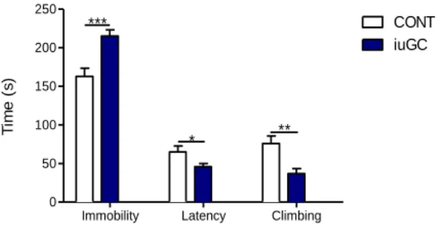
In order to assess depressive-like behavior, animals performed the forced swimming test (FST). As expected, iuGC animals presented an increase in total time immobile (Fig.6, t=4.095, P=0.0005), decreased latency to immobility (Fig.6, t=2.207, P=0.038) and reduced climbing (Fig.6, t=3.387, P=0.0027) when compared with CONT animals.
Immobility Latency Climbing
0 50 100 150 200 250 CONT iuGC *** * ** T im e ( s )
Figure 6 - Prenatal administration of iuGC leads to depressive-like-behavior in the FST test. iuGC present
increased immobility time, decrease latency to immobility and reduced climbing time. nCONT=12; niuGC=12;
CONT=Controls; iuGC=in utero glucocorticoids; *P <0.05, ** P <0.01, *** P <0.001.
Depression leads to a lack of motivation and diminished pleasure feelings (Frank et al., 2007, Der-Avakian and Markou, 2012). In order to assess motivation/pleasure, we analysed
36
the emission of USVs in an enriched environment in socially isolated animals. In addition, we also measured USVs in response to a cue that anticipates food in food deprived animals.
During the enrichment test (arena with toys), iuGC animals emitted less 50 kHz USVs when compared with CONT (Fig.7, F1,41= 5.58, P=0,023), this difference was more accentuated
in the initial day.
3 4 5 0 20 40 60 80 CONT iuGC 1 2 Days N u m b e r o f 5 0 k H z U S V s
Figure 7 - Prenatal administration of iuGC leads to a diminished affective state in a novel enriched environment.
iuGC emitted less 50 kHz USVs than CONT animals, throughout time. nCONT=8; niuGC=8; CONT=Controls; iuGC=in
utero glucocorticoids.
Relative to anticipatory behavior to food, animals were exposed to a cue, in this case a light, for 6 consecutive days, that predicted a bowl of food, and during this period the USVs were measured. Both groups increased the emission of positive calls over time (Fig.8A, F5,82=5.83, P=0,0001), suggesting that they learned the task. Over the days, number of USVs
was substantially different between groups, with the iuGC animals emitting significantly less USVs than CONT animals (Fig.8A, F1,82=13,95, P=0.0003). On the seventh day, we also
measured the USVs during the extinction period, i.e., after the light was off, the animal was presented with an empty recipient. Comparing both groups during cue exposure and extinction period, we observe that CONT rats emitted less in the second period (Fig.8B, t=2.659, P=0.0187), whereas the iuGC subjects emitted approximately the equal number of calls in the two periods (Fig.8B, t=0.7442, P=0.4691).
37 0 1 2 3 4 5 6 7 0 20 40 60 80 100 120 CONT iuGC *** Days N u m b e r o f 5 0 k H z U S V s
CONT iuGC CONT iuGC
0 20 40 60 80 100 120
Cue Exposure Extinction
* * N u m b e r o f 5 0 k H z U S V s A B
Figure 8 - Prenatal administration of iuGC leads to a lack of motivation/pleasurable feelings in anticipation to
food. (A) Number of positive calls over the six consecutive days, during cue exposure, emitted by iuGC and CONT rats. Both animals increase the emission of positive calls over time although iuGC emitted less than CONT animals. (B) Number of 50 kHz USVs, during cue exposure and extinction periods (7th day). iuGC animals emitted
almost the same number of USVs in the two periods, while the CONT emitted significantly less in the second period when compared with the first. nCONT=8; niuGC=8; CONT=Controls; iuGC=in utero glucocorticoids; *P <0.05, **
P <0.01, *** P <0.001.
Anxiety
In order to assess if iuGC treated animals presented an anxious phenotype, we performed the light/dark box test. iuGC spend more time in the dark box than CONT animals (Fig.9A, t=2.215, P=0.0407). Regarding the distance travelled during the test, no differences were found (Fig.9B, dark: t=1.384, P=0.1854, light: t=1.825, P=0.0867).
Dark Light 0 200 400 600 CONT iuGC * T im e ( s ) Dark Light 0 1000 2000 3000 4000 CONT iuGC Di s ta n c e ( c m ) A B
Figure 9 - Prenatal administration of dexamethasone leads to the development of anxious phenotype. Time (A)
38
with CONT, concerning the distance no differences were found. nCONT=10; niuGC=10; CONT=Controls; iuGC=in utero
glucocorticoids; *P <0.05, ** P <0.01, *** P <0.001.
An additional paradigm was performed in order to assess the anxious phenotype. Adult animals were placed inside of a novel and confined cage for 11 min. Control animals emitted very few negative 22 kHz calls, but iuGC animals emitted significantly more (Fig.10A, U=9, P=0.0202). Regarding freezing behavior, no differences were found (Fig.10B, t=0.06417, P=0.9498). After habituation to the test box, iuGC animals no longer emitted 22 kHz vocalizations (Fig.10A), and a slight decrease in the percentage of freezing was observed (Fig.10B, U=25.5, P= 0.8162).
CONT iuGC CONT iuGC
0 50 100 150 * 1º Day 2º Day N u m b e r o f 2 2 k H z U S V s
CONT iuGC CONT iuGC
0 1 2 3 4 5 6 7 1º Day 2º Day % o f fr e e z in g A B
Figure 10 - Prenatal administration of dexamethasone leads to the development of an anxious phenotype.
Number of 22 kHz calls (A) and percentage of freezing time (B) in a novel and confined cage. iuGC animals in an anxiogenic environment emitted more negative calls than CONT, while no differences were found in the freezing behavior, on the first day. In the second day, both groups did not emit any 22 kHZ calls, and no differences were found in the percentage of freezing. nCONT=8; niuGC=8; CONT=Controls; iuGC=in utero glucocorticoids; *P <0.05, **
P <0.01, *** P <0.001.
Fear
We also assessed fear behavior, by conditioning the animals to a cue (light) paired with electric shocks. After the first 3 min of cage habituation, we did not observe either emission of negative calls or any time immobile. During the period of 6 light/shock pairs, as expected, both groups emitted several 22 kHz calls and spent more time immobile than in the initial phase, but iuGC to a greater extent (Fig.11A, t=2.611, P=0.0242; Fig.11C, t=2.355, P=0.0336, respectively). On the following day, the test day, animals were exposed to a cue again, but no