Amphotericin B fungicidal activity:
a comprehensive analysis of fungal recovery
Rita Teixeira dos Santos
D
2017
Rita
Teixeira dos Santos
Amphotericin B fungicidal activity:
a comprehensiv
e anal
ysis of fungal reco
ver
y
D.
FMUP
2017
Amphotericin B fungicidal activity:
a comprehensiv
e anal
ysis of fungal reco
ver
y
Rita
Teixeira dos Santos
FA CULD ADE DE MEDICINA D A UNIVER SID ADE DO POR T O
Dissertação de candidatura ao grau de doutor em Biomedicina,
apresentada à Faculdade de Medicina da Universidade do Porto
Programa Doutoral em Biomedicina
Medicina da Universidade do Porto, Portugal.
Orientação
Professora Doutora Cidália Pina Vaz
Co-orientação
Professor Doutor Acácio Gonçalves Rodrigues
Júri da Prova de Doutoramento em Biomedicina
Presidente
Doutor José Eduardo Torres Eckenroth Guimarães, professor catedrático da Faculdade de Medicina da Universidade do Porto
Vogais
Doutor José António Martinez Souto de Oliveira, professor catedrático da Faculdade de Ciências da Saúde da Universidade da Beira Interior
Doutora Cidália Irene Azevedo Pina Vaz, professora associada da Faculdade de Medicina da Universidade do Porto
Doutora Teresa Maria Fonseca Oliveira Gonçalves, professora auxiliar da Faculdade de Medicina da Universidade de Coimbra
Doutor Manuel Joaquim Lopes Vaz da Silva, professor auxiliar da Faculdade de Medicina da Universidade do Porto
Doutor Luís Filipe Duarte Reino Cobrado, professor auxiliar da Faculdade de Medicina da Universidade do Porto
Doutora Cármen Maria Lisboa da Silva, professora auxiliar da Faculdade de Medicina da Universidade do Porto
Artigo 48°, § 3° - “A Faculdade não responde pelas doutrinas expendidas na Dissertação.”
(Regulamento da Faculdade de Medicina da Universidade do Porto – Decreto de Lei n° 19337 de 29 de Janeiro de 1931)
ix
“Aqueles que passam por nós, não vão sós, não nos deixam sós. Deixam um pouco de si, levam um pouco de nós.”
Antoine de Saint-Exupéry
E porque não fiz este caminho sozinha gostaria de expressar a minha enorme gratidão…
À Professora Cidália Pina Vaz, minha orientadora, pela sua orientação ao longo deste trabalho, pelo seu entusiamo contagiante, por ouvir com interesse as minhas questões, e sobretudo, pela confiança.
Ao Professor Acácio Rodrigues, meu co-orientador e Director do Serviço e Laboratório de Microbiologia, agradeço pela forma como me acolheu neste Serviço, por proporcionar as condições para o desenvolvimento deste trabalho, pela disponibilidade e orientação, e pela partilha de conhecimentos.
À Susana Guerreiro e ao Ricardo Branco, pela colaboração neste trabalho e pela disponibilidade e atenção que tiveram comigo.
A todos os colegas do Serviço e Laboratório de Microbiologia, pela vossa disponibilidade e partilha de conhecimentos. Foi convosco que cresci, profissionalmente e pessoalmente, ao longo dos últimos anos. Agradeço em especial à Isabelita, não só por todo o seu apoio técnico durante a realização deste trabalho, mas também pelo apoio emocional e amizade de todos os dias. Agradeço à Elisabete, pelo seu apoio na realização deste trabalho e pela amizade.
Aos amigos de sempre e aos novos que surgiram pelo caminho. Em mim existe muito de Vós. Ao João e à Inês pelo apoio e amizade. À Nádia, por todo o apoio diário nesta fase final e por fazer lembrar-me todos os dias o sentido da amizade.
Aos Tios, à Carla e à Rute, por darem sentido à palavra família, pela confiança e amor.
À Teresa e ao Serafim, pelo carinho e amizade.
À Susana e ao Tomás, pelo amor e pela vossa presença no meu caminho. Ao Tomás agradeço ainda pela inspiração e por me mostrar o lado mais genuíno da Vida.
x
À minha Avózinha, pelo amor, pela preocupação de todos os dias, por todos os ensinamentos e por acreditar em mim e na minha força. Ao meu Avô, que acompanha todos os meus passos e que, de certo, acompanhou mais esta conquista.
Aos meus Pais, a quem devo tudo o que sou e onde cheguei. Agradeço pelo amor e apoio incondicional. E a ti Mãe, que acompanhaste de perto os bons e maus momentos deste caminho, que cuidas de mim e de todos Nós, Obrigada por tudo e por seres tão especial!
“Nada acontece por acaso. Não existe a sorte.
Há um significado por detrás de cada pequeno acto.
Talvez não possa ser visto com clareza imediatamente, mas sê-lo-á antes que passe muito tempo.”
xiii
Ao abrigo do artigo 8° do Decreto-Lei n.o 388/70, fazem parte desta dissertação as seguintes publicações:
Manuscripts
I. Teixeira-Santos R., Rocha R., Moreira-Rosário A., Monteiro-Soares M., Cantón E.,
Rodigues A. G., Pina-Vaz C. (2012). Novel method for evaluating in vitro activity of anidulafungin in combination with amphotericin B or azoles. J Clin Microbiol. 50(8):2748-54.doi: 10.1128/JCM.00610-12.
II. Teixeira-Santos R., Ricardo E., Gomes-Guerreiro S., Costa-de-Oliveira S.,
Rodrigues A. G., Pina-Vaz C. (2015). New Insights Regarding Yeast Survival following Exposure to Liposomal Amphotericin B. Antimicrob Agents Chemother 59: 6181-6187. doi: 10.1128/AAC.00575-15.
III. Teixeira-Santos R., Ricardo E., Branco R. J., Azevedo M. M., Rodrigues A. G.,
Pina-Vaz C. (2016). Unveiling the Synergistic Interaction Between Liposomal Amphotericin B and Colistin. Front Microbiol 7:1439. doi: 10.3389/fmicb.2016.01439.
Abstracts published in conference proceedings
I. Teixeira-Santos R., Silva A. P., Costa-de-Oliveira S., Rodrigues A. G., Pina-Vaz C.
(2013). Uncovering yeast recovery pathway to liposomal amphotericin B-induced stress. Mycoses, Vol. 56, Suppl.3, 68-68.
xv
L
IST OF
A
BBREVIATIONS
5-CFDA 5-Carboxyfluorescein diacetate
5-FC 5-flucytosine
AIDS Acquired immunodeficiency syndrome
AMB Amphotericin B
AND Anidulafungin
ATB Automated Topology Builder AUC Area under the curve
AZM Azithromycin
BMD Broth microdilution method BSI Blood stream infection CBP Clinical breakpoint CFU Colony forming unit
CI Confidence interval
CL Clearance
CLR Clarithromycin
CLSI Clinical and Laboratory Standards Institute Cmax Maximum (or peak) plasma concentration
CSF Caspofungin
CST Colistin
DAPI 4',6-diamidino-2-phenylindole
DC Depolarized cells
DCF 2’,7’- dichlorodihydrofluorescein DCFH-DA 2,7-dichlorofluorescin diacetate
DiBAC4(3) Bis-(1,3-Dibutylbarbituric Acid) Trimethine Oxonol DNA Deoxyribonucleotide Acid
ECMM European Confederation of Medical Mycology ECV Epidemiological cutoff value
ERG Ergosterol
EUCAST European committee on antimicrobial susceptibility testing
FC Flow cytometry
FI Fluorescence intensity
FICI Fractional inhibitory concentration index
FLU Fluconazole
FUN-1 2-chloro-4-(2,3-dihydro-3-methyl-[benzo-1,3-thiazol-2-yl]-methylidene)-1 HIV Human immunodeficiency virus
Hsp Heat shock protein
xvi
L-AMB Liposomal amphotericin B
LPS Lipopolysaccharide
LZD Linezolid
MCF Micafungin
MD Molecular dynamics
MIC Minimal inhibitory concentration MIF Mean intensity of fluorescence NPT Isothermal-isobaric nwt non-wild-type PA Proportion of agreement PBS Phosphate-buffered saline PI Propidium iodide PMT Photomultiplier POS Posaconazole RIF Rifampicin
RNA Ribonucleic acid
RNAP RNA polymerase
ROS Reactive oxygen species
SDA Sabouraud dextrose agar
SI Staining index
TET Tetracycline
TUNEL Terminal deoxynucleotidyl transferase dUTP nick end labeling
VOR Voriconazole
wt wild-type
xvii
L
IST OF
T
ABLES AND
F
IGURES
List of Tables
Chapter III. New insights regarding yeast survival following exposure to Liposomal Amphotericin B
Table 1 - Yeast strains used in this study. Liposomal amphotericin B susceptibility test
results and membrane potential of yeast cells treated with L-AMB plasma concentrations after 3, 6 and 24 h, expressed as percentage of depolarized cells.
Chapter IV. Novel method for evaluating in vitro activity of anidulafungin in combination with Amphotericin B or Azoles
Table 1 - In vitro interaction of anidulafungin and amphotericin B by the checkerboard
and flow cytometry methods against 39 Candida species.
Table 2 - In vitro interaction of anidulafungin and fluconazole by the checkerboard and
flow cytometry methods against 36 Candida species.
Chapter V. Unveiling the synergistic interaction between Liposomal Amphotericin B and Colistin
Table 1 - Minimal inhibitory concentration (MIC) of liposomal amphotericin B alone and
xviii
Chapter II. IntroductionFigure 1 - Mechanism of action of antifungal drugs available for the treatment of
invasive fungal infections. Polyenes bind to ergosterol, a main component of the cell
membrane, forming pores in the membrane. Azoles antifungals inhibit the enzyme lanosterol 14α-demethylase, involved in the synthesis of ergosterol. This inhibition leads to the production of toxic compounds altering cell membrane structure and permeability. Echinocandins inhibit the enzyme 1,3-β-D-glucan synthase leading to alterations in the cell wall structure. Pyrimidine analogues induce the production of toxic compounds that interfere with the nucleic acid and protein synthesis.
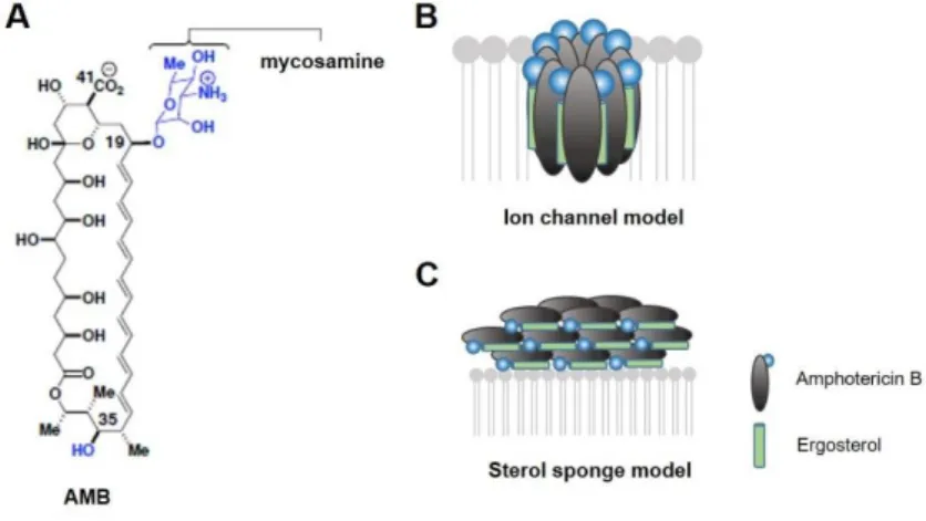
Figure 2 - Models for the structure and function of amphotericin B. (A) Molecular
structure of AMB. (B) The classic ion channel model for the structure and function of AMB. (C) A new sterol sponge model, in which AMB primarily exists in the form of large extramembranous aggregates that extract ERG from lipid bilayers (Anderson et al., 2014).
Chapter III. New insights regarding yeast survival following exposure to Liposomal Amphotericin B
Figure 1 - Effect of liposomal amphotericin B on S. cerevisiae BY4741 and C.
albicans 596. (A) Study design scheme. (a) Plasma concentration-time curve obtained
after a first infusion of AMB at 3 mg/L. (b) The scheme proposed in accordance with L-AMB plasma levels described by Walsh et al. (Walsh et al., 1998); (c) Dashed line represents the treatment with a constant concentration of 3 mg/L L-AMB during 24 hours. (B) Viability assessment by CFU enumeration of S. cerevisiae BY4741 and C. albicans 596 cells exposed to treatment conditions (b) and (c). Data at respective time points are given as mean ± standard deviations. An asterisk indicates significant differences between the two treatment conditions.
xix
Figure 2 - Effect of liposomal amphotericin B on S. cerevisiae BY4741 physiological parameters. (A) Cell membrane integrity was assessed with propidium iodide (PI). (B)
Cell membrane potential was evaluated using DiBAC4(3). (C) Metabolic activity was determined by 5-CFDA staining. (D) Endogenous reactive oxygen species (ROS) production as determined by DCFH-DA staining. An asterisk indicates significant differences (P < 0.05) between the two treatment conditions.
Figure 3 - TUNEL staining of S. cerevisiae BY4741 cells exposed to liposomal amphotericin B. (A) Fluorescence microscopy imaging showing TUNEL-positive cells
after treatment with L-AMB (a) plasma concentrations and with 3 mg/L (b) after 3, 6 and 24 h. (B) Percentage of cells exhibiting damaged DNA (i.e., cells positive by TUNEL) after treatment with L-AMB, as assessed by flow cytometry. An asterisk indicates significant differences (P < 0.05) between the two treatment conditions. (C) Nuclear fragmentation as shown by DAPI staining. Fluorescence microscopy imaging with (a) and without (b) DAPI filter. S. cerevisiae cells exposed to L-AMB exhibit an irregular shape and fragmented DNA, two findings in accordance with DNA damage during apoptosis.
Chapter IV. Novel method for evaluating in vitro activity of Anidulafungin in combination with Amphotericin B or Azoles.
Figure 1 - In vitro antifungal activities of anidulafungin and amphotericin B.
Distribution of fluorescence intensity of the C. albicans 0207 AND-susceptible strain (A), C. parapsilosis 0136 AND-nonsusceptible strain (B), C. albicans O207 AMB-susceptible strain (D), and C. lusitaniae D51 AMB-nonsusceptible strain (E). In each histogram, the autofluorescence is represented by line (a); line (b) represents the fluorescence of untreated cells stained with DiBAC4(3); line (c) is the fluorescence of cells treated with 70% ethanol and stained with DiBAC4(3) (positive control); line (d) is the fluorescence of cells treated with 1 mg/L of antifungal drugs during 1 h and stained with DiBAC4(3). (C, F) Determination of the number of CFU (CFU/mL) of cell suspensions treated with different antifungal concentrations under conditions identical to those of the flow
xx
susceptible strain by the light-gray bars.
Figure 2 - In vitro antifungal activities of anidulafungin and fluconazole. Distribution
of fluorescence intensity of C. albicans 0207 AND-susceptible strain (A), C. parapsilosis 0136 AND-nonsusceptible strain (B), C. albicans O223 FLU-susceptible strain (D), and C. albicans O216 FLU-nonsusceptible strain (E). In each histogram, the autofluorescence is represented by line (a); line (b) represents the fluorescence of untreated cells stained with FUN-1; line (c) is the fluorescence of cells treated with 70% ethanol and stained with FUN-1 (positive control); line (d) is the fluorescence of cells treated with antifungal drugs (1 mg/L of AND and 16 mg/L of FLU) during 1 h and stained with FUN-1. (C, F) Determination of the number of CFU (CFU/mL) of cell suspensions treated with different antifungal concentrations under conditions identical to those of the flow cytometric assay. The nonsusceptible strain is represented by the dark-gray bars and the susceptible strain by the light-gray bars.
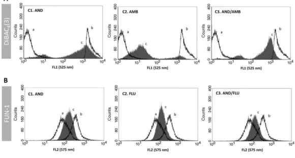
Figure 3 - Evaluation of antifungal combination effect using flow cytometry. (A)
Flow cytometric analysis of the combination effect between anidulafungin and amphotericin B on the C. albicans O215 strain, an example of synergistic association. Line (a), fluorescence of untreated cells stained with DiBAC4(3); line (b), fluorescence of cells treated with 70% ethanol and stained with DiBAC4(3); line (c), fluorescence of cells treated with antifungal drugs and stained with DiBAC4(3); C1, cells treated with a subinhibitory concentration of AND (0.5× MIC); C2, cells treated with a subinhibitory concentration of AMB (0.5× MIC); and C3, cells treated with a subinhibitory concentrations of both antifungal drugs in association (AND 0.5× MIC + AMB 0.5× MIC). (B) Flow cytometric analysis of the combination effect between anidulafungin and fluconazole on the C. albicans OL196 strain, an example of indifferent association. Line (a), fluorescence of untreated cells stained with FUN-1; line (b), fluorescence of cells treated with 70% ethanol and stained with FUN-1; line (c), fluorescence of cells treated with antifungal drugs and stained with FUN-1; C1, cells treated with a subinhibitory concentration of AND (0.5× MIC); C2, cells treated with a subinhibitory concentration of
xxi
FLU (0.5× MIC); and C3, cells treated with subinhibitory concentrations of both antifungal drugs in association (AND 0.5× MIC + FLU 0.5× MIC).
Chapter V. Unveiling the synergistic interaction between Liposomal Amphotericin B and Colistin
Figure 1 - Effect of liposomal amphotericin B in combination with colistin upon C.
albicans (strain 596) physiological parameters. (A) Viability assessment by colony
forming units (CFU) enumeration of C. albicans cells exposed to (■) L-AMB, (■) CST, and (■) L-AMB in association with CST. The CFU counts of cells non-treated are represented as (■). (B) Cell membrane potential was evaluated using DiBAC4(3) staining. (C) Cell membrane integrity was evaluated using propidium iodide (PI) staining. (D) Cell metabolic activity was evaluated using 5-CFDA staining. Data at respective time points corresponds to mean ± standard deviations. *p values < 0.05, significant differences between the treatments with L-AMB alone and CST alone vs. the control (non-treated cells); and between the association L-AMB/CST and L-AMB alone.
Figure 2 - Effect of liposomal amphotericin B (L-AMB) in association with colistin upon endogenous ROS production. (A) Endogenous reactive oxygen species (ROS)
production determined by DCFH-DA staining, assessed by flow cytometry. (B) Fluorescence microscopy imaging showing ROS-positive cells after treatment with b) L-AMB and c) CST alone and d) in association after 24 h of exposure; a) control, non-treated cells. *p values < 0.05, significant differences between the treatments with L-AMB alone and CST alone vs. the control (non-treated cells); and between the association L-AMB/CST and L-AMB alone.
Figure 3 - Self-assembly of amphotericin B (AMB) and colistin (CST) (represented in sticks) in water medium (represented in lines) through molecular dynamics (MD) simulation studies. (A) Complex conformation snapshot after 20 ns of simulation,
including water molecules within 4 Å of the complex. (B) Time evolution of the h-bond distance between two molecules during the complex formation.
xxiii
A
BSTRACT
Invasive fungal infections (IFIs) have increased significantly over the last decades. Amphotericin B (AMB) represents a very important therapeutic option for the treatment of these infections. Interestingly, despite more than 50 years of use in clinical setting, emergence of resistance to AMB is an extremely rare event. However, although the often confirmed in vitro susceptibility, the clinical response to AMB is sometimes reduced. While AMB is effective as a fungicidal drug, several factors can impair its clinical utility, namely underlying diseases, other concomitant administered drugs, timing of antifungal therapy, as well as AMB pharmacokinetics and distribution. The present study sought to understand this long-standing question in a biological perspective. It was evaluated the cascade of functional changes occurring in yeast cells, induced by exposure to liposomal amphotericin B (L-AMB), in a variable concentration simulating the human plasma concentration, along 24 h. Indeed, under such condition, yeast cells developed compensatory responses at distinct levels like membrane permeability, metabolic activity, and production of endogenous reactive oxygen species (ROS) during the first 6 h after the exposure to high plasma concentrations (20 to 5 mg/L); in the remaining 18 h, when exposed to much lower L-AMB concentrations (at or below minimal inhibitory concentration [MIC]), cells revealed almost full recovery with no evidence of fungicidal activity. These results highlighted the importance of monitoring and maintaining L-AMB at sufficient concentrations in plasma and tissue to ensure it produces its fungicidal effect.
Different strategies aiming to improve the clinical efficacy of AMB were also explored. Combined antifungal therapy could be a promising approach for the treatment of IFIs. However, late diagnosis and poor clinical response to antifungal monotherapy frequently promote the use of empirical antifungal combination as salvage therapy, even without scientific base. Since the methodologies available for in vitro evaluation of drug associations are very laborious, and thus not compatible with the daily routine of the microbiology laboratory, it was developed a novel assay using flow cytometry, based upon the classic checkerboard method. A cytometric algorithm was determined for the classification of the association between anidulafungin (AND) and AMB or azoles against yeasts as synergistic, indifferent, or antagonistic interaction. This novel protocol exhibited high agreement with the traditional checkerboard method, having the advantage of
xxiv
susceptibility patterns to AND, AMB and azoles, was evaluated using this new protocol. The association of AND with AMB or azoles was synergistic for a vast amount of the isolates tested. However, an indifferent effect was frequently found, and a few cases of antagonism occurred. Although this therapeutic approach seems to be promising, these results stress that the association of antifungal drugs should be conveniently evaluated in vitro before its clinical use.
Likewise, the combination of antifungals with antibacterial agents deserved a particular attention. The combination of L-AMB with several antibacterial agents was investigated. The association of compounds that act by inhibiting RNA/protein synthesis, namely rifampicin, azithromycin, clarithromycin, and tetracycline with L-AMB was synergistic against Candida spp. and Aspergillus fumigatus. The most effective association involved L-AMB and colistin (CST), a drug that acts by disrupting the cell membrane of gram-negative bacteria. This association was investigated at a functional and molecular level. Molecular dynamics studies demonstrated that CST forms a stable complex with L-AMB acting together in the fungal membranes. The formation of this complex enhanced and catalyzed the fungicidal activity of L-AMB, as demonstrated by functional studies.
In summary, this dissertation presented a comprehensive analysis of the AMB fungicidal activity and the fungal cell recovery mechanisms, that may justify the fungal survival and treatment failure. In addition, it elucidated about the potential beneficial effects of the combined therapy, either involving antifungal or antibacterial compounds.
xxv
R
ESUMO
As infecções fúngicas invasivas (IFIs) aumentaram significativamente nas últimas décadas. A anfotericina B (AMB) representa uma opção terapêutica muito importante para o tratamento destas infecções. Curiosamente, após mais de 50 anos de uso na prática clínica, a emergência de resistência à AMB é um evento extremamente raro. Apesar de ser frequentemente confirmada susceptibilidade in vitro, a resposta clínica à AMB é por vezes reduzida. Embora a AMB seja um fármaco fungicida, vários factores podem comprometer a sua eficácia clínica, nomeadamente doenças subjacentes, outros fármacos concomitantemente administrados, assim como o seu perfil farmacocinético. O presente estudo procurou compreender esta questão numa perspectiva biológica. Foi avaliada a cascata de alterações funcionais que ocorreram nas células de levedura, induzidas pela exposição à anfotericina B lipossómica (L-AMB), numa concentração variável que simula a concentração plasmática humana, ao longo de 24 horas. De facto, nestas condições, as células de levedura desenvolveram respostas compensatórias a níveis distintos, tais como permeabilidade de membrana, actividade metabólica e produção endógena de espécies reactivas de oxigénio (ROS), durante as primeiras 6 horas após a exposição a concentrações plasmáticas elevadas (20 a 5 mg/L); nas restantes 18 horas, quando expostas a concentrações de L-AMB muito menores (iguais ou inferiores à concentração mínima inibitória), as células mostraram recuperação quase completa, não havendo evidência da actividade fungicida do fármaco. Estes resultados destacaram a importância de monitorizar e manter a L-AMB em concentrações suficientes no plasma e nos tecidos, para garantir que esta exerce a sua actividade fungicida.
Foram também exploradas diferentes estratégias com o objectivo de melhorar a eficácia clínica da AMB. A terapêutica antifúngica combinada poderá ser uma abordagem promissora para o tratamento das IFIs. No entanto, o diagnóstico tardio e a pobre resposta clínica à monoterapia antifúngica promovem frequentemente o uso da combinação empírica de antifúngicos como terapêutica de salvação, mesmo sem base científica. Uma vez que as metodologias disponíveis para a avaliação in vitro das associações de fármacos são muito trabalhosas e, por isso, incompatíveis com a rotina diária de um laboratório de microbiologia, foi desenvolvido um novo ensaio por citometria de fluxo, baseado no método clássico checkerboard.
xxvi
anidulafungina (AND) e a anfotericina B ou azoles contra leveduras como uma interação sinérgica, indiferente ou antagonista. Este novo protocolo revelou uma elevada concordância com o método tradicional checkerboard, tendo a vantagem de fornecer resultados quantitativos em menos de 2 horas. Um grande número de isolados do género Candida com diferentes padrões de susceptibilidade à AND, AMB e azoles, foi avaliado usando este novo protocolo. A associação da AND com AMB ou azoles foi sinérgica para grande parte dos isolados testados. No entanto, foi frequentemente encontrado um efeito indiferente e raros casos de antagonismo. Embora esta abordagem terapêutica pareça ser promissora, estes resultados salientam que a associação de fármacos antifúngicos deve ser convenientemente avaliada in vitro antes da sua utilização clínica.
Do mesmo modo, a combinação de antifúngicos com agentes antibacterianos mereceu uma atenção particular. A combinação da L-AMB com vários agentes antibacterianos foi investigada. A associação de compostos que actuam pela inibição da síntese de RNA/proteínas, nomeadamente rifampicina, azitromicina, claritromicina e tetraciclina com L-AMB foi sinérgica contra Candida spp. e Aspergillus fumigatus. A associação mais eficaz envolveu L-AMB e colistina (CST), um fármaco que actua na membrana celular de bactérias gram negativo. Esta associação foi investigada a nível funcional e molecular. Estudos de dinâmica molecular demonstraram que a CST forma um complexo estável com a L-AMB, atuando em conjunto na membrana da célula fúngica. A formação deste complexo potenciou e catalisou a actividade fungicida da L-AMB, como demonstrado pelos estudos funcionais.
Em resumo, esta dissertação descreve uma análise abrangente da actividade fungicida da AMB e dos mecanismos de recuperação da célula fúngica, os quais podem justificar a sua sobrevivência e a falha da terapêutica. Além disso, elucida sobre os potenciais efeitos benéficos da combinação de fármacos, envolvendo compostos antifúngicos e antibacterianos.
xxvii
C
ONTENTS
Acknowledgments/ Agradecimentos ix
List of Publications xiii
List of Abbreviations xv
List of Tables and Figures xvii
Abstract xxiii
Resumo xxv
Contents xxvii
CHAPTER I:AIMS AND OUTLINE OF THE THESIS 1
CHAPTER II:INTRODUCTION 7
Epidemiology of Invasive Fungal Infections 9
Candida species 9
Aspergillus species 11
Zygomycetes 12
Antifungal therapy 13
Amphotericin B: A milestone in antifungal therapy 15
Mechanism of action 15
Pharmacokinetics and distribution 17
Spectrum of activity 18
Development of resistance 19
Antifungal Susceptibility testing 20
Clinical outcome 22
Combination therapy: The effect of AMB in association with other compounds 22
i. With antifungal compounds 22
ii. With antibacterial compounds 23
CHAPTER III: NEW INSIGHTS REGARDING YEAST SURVIVAL FOLLOWING EXPOSURE TO
LIPOSOMAL AMPHOTERICIN B 25
Abstract 27
Background 28
Material and Methods 30
Results 34
xxviii
Abstract 45
Background 46
Material and Methods 48
Results 51
Discussion 58
CHAPTER V: UNVEILING THE SYNERGISTIC INTERACTION BETWEEN LIPOSOMAL
AMPHOTERICIN B AND COLISTIN 610
Abstract 63
Background 64
Material and Methods 66
Results 71
Discussion 78
CHAPTER VI:OVERALL DISCUSSION 81
CHAPTER VII:CONCLUDING REMARKS AND FUTURE PERSPECTIVES 91
CHAPTER VIII:REFERENCES 95
Chapter I
3
A
IMS OF THE STUDY
Despite the clinical success of amphotericin B (AMB) an insufficient response is sometimes reported. The aims of the present work address the fungal survival mechanisms in response to decreasing concentrations of AMB, simulating those obtained in human plasma, and to investigate strategies to improve AMB efficacy, namely antimicrobial associations. Flow cytometric studies were performed in order to characterize different physiological statuses induced by single AMB exposure, and in association with other antimicrobial drugs. In addition, computational molecular dynamics studies were carried out, aiming to unveil the underlying mechanisms of the association between AMB and synergic antimicrobials.
The specific aims of the present work were:
i. To investigate the mechanism of yeast “escape” to AMB effect, by studying the different physiological statuses induced by AMB exposure at decreasing concentrations, along a timeframe;
ii. To determine whether cell survival following AMB exposure is a phenomenon transversal through different yeast species, some of which have serious clinical implications;
iii. To develop a new, rapid and convenient method to evaluate antifungal associations, validating its in vitro efficacy;
iv. To improve AMB efficacy by proposing antibacterial compounds that may act synergistically with AMB, enhancing its fungicidal effect;
v. To characterize the AMB/most additive antimicrobial association at a phenotypic and functional level, and to unveil the mechanisms of synergism using computational molecular dynamics simulations.
4
O
UTLINE OF THE THESIS
The present dissertation is divided in chapters, which include the different manuscripts published in international peer reviewed journals and works presented throughout the doctoral program in international conferences as poster presentations or abstracts published online.
Chapter I details the aims of the work and presents a brief explanation of the structure of the
thesis, in order to facilitate the reading and understanding of the text.
Chapter II corresponds to the introduction section. This chapter presents the most important facts
published in the last years related to the present work. It constitutes a theoretical background to support the understanding and the future discussion of the information presented in the following chapters.
Chapter III includes the second paper published in an international peer reviewed journal. It
describes, for the first time, the yeast physiological mechanisms of liposomal amphotericin B (L-AMB)-induced action at plasma concentrations and a constant fixed concentration, along a timeframe. The mechanism of yeast survival to L-AMB action was explored and it has been shown that yeast cells can respond to L-AMB following different perspectives; the adoption of different stress responses can allow its full recovery.
Chapter IV includes the first paper published in an international peer reviewed journal. It
describes a rapid alternative method to conventional approaches to test antimicrobial drug interactions. The flow cytometric protocol was developed to evaluate critical antifungal associations like amphotericin B or azoles with the echinocandin anidulafungin.
Chapter V includes the third paper, published in an international peer reviewed journal. It
describes the improvement of the fungicidal effect of liposomal amphotericin B at subinhibitory concentrations when combined with distinct antibacterial drugs. The most synergic interaction
5
was scrutinized and the underlying mechanism characterized at a phenotypic, functional and molecular level.
Chapter VI includes a global discussion of the different studies presented in the previous
chapters.
Chapter VII presents the general conclusions of this dissertation and describes the future
perspectives aiming to establish new research topics based on the current findings for a more comprehensive study of AMB action, at a biological and clinical level.
Chapter VIII lists the bibliography accessed throughout the development of the work and thesis.
Chapter II
9
E
PIDEMIOLOGY OF
I
NVASIVE
F
UNGAL
I
NFECTIONS
Over the past two decades, the incidence of invasive fungal infections (IFIs) has increased significantly in nosocomial settings throughout the world (Pfaller et al., 2006; Pfaller and Diekema, 2007; Richardson and Lass-Florl, 2008). The emergence of such infections invariably associated with substantial morbidity and mortality rates, is directly related to: i) the increase of populations at risk, including haematopoietic stem cell transplant, solid organ transplant recipients, patients with neoplastic disease, HIV/AIDS, advanced age (>70 years), intensive care unit (ICU), surgical and burn patients (Pfaller et al., 2006; Lass-Florl, 2009); ii) the increased use of immunosuppressive and antineoplastic agents or broad-spectrum antimicrobials for a better control of underlying diseases (Pfaller and Diekema, 2007; Richardson and Lass-Florl 2008; Lass-Florl, 2009); and iii) the developments in the medical field and the practice of a more interventionist medicine, which resulted in improved survival of individuals with life-threatening illnesses, but has also contributed to a higher risk of acquisition of opportunistic fungal infections (Richardson, 2005; Shao et al., 2007; Lass-Florl, 2009).
Species of Candida and Aspergillus remain the most common causes of invasive fungal infections. However, the epidemiology of IFIs has shifted in recent years as Zygomycetes, Fusarium spp. and Scedosporium spp. have become increasingly important pathogens (Lai et al., 2008; Richardson and Lass-Florl, 2008; Oren and Paul, 2014). The reasons for the changing epidemiology of IFIs are not entirely known, but possibly are related to patient demographics and comorbidities, changes in treatment strategies, and the increased use of antifungal prophylaxis (Lass-Florl, 2009).
The complexity of the risk population and the diversity of fungal pathogens taken together with difficult and late diagnosis of IFIs often result in a poor clinical outcome, despite the antifungal treatment.
Candida species
Candida is a common inhabitant of the normal human flora (e.g. skin, gastrointestinal tract, genitourinary tract) and is also found in the environment. Despite the main reservoir of Candida (particularly C. albicans) is endogenous, infections can also arise from exogenous
10
sources (Pfaller et al., 2006). Candidosis can manifest as a wide range of clinical pictures, from mucocutaneous to bloodstream infections (BSIs), being the latter the most frequent manifestation of invasive candidosis (Pfaller et al., 2006; Lass-Florl, 2009).
Over the last 20 years, Candida spp. have become the fourth leading cause of BSIs, accounting for 8% to 10% of all BSIs acquired in hospital (Pfaller and Diekema, 2007; Oren and Paul, 2014). The annual incidence of Candida-associated BSIs ranged from 6 to 23 per 100 000 persons in the USA, and from 2.53 to 11 per 100 000 persons in European countries (Clark et al., 2004; Hajjeh et al., 2004; Pfaller et al., 2006; Tortorano et al., 2006). The European Confederation of Medical Mycology (ECMM) survey conducted between 1997 and 1999, reported rates of 0.20 to 0.38 per 1000 admissions by participating countries (France, Germany/Austria, Italy, Spain, Sweden and UK) (Tortorano et al., 2006). A French study performed between 2001-2002 has described an annual incidence of candidemia of 6.7 per 1000 admissions (Bougnoux et al., 2008). In Spain, the number of candidemia episodes between 2000 to 2009 has increased from 0.57 per 1000 admissions per year in 2000 to 1.52 in 2009 (Fortún et al., 2012); then, decreased to 0.89 per 1000 admissions between 2010 and 2011 (Puig-Asensio et al., 2014). In Portugal, the incidence of fungemia has decreased from 2.7 per 1000 admissions in 2004 to 0.88 in 2012, being 95% of such infections caused by Candida spp. (Costa-de-Oliveira et al., 2008; Faria-Ramos et al., 2014).
Population at risk for candidemia includes mostly patients admitted in the ICU, patients with solid or haematological malignancy and those undergoing abdominal surgery mainly involving the colon. Mucous membrane colonization seems to be a requisite for development of IFIs due Candida species, being significantly increased among critically ill patients receiving broad-spectrum antibiotics (Lass-Florl, 2009). Other risk factors for acquisition of invasive candidosis include the use of intravascular devices, catheters and parenteral nutrition, neutropenia, and the combination of such risk factors (Lass-Florl, 2009; Oren and Paul, 2014).
Candida albicans is the main cause of candidemia worldwide and was responsible for more than half of cases of infection in several reports (Pfaller et al., 2006; Tortorano et al., 2006; Lass-Florl, 2009). However, its relative frequency is decreasing, while the frequency of the non-albicans Candida species such as C. glabrata, C. parapsilosis, C. tropicalis and C. krusei is increasing as causative pathogens in BSI (Lass-Florl, 2009; Oren and Paul, 2014). Candida
11
species distribution is influenced by geographical area and patient’s characteristics. C. glabrata infections are more common in the elderly; C. parapsilosis is more common in children and neonates, while C. tropicalis in patients with cancer, especially leukemia and among patients with neutropenia. C. krusei is frequent in immunocompromised patients and similar to C. tropicalis, in patients with neutropenia (Pfaller et al., 2006; Oren and Paul, 2014). In the Candida Surveillance Study conducted between 2004 and 2007, the distribution of Candida species in United States was C. albicans the most prevalent species with 43.5%, followed by C. glabrata with 24.8%, C. parapsilosis with 17.8%, C. tropicalis with 8.9%, C. krusei with 1.9%, C. lusitaniae with 1.3%, and other Candida species with 1.9% (Lyon et al., 2010). In the ECMM survey, the incidence rates for non-albicans species were 14% each for C. glabrata and C. parapsilosis, 7% for C. tropicalis and 2% for C. krusei (Tortorano et al., 2006). A Portuguese multicenter survey conducted between 2011 and 2012, has reported the following Candida species distribution: C. albicans with 40.4%, C. parapsilosis with 22.9%, C. glabrata with 13.3%, C. tropicalis with 6.3%, and C. krusei with 5% (Faria-Ramos et al., 2014). In several reports, the use of prophylactic fluconazole is described as a risk factor for non-albicans species, e.g. C. krusei and C. glabrata (Lass-Florl, 2009; Pfaller et al., 2014).
The reported mortality rates from Candida fungaemia range from 28% to 42% in United States, and from 28 to 59% in European surveys, and depend on the species and geographical location (Tortorano et al., 2006; Lass-Florl, 2009; Ha et al., 2012).
Aspergillus species
Aspergillus species are ubiquitous in water, soil, food and decaying materials. Their spores are frequently inhaled by humans (Shao et al., 2007; Gregg and Kauffman, 2015; Gautier et al., 2016). Aspergillosis encompasses a broad spectrum of diseases, including allergic bronchopulmonary aspergillosis, aspergilloma, chronic necrotizing aspergillosis, and invasive aspergillosis (IA) (Perfect et al., 2001). Invasive aspergillosis is a serious opportunistic infection and the second most common IFI, its incidence having increased over the last 20 years (Meersseman and Wijngaerden, 2007; Oren and Paul, 2014). The IA incidence is probably underestimated due to limitations in the diagnosis of this infection; it varies according with patient population and their environment, reaching rates of 7% in Europe (Lass-Florl, 2009; Meersseman
12
et al., 2004). The main affected population involves neutropenic patients with haematological malignancies (5 - 24%) and/or those receiving haematopoietic stem cell transplantation (2 - 26%) (Weber et al., 2009; Nicolle et al., 2011); patients with chronic granulomatous disease (25 – 40 %) (Denning et al., 1998), patients receiving solid organ transplantation (1 – 15%) or advanced acquired immunodeficiency syndrome (0.5 - 10 %) (Denning et al., 1998; Tong et al., 2009; Weber et al., 2009). Risk factors for IA including neutropenia, antibiotic therapy, corticosteroid therapy, cytotoxic chemotherapy, and other immunosuppressive agents (Maschmeyer et al., 2007; Shao et al., 2007; Lass-Florl, 2009; Gregg and Kauffman, 2015).
Although Aspergillus fumigatus is the most common cause of aspergillosis, non-fumigatus Aspergillus species such as A. flavus, A. niger and A. terreus are becoming more frequent (Shao et al., 2007; Lass-Florl, 2009). According with an international multicenter study conducted between 2000 and 2011 in critically ill patients, the incidence rates for Aspergillus species were: 92% for A. fumigatus, 3% for A. flavus, 1% for A. niger and 3% for another Aspergillus spp. (Taccone et al., 2015).
Mortality rates of IA are very high (50-95%), partly due to diagnostic difficulties, limited antifungal treatment options, underlying diseases of patients at risk, and also due to the lack of understanding of virulence factors involved in fungal pathogenicity and possible interaction of the pathogen with the host immune system (Binder and Lass-Florl, 2013).
Zygomycetes
Zygomycetes have emerged as increasingly important pathogens associated with high mortality rates (up to 90%) (Petrikkos et al., 2014). Microorganisms belonging to the order Mucorales (e.g. Rhizopus, Mucor, Rhizomucor) are most frequently implicated in human disease (Lass-Florl, 2009). Mucormycosis is the second most frequent invasive mould infection and its incidence increased from 0.7 per million in 1997 to 1.2 per million in 2006, in Europe (Petrikkos et al., 2014).
The host risk factors for mucormycosis including malignant hematological disease, prolonged and severe neutropenia, poorly controlled diabetes mellitus, iron overload, burns, and prolonged use of corticosteroids (Lass-Florl, 2009; Petrikkos et al., 2014). The clinical
13
manifestations and outcome of these infections is determined by the underlying diseases of patients (Petrikkos et al., 2012).
A
NTIFUNGAL THERAPY
The therapeutic options for invasive fungal infections are narrow, comprising only four chemical classes, divided according to their mechanism of action (Perlin et al., 2015).
The polyenes, the most important member of which is amphotericin B (AMB), bind to ergosterol (the major sterol in the fungal cell membranes), and form complexes that induce membrane damage (Figure 1). The polyene antibiotics have broad antifungal activity against microorganisms ranging from yeasts to filamentous fungi (Carrillo-Muñoz et al., 2006).
The azoles, i.e., fluconazole (FLU), voriconazole (VOR), posaconazole (POS), impair sterol biosynthesis by inhibiting sterol 14α-demethylase, which results in ergosterol depletion and accumulation of toxic methylated sterols (Figure 1). Fluconazole is one of the antifungal agents mostly used in both prophylactic and therapeutic protocols (Tortorano et al., 2006). Its spectrum of activity includes C. albicans, most strains of C. tropicalis, and C. parapsilosis. Conversely, C. krusei is intrinsically resistant, and C. glabrata demonstrates reduced susceptibility to FLU (Tortorano et al., 2006). Voriconazole is indicated for the primary treatment of aspergillosis, for salvage therapy in case of severe fungal infections due to Fusarium sp. and Scedosporium sp., and in patient’s refractory or intolerant to other antifungals. It exhibits a large spectrum of activity against Candida spp., including C. krusei and C. glabrata (Johnson and Kaufman, 2003; Kontoyiannis et al., 2005). Posaconazole has a broad spectrum of activity towards yeasts, filamentous and dimorphic fungi. However, it is less active in vitro against FLU-resistant Candida spp., especially C. glabrata and C. krusei (Nagappan and Deresinski, 2007).
The echinocandins, i.e., anidulafungin (AND), caspofungin (CSF), micafungin (MCF), target the 1,3-β-D-glucan synthase, an enzyme necessary for synthesis of 1,3-β-D-glucan, an important constituent of the fungal cell wall, impairing cell wall synthesis. This antifungal class exhibits good in vitro and in vivo activity against a range of Candida species and is recommended as an alternative for Aspergillus infections (Perlin, 2011).
14
Finally, the pyrimidine analog flucytosine, which is taken up into the cell by a specific transporter and then converted into 5-fluoro-uridine monophosphate by the sequential action of the enzymes cytosine deaminase and uracil phosphoribosyltransferase, causes the production of toxic nucleotides and disruption of DNA and protein synthesis (Figure 1). 5-Flucytosine (5-FC) remains an option for the treatment of Candida infection which are life threatening or in the cases where drug penetration may be problematic (Hope et al., 2004). However, monotherapy with 5-FC is limited due to the frequent development of resistance and its narrow spectrum of activity (Vermes et al., 2000).
Figure 1 - Mechanism of action of antifungal drugs available for the treatment of invasive fungal infections.
Polyenes bind to ergosterol, a main component of the cell membrane, forming pores in the membrane. Azoles antifungals inhibit the enzyme lanosterol 14α-demethylase, involved in the synthesis of ergosterol. This inhibition leads to the production of toxic compounds altering cell membrane structure and permeability. Echinocandins inhibit the enzyme 1,3-β-D-glucan synthase leading to alterations in the cell wall structure. Pyrimidine analogues induce the production of toxic compounds that interfere with the nucleic acid and protein synthesis.
However, many of these antifungal drugs demonstrate reduced clinical efficacy, high toxicity, and are prone to the development of resistance, what limit the antifungal options, and result in a poor clinical outcome (Zeidler et al., 2013).
15
A
MPHOTERICIN B
:
A MILESTONE IN ANTIFUNGAL THERAPY
Despite the availability of new antifungal drugs over the last thirty years, amphotericin B (AMB) has remained as the last line of defense in the treatment of life-threatening systemic fungal infections (Gray et al., 2012; Anderson et al., 2014). AMB is produced naturally by Streptomyces nodosus and was first isolated in 1955 by Gold et al. (Gold et al., 1955). Its molecular structure is characterized by a lactone ring with 38 carbon atoms, encompassing a hydrophilic and a heptaenic chains parallel to each other (Figure 2). The hydrophilic chain of AMB contains several hydroxyl groups; in the “polar head” of the molecule are situated carboxyl and mycosamine groups (C16 and C19, respectively); in the hydrophobic portion of the molecule at position C35 another hydroxyl group is located, which confers amphiphilic properties to the AMB molecule (Figure 2) (Sternal et al., 2004; Mouri et al., 2008). This polyene has been one of the most widely used antifungal drug worldwide in the clinical practice, due to its broad spectrum of fungicidal activity, and extremely rare acquisition of resistance. Its use along more than 50 years is a case study in clinical medicine. However, its use is somewhat hampered by the incidence of side effects such as nephrotoxicity. Additionally, AMB exhibits hepatotoxic activity, and in higher doses neurotoxic and hemolytic activities (Gagós and Arczewska, 2010). Such faits render the conventional amphotericin B deoxycholate formulation unsuitable for treatment. In an attempt to improve the delivery of the drug and the therapeutic index of amphotericin B, three lipid-associated formulations were developed, including liposomal amphotericin B (L-AMB), amphotericin B lipid complex, and amphotericin B colloidal dispersion. The lipid composition of all three of these preparations differs considerably and contributes to different pharmacokinetic parameters (Hamill, 2013).
Mechanism of action
Studies on amphotericin B mechanism of action revealed that its antifungal activity is very complex. For decades, the prevailing mechanism of action has been that AMB binds to ergosterol, inserting into the fungal cytoplasmic membrane, and forms pore-like structures, resulting in osmotic instability, loss of membrane integrity, metabolic disruption, ultimately killing fungal cells (Gray et al., 2012; Anderson et al., 2014; Gonçalves et al., 2016). Additional studies also propose
16
the hypothesis that AMB induces common oxidative damage death pathways (Belenky et al., 2013).
Figure 2 - Models for the structure and function of amphotericin B. (A) Molecular structure of AMB. (B)
The classic ion channel model for the structure and function of AMB. (C) A new sterol sponge model, in which AMB primarily exists in the form of large extramembranous aggregates that extract ERG from lipid bilayers (Anderson et al., 2014).
Transcriptomic studies supported the classic AMB action; it was demonstrated that AMB induced alterations in expression of genes involved in cell stress, membrane reconstruction, transport, and cell wall integrity (Zhang et al., 2002; Agarwal et al., 2003; Liu et al., 2005; Ko et al., 2009). Proteomic analysis has been also used to unveil the adaptive response of Candida to AMB. Hoehamer et al., have demonstrated that exposure to AMB impaired the abundance of 43 proteins, including those associated with oxidative stress, osmotic tolerance, and carbohydrate metabolism (Hoehamer et al., 2010). The changes of these functional classes of genes/proteins are consistent with the mechanism of action previously proposed. According to this model, the cytotoxic activity of AMB is dependent on the ion channel-forming (Anderson et al., 2014). Importantly, it has long been known that lipid membranes containing sterols are extremely vulnerable to permeabilization by AMB, being the AMB affinity higher for ergosterol (ERG) compared with its affinity for cholesterol; however, there was the doubt whether AMB effect was due to indirect sterol-mediated general changes in membrane properties or direct sterol binding. Recent structural, molecular and biophysical studies, including computer modeling, spectroscopy, microscopy, show that the channel forming capacity of AMB is not required for fungicidal activity, whereas binding ergosterol is crucial (Bolard, 1986; Baginski et al., 2002;
17
Milhaud et al., 2002; Volmer et al., 2010). Indeed, the ergosterol plays an important role in fungal cell physiology such as vacuole fusion, cell division, endocytosis, cell signaling, membrane compartmentalization, and functional regulation of membrane proteins. Therefore, ergosterol binding prevents its participation in multiple cellular functions, thus leading to fungal cell death. These studies suggest that AMB primarily kills fungal cells by simply binding ergosterol, and the membrane permeabilization via channel formation represents a second complementary mechanism that further increases drug potency and the rate of yeast killing. Anderson et al. have explored the primary structure and function of AMB in the presence or ERG-containing phospholipid membranes and reported that AMB exists primarily in the form of large, extramembranous aggregates, which kill fungal cells by extracting ergosterol from lipid membranes (Figure 2) (Anderson et al., 2014). The membrane-inserted ion channels are relatively minor contributors, both structurally and functionally, to the antifungal action of this natural product. Moreover, the ion channels formation is strongly dependent on the molecular organization of AMB; only AMB dimers and N-aggregates can cross through the lipid membrane (Gagós and Arczewska, 2010).
Pharmacokinetics and distribution
The effectiveness of the treatment of fungal infections depends on the drug pharmacokinetics and distribution and its adequate penetration and retention at the sites of infection. Liposomal amphotericin B (AmBisome) is a lipid formulation that consists in amphotericin B in small, unilamellar lipossomes (Bekersky et al., 2002). Amphotericin B lipid formulation is safer than conventional AMB (amphoterin B deoxycholate), having the advantage to alter the disposition of AMB in the body and thereby decrease the toxic side effects. The reduced toxicity of L-AMB allows to increase the dose and may contribute to improved antifungal effectivity (Heinemann et al., 1997). Data on the pharmacokinetics of L-AMB are scarce in the literature. However, studies in animals and humans have shown that liposomal amphotericin B results in higher drug levels in plasma than other formulations as well as higher levels of amphotericin B in tissue (Walsh et al., 1998; Bekersky et al., 2002). This is a result of the unique composition of the L-AMB liposomes, which contain rigid, charged phospholipids and cholesterol to retain amphotericin B within the bilayer membranes of the circulating liposomes. Liposomal
18
AMB remains unchanged in the circulation and distributes as intact liposomes to tissues (Adler-Moore and Proffitt, 1993, 1998). Therefore, the pharmacokinetics of L-AMB differ significantly from those of conventional AMB. According with a study performed by Bekersky et al. in healthy volunteers, the plasma concentrations during the first 24h are 8- to 16-fold higher with L-AMB (Cmax, 22.9 ± 10 mg/L) than with amphotericin B deoxycholate (Cmax, 1.4 ± 0.2 mg/L); although the two formulations had similar half-lives, the urinary and fecal clearances (CL) of L-AMB are 10-fold lower than those of AMB deoxycholate (Bekersky et al., 2002). Another study involving critically ill patients has demonstrated that when the patients were administrated L-AMB in a dose 3-fold higher than AMB deoxycholate, the median Cmax and AUC values were 8.4-fold (14.4 versus 1.7 mg/L) and 9-fold (171 versus 18.65 mg · h/L) higher, respectively, for L-AMB than for AMB deoxycholate. This study also has demonstrated that the median V (volume of distribution) and CL were lower for L-AMB than for AMB deoxycholate (Heinemann et al., 1997). Walsh et al., in a study with neutropenic patients obtained similar results (Walsh et al., 1998). The authors have suggested that the low volume of distribution of L-AMB occurs due to a decreased interaction of AMB with proteins and/or membrane cholesterol, which contribute to a greater peak plasma concentrations and AUC values; the reduced clearance might be related with the fact that L-AMB is eliminated from plasma mainly by the reticuloendothelial system (Heinemann et al., 1997; Walsh et al., 1998; Bekersky et al., 2002). It is also important to stress that most fungal infections are located in the tissue, such as the lung, kidney, liver, and spleen. In these organs, AMB levels given as L-AMB are maintained above the MIC for many species for at least 1 week and longer, depending of the tissue (Adler-More and Proffitt, 2003; Smith et al., 2007).
Spectrum of activity
Amphotericin B exhibits a broad spectrum of activity. It is active against most Candida species and Cryptococcus neoformans as well as many molds, including Aspergillus spp., Fusarium spp. and Zygomycetes (Chandrasekar, 2011). Most C. albicans and C. parapsilosis might be considered to be fully susceptible to AMB, although there have been occasional reports of resistant clinical isolates (Ellis, 2002). While C. tropicalis and C. guilliermondii are also considered susceptible, can C. glabrata and C. krusei exhibit decreased susceptibility to AMB (Ellis, 2002; Pfaller and Diekema, 2007). Despite some strains of C. lusitaniae have been reported
19
to be resistant to AMB, most strains appear to be susceptible when tested in laboratory. Regarding the molds, most species of Aspergillus are susceptible to AMB; however, a reduced susceptibility to amphotericin B has been described among isolates of A. flavus and A. terreus (Barchiesi et al., 2013; Gregg and Kauffman, 2015).
Development of resistance
After more than 50 years of use as monotherapy, the acquisition of amphotericin B resistance remains extremely rare, contrary to azoles and echinocandin drugs. Resistance to AMB is an uncommon phenomenon in Candida (1-3%, particularly in C. lusitaniae), and Aspergillus, although a considerable proportion of A. terreus and A. flavus has higher MIC values (McClenny NB et al., 2002; Pfaller and Diekema, 2007; Cuenca-Estrella, 2014, Gonçalves et al., 2016). Because the lack of clinical strains with resistance to amphotericin B, few studies addressing the molecular resistance mechanisms have been reported.It has been published that resistance to amphotericin B is associated with mutants with low levels of ergosterol and/or prior exposure to azole antifungals, and disturbance of the levels and composition of the phospholipids in the membrane. Some of these changes have been associated with mutations in genes involved in ergosterol biosynthesis ERG2, ERG3, ERG5, ERG6, and ERG11 (Vincent et al., 2013). Furthermore, it was described that perturbations in ergosterol biosynthesis can lead to general increases in the accumulation of diverse small molecules involved in stress response. One of the most powerful and highly conserved adaptation mechanisms comprises the heat shock proteins (Hsps). Hsps function as molecular chaperones, which repair and adapt to cell damage caused by aggregated proteins and ensure proper folding of newly synthesized proteins (Blatzer et al., 2015). It is already described the critical role of Hsp90 in the evolution of AMB resistance in Candida and Aspergillus (Vincent et al., 2013; Blatzer et al., 2015). Hsp90 also promotes the maturation of a diverse array of metastable signal transduction proteins (known as Hsp90 clients) that function in many stress response pathways. Normally, these client proteins are nonessential stress responses; however, they become essential in AMB-resistant strains. Vincent et al., has demonstrated that Hsp clients, calcineurin and protein kinase C pathways are required to tolerate the stresses imposed by resistance mutations. In turn, the MAP-Kinase Hog1 only is required to tolerate the stress induced by AMB exposure (Vincent et al., 2013; Cohen, 2014). In addition, the
20
polyenes induce oxidative stress in fungal cells that lead to development of robust stress responses to counteract the antimicrobial action. It was described that the less susceptible isolates can have higher levels of antioxidative enzymes and/or alterations in the production of free radicals (Cohen, 2014; Cuenca-Estrella, 2014; Gonçalves et al., 2016). According to Vincent et al., every mutation that can confer robust AMB resistance came at great cost to the pathogen, and therefore these phenotypes do not become prevalent in the clinic (Vincent et al., 2013). Therefore, its expression at a clinical level may ultimately be somewhat reduced and such strains are not prone to thrive and colonize the human host.
Antifungal susceptibility testing
In recent years we have seen major advances in susceptibility testing for fungi. The need for reproducible and clinically relevant antifungal susceptibility testing has been encouraged by the increasing number of IFIs, the expanding use of antifungal agents, and the development of antifungal resistance (Ellis, 2002; Pfaller and Diekema, 2012). There are two internationally recognized standard methods for the performance of antifungal susceptibility testing of yeast and molds, both using broth microdilution (BMD): the Clinical and Laboratory Standards Institute (CLSI) (CLSI 2008a,b, 2012) and the European Committee on Antimicrobial Susceptibility Testing (EUCAST) (EUCAST 2017a,b). Although the CLSI method has provided reliable and reproducible results, it generates a restricted range of amphotericin B minimal inhibitory concentrations (MICs), precluding reliable discrimination between susceptible and resistant isolates of Candida species and preventing the development of clinical interpretive breakpoints (CBPs) for AMB in vitro testing (Park et al., 2006).Analyses of both clinical trial data and clinical and microbiological data from population-based surveillance studies for Candida species also have failed to establish any clinical correlation between amphotericin B MICs, as determined by CLSI BMD and clinical outcome (Pfaller and Diekema, 2012). While for the CLSI protocol there are no CBPs defined for the Candida species, the epidemiological cutoff value (ECV) for AMB is 2 mg/L for all species. Whenever the MIC value is ≤ 2 mg/L the strain is wild-type (WT, without mutational or acquired resistance mechanisms); whenever the MIC value is > 2 mg/L the strain is non-WT, having mutational or acquired resistance mechanisms (Pfaller and Diekema, 2012). Regarding Aspergillus spp., the ECVs for the CLSI method are 2 mg/L for A. fumigatus and A. flavus, and 4
21
mg/L for A. terreus; whereas the CBP is 1 mg/L for all Aspergillus species (Elefanti et al., 2014). The EUCAST method has already determined AMB clinical breakpoints for Candida spp.; C. albicans, C. glabrata, C. krusei, C. parapsilosis and C. tropicalis are S whenever MIC ≤ 1 mg/L and R when MIC > 1 mg/L (Lass-Florl et al., 2011). For Aspergillus spp., the ECVs are 1 mg/L in case of A. fumigatus and 4 mg/L for A. flavus and A. terreus; in turn, the susceptible breakpoint is 1 mg/L for all species (Arendrup et al., 2012; Elefanti et al., 2014). In addition to lack of consensus between CLSI and EUCAST methods, its essential agreement is often poor for some species of Candida, namely C. albicans and C. glabrata (Pfaller et al., 2014). The standardization of reference methods by CLSI and EUCAST has led to the development of several automated or semiautomated commercial systems like the Sensitrite YeastOne, Etest, and Vitek 2. The first is a broth microdilution method that determines the susceptibility profile of yeast and filamentous fungi; the Etest is a susceptibility method using a strip with a predefined concentration gradient of the antimicrobial agent that provides the MIC value, which is difficult to interpret for both yeast and molds; Vitek 2 is an automated commercial method that allows both yeast identification and MIC determination (Ellis, 2002; Canton et al., 2009; Cuenca-Estrella et al., 2010). Since they all dependent of cell growth, all these methods are time consuming, and often provide contradictory results. Recently, flow cytometry (FC) has demonstrated to be a valuable tool for evaluation of antifungal susceptibility testing in yeasts, since it can be used to detect different cellular physiological status induced by the antifungal action by means the use of appropriate fluorescent dyes (Chaturvedi et al., 2004; Czechowska et al., 2008; Pina-Vaz and Rodrigues, 2010). FC susceptibility testing to azoles, amphotericin B, and echinocandins has already been described (Kirk et al., 1997; Ramani et al., 1997; Pina-Vaz et al., 2001a, 2001b and 2010;Rudensky et al., 2005). This new methodology has the advantage of providing timely results (4h versus 24 or 48h of available methods). Several authors have considered that routine antifungal susceptibility testing can serve as an adjunct in the treatment of IFIs (Collins et al., 2007; Perkins et al., 2005); however, susceptibility testing is not always performed routinely because the available methods are cumbersome and the time to results is high, what means that patients are often treated empirically. In addition, the lack of well-established clinical breakpoints for AMB and the consequently poor correlation with the clinical outcome may impair the clinical success.
22
Clinical outcome
Despite the confirmed in vitro susceptibility (0.125 to 1 mg/L), in vivo response to liposomal amphotericin B is somewhat reduced and an unfavorable outcome is reported in about 40% of the treated patients (Ullmann et al., 2006; Moen et al., 2009). The exact reasons for such dismal response remain unclear. Some authors do not associate this response failure with target modification as it happens with other antifungal drugs, but with inappropriate concentration of amphotericin B at the infection site (Liao et al., 2003; Shapiro et al., 2011; Vincent et al., 2013).
Although the underlying diseases, immunosuppression, concomitant therapies, toxicity, and timing of antifungal therapy affect the mortality of these infections, pathogen susceptibility and AMB plasma and tissue concentrations also represent crucial contributing factors for the success of therapy, that deserve future elucidation in order to obtain an improved clinical response (Elefanti et al., 2014).
C
OMBINATION THERAPY
:
THE EFFECT OF AMPHOTERICIN
B
IN ASSOCIATION
WITH OTHER COMPOUNDS
Monotherapy regimens are often ineffective against IFIs, resulting in an unfavorable outcome. Combination therapy may thus represent a beneficial therapeutic option, although there are few scientific studies showing the potential advantages of such approach.
i.
With antifungal compounds
The association of AMB with anidulafungin was described as having a synergic interaction against Candida spp. (Rosato et al., 2012; Valentin et al., 2012). Several studies have also described that the combination of AMB with caspofungin, another echinocandin, resulted in a synergistic effect against Aspergillus spp., Fusarium spp. and C. glabrata (Arikan et al., 2002; Kiraz et al., 2009; Liu et al., 2012). Nishi et al., have described that the combination of micafungin and AMB also had a benefic effect against C. glabrata (Nishi et al., 2009). Likewise, the combination of 5-fluorocytosine and AMB has showed to improve the antifungal effect upon Cryptococcus neoformans, even in flucytosine-resistant isolates (Schwarz et al., 2007). Recently, Gazzoni et al., have described that voriconazole in association with AMB results in a synergistic interaction against C. neoformans (Gazzoni et al., 2012).