Sonic hedgehog in temporal control of
somite formation
Tatiana P. Resendea, Mónica Ferreiraa, Marie-Aimée Teilletb,c, Ana Teresa Tavaresd,e, Raquel P. Andradea,1, and Isabel Palmeirimf,1,2
aLife and Health Sciences Research Institute, School of Health Sciences, University of Minho, 4710-057 Braga, Portugal;bCentre National de la Recherche Scientifique, Unité Mixte de Recherche 7622, Laboratoire de Biologie du Développement, F-75005, Paris, France;cUniversité Pierre et Marie Curie, University of Paris 06, Unité Mixte de Recherche 7622, Laboratoire de Biologie du Développement, F-5005 Paris, France;dInstituto Gulbenkian de Ciência, 2781-901 Oeiras, Portugal;eCentro de Investigação Interdisciplinar em Sanidade Animal, Faculdade de Medicina Veterinária, 1300-477 Lisboa, Portugal; and fIBB-Institute for Biotechnology and Bioengineering, Centre for Molecular and Structural Biomedicine, University of Algarve, Campus de Gambelas, 8005-139 Faro, Portugal
Edited by Kathryn V. Anderson, Sloan-Kettering Institute, New York, NY, and approved June 7, 2010 (received for review February 2, 2010)
Vertebrate embryo somite formation is temporally controlled by the cyclic expression of somitogenesis clock genes in the preso-mitic mesoderm (PSM). The somitogenesis clock is believed to be an intrinsic property of this tissue, operating independently of embryonic midline structures and the signaling molecules pro-duced therein, namely Sonic hedgehog (Shh). This work revisits the notochord signaling contribution to temporal control of PSM segmentation by assessing the rate and number of somites formed and somitogenesis molecular clock gene expression oscillations upon notochord ablation. The absence of the notochord causes a delay in somite formation, accompanied by an increase in the period of molecular clock oscillations. Shh is the notochord-derived signal responsible for this effect, as these alterations are recapi-tulated by Shh signaling inhibitors and rescued by an external Shh supply. We have characterized chick smoothened expression pat-tern and have found that the PSM expresses both patched1 and smoothened Shh signal transducers. Upon notochord abla-tion, patched1, gli1, and fgf8 are down-regulated, whereas gli2 and gli3 are overexpressed. Strikingly, notochord-deprived PSM segmentation rate recovers over time, concomitant with raldh2 overexpression. Accordingly, exogenous RA supplement rescues no-tochord ablation effects on somite formation. A model is presented in which Shh and RA pathways converge to inhibit PSM Gli activity, ensuring timely somite formation. Altogether, our data provide ev-idence that a balance between different pathways ensures the ro-bustness of timely somite formation and that notochord-derived Shh is a component of the molecular network regulating the pace of the somitogenesis clock.
somitogenesis
|
molecular clock|
notochordV
ertebrate body segmented organization relies on the pro-gressive formation of early embryonic vertebrae precursor structures—somites—flanking bilaterally the axial organs, noto-chord, and neural tube. Somitogenesis is a reiterated process oc-curring over time with strict periodicity. In the trunk of the chicken embryo, a pair of somites is formed every 90 min from the anterior tip of presomitic mesoderm (PSM). As development proceeds, ventral somitic cells migrate around the axial organs, giving rise to segmented structures such as vertebrae, intervertebral disks, and ribs, whereas more dorsal somitic cells give rise to the dermis of the back and all of the striated muscles of the adult body (1). Un-derlying somitogenesis periodicity is a molecular clockfirst evi-denced by the cyclic expression of hairy1 in chick PSM cells with 90-min periodicity, corresponding to the time of somite formation (2). Currently, multiple genes belonging to Notch, Wnt, and Fgf sig-naling pathways are known to have similar oscillatory behavior and to participate in the embryo segmentation clock (1, 3). Posterior PSM cells in an undetermined state are influenced by high FGF/ Wnt levels, which fade away anteriorly, counteracted by an op-posing gradient of RA (4–6). A determination front is established in the intersection of these opposite gradients at the level ofsomites–III to –IV, and cells located anteriorly to this front are determined to form somites (6).
Packard et al. described somite formation to be independent of the neural tube and notochord (7, 8) and today this is widely accepted. The somitogenesis clock is also believed to operate independently of these axial structures (2). However, there is an intriguing report showing that when a quail PSM is grafted into a chicken embryo, the grafted tissue progressively adjusts somite formation to that of the host contralateral part (9). This is par-ticularly evident in the somites formed by cells that were located posterior to the donor determination front when the graft was performed, strongly suggesting that undetermined PSM requires a signal coming from the embryo midline structures for subse-quent somite formation. Notochord and neural tubefloor plate produce Sonic hedgehog (Shh), a morphogen central to growth, patterning, and morphogenesis (10), implicated in somite scle-rotome induction (11) and in survival of medial somitic cells (12, 13). These functions have been described to account for the lack of spinal column observed in Shh KO mice (14). However, it is commonly believed that Shh signaling does not play a role in PSM segmentation or in the somitogenesis clock.
In the present work, we evaluated the notochord and Shh signaling contribution to PSM segmentation preceding somite formation. We show that notochord ablation delays somite for-mation and molecular clock oscillations, and that Shh is the notochord-derived signal regulating these events. Furthermore, our results show that the delay in somite formation in notochord-deprived PSMs is overcome in time, mediated by RA signaling. Altogether, our data identify embryo midline-derived Shh as a component of the molecular network regulating the pace of the somitogenesis clock and reveal a cross-talk with the RA pathway to ensure the robustness of somite formation.
Results
Timely Somite Segmentation of Undetermined PSM Tissue Requires
Presence of Notochord.To evaluate the role of the notochord in
PSM segmentation, the posterior part of chicken embryos was cut along the embryo midline, generating two embryo explants (Fig. 1A): a control explant containing half of the neural tube, the floor plate, and the notochord (No+), and an experimental ex-plant with half of the neural tube but without thefloor plate and notochord (No−). Explant pairs were cultured separately for dif-ferent time periods (4.5, 7.5, 9, and 12 h), and the number of somites formed in culture was assessed (Fig. 1 B and C). The
Author contributions: R.P.A. and I.P. designed research; T.P.R., M.F., M.-A.T., and A.T.T. performed research; A.T.T. contributed new reagents/analytic tools; T.P.R., R.P.A., and I.P. analyzed data; and T.P.R., R.P.A., and I.P. wrote the paper.
The authors declare no conflict of interest. This article is a PNAS Direct Submission.
1R.P.A. and I.P. contributed equally to this work.
2To whom correspondence should be addressed. E-mail: [email protected].
This article contains supporting information online atwww.pnas.org/lookup/suppl/doi:10. 1073/pnas.1000979107/-/DCSupplemental.
DEVELOPMENTA
L
presence or absence of notochord on the control and experi-mental explants was randomly confirmed by in situ hybridization using a sonic hedgehog (shh) antisense RNA probe (Fig. 1B).
Explants incubated for 4.5 h formed three somites at the expected rate of one somite/90 min, even in the absence of the notochord (n = 68) (Fig. 1 B and C). Surprisingly, for greater incubation periods of 7.5 h (n = 33) and 9 h (n = 100), the PSM tissue did not segment to the same extent in the absence of notochord as in control explants (Fig. 1 B and C). In fact, somite formation considerably slowed down from 4.5 h to 9 h of in-cubation. Both PSM cell apoptosis and proliferation levels were assessed, and showed that the results obtained were not caused by alterations in either of these cellular processes (Fig. S1). Strikingly, from 9 h to 12 h (n = 25), an average of two new somites was formed even in the absence of the notochord, sug-gesting a recovery of the system (Fig. 1 B and C). Our experi-mental approach was reproduced in ovo; upon 9 h (n = 28) or 15 h (n = 24) of reincubation, the results obtained recapitulate those observed in explant cultures: the side without notochord consistently formed less somites than the control part (Fig. 2 and
Movie S1). Moreover, somite formation rate is recovered over
time and, accordingly, a difference of two somites in control and experimental sides is maintained after 9, 12, or 15 h of incubation (Figs. 1C and 2C). In some embryos the size of the slit allowed to observe a delay in somite segmentation in the PSM facing the incision, whereas timely bilateral somite formation occurred caudal to the slit, leaving a rostral gap of unsegmented tissue with approximately two somites length (Fig. 2D). Because somite formation periodicity in the absence of the notochord is already reestablished at 15 h (Figs. 1C and 2C), No−tissue will undergo further segmentation in a timely fashion, concomitantly with the generation of new somites caudal to the slit. Accordingly, after a 24 h incubation, both control and experimental PSMs are com-pletely segmented indicating that notochord deprivation does not impair, but transiently delays somite formation (Fig. S2 A and B). Altogether, we show that the undetermined PSM tissue depends on the presence of the notochord for timely somite segmentation. Moreover, as long as the PSM cells retain contact with the noto-chord, they preserve autonomous correct temporal information.
PSM rostral to the determination front no longer requires the no-tochord to segment at normal rate.
Notochord Ablation Alters Clock and Determination Front Gene
Expression.Because notochord ablation affected temporal control
of morphological somite formation, we evaluated the expression of genes involved in the somitogenesis clock and determination front establishment. Explants such as those previously described (No+and No−) were incubated for 4.5 or 9 h and hybridized with probes for the clock genes hairy2 and lunatic fringe (lfng), and determination front genes fgf8 and raldh2 (Fig. 3). Molecular clock gene expression was significantly altered in the absence of the no-tochord both after 4.5 h (lfng, n = 4/4; hairy2, n = 8/11) and 9 h (lfng, n = 19/22; hairy2, n = 15/18) (Fig. 3). The same could be observed upon in ovo microsurgery (Fig. S2C). Furthermore, in notochord-deprived explants the expression domains of both fgf8 (Fig. 3) and its downstream target sprouty1 (Fig. S3) were shifted caudally. However, this was not the primary cause for somite formation delay, because FGF8 supplementation did not rescue somite formation in No−explants and inhibition of FGF signaling in No+explants did not mimic Shh deprivation (Fig. S4). Raldh2 expression was un-altered by notochord ablation upon 4.5 h of incubation (n = 9/10) but was highly overexpressed after 9 h (n = 4/5) (Fig. 3). The ex-pression of meis2, a RA downstream target, further confirms these results and was already slightly up-regulated after 4.5 h (Fig. S3). As a control, two equivalent No+explants were generated by splitting the notochord in half, and no alterations in segmentation rate or gene expression were observed (Fig. S5). In conclusion, somite formation delay in the absence of the notochord is accompanied by alterations in both molecular clock and determination front gene expression.
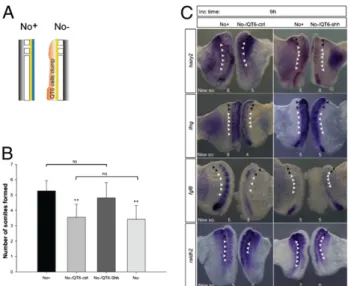
Ectopic Shh Replaces Notochord in Temporal Control of Gene Expression
and Somite Formation.Shh is one of the most well-studied signaling
molecules produced by the notochord. Shh-producing (QT6-Shh) or control (QT6-ctrl) QT6 cells were juxtaposed to No−explants which were cultured for 9 h in parallel to their No+counterpart (Fig. 4A). No−/QT6-ctrl (n = 43) failed to form the expected number of somites and presented gene expression perturbations, similarly to what had been previously observed for the No−
A
C
ns ns ** ** ** ns ns Determined PSM tissue Undetermined PSM tissueB
Time of incubation (hours)
4.5 6 7.5 9 10.5 12 N u m b er of so m ite s for me d 0 2 4 6 8 No+ No-hh s 3 3 4 3 6 4 8 5 New so: No: + - + - + - + -Inc time: 4.5h 7.5h 9h 12h
Fig. 1. Notochord removal impairs timely somite segmentation of the undetermined PSM in vitro. (A) Schematic representation of explant delimitation and excision. No, notochord; NT, neural tube; So, somite; PSM, presomitic mesoderm. (B) Evidence for the presence (No+)/absence (No−) of the notochord in control and experimental explants, by in situ hybridization for shh. Black arrowheads indicate somites formed before culture; white arrowheads indicate somites formed during incubation (New so). (C) Graphic representation of mean number of somites formed de novo upon 4.5, 7.5, 9, or 12 h of incubation. Data are mean± SD. **P < 0.05. ns, not statistically significant.
explants (Fig. 4 B and C). Notably, No−/QT6-Shh explants and No+ controls presented the same number of somites and com-parable expression patterns for the molecular clock genes hairy2 (n = 9/14) and lfng (n = 9/12) and for the determination front genes fgf8 (n = 4/6) and raldh2 (n = 3/3) (Fig. 4 B and C). Al-together, the results indicate that Shh is able to replace the no-tochord in control of timely somite segmentation and underlying gene expression dynamics.
Shh Signaling Controls Clock Pace and Somite Formation.Shh
sig-naling is transduced by a receptor complex that includes two transmembrane proteins, Patched (Ptch) and Smoothened (Smo). Shh association with Ptch allows Smo activity, whereas in the
absence of Shh, Ptch binds and inhibits Smo. Among the many target genes regulated by Shh signaling are ptch and gli signal transducers (15). To investigate the Shh pathway components op-erating in somite segmentation, we characterized the expression patterns of smo and ptch1/2 (Fig. S6). We found that notochord-adjacent PSM cells express both smo and ptch1/2, enabling them to respond to notochord-derived Shh.
We performed chemical inhibition of Shh pathway in No+ explants and the results obtained mimicked the absence of noto-chord, leading to a similar delay in somite formation (Fig. 5, A–C). Furthermore, the expression patterns of molecular clock genes are different in control and Shh-inhibited explants (Fig. 5B). Shh inhibition, however, does not halt molecular clock oscillations: cyclopamine-treated explants incubated for time periods differing by 30 min (6 h and 6.5 h) presented dissimilar phases of molecular clock gene expression (Fig. 5D). In accordance to the observed delay in somite formation, Shh-inhibited explants incubated for time periods differing by 90 min (6 h and 7.5 h) differed in the phases of molecular clock gene expression (Fig. 5E). Taken to-gether, these results indicate that, in the absence of Shh signaling, the period of the clock is no longer 90 min.
To evaluate the delay in the molecular clock, we cultured pairs of Shh-inhibited explants for time periods differing by 1 h 10 min, 2 h, 2 h 15 min, or 2.5 h. All explant pairs presented dif-ferent phases of expression of the clock and the same number of somites. When explants were incubated with a time difference of 2 h 45 min (Fig. 5F), 27% of the cases presented similar clock gene expression, but no extra somite formed. Finally, when dif-fering by 3 h, 85% of the explants had formed one extra somite but already presented different molecular clock patterns. These results indicate that clock oscillations are severely delayed in the absence of Shh, with a new time period between 2 h 45 min and 3 h. The imprecision in the determination of the new oscillation period could be due to differing levels of perturbation of the system imposed by Shh deprivation, which can be dependent on the phase of the molecular clock at the moment of notochord ablation. In conclusion, Shh ensures timely somite segmentation by setting the pace of the somitogenesis molecular clock.
Retinoic Acid Is Able to Recover Somite Formation Rate in the Absence
of Notochord.Notochord-deprived PSMs present delayed somite
formation from 4.5 h of incubation onward, accompanied by an overexpression of gli2 (n = 4/4) and gli3 (n = 11/11), a slight up-regulation of smo (n = 5/5) and down-up-regulation of both ptch1 (n = 5/5) and gli1 (n = 3/3) (Fig. 6). Timely somite formation is resumed after 9 h and this is not due to a re-establishment of Shh pathway components throughout this time period, as their levels of expression do not revert to basal state (Fig. 6). At 9 h, an in-triguing overexpression of raldh2 is observed that is not present at 4.5 h (Fig. 3). To test whether increased amounts of retinoic acid (RA) could mediate the recovery of somite formation rate, No− explants were incubated in RA-conditioned medium, and the number of formed somites after 9 h was assessed (Fig. 7 A and B). Noticeably, a greater number of somites is formed in No−explants when the medium is supplemented with RA. Moreover, the total number of somites formed in these conditions is not significantly
A
C
****
Time of incubation (hours)
9 15 Number of somi te s fo rmed 0 2 4 6 8 10 12 No+
No-B
New so: 7 5 10 8 No: + -Inc time: 9h 15h + -D
6+2 10 No: Inc time: 15h + -New so:Fig. 2. Notochord ablation in ovo delays somite segmentation of the undetermined PSM. (A) Schematic representation of embryo microsurgical operation. Only one of the embryo’s PSMs (PM) was left in contact with the notochord (No). HN, Hensen’s node; FP, floor plate; NP, neural plate. (B) Representative results obtained in operated embryos after a reincubation period of 9 or 15 h. (C) Graphic representation of the mean number of somites formed after 9 or 15 h. Data are mean± SD. **P < 0.001. (D) Operated embryo reincubated for 15 h where a gap in somite formation is observed, resulting from the absence of notochord (No−) in the PSM tissue juxtaposed to the surgical slit, whereas reestablished contact of the posterior PSM with the notochord reinstates timely somite formation. Presence (No+) or absence (No−) of notochord is indicated. Black arrowheads indicate somites formed before the surgical intervention; white arrowheads indicate somites formed during rein-cubation (New so).
5 3 5 3 5 4 6 4 No: + -In c ti m e : 9h h 5. 4 New so: New so: 3 3 3 3 3 3 3 3 hairy2 lfng fgf8 raldh2 + - + - +
-Fig. 3. Notochord ablation alters somitogenesis clock and determination front gene expression. Expression patterns of hairy2, lfng, fgf8, and raldh2, evaluated by in situ hybridiza-tion in explants cultured for 4.5 or 9 h. Presence (No+) or ab-sence (No−) of the notochord is indicated. Black arrowheads indicate somites formed before culture; white arrowheads indicate somites formed during incubation (New so).
DEVELOPMENTA
L
different from that observed in the No+ explant (Fig. 7B). Our results strongly suggest that an increase in RA signaling can mediate the recovery of somite formation in the absence of Shh. However, greatly increasing the amounts of RA, Shh, or both could not ac-celerate somite formation rate (Fig. S7).
Discussion
Somite Segmentation Relies on Axial Structures. Segmentation of
paraxial mesoderm has been classically described as an intrinsic mechanism independent of axial structures (16). PSM tissue
in-cubated for 14 h, with or without the axial structures, formed the same number of somites (7, 8), suggesting that these structures do not contribute to somite formation. However, the rate of PSM seg-mentation during the incubation period was not evaluated. When the molecular clock genes were described, their expression oscillations were also shown to occur independently of axial structures (2, 17, 18). Nevertheless, these studies were performed only up to 4.5 h of in-cubation, and we now know that this accounts only for determined PSM segmentation (6). Only one report has suggested that contact with tissues near the midline influences somite formation (9).
We have revisited this issue by evaluating somite formation rate over time in the presence or absence of the notochord. The tem-poral framework used allowed us to assess segmentation of both determined and undetermined PSM. Our results clearly show that the anterior third of the PSM does not require the notochord for timely segmentation, indicating that this tissue is determined not only spatially (6) but also temporally (this work). Segmentation of the undetermined PSM, however, is significantly delayed in the absence of the notochord. This is particularly evident for thefirst three undetermined somites, which resume proper segmentation only after 9–12 h of incubation. Hence, although the absence of the notochord does not impair segmentation (this work; 7, 8), we show that the strict, characteristic rate of embryo somite formation is dependent upon a signal from the notochord.
Shh Produced by Embryo Midline Structures Ensures Somite Formation
Rate.Shh signaling pathway is not classically associated with somite
segmentation. In fact, neither the Shh downstream Gli effectors nor the pathway’s membrane receptors have been reported to be expressed throughout the PSM (19, 20). We show that ptch1 is expressed in the medial PSM tissue and that smo is ubiquitously present in the PSM, creating the conditions for Shh signaling readout in this tissue.
Embryo midline structures produce diffusible Shh. Our results indicate that the notochord role on somite formation rate may be the production of a Shh signal delivered to the adjacent PSM. In fact, exogenous supply of this protein rescued timely somite formation in the absence of the notochord. Moreover, in the presence of canonical Shh inhibitors somite formation rate was perturbed to the same extent as when the notochord was re-moved. These experiments strongly suggest that embryo midline structures ensure strict control of somite formation rate through the supply of canonical Shh signal to the PSM.
Somitogenesis Clock Periodicity Is Modulated by Shh Signaling.Could
a perturbation in the clock period underlie the delay in somite Fig. 4. Exogenous Shh supply replaces the notochord in temporal control of
somite formation and gene expression. (A) Schematic representation of a clump (pink) of control QT6fibroblasts (QT6-ctrl) or QT6-producing Shh cells (QT6-Shh) juxtaposed to the neural tube (yellow) of explants without notochord (No−). (B) Graphic representation of mean number of somites formed by explants with notochord (No+), without notochord (No−), and without notochord but with control (No−/QT6-ctrl) or Shh-producing (No−/ QT6-Shh) cells. **P< 0.05 vs. No+or No−/QT6-Shh. ns, not statistically sig-nificant. Data are mean ± SD. (C) Explants with juxtaposed cells were hy-bridized for hairy2, lfng, fgf8, and raldh2. Black arrowheads indicate somites formed before culture; white arrowheads indicate somites formed during incubation (New so).
A
C
** ** **
No+ No- +cyclopamine +forskolin
N u m b e r o fs o m it e s fo rm e d 0 1 2 3 4 5 6 7 No
D
6.5h Inc time: 6h No: + + cyclopamine 3 3 New so: 2yri ahB
5 2 5 2 New so: O S M D O S M D en i ma po lc yc nil ok sr of No: + + + + Inc time: 9h 2y ri ahE
F
No: + + cyclopamine 3 3 Inc time: 6h 8h45 No: + + cyclopamine 3 3 Inc time: 6h 9h No: + + cyclopamine 3 4 Inc time: 6h 7.5h cyclopamineG
6.5hFig. 5. Inhibition of Shh signaling delays the pace of the clock. (A) Schematic rep-resentation of explant delimitation and excision. (B) Bisected-notochord explants were incubated for 9 h with DMSO (con-trol explant) and with either 10 μg/mL cyclopamine or 25 μg/mL forskolin (perimental explant). Forskolin-treated ex-plant and corresponding control were hybridized for hairy2. (C) Graphic repre-sentation of mean number of somites formed by explants with notochord (No+), without notochord (No−), and with noto-chord but incubated in the presence of cyclopamine or forskolin after 9 h of in-cubation. **P< 0.05 vs. No+. ns, Not sta-tistically significant. Data are mean ± SD. (D–G) Explants with bisected notochord treated with cyclopamine were cultured for the indicated time periods and hy-bridized with hairy2 probe. Black arrow-heads indicate somites formed before culture; white arrowheads indicate somites formed during incubation (New so).
formation observed in the absence of Shh? When PSM tissue is deprived of Shh signaling, either by notochord ablation or chemical inhibition, hairy2/lfng expression patterns are significantly different from those obtained in control conditions. Moreover, an exogenous Shh supply to notochord-ablated explants recovered molecular clock expression patterns. Hence, we could detect a clear correla-tion between the presence of Shh and both timely somite formacorrela-tion and molecular clock gene oscillations. Our group has previously shown that only the medial-most PSM (M-PSM) cells possess in-trinsic information for both somite formation and molecular clock gene expression (21). The study suggested that a signal supplied by M-PSM travels along the medial-lateral PSM axis, ensuring both timely segmentation and cyclic clock expression. However, the nature of this signal was not identified (21). The results obtained throughout the present work suggest that M-PSM cells are exposed to notochord-derived Shh signaling, as evidenced by ptch1/2 re-stricted expression, providing them crucial information for timely clock oscillations and somite formation. M-PSM cells are then ca-pable of instructing and recruiting their lateral neighbors for seg-mentation. This hypothesis is supported by the observation that somites formed in the absence of Shh are smaller along the medial-lateral (M-L) axis (Fig. S2B). How this is achieved in vivo is subject of ongoing studies in the laboratory.
Inhibition of Wnt signaling has also been described to perturb the molecular clock (22). We found that axin2 expression is un-altered in 60% of the notochord-ablated explants (Fig. S8) and the other 40% are only slightly down-regulated after 4.5 h. Because clock expression is perturbed in a much higher percentage of cases (80%), somite formation delay in the absence of Shh should not be due primarily to an impairment of Wnt signaling. Moreover, Wnt does not seem to be involved in the recovery of timely seg-mentation, as axin2 is clearly down-regulated after 9 h (Fig. S8).
Somitogenesis clock gene expression perturbation in the ab-sence of Shh signaling was not due to a halt in gene oscillations, but to an alteration of the clock period. In fact, the period of hairy2 expression cycles is considerably enlarged in the absence of Shh signaling, from 1.5 h to a time point between 2 h 45 min and 3 h. Significantly, somite formation in these conditions is also greatly delayed, reinforcing the dependence of timely somite formation on the pace of clock oscillations. Interestingly, somite formation delay observed upon Shh inhibition was dose de-pendent, as fewer somites were formed with increased inhibitor concentrations (Fig. S9). In situ hybridization on sections sug-gests the existence of a ptch1 expression gradient, with higher levels in the determined PSM (Fig. S6D). It is tempting to pos-tulate that an anterior–posterior gradient of Shh activity could account for the different segmentation rates observed for rostral (early) and caudal (late) somites. Shh knock-out mice present a striking absence of the entire spinal column and most of the ribs (14). Our data suggest that a perturbation in somite seg-mentation could be contributing to this phenotype.
Shh and RA Pathways Interplay with the Molecular Clock to Ensure
Timely Somite Formation.This work shows that Shh produced by the
notochord/floor plate is required to maintain timely operation of the molecular clock and the correct pace of somite formation (Fig. 7C, Left). An important consequence of PSM isolation from its source of Shh is the up-regulation of gli2 and gli3 (Fig. 7C, Middle), which corroborates previous reports (19). This strongly suggests
that low gli levels in the PSM are not due to the lack of Shh sig-naling readout, as is commonly believed, but rather are actively restricted by notochord-derived Shh. Another striking molecular consequence of notochord ablation is the down-regulation of fgf8 expression in the PSM, showing that Shh signaling is required to preserve the fgf8 gradient underlying the determination front. These molecular changes, observed in the PSM after 4.5 h of incubation in the absence of Shh, most probably account for the subsequent delay in somite formation rate and molecular clock oscillations.
The PSM fgf8 gradient counteracts an opposing RA gradient (5). Hence, the reduction of fgf8 levels in the absence of Shh could allow the observed up-regulation of Raldh2 after 9 h of incubation (Fig. 7C, Right). Under these molecular conditions, there is a re-covery of somite formation rate. Accordingly, we show that an external RA supply is able to rescue timely somite formation in the absence of Shh (Fig. 7 A and B). Although RA has been shown to
7 4 7 5 6 3 5 4 No: + -In c time : 9h h5. 4 New so: New so: 3 3 2 2 3 3 4 3
patched1 gli1 gli2 gli3
+ - + - + -3 3 6 5 smo + -* -* * *
Fig. 6. Expression of Shh pathway components in the PSM is altered by notochord ablation. Expression patterns of smo, ptch1, gli1, gli2, and gli3 genes evaluated by in situ hybridization in explants cultured for 4.5 or 9 h. *Neural tube removal from ex-plants for better visualization of PSM gli3 expression. Presence (No+) or absence (No−) of the notochord is indicated. Black arrowheads indicate somites formed before culture: white arrowheads indicate somites formed during incubation (New so).
A
B
ns**
No+ No- No-/RA+
Number of so mites form ed 0 1 2 3 4 5 6 7 6 6 New so: O S M D A R
C
No: Inc time: 9h +-Fig. 7. Shh and RA signaling pathways interplay in the PSM to ensure timely somite formation. (A and B) Retinoic acid mediates the recovery of timely somite formation in absence of notochord. (A) Explants with (No+) or with-out (No−) notochord were cultured for 9 h with DMSO (control explant) or with 15μM of retinoic acid (RA) (experimental explant). Black arrowheads indicate somites formed before culture; white arrowheads indicate somites formed during incubation period (New so). (B) Graphic representation of mean number of somites formed by explants with notochord (No+), without notochord (No−) or without notochord but with RA (No−/RA+). **P< 0.05 vs. No+or No−/RA. ns, Not statistically significant. Data are mean ± SD. (C) Proposed model for mo-lecular events underlying temporal control of somite formation in the presence (No+) or absence (No−) of notochord over time. In the presence of notochord-derived Shh, gli2/3 expression in the PSM is repressed, and timely molecular clock oscillations and somite formation occur. When Shh signaling is disrupted, gli2/3 are overexpressed, and there is a delay in both the clock period and PSM mor-phological segmentation. The normal rate of these events is reestablished over time due to an up-regulation of RA that counteracts Gli activity (detailed in text).
DEVELOPMENTA
L
inhibit gli2 expression (23), this does not occur within our experi-mental time frame. Because RA also modulates Gli activity (24), our results suggest that raldh2 overexpression after 9 h of noto-chord ablation rescues somite formation through an inhibition of Gli2/3 activity in the PSM (Fig. 7C, Right). It is plausible that so-mite formation recovery may further involve the inhibition of gli2 expression levels later in time. Our work strongly suggests that Shh and RA pathways interplay in the PSM to ensure timely somite formation and positions Gli2/3 at the intersection of these two pathways. Gli2 was recently reported to exert direct transcriptional regulation on the murine hes gene in retinal explants (25). In our experimental setup, gli2 overexpression was concomitant with a deregulation in the tempo of hairy2 transcriptional bursts, sug-gesting that in the PSM the same transcriptional regulation could take place. Hence, Shh-mediated restriction of Gli activity in the PSM seems to be essential to ensure the pace of both molecular clock oscillations and somite formation. Interestingly, Shh has been recently described to be essential for proper timing of neural progenitor differentiation, and RA was proposed to compensate for Shh deficiency in this developmental system (26).
Somitogenesis is a robust developmental mechanism. This can be achieved only by the integration of multiple signaling pathways. In fact, Notch, Wnt, Fgf, and RA signaling pathways operate in the PSM molecular clock and/or determination front (3, 4, 6). In the present work, we provide conclusive evidence that Shh sig-naling is a component of the intricate molecular machinery re-sponsible for temporal control of somite formation.
Multiple congenital vertebral malformations have been asso-ciated with mutations in somitogenesis clock genes (1, 3). Further-more, there are reports associating the Shh signaling pathway with congenital vertebral anomalies (27, 28). We report a previously uncharacterized association between these two molecular
sys-tems operating in the formation of early embryonic vertebrae precursor structures. A deeper understanding of the multiple mo-lecular interactions occurring during the somitogenesis process is crucial for an adequate scientific contribution to the medical community, striving to better comprehend and treat such human malformations.
Materials and Methods
Detailed materials and methods can be found inSI Materials and Methods. Chick embryos were staged according to Hamburger and Hamilton (HH) (29), and somites were considered to be formed when a definite cleft was ob-served (30). Antisense digoxigenin-labeled RNA probes were produced as described: shh (31), hairy2 (17), lunatic fringe (32), fgf8 (33), raldh2 (34), patched-1 (20), gli1 and gli2 (35), and gli3 (19). A smoothened-specific probe was generated. For explant cultures, ventrally positioned embryos were di-vided into two halves by cutting across the three germ layers at the midline level, either parallel to the embryonic axis or bisecting the axial structures. In Shh-graft experiments, clumps of either QT6-ctrl or QT6-Shh cells (36) were juxtaposed to the No− experimental explants. For chemical treatments, 15μM of RA (Sigma), 10 μg/mL cyclopamine (Calbiochem), 25 μg/mL forskolin (Sigma), or DMSO was added to the culture medium. In situ hybridization was performed as previously described (37).
ACKNOWLEDGMENTS. We thank R. Moura, C.J. Sheeba, F. Bajanca, and O. Martinho for helpful input regarding this work, and N. Le Dourain and L. Saúde for critical reading of the manuscript. We also wish to acknowledge stimulating insights provided by the Reviewers. T.P.R was supported by Fun-dação para a Ciência e a Tecnologia (FCT), Portugal (SFRH/BD/27796/2006); R.P.A. is funded by a Ciencia2007 Program Contract (Portuguese Govern-ment). This work was supported by FCT, Portugal (Projects PTDC/SAU-OBD/ 099758/2008 and PTDC/SAU-OBD/105111/2008), the EU/FP6—Network of Ex-cellence—“Cells into Organs” and IBB/CBME, LA, FEDER/POCI 2010.
1. Andrade RP, Palmeirim I, Bajanca F (2007) Molecular clocks underlying vertebrate embryo segmentation: A 10-year-old hairy-go-round. Birth Defects Res C Embryo Today 81:65–83.
2. Palmeirim I, Henrique D, Ish-Horowicz D, Pourquié O (1997) Avian hairy gene expression identifies a molecular clock linked to vertebrate segmentation and somitogenesis. Cell 91:639–648.
3. Dequéant ML, et al. (2006) A complex oscillating network of signaling genes underlies the mouse segmentation clock. Science 314:1595–1598.
4. Aulehla A, et al. (2003) Wnt3a plays a major role in the segmentation clock controlling somitogenesis. Dev Cell 4:395–406.
5. Diez del Corral R, et al. (2003) Opposing FGF and retinoid pathways control ventral neural pattern, neuronal differentiation, and segmentation during body axis extension. Neuron 40:65–79.
6. Dubrulle J, McGrew MJ, Pourquié O (2001) FGF signaling controls somite boundary position and regulates segmentation clock control of spatiotemporal Hox gene activation. Cell 106:219–232.
7. Packard DS, Jr (1980) Somitogenesis in cultured embryos of the Japanese quail, Coturnix coturnix japonica. Am J Anat 158:83–91.
8. Packard DS, Jr, Jacobson AG (1976) The influence of axial structures on chick somite formation. Dev Biol 53:36–48.
9. Packard DS, Jr, Zheng R-Z, Turner DC (1993) Somite pattern regulation in the avian segmental plate mesoderm. Development 117:779–791.
10. Ingham PW, McMahon AP (2001) Hedgehog signaling in animal development: Paradigms and principles. Genes Dev 15:3059–3087.
11. Fan C-M, Tessier-Lavigne M (1994) Patterning of mammalian somites by surface ecto-derm and notochord: Evidence for sclerotome induction by a hedgehog homolog. Cell 79:1175–1186.
12. Rong PM, Teillet M-A, Ziller C, Le Douarin NM (1992) The neural tube/notochord complex is necessary for vertebral but not limb and body wall striated muscle differentiation. Development 115:657–672.
13. Teillet MA, LaPointe F, Le Douarin NM (1998) The relationships between notochord andfloor plate in vertebrate development revisited. Proc Natl Acad Sci USA 95: 11733–11738.
14. Chiang C, et al. (1996) Cyclopia and defective axial patterning in mice lacking Sonic hedgehog gene function. Nature 383:407–413.
15. Varjosalo M, Taipale J (2008) Hedgehog: Functions and mechanisms. Genes Dev 22: 2454–2472.
16. Palmeirim I, Rodrigues S, Dale JK, Maroto M (2008) Development on time. Adv Exp Med Biol 641:62–71.
17. Jouve C, et al. (2000) Notch signalling is required for cyclic expression of the hairy-like gene HES1 in the presomitic mesoderm. Development 127:1421–1429.
18. McGrew MJ, Dale JK, Fraboulet S, Pourquié O (1998) The lunatic fringe gene is a target of the molecular clock linked to somite segmentation in avian embryos. Curr Biol 8:979–982.
19. Borycki A-G, Brown AMC, Emerson CP, Jr (2000) Shh and Wnt signaling pathways converge to control Gli gene activation in avian somites. Development 127:2075–2087. 20. Marigo V, Tabin CJ (1996) Regulation of patched by sonic hedgehog in the
developing neural tube. Proc Natl Acad Sci USA 93:9346–9351.
21. Freitas C, Rodrigues S, Charrier JB, Teillet MA, Palmeirim I (2001) Evidence for medial/ lateral specification and positional information within the presomitic mesoderm. Development 128:5139–5147.
22. Gibb S, et al. (2009) Interfering with Wnt signalling alters the periodicity of the segmentation clock. Dev Biol 330:21–31.
23. Ribes V, Le Roux I, Rhinn M, Schuhbaur B, Dollé P (2009) Early mouse caudal deve-lopment relies on crosstalk between retinoic acid, Shh and Fgf signalling pathways. Development 136:665–676.
24. Goyette P, et al. (2000) Regulation of gli activity by all-trans retinoic acid in mouse keratinocytes. Cancer Res 60:5386–5389.
25. Wall DS, et al. (2009) Progenitor cell proliferation in the retina is dependent on Notch-independent Sonic hedgehog/Hes1 activity. J Cell Biol 184:101–112. 26. Oh S, Huang X, Liu J, Litingtung Y, Chiang C (2009) Shh and Gli3 activities are required
for timely generation of motor neuron progenitors. Dev Biol 331:261–269. 27. Celli J, van Bokhoven H, Brunner HG (2003) Feingold syndrome: Clinical review and
genetic mapping. Am J Med Genet A 122A:294–300.
28. Kim PC, Mo R, Hui Cc C (2001) Murine models of VACTERL syndrome: Role of sonic hedgehog signaling pathway. J Pediatr Surg 36:381–384.
29. Hamburger V, Hamilton HL (1992) A series of normal stages in the development of the chick embryo. 1951. Dev Dyn 195:231–272.
30. Pourquié O, Tam PPL (2001) A nomenclature for prospective somites and phases of cyclic gene expression in the presomitic mesoderm. Dev Cell 1:619–620.
31. Riddle RD, Johnson RL, Laufer E, Tabin C (1993) Sonic hedgehog mediates the polarizing activity of the ZPA. Cell 75:1401–1416.
32. Sakamoto K, et al. (1997) Identification of a chick homologue of Fringe and C-Fringe 1: Involvement in the neurogenesis and the somitogenesis. Biochem Biophys Res Commun 234:754–759.
33. Crossley PH, Minowada G, MacArthur CA, Martin GR (1996) Roles for FGF8 in the induction, initiation, and maintenance of chick limb development. Cell 84:127–136. 34. Tsukui T, et al. (1999) Multiple left-right asymmetry defects in Shh(-/-) mutant mice
unveil a convergence of the shh and retinoic acid pathways in the control of Lefty-1. Proc Natl Acad Sci USA 96:11376–11381.
35. Marigo V, Scott MP, Johnson RL, Goodrich LV, Tabin CJ (1996) Conservation in hedgehog signaling: Induction of a chicken patched homolog by Sonic hedgehog in the developing limb. Development 122:1225–1233.
36. Duprez D, Fournier-Thibault C, Le Douarin N (1998) Sonic Hedgehog induces proliferation of committed skeletal muscle cells in the chick limb. Development 125: 495–505.
37. Henrique D, et al. (1995) Expression of a Delta homologue in prospective neurons in the chick. Nature 375:787–790.