Faculdade de Ciências
Histamine modulates dopaminergic neuronal
survival by boosting microglial activity
Tatiana Filipa Melo Saraiva
Dissertação para obtenção do Grau de Mestre em
Bioquímica
(2º ciclo de estudos)
Orientadora: Prof. Doutora Liliana Inácio Bernardino
iii
Acknowledgements
Desde já, agradeço à Prof.ª Doutora Liliana Bernardino, orientadora deste trabalho, pelo apoio, disponibilidade, confiança e empenho ao longo de todo o ano e que foi essencial para que todo este trabalho fosse possível.
Um agradecimento muito especial à Sandra Rocha, pela sua extraordinária capacidade de orientação ao longo de todo o trabalho. Com a sua ajuda, disponibilidade constante e paciência tornou este ano fantástico, deixando muitas saudades e vontade de seguir em frente.
Também agradeço á Rita pela disponibilidade e conhecimentos transmitidos neste ano. Aos meus colegas do mestrado pelo bom ambiente mantido aos longo do ano, em especial à Marta que partilhou comigo vários momentos de aprendizagem e que sempre se mostrou disponível para ajudar.
Um grande “obrigado” à minha mãe por ser um exemplo de mulher, sem se aperceber aqueles abraços dela que, mesmo sem palavras, me dão força e vontade se erguer a cabeça e seguir em frente; ao meu pai pelo apoio constante e à minha irmã pela presença e animação demonstradas desde sempre.
Agradeço especialmente ao Tiago por toda a paciência, compreensão, apoio incondicional e palavras de otimismo que me fazem continuar a lutar pelos meus sonhos.
Para finalizar, agradeço à minha família pelo apoio incondicional, pelas palavras de apoio e especialmente pela nossa união que me dá força para a ultrapassar todos os obstáculos na vida.
iv
Resumo
As células microgliais são os principais intervenientes na resposta inflamatória inata no cérebro adulto. Num contexto de lesão cerebral, a resposta das células da microglia envolve mecanismos de fagocitose de neurónios mortos ou danificados, libertação de fatores tróficos e/ou inflamatórios, e a produção de espécies reativas de oxigénio (ROS).
A histamina é uma amina encontrada em grandes quantidades em mastócitos, neurónios histaminérgicos, e leucócitos. No Sistema Nervoso Central (SNC), a histamina também é libertada por células da microglia e exerce as suas funções através da ativação de quatro subtipos de recetores acoplados a proteínas G: H1, H2, H3 e H4. Previamente, mostramos que a histamina modula a motilidade microglial e a libertação de citocinas. Os principais objetivos deste trabalho foram: i) avaliar o papel da histamina na atividade fagocítica microglial e na produção de ROS; e ii) explorar as consequências da inflamação microglial induzida pela histamina na sobrevivência neuronal dopaminérgica.
Inicialmente, verificamos que a histamina, através da ativação do recetor H1R, induziu um aumento de fagocitose na linha celular N9 de microglia, quando comparada com a condição controlo. Este efeito foi acompanhado por um rearranjo do citoesqueleto microglial monitorizado através da imunomarcação para a faloidina e a tubulina acetilada. A histamina também induziu um aumento da produção de ROS através da ativação dos recetores H1R e do H4R. A apocinina, um inibidor do NADPH oxidase, foi capaz de inibir totalmente a fagocitose e a produção de ROS mediada pela histamina. A incubação com lipopolissacarídeo (LPS), utilizado como controlo positivo, também induziu um aumento significativo de fagocitose e produção de ROS, quando comparado com culturas controlo.
Por outro lado, a injeção estereotáxica de histamina ou LPS na substancia nigra de murganhos adultos da estirpe C57BL/6 durante 7 dias, induziu um aumento da reatividade glial e uma diminuição robusta na sobrevivência neuronal dopaminérgica. Tanto a apocinina como a anexina V (usada como inibidor de fagocitose induzida pela fosfatidilserina) inibiram completamente a toxicidade dos neurónios dopaminérgicos induzida pela histamina.
Surpreendentemente, valores semelhantes à condição controlo, nos parâmetros avaliados
invitro (fagocitose e produção de ROS) e in vivo (sobrevivência neuronal dopaminérgica),
foram encontrados quando se procedeu à co-administração de histamina e LPS.
Em geral, os nossos resultados sugerem que a histamina induz a reatividade da microglia e que este efeito pode modular a sobrevivência neuronal dopaminérgica. Histamina per se atua principalmente como um agente pro-inflamatório induzindo neurotoxicidade. Contudo, na presença de LPS, a histamina pode exercer atividade anti-inflamatória e neuroprotetora.
Palavras-chave
Microglia, Histamina, LPS, Fagocitose, Espécies Reativas de Oxigénio, Neurotoxicidade, Neurónios dopaminérgicos
v
Resumo Alargado
Em condições fisiológicas, as células microgliais apresentam uma estrutura ramificada caracterizada pela baixa expressão de moléculas imunológicas. Estas células gliais são os principais intervenientes na resposta inflamatória inata, participando na primeira linha de defesa em resposta a vários estímulos, tais como as infeções, trauma, doenças neurodegenerativas, entre outros. Num contexto de lesão cerebral, as células da microglia tornam-se reativas, libertando fatores tróficos e/ou inflamatórios, e produzindo espécies reativas de oxigénio (ROS). A sua morfologia também é alterada adquirindo um estado ameboide responsável por processos de migração em direção ao local de lesão e ativação de mecanismos de fagocitose de neurónios mortos ou danificados. A microglia expressa diferentes tipos de recetores na sua superfície que estão envolvidos, por exemplo, na eliminação de micróbios e de material apoptótico ou, na indução da fagocitose (processo que envolve o rearranjo do citoesqueleto).A ativação microglial em resposta a um estímulo neurotóxico está geralmente associada a um aumento da expressão de citocinas pro-inflamatórias capazes de provocar degeneração neuronal. Por outro lado, dependendo da natureza e da intensidade do estímulo, as células da microglia podem libertar citocinas anti-inflamatórias e factores neurotróficos envolvidos em mecanismos celulares de protecção e reparação neuronal.
A histamina é uma amina neurogénica detetada precocemente no cérebro em desenvolvimento. Esta molécula, para além de ser o maior mediador das reações de hipersensibilidade imediata é também um interveniente importante em respostas imunes celulares e humorais. No sistema periférico, a histamina é produzida principalmente por mastócitos e não é capaz de atravessar a barreira hematoencafálica. No cérebro humano, esta amina é sintetizada pelos neurónios histaminérgicos localizados especificamente no núcleo tuberomamilar. A histamina também é libertada por células da microglia e exerce as suas funções através da ativação de quatro subtipos de recetores acoplados a proteínas G: H1, H2, H3 e H4. Previamente, mostramos que a histamina modula a motilidade microglial e a libertação de citocinas.
Com este trabalho pretendemos determinar a papel da histamina na fagocitose microglial e na produção de ROS em linhas celulares de microglia. Pretendemos também avaliar o efeito da actividade microglial induzida por esta mina na sobrevivência neuronal dopaminérgica. Inicialmente, verificamos que a histamina, através da ativação do recetor H1R, induz um aumento de fagocitose na linha celular N9 de microglia, quando comparada com a condição controlo. Este efeito foi acompanhado por um rearranjo do citoesqueleto microglial monitorizado através da imunomarcação para a faloidina e a tubulina acetilada. Em adição, também verificamos que a histamina induz um aumento da produção de ROS através da
vi
ativação dos recetores H1R e do H4R. A pré-administraçãode de apocinina, um inibidor do NADPH oxidase, inibiu totalmente a fagocitose microglial e a produção de ROS mediada pela histamina. A incubação com lipopolissacarídeo (LPS), utilizado como controlo positivo, também induziu um aumento significativo de fagocitose e produção de ROS, quando comparado com culturas controlo.
Por outro lado, a injeção estereotáxica de histamina ou LPS na substancia nigra de murganhos adultos da estirpe C57BL/6 durante 7 dias, induziu um aumento da reatividade glial e uma diminuição robusta na sobrevivência neuronal dopaminérgica. Tanto a apocinina como a anexina V (usada como inibidor de fagocitose induzida pela fosfatidilserina) inibiram completamente a toxicidade dos neurónios dopaminérgicos induzida pela histamina.
Surpreendentemente, valores semelhantes à condição controlo, nos parâmetros avaliados in
vitro (fagocitose e produção de ROS) e in vivo (sobrevivência neuronal dopaminérgica), foram
encontrados quando se procedeu à co-administração de histamina e LPS.
Em geral, os nossos resultados sugerem que a histamina induz a reatividade da microglia e que este efeito pode modular a sobrevivência neuronal dopaminérgica. Histamina per se atua principalmente como um agente pro-inflamatório induzindo neurotoxicidade. Contudo, na presença de LPS, a histamina pode exercer atividade anti-inflamatória e neuroprotetora.
Palavras-chave
Microglia, Histamina, LPS, Fagocitose, Espécies Reativas de Oxigénio, Neurotoxicidade, Neurónios dopaminérgicos
vii
Abstract
Microglial cells are the main players involved in the innate inflammatory responses in the adult brain. The response of microglia to brain injury involves the phagocytosis of death or damaged neurons, release of trophic and/or inflammatory factors, and the production of reactive oxygen species (ROS). Histamine is an amine found in high amounts in mast cells, histaminergic neurons, and leukocytes. In the Central Nervous System (CNS), histamine is also released by microglial cells and exerts its functions through the activation of four subtypes of G-protein coupled receptors: H1, H2, H3 and H4. Previously, our group showed that histamine modulates microglial motility and cytokines release. The main aims of this work were: i) to evaluate the role of histamine in microglial phagocytic activity and ROS production and ii) to explore the consequences of histamine-induced microglia inflammation in dopaminergic neuronal survival.Initially, we showed that histamine induced an increase of phagocytosis via H1R activation in a N9 murine microglial cell line, as compared to control. This effect was accompanied by the rearrangement of microglial cytoskeleton monitored through phalloidin and acetylated tubulin immunostaining. Histamine also induced an increase of ROS production
via H1R and H4R activation. Apocynin, a NADPH oxidase inhibitor, was able to fully inhibit
phagocytosis and ROS production mediated by histamine. Incubation with lipopolysaccharide (LPS), used as a positive control, also increased phagocytosis and ROS production, as compared with control cultures.
On the other side, the stereotaxic injection of histamine or LPS in the substantia nigra of adult C57Bl6 mice for 7 days induced an increase of glial reactivity and a robust decrease in dopaminergic neuronal survival. Both apocynin and annexin V (used as inhibitor of phosphatidylserine-induced phagocytosis) fully abolished the histamine-induced neurotoxicity of dopaminergic neurons.
Surprisingly, values similar to controls were found in cells co-treated with histamine and LPS, both in in vitro (phagocytosis and ROS production) and in vivo (dopaminergic survival).
Overall, our results suggest that histamine induce microglial reactivity both and that this effect may modulate dopaminergic neuronal survival. Histamine per se may act as a pro-inflammatory stimulus leading to neurotoxicity, whereas, in the presence of LPS, it acts as an anti-inflammatory and neuroprotective agent.
Keywords
Microglia, Histamine, LPS, Phagocytosis, Reactive Oxygen Species, Neurotoxicity, Dopaminergic neurons
viii
Table of contents
ACKNOWLEDGEMENTS III RESUMO IVII RESUMO ALARGADO V ABSTRACT VII LIST OF FIGURES XLIST OF TABLES XII
LIST OF ABBREVIATIONS XIII
CHAPTER I - INTRODUCTION 1
1. Microglial cells: the “housekeepers” of the brain 1
1.1. Microglial migration/motility 4
1.2. Release of soluble mediators 5
1.3. Microglial phagocytosis 8
1.3.1.The mechanisms of phagocytosis 9
2. Histamine 12
3. Neuroinflammation in Parkinson’s Disease 15
3.1. Animal models of Parkinson’s disease 16
CHAPTER II - OBJECTIVES 19
CHAPTER III - MATERIALS AND METHODS 20
In Vitro assays 20
3.1. Cell line cultures 20
3.2. Phagocytosis assay 20
Beads 20
Phosphatidylserine/ Phosphatidylcholine containing liposomes 21
ix 3.4. Immunocytochemistry 22 3.5. Western Blot 23 In Vivo assays 24 3.6. Animals 24 3.7. Stereotaxic injections 24
3.8. Preparation of the brain tissue 24
3.9. Immunohistochemistry against glial markers 25
3.10.Free-Floating immunohistochemistry for Tyrosine Hydroxylase 25
3.10.1. Cell counting and quantitive analysis 26
3
3..1100..11..11.. Data analysis 26
CHAPTER IV - RESULTS 27\
In Vitro assays 27
4.1. Histamine induced microglial phagocytosis of opsonized latex beads through H1
receptor activation 27
4.2. Histamine induced phagocytosis of PS-liposomes 29
4
4..33.. Histamine induced ROS production via H1R/H4R activation 32
4.4. Histamine-induced phagocytosis requires cytoskeleton alterations 33
In Vivo assays 35
4.5. Histamine increased glial reactivity in vivo 35
4.6. Histamine modulates dopaminergic neuronal survival 36
CHAPTER V - DISCUSSION 40
CHAPTER VI – CONCLUSIONS AND FUTURE PERSPECTIVES 44
x
List of Figures
Chapter I
Figure 1 – Microglial cells origin
Figure 2 – Microglial colonization during the brain development Figure 3 – Microglial morphology
Figure 4 – Receptors on microglia cell surface responsible by the propagation of
neuroimmune responses
Figure 5 - Microglial phenotypes
Figure 6 – Model summarizing the role of ion channels and transporters in controlling
microglial migration
Figure 7 - Microglia play distinct roles depending on the stimulus Figure 8 - NADPH oxidase enzyme
Figure 9 - Microglial phagocytic receptors
Figure 10 - Three-step model of microglial phagocytosis Figure 11 - The histaminergic system in the human brain Figure 12 - Biosynthesis and metabolism of brain histamine Figure 13 - The pathology of Parkinson’s disease.
Figure 14 - Schematic representation of LPS-induced and glial activation-mediated DA
neurodegeneration
Chapter III
Figure 15 – Treatment of N9 microglia cell cultures for phagocytosis assays in vitro
Chapter IV
Figure 16 – Histamine induced bead phagocytosis by microglial cells
Figure 17 – Fluorescent immunostainning to reveal phagocytosed liposomes (in red) in
microglial cellsFigure
18 - Quantification of fluorescence intensity of the liposomes phagocyted per cell Figure 19 – Histamine increased ROS production via H1R and H4R activation
xi
Figure 20 – Immunostaining against cytoskeleton proteins (phalloidin and α-acetylated
tubulin) in microglial cells
Figure 21 – Quantification of the acetylated α-tubulin protein levels in microglia cells
exposed with LPS or histamine
Figure 22 - Immunostainings to reveal astrocytes and microglia in SN brain slices of mice Figure 23 - Representative immunostainings for TH in the SN of mice
xii
List of Tables
xiii
List of Abbreviations
ATP Adenosine triphosphate
BDNF Brain-derived neurotrophic factor
CCL Chemokine (C-C motif)
CNS Central nervous system
CRs Complement Receptors
CXCL10 C-X-C motif chemokine 10
CCL21 Chemokine (C-C motif) ligand 21
CD11b Alpha chain of αMβ2-integrin or cluster of differentiation molecule 11B
COX Cyclo-Oxygenase
DA Dopamine
FBS Fetal Bovine Serum
FcR Fc-Receptors
GDNF Neurotrophic factor derived from a glial cell line
GFAP Glial Fibrillary Acid Protein
HRs Histamine receptors
IGF Insulin-like growth factor
IL Interleukin
iNOS Inducible nitric oxide synthase
i.p. Intraperitoneal
i.v. Intravenouse
LBs Lewys Bodys
LPS Lipopolysaccharide
NADPH (NOX) Nicotinamida Adenine Dinucleotide Phosphate (NADPH Oxidase)
NGF Nerve growth factor
NO Nitric Oxide
MAPK Mitogen-activated protein kinase
MHC Major histocompatibility complex
MPO Myeloperoxidase
xiv
PC Phosphatidylcoline
PD Parkinson’s Disease
PFA Paraformaldehyde
PKA Protein kinase A
PLA2 Phospholipase A2
PLC Phospholipse C
PRs Purine receptors
PS Phosphatidylserine
PSRs Phosphatidylserine receptors
TGFβ Transforming growth factor β
TH Tyrosine Hydroxylase
TLR Toll like receptors
TNF Tumour necrosis factor
TREM Triggering receptor expressed on myeloid cells
ROS Reactive oxygen species
RT Room temperature
SN Substantia nigra
SNpc Substantia nigra pars compacta
SRs Scavenger receptors
1
C
HAPTER
I
Introduction
1. Microglial cells: the “housekeepers” of the brain
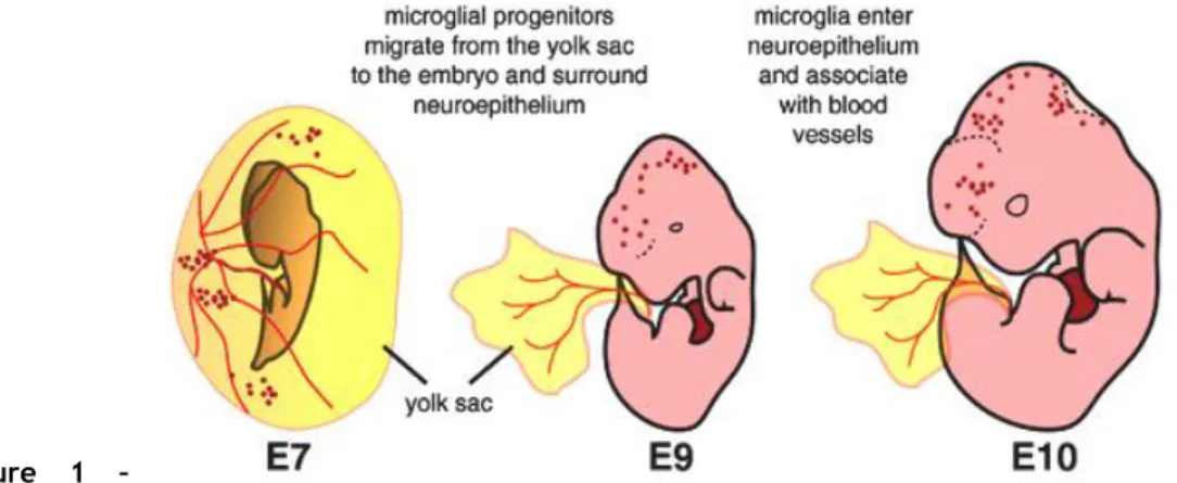
Microglial cells are originated from myeloid/mesenchymal progenitors that migrate from the yolk sac to the embryo and surround the neuroepithelium (Figure 1).
Figure 1 – Microglial
cells origin. Microglial cells originate from myeloid precursors in the yolk sac, which migrate into the
neuroepithelium by the embryonic day 10 (E10) (Adapted from Arnold and Betsholtz, 2013).
In the neuroepithelium, the microglial population rapidly expands and colonizes the brain from the dorsal to the ventral side (Figure 2). Over time, as early microglia move deeper into the developing parenchyma, they begin to differentiate, becoming more branched and expressing markers of mature microglia (Pont-Lezica et al. 2011).
Figure 2 – Microglial colonization during the brain development. At E12, microglia can be detected in
the brain mesenchyma, in the meninges and scattered in the neuroepithelium. (Adapted from
2
In the mature brain, approximately 12% of the total cells are microglial cells but they are not uniformly distributed (Block et al., 2007; Walter and Neumann, 2009). These cells exist in higher density in areas such as the hippocampus, olfactory telencephalon, basal ganglia and substantia nigra (SN) (Block et al., 2007; Walter and Neumann, 2009).
In physiologic conditions, these cells remain in a “resting” stage that is characterized by a ramified structure (Figure 3) and low expression of immunological molecules such major histocompatibility complex molecules (MHC), chemokine receptors, and several other markers (Walter and Neumann, 2009; Zhang et al., 2010). These receptors expressed constitutively at low levels are essential to the initiation and propagation of immune responses (Figure 4).
Figure 3 – Microglial morphology. Resting and ramified microglia in mixed glial cultures. Bright field
image of a murine primary cortical mixed glial culture stained with the microglial marker Tomato lectin (brown) and counterstained with hematoxylin (blue). Three of them, identified with arrows, are round microglial cells with a strong lectin staining. In contrast, there are several microglial cells with ramified morphology and less intense lectin staining (Adapted from Saura, 2007).
3 Figure 4 – Receptors on microglia cell surface responsible by the propagation of neuroimmune responses. Microglial cells can be activated by binding of various ligands to various cell-surface innate
immune receptors: CD14 binds lipopolysaccharide (LPS) and components of Protollin; Toll-like receptor 2 (TLR2) and 4 (TLR4) bind Protollin components; MHC class II molecules interact with T-cell receptors; CD40 binds CD40 ligand expressed by T cells and astrocytes; complement receptors bind complement components such as C1q; and Fc receptors (FcRs) bind amyloid-β-specific antibodies (Adapted from
Weiner et al., 2006)
The ramified morphology is a cytoarchitectural reflection of their surveillance function in the healthy adult tissue. In fact, microglia cells are not passive agents. Instead, they are highly dynamic cells, always patrolling the brain parenchyma, extending and retracting their processes, searching for any neuronal lesion or infection (Hanisch, 2013). In addition, they contact with neighbouring cellular elements, including neurons and astrocytes, in order to maintaining the structural and functional integrity of the CNS (Tremblay et al., 2011;
Kettenmann et al., 2011).
Microglia are considered to be a first line of brain defence and respond quickly to diverse stimulus, such as infection, trauma, ischemia, neurodegenerative diseases, or altered neuronal activity which can cause changes in brain homeostasis (Suzumura, 2013). These changes that may be potentially dangerous to the CNS leads to “microglia activation”, which is characterized by rapid change in the ramified structure to the amoeboid morphology, migration of these cells to the site of injury or invading pathogens where they proliferate to increase the number of fighter cells and phagocyte cell debris or invading agents (Walter and
Neumann, 2009; Kettenmann et al., 2011; Sierra et al., 2013)
In a classic activation paradigm, the so-called M1 phenotype, microglia are activated by the detection of pathogen-associated molecular patterns (PAMP’s) and pro-inflammatory cytokines resulting in an increased expression of Toll-like receptors (TLR), tumour necrosis factor α (TNFα), coregulatory molecules for antigen presentation and an increase of reactive species of oxygen (ROS) production (Figure 5). This phenotype leads mainly to a pro-inflammatory status. The administration of LPS, an endotoxin derived from Gram-negative bacteria, is the well-studied stimulus leading to a M1 microglia phenotype. LPS triggers microglial activation, release a variety of pro-inflammatory cytokines and chemokines (as IL-1β, IL-1, IL-10), nitric oxide (NO), transforming growth factor β (TGFβ) and TNFα (Kim et al.
2000; Kim and de Vellis 2005; Kettenmann et al., 2011).
On the other hand, the alternative activation or M2 phenotype is induced by interleukin 4 (IL-4) or interleukin 13 (IL-13), resulting in an increased production of interleukin 10 (IL-10) and TGFβ and, higher expression of scavenger receptors (Sierra et al., 2013) (Figure 5). It was proposed that this phenotype is associated with an anti-inflammatory and neuroprotective status.
4 Figure 5 - Microglial phenotypes. Microglia can be classified in a simplified manner into two subsets of
phenotypes and effector functions depending on the activation pathway (Adapted from Czeh et al.,
2011)
1.1. Microglial migration/motility
The microglia migration plays a central role in many physiological and pathophysiological processes; with particular relevance on the clearance of microbes and other invading agents or neuronal debris (Walter and Neumann, 2009; Kettenmann et al., 2011).
The highly ramified microglial processes are remarkably motile, continuously and randomly undergoing cycles of filopodia-like protrusion formation, extension and withdrawal of bulbous tips (Walter and Neumann, 2009; Kettenmann et al., 2011). Due to this mobility, microglia are capable of monitoring the local microenvironment surroundings and possibly to endocytose small cellular debris or budded vesicular structures, including that from apoptotic cells (Nimmerjahn et al., 2005; Kress et al., 2007; Neumann et al., 2009).
During pathological processes, injured neurons release various signals responsible for the attraction of microglia to the sites of injury, such as a triphosphate (ATP), chemokines as C-X-C motif chemokine 10 (C-X-CXC-X-CL10) and C-X-C-C-X-C motif ligand 21 (C-X-CC-X-CL21), grown factors as nerve growth factor (NGF), β-amyloid (Aβ), cannabinoids, morphine, lysophosphatidic acid and bradykinin (Neumann et al., 2009; Walter and Neumann, 2009; Kettenmann et al., 2011).
M2 phenotype
M1 phenotype
5
Likewise, ion channels and transporters play an important role in controlling microglial cell migration, such as potassium (K+) and chlorine (Cl-) channels, sodium/hydrogen (Na+/H+) and
chlorine/bicarbonate (Cl-/HCO3-) exchanger, and Na+/HCO3- cotransporter, which all are
linked to actin cytoskeleton dynamics (Figure 6) (Kettenmann et al., 2011; Harry, 2013).
Figure 6 – Model summarizing the role of ion channels and transporters in controlling microglial migration. The cytosolic calcium (Ca2+) signals induced by activation of metabotropic receptors and InsP3 cascade and/or by Ca2+ entry through ionotropic receptors or reverse mode of Na+/Ca2+ exchanger induces the retraction of the rear part of a migrating cell, which is paralleled by massive K+ efflux via Ca2+-dependent K+ channels and shrinkage of the cell at the rear (retraction site). Transporters such as Na+/H+ and Cl-/HCO
3- exchangers at the front of migrating cells (protrusion site) are reported to contribute to the extension of the actin projection (lamellipodium) by mediating salt and osmotically obliged water uptake (Adapted from Kettenmann et al., 2011).
1.2. Release of soluble mediators
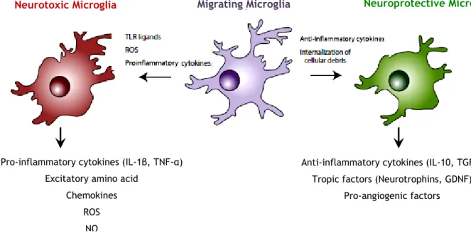
Another consequence of microglia activation is the release of inflammatory/neurotrophic factors which regulate the inflammatory response. The type of soluble factors released by microglial cells dependents on the initial stimulus that microglia cells receive.
Normally, the microglial activation in response to a strong neurotoxic stimulus results in the increase of the expression and release of pro-inflammatory cytokines, ROS and NO, that can cause further neuronal death (Figure 7; Konsman et al. 2002; Walter and Neumann, 2009;
Kettenmann et al., 2011; Fricker et al., 2012; Suzumura, 2013).
However, microglial activation can also induce neuroprotective actions by the release of anti-inflammatory cytokines such as TGFβ and IL-10, the release of neurotrophins such as NGF, brain-derived neurotrophic factor (BDNF) and neurotrophic factor derived from a glial cell line (GDNF) and/or inhibition of antigen presentation and release of pro-inflammatory
6
cytokines and reactive oxygen intermediates. The release of these trophic/anti-inflammatory factors contributes to the creation of an environment conducive for regeneration. These soluble factors can also attract phagocytic and repair-promoting effector and precursor cells, which are able to repair the damaged tissue (Figure 7; Honda et al., 1999; Lai and Todd,
2008; Neumann et a., 2009; Garden and La Spada, 2012).
A typical example of this duality of effects is the fact that the components of pathogens such as LPS are typically neurotoxic agents because it rapidly induce the production of interleukin-1 beta (IL-interleukin-1β) and TNFα by microglia; but when microglial cells are pretreated with IL-4, occurs a downregulation of TNFα and an upregulation of insulin-like growth factor-1 (IGF-1) gene transcripts, resulting in a neuroprotective effect (Figure 7; Neumann et a., 2009).
Figure 7 - Microglia play distinct roles depending on the stimulus. In the healthy CNS, microglia
survey their microenvironment, and in this “resting state”, do not express inflammatory mediators. However, after exposure to a number of chemical signals from damaged neurons, microglia respond rapidly and physically migrate to the site of injury. Responding microglia may then adopt a pattern of behavior similar to proinflammatory macrophages (left), as they release neurotoxic cytokines, chemokines, ROS, and NO. The release of cytokines and chemokines can lead to the recruitment of additional inflammatory cells from adjacent blood vessels, and may also engage astrocytes in the proinflammatory response. Alternatively, activated microglia may have neuroprotective behavior (right), secreting molecules that promote tissue repair, and internalizing cellular debris including aggregated, misfolded proteins such as β-amyloid, through phagocytosis. Whether two distinct populations of microglia exist that are committed to either of these response patterns, or all microglia can be induced to exhibit either response behavior when exposed to the correct combination of signals, remains to be determined (Addapted from Lai and Todd, 2008; Neumann et al, 2009; Garden and La
Spada, 2012)
It is also known that microglia have an antimicrobial activity due to production and release of toxic oxygen-derived and nitrogen-derived products, which are generated in a process known
Anti-inflammatory cytokines (IL-10, TGFβ) Tropic factors (Neurotrophins, GDNF)
Pro-angiogenic factors Pro-inflammatory cytokines (IL-1β, TNF-α)
Excitatory amino acid Chemokines
ROS NO
7
as the respiratory or oxidative burst. This production is due to situations of tissue damage or during defence against pathogens that have to be eliminated from brain (Sun et al., 2008;
Walter and Neumann, 2009; Hirsch and Hunot, 2009; Czeh et al., 2011; Fricker et al. 2012; Peterson and Flood, 2012).
This oxidative process is regulated by several enzymatic systems, principally the nicotinamide adenine dinucleotide phosphate-oxidase (NADPH oxidase/NOX) and inducible nitric oxide synthase (iNOS), and to a lesser extent by mitochondrial oxidases, cytochrome P450c, cyclooxygenases, myeloperoxidase (MPO) (Qin et al., 2005; Barger et al., 2007; Drechsel and
Patel, 2008; Hirsch and Hunot, 2009; Mead et al., 2012).
After microglial activation, the four regulatory cytoplasmic subunits (p47 phox, p67 phox, p40 phox
and Rac proteins) present in the NOX translocate to the plasma membrane linking to the other two subunits (p22 phox and gp91phox/Nox2) present there, forming the functional enzyme
that catalyses the reaction of NADPH and oxygen to form NAD+, protons and O2- (Figure 8;
Walter and Neumann, 2009; Sierra et al., 2013). Due to acidic pH into phagosome, the O2- is
dismuted into hydrogen peroxide (H2O2) and, later, into hypochlorous (HOCl-) that actively
participate in the modulation of signalling pathways involving microglial phagocytosis, for example in the phagocytic neutralization of microorganisms and promotion of neuronal death in animal models of neurodegenerative diseases (Figure 8; Block et al., 2007; Sun et al.,
2008; Chéret et al., 2008; Walter and Neumann, 2009; Peterson and Flood, 2012; Sierra et al., 2013)
Figure 8 - NADPH oxidase enzyme. The integral membrane of the phagocyte consists of two subunits:
p22phox and gp91phox which respectively produce the smaller and larger chain of the cytochrome-b558. Two cytosolic subunits: p67phox and p47phox; a p40phox accessory protein and a Rac-GTP binding protein then translocate to the cell membrane upon cell activation to form the NADPH oxidase complex which generates a respiratory burst. Superoxide can react to form hydrogen peroxide and hypochlorus acid, which together participate in bacterial killing (Adapted from Assari T., 2006).
8
Several studies demonstrated that higher levels of inflammatory mediators due to activated microglial cells, particularly ROS and NO, are responsible for the loss of the majority of DA neurons in Parkinson's disease (PD) patients. This fact suggests that the oxidative stress response that comes from microglial activation may be an important component in the neurodegenerative diseases and in the maintenance of the chronic pro-inflammatory response in PD patients (Drechsel and Patel, 2008; Hirsch and Hunot, 2009; Peterson and Flood, 2012).
1.3. Microglial phagocytosis
The phagocytosis comprises the first line of the innate immune defence against multicellular organisms and is mostly performed by specialized phagocytes, such as macrophages, dendritic cells, and neutrophils (Sierra et al., 2013). In the CNS, the innate immune response is mediated by microglia (Czeh et al., 2011; Sierra et al., 2013).
Microglia express different types of receptors on their surface that are involved in scavenging particles, debris, apoptotic material and microbes, or induction of phagocytic signaling, an active process involving rearrangement of the cytoskeleton (Walter and Neumann, 2009). More specifically, there are two functional types of phagocytic receptors, the receptors recognizing microbes such as to TLRs and Fc receptors (FcR’s) which support removal of pathogens and simultaneously stimulates a pro-inflammatory response in the phagocytes, and, receptors recognizing apoptotic cellular material such as receptors that recognize phosphatidylserine (PS) and which are important for ingesting apoptotic cell and stimulate an anti-inflammatory response in phagocytes (Figure 9; Ravichandran, 2003; Walter and
Neumann, 2009). The phagocytosis of apoptotic debris is essential and beneficial for the CNS
because it reduces the secretion of pro-inflammatory cytokines, chemoattraction and migration of T lymphocytes (Tremblay et al., 2011).
9 Figure 9 - Microglial phagocytic receptors. (Left) Phagocytosis is associated with inflammation during
uptake of microbes, while phagocytosis of apoptotic cells is executed without inflammation (Right). Recognition and phagocytosis of apoptotic cells induces an anti-inflammatory cytokine profile in microglia (Adapted from Neumann et al., 2009)
The phagocytic process is mediated by a number of receptors. Actually, some studies that focus on the action of the FcR's that are responsible to generate signals that regulate phagocytosis of immunoglobulin G (IgG)-coated particles. This process occurs when the Fc regions of the IgG molecules, that are formed when a small particle (eg. beads) or erythrocyte is opsonized with IgG, bind to FcR in the macrophage plasma membrane and initiate a phagocytic response forming a cup-shaped folds of plasma membrane extend outward from the macrophage around the particle and constrict at its distal margin, closing in a few minutes into a plasma membrane-derived phagosome. During the next hour, interactions between the phagosome and other membranous organelles change its internal and surface chemistries in a maturation process that typically leads to degradation of the phagosome contents by acid hydrolases. Throughout this event, the reduced NADPH oxidase complex is activated to deliver ROS into the phagosome by producing O2- from the oxidation
of NADPH and reduction of molecular oxygen (Kerrigan and Brown, 2009; Jaumouillé and Grinstein, 2010)
1.3.1. The mechanisms of phagocytosis
Microglial phagocytosis is a highly efficient process that maintains brain homeostasis. Targets for phagocytosis include: apoptotic cells, synapses, degenerated neuronal debris, or proteins with very high turnover such as Aβ protein.
Recent studies have demonstrated that damaged neurons are not merely passive targets but they regulate the microglia activity by releasing several signaling molecules. Specifically, degenerated neurons release nucleotides, cytokines and chemokines, to recruit microglia and enhance their activities. In literature, these molecules are described as “find-me”, “eat-me” and “digest-me” signals (Figure 10; Tremblay et al, 2011; Suzumura, 2013; Sierra et al.,
2013).
10 Figure 10 - Three-step model of microglial phagocytosis. (A) In physiological conditions, microglial
processes are highly motile and respond to chemoattractant molecules released by damaged or apoptotic cells - “find-me” signals - such as fractalkine and extracellular nucleotides (ATP, UDP). (B) An engulfment synapse is formed between a series of microglial receptors and their ligands in the membrane of the apoptotic cell - “eat-me” signals, leading to the tethering and engulfing of the apoptotic cell in a phagosome. (C) The phagosome becomes mature by fusing with lysosomes and other organelles, and the apoptotic cell is fully degraded in the phagolysosome in less than 2h -“digest-me” signals (Adapted from Sierra et al., 2013).
“Find-me” signals (Figure 10A)
It is known that the role played by phagocytic microglia occurs due to constant surveillance in the brain. So, phagocytosis is initiated when the phagocyte encounters a target cell by the presence of signals released by these cells. For instance, apoptotic cells release extracellular nucleotides (ATP and UTP) and other chemotactic signals fractalkine (CX3CL1) that are recognized by the receptors P2Y6 and CX3C chemokine receptor 1 (CX3CR1) respectively, on the surface of microglia, facilitating phagocytosis (Sierra et al., 2013).
“Eat-me” signals (Figure 10B)
Microglial cells have a series of receptors on their surface which are responsible for the different steps of phagocytosis. One group of receptors is responsible for recognition of target cells while another group is responsible for the internalization of these cells. These steps are the most important in the process of phagocytosis, leading to the formation of the phagocytic cup (Fricker et al., 2012; Sierra et al., 2013).
There is a group of receptors which are called “pathogen-associated molecular patterns” (PAMPS) that are mediated through scavenger receptors in conjunction with TLRs such as the CD14/TLR4 complex, or receptors of the immunoglobulin superfamily (e.g., c-type lectins). On the other hand, there is another group of receptors called “apoptotic cells-associated cellular patterns” (ACAMPs) which detects PS residues on the surface of microglial cells; this process is regulated by receptors as brain-specific angiogenesis inhibitor 1 (BAI-1) and by
11
linking with molecules, as milk fat globule-epidermal growth factor (MFG-E8), soluble opsonins and peroxynitrite (Armstrong and Ravichandran, 2011; Neher et al., 2011; Fricker et
al., 2012; Sierra et al., 2013).
These pathways lead to the remodeling of the microglial cytoskeleton through actin polymerization triggering the formation of pseudopodia that form a phagocytic cup engulfing the target (Lee et al., 2007; Sierra et al., 2013)
“Digest-me” signals (Figure 10C)
The phagosome formation occurs after closing the phagocytic cup. The phagosome merges with the early and late endosome and lysosome forming the phagolysosome which contains hydrolases and proton pumps responsible for digestion of the target and acidification of the medium, respectively. The acidic pH (pH≤5) allows lysosomal degradation, besides being an optimum environment for hydrolases. In addition, the low pH deactivates the production of free radicals resulting from the oxidative burst (Li et al, 2010; Sierra et al., 2013). This degradation process leads to subsequent antigen presentation, respiratory burst and release of anti-inflammatory factors.
The rapid elimination of apoptotic cells prevents them from becoming necrotic cells which can lead to loss of cell membrane permeability and spillover of intracellular contents. In fact, others have showed that the blockade of phagocytosis of microglia and polymorphonuclear neutrophils that infiltrate the brain parenchyma after focal ischemia, decreases neuronal viability in organotypic slices (Neumann et al, 2008).
Currently, the most recent method used to block microglial phagocytosis is the systemic administration of annexin V, which binds to the PS residues causing the accumulation of apoptotic debris (Lu et al., 2011; Fricker et al., 2012; Sierra et al., 2013). Other known compounds able to inhibit microglia phagocytosis include vitronectin receptor blockers, such as mutant MFG-E8 and vitronectin antagonists (Neher et al., 2011; Fricker et al., 2012)
12
2. Histamine
Histamine is one of the first neuroactive molecules to be detected in the early development of the brain. This biogenic amine is the major mediator of immediate-type hypersensitivity reactions as well as a modulator of cellular and humoral immune responses occurring in the pherypheric vascular system, but it is not transported into the brain across the blood–brain barrier (BBB). Most of the histamine is stored in master cells but it is also present in basophils, gastric enterochromaffin-like cells, leukocytes, platelets and even tumor cells. In the brain, histamine is synthesized in histaminergic neurons distributed in a posterior basal hypothalamus region - the tuberomammillary nucleus- and their axonal ramifications covers all over the CNS (Figure 11; M. L. Vizuete et al., 2000; R.E. Brown et al., 2001; N. Adachi,
2005; Molina-Hernández et al., 2012, 2013; Walker et al, 2013).
Figure 11 - The histaminergic system in the human brain. The histaminergic fibers emanating from
the tuberomamillary nucleus project to and arborize in the whole central nervous system (Adapted from
Haas, Sergeeva, and Selbach, 2008).
During neuronal differentiation in cerebral cortex, the fibers from the histaminergic neurons can be detected in the mesencephalon, passing through the ventral tegmental area and within the medial forebrain bundle and the optic tract, to reach the frontal and the parietal cortices, earlier than other monoaminergic systems (Molina-Hernández et al., 2012).
Histamine is synthesized from L-histidine by the enzyme L-histidine descarboxylase and converted into tele-methylhistamine by histamine-N-methyltransferase. By action of Monoamine oxidase B, tele-methylhistamine is converted into tele-methylimidazoleacetic acid (Figure 12; Brown et al., 2001; Adachi, 2005).
13 Figure 12- Biosynthesis and metabolism of brain histamine. In the brain, histamine is formed from
l-histidine by a specific enzyme, l-l-histidine decarboxylase. There are two major pathways of histamine metabolism; ring methylation and oxidative deamination by diamine oxidase. In the brain, most of histamine is catalized by histamine- N-methyltransferase to form tele-methylhistamine, which is converted by monoamine oxidase B to tele-methylimidazoleacetic acid (Adapted from Adachi, 2005).
Histamine exerts its functions through the activation of four subtypes of G-protein coupled receptors: H1R, H2R, H3R and H4R.
The H1Rs are expressed in regions related to behavioural, nutritional sate control and neuroendocrine but also plays an important role in inducing anaphylactic responses, such as bronchospasm, an increase in vascular permeability, and hypotensive shock. In contrast, the H2Rs mediates gastric acid production besides contributing to depress immunological processes by suppressing lymphocyte proliferation, cytokine production, and neutrophil accumulation. The H3Rs are heterogeneously distributed in brain and it is responsible to mediate feedback inhibition of the release and synthesis of histamine. Finally, the H4R is predominantly expressed in hematopoietic cells and is involved in or controlling the activities of eosinophils, master cells, monocytes, dendritic cells and T cells (Table 1) (O’Reilly et al.
2002; Adachi, 2005; Dijkstra et al. 2008; Jadidi-Niaragh and Mirshafiey, 2010; Molina-Hernández et al., 2012).
14 Table 1 – Properties of histamine receptors (Adapted from Jadidi-Niaragh and Mirshafiey, 2010)
Characte
-ristics H1R H2R H3R H4R
G-protein
coupling Gq/11 Gs Gi/o Gi/o
CNS expressio n Thalamus, hippocampus, cortex, amygdala, basal forebrain Basal ganglia, hippocampus, amygdala,
pyramidal cells, raphe nuclei, SN Nucleus, accumbens, striatum, basal ganglia, olfactory tubercles, SN, amygdala Cerebellum, hippocampus General function Wakefulness, inflammatory responses, decreasing blood pressure Regulation of gastric acid secretion, decreasing blood pressure, relaxation of airway and vascular
smooth muscle, excitation, fluid balance,
regulation of hormonal secretion Regulation of production and release of histamine Modulation of immune system Signaling
pathway PLC Activation of PKA
Inhibition of PKA, activation of PLA2, MAPK Inhibition of PKA, activation of PLC, MAPK
In CNS, histamine can be also released by microglial cells (Katoh et al., 2001). Recently, our group showed a dual role for histamine in the regulation of microglia activity by modulating cell recruitment and the release of pro-inflammatory cytokines, such as IL-1β and TNF α
(Ferreira et al, 2012).
The more than two decades ago, Francis et al. demonstrated that, while, the receptors specific for the C3bi cleavage fragment of the third component of complem ent (CR3) promote adhesion, histamine and our receptors inhibited the ability of CR3 to cluster on plasma membranes of neutrophils adherent to C3-coated surfaces (Francis et.el. 1991). Based on this fact, Azuma et al., demonstrated that histamine can inhibits phagocytosis through expression of complement receptor 3 in macrophages and it may affect the flow through the membrane and the expression of Fcγ receptors.
Other studies showed that histamine releasing peptide (HRP) promotes chemotaxis of leukocytes and enhances macrophage phagocytosis, and, in a presence of acute cutaneous inflammatory response promotes an increased of the level of HRP. These results suggested that HRP is a pro-inflammatory peptide that helps amplify and perpetuate the inflammatory response (Jaumouillé and Grinstein, 2011).
15
3. Neuroinflammation in Parkinson’s Disease
Parkinson’s disease (PD) is the second most common neurodegenerative disorder after Alzheimer’s disease (AD) and it is the most common movement disorder that affects approximately 1% of the population at the age of 55/60 and increases in prevalence to 4/5% by the age of 80/85 (Block et al., 2007; Hirsch and Hunot, 2009; Glass et al., 2010;
Labandeira-Garcia et al., 2011).
PD is a proteinopathy, such as AD, characterised by the presence of intraneuronal proteinaceous cytoplasmic inclusions known as Lewy bodies (LBs) and, by progressive and selective degeneration of DA-containing neurons in the substantia nigra pars compacta (SNpc) (Figure 13). It is known that these effects result from multiple molecular and cellular alterations that might be induced by abnormal protein handling, mitochondrial dysfunction, excitotoxicity, apoptotic processes, oxidative stress, inflammation and impairment of the ubiquitin-proteosome system (Hirsch and Hunot S., 2009; Glass et al., 2010; Neher et al,
2011; Labandeira-Garcia et al., 2011; Morroni et al, 2013).
Figure 13 - The pathology of Parkinson’s disease. The image represents the main neuropathological
events in PD at three levels from left to right. At the level of the brain, a major pathway is degeneration of the dopaminergic projections from the SN (in black) to the striatum (in purple), both of which are in the midbrain underneath the cerebral cortex. At the level of SN, the neurons that form the presynaptic portion of this pathway are normally melanized and are easily identified by this pigment in control brains (upper panel). In contrast, the loss of neurons in this region is so substantial that the whole area becomes depigmented in PD cases (lower panel). Of the few remaining cells, many show pathological changes, including the accumulation of proteins and lipids in Lewy bodies (Adapted from
16
The degeneration of the dopaminergic signalling present in the nigrostriatal pathway is responsible for the symptoms of motor dysfunction such as rigidity tremor, slowness of motion, difficulty to initiate movements and loss of balance. PD also presents non-motor-related symptoms as olfactory deficits, autonomic dysfunction, depression, cognitive deficits, and sleep disorders (Pei at al., 2007; Block et al., 2007; Hirsch E.C. and Hunot S., 2009; Glass
et al., 2010; Morroni et al, 2013).
Several evidences support that neuroinflammation can be involved in the loss of DA neurons that occurs in PD (Block et al., 2007; Brown and Neher, 2010; Glass et al., 2010; Neher et al,
2011; Labandeira-Garcia et al., 2011). Microglia activity can be detrimental to DA neurons by
regulating the activity of several enzymatic systems, among which NOX, iNOS, and MPO, are responsible for the production of O2–, NO free radicals, and HOCl-. In PD, these compounds are
increased in the SN (Figure 13; Hirsch and Hunot, 2009; Glass et al., 2010; Brown and Neher,
2010; L’Episcopo et al. 2010; L’Episcopo et al., 2010). Moreover, the SN is highly enriched in
microglial cells, making this brain region highly vulnerable to inflammatory reactions.
3.1. Animal models of Parkinson’s disease
Over several decades, has been extremely important to use animal models of PD that allow the pathological study of the disease and the development of therapeutic strategies to treat motor symptoms or, even one day, prevent to some extent the development of this disease neurodegenerative (J. Bové and C. Perier, 2012).
All models of PD are formulated based on the loss of DA neurons in the SN, although many of them have similar characteristics to the disease itself, can’t produce all the features presented in chronic neurodegenerative human PD. However, these animal models must possess special requirements such as having the ability to induce an injury replicable in the SN, the loss of DA neurons should be stable over time without the occurrence of spontaneous recovery, and must be able to "treated" based neuroprotective strategies (Emborg, 2004). The toxins 1-methyl-4-phenyl-1,2,3,6-tetrahydropyridine (MPTP) and 6-hydroxydopamine (6-OHDA) are two compounds commonly used and best characterized with respect to the development of PD in animals once they are responsible for loss of DA neurons. In recent decades have been discovered compounds able to produce similar effects as rotenone, paraquat, dieldrin and maneb (Emborg, 2004; Bové et al, 2005; Dutta et al., 2008; Drechsel
and Patel, 2008; Cristóvão et al., 2009; D.M. Crabtree, Zhang, 2012; Bové and Perier, 2012).
In the same sense, the lipopolysaccharide (LPS) has been the most extensively used to determine whether direct activation of microglia promotes a progressive and selective degeneration of DA neurons in rodents.
17
LPS an endotoxin found in the outer membrane of gram-negative bacteria is known as a potent activator of the innate immune response. It is composed by the O-antigen with multiple repeating units of monosaccharides, a polysaccharide core with an unusual sugar (2-keto-3-deoxyoctonate), and lipid A consisting of a unique diglucosamine backbone to which six fatty acid chains are attached (Figure 14; Qin et al, 2004; Dutta et al., 2008).
The binding of LPS to the soluble LPS binding protein (LBP) and CD14, which is anchored to the outer leaflet of the plasma membrane, promotes signal transduction through the plasma membrane, making possible the interaction of the complex LPS-CD14 with the TLR4 and extracellular accessory protein MD2. This interaction leads to the activation of kinases of various intracellular signaling pathways and upregulation of gene transcription for a variety of proinflammatory factors and free radical-generating enzymes. Consequently, this endotoxin is a potent stimulator of the microglia that able to promote the release of various immunoregulatory and proinflammatory cytokines and free radicals (Figure 14; Qin et al.,
2004; Dutta et al., 2008).
Figure 14 - Schematic representation of LPS-induced and glial activation-mediated DA neurodegeneration. LPS binding protein works as a chaperon that enhances the binding of LPS to its
intermediate receptor CD14. The association of the LPS-CD14 complex with TLR4, together with the accessory adaptor protein MD2 initiates a plethora of downstream signalling events that involve mitogen-activated protein kinases (MAPK) and transcription factors such as nuclear factor-kappa B. Upregulation of gene transcription leads to the production and release of cytokines such as TNFα and IL1b. Induction of cyclo-oxygenase-2 and iNOS expression results in the biosynthesis and release of prostaglandins and NO. Activation of the multi-subunit phagocyte oxidase complex (PHOX), also called NADPH oxidase generates superoxide anion that combines with NO from iNOS to form the more
18
damaging peroxynitrite (ONOO-) free radical. The collective insult of microglia-released cytokines, ROS and lipid metabolites eventually leads to the demise of the oxidative stress-vulnerable DA neurons
(Adapted from G. Dutta et al., 2008).
It is described that both the administration of LPS in vitro and in vivo, is responsible for the microglial cell activation, which the release of ROS promoting the selective and progressive degeneration of DA neurons. In the same way, some reports suggest that a brief episode (±2 weeks) of neuroinflammation that occurs early in life is capable of inducing significant glial activation accompanied by a delayed, progressive and preferential degeneration of SNpc DA neurons (Pei et al., 2007; Neher et al., 2011; Sanchez-Guajardo V., 2013).
19
C
HAPTER
II
Objectives
Microglial cells act as resident macrophages on the CNS. They are responsible for the constant monitoring of the brain microenvironment through elimination of toxic compounds and pathogenic substances. Several studies demonstrated that microglial activity can be related with the loss of DA neurons in the SN, a hallmark of PD.
In the brain, histamine is synthesized in histaminergic neurons present in the hypothalamus; however, it can be also released by microglial cells. Recently, our group showed a dual role for histamine in the regulation of microglia activity by modulating cell recruitment and the release of pro-inflammatory cytokines, such as IL-1β and TNF-α (Ferreira et al, 2012).
In general, this thesis aimed to determine the role of histamine in microglial phagocytosis and ROS production in a murine N9 microglia cell line. In the same way, we proposed to evaluate the effect of histamine-induced microglial activity on dopaminergic neuronal survival.
Specific aims included:
The evaluation of the role of histamine and its receptors in microglia phagocytic activity and ROS production, with or without the presence of an inflammatory stimulus – LPS;
The characterization of cytoskeleton alterations driven by histamine and /or LPS in microglial cells;
To investigate the role histamine and/or LPS-induced microglia activation on DA neuronal survival.20
Chapter III
Materials and Methods
In Vitro assays
3.1. Cell line cultures
A murine N9 microglia cell line (a kind gift from Prof. Claudia Verderio, CNR Institute of Neuroscience, Cellular and Molecular Pharmacology, Milan, Italy) was grown in modified RPMI medium during 24h to 37°C in a 95% atmospheric air and 5% CO2 humidified atmosphere.
Cells were plated at a density of 2×10⁴ cells per well in 24-well trays (phagocytic studies and
immunocytochemistry), 5×10⁵ cells per well in 6-well trays (protein extraction) or 5×10⁴ cells
per well in 96-well trays (ROS quantification).
Cell treatments included the following incubation setup: LPS (100 ng/ml, Sigma Aldrich), Histamine dihydrochloride (1–100 μM, Sigma), H1 receptor antagonist (mepyramine maleate, 1 μM), H4 receptor antagonist, (JNJ7777120, 5 μM), H1 receptor agonist, (2-Pyridylethylamine dihydrochloride, 100 μM) H4 receptor agonist, (4-methylhistamine dihydrochloride, 20 μM) (all from Tocris, Ballwin, MO, USA), apocynin (5 μM, Sigma). All histamine receptor antagonists/agonists and apocynin were added 30 min and 1h, respectively, prior to cell treatment and maintained during the course of experiments.
3.2. Phagocytosis assay
Beads
The murine N9 microglia cell lines were plated in a MW24 with at the density of 2×10⁴ cells
per well containing sterile glass coverslips (10 mm). Cells were allowed to grew for 24h and then treated for further 6h with LPS (100 ng/mL) and/or histamine (100 µM). Latex beads (Sigma Aldrich) were opsonized with rabbit IgG (1 μg/ml, Sigma Aldrich) under constant agitation overnight at 4ºC. Then, the beads were ressuspended in modified RPMI medium without NaHCO3 (Sigma Aldrich), and distributed at a density of 1×10⁵ beads per well.
After 40 min of incubation, cells were washed with 1PBS and fixed with 4% paraformaldehyde
(PFA, Sigma) or methanol/acetone (1:1, Fisher/Labsolve) for 30 min at room temperature (RT) or at -20ºC, respectively.Extracellular and/or adherent beads were labeled with
1
21
secondary antibody Alexa Fluor 594 donkey anti-rabbit (1:500; Molecular Probes, Oregon,
USA), in PBS, for 1h at RT. For nuclear labeling, cell preparations were stained with Hoechst
33342 (2 μg/ml) (Molecular Probes, Eugene, Oregon, USA) in PBS, for 5 min at RT. Coverslips were then mounted in Dako fluorescent medium (Dakocytomation Inc., California, USA). Fluorescent images were acquired using an AxioObserverLSM710 confocal microscope (Zeiss) under a 63×/1.40 oil objective. For each field, five photomicrographs were acquired in order to capture stained nuclei (in blue), extracellular and/or adherent beads (in red) and total number of beads (differential interference contrast image). The location of each bead was analyzed by comparing the three separate images simultaneously. Only beads without fluorescent labeling were considered as internalized particles.
Phosphatidylserine/ Phosphatidylcholine containing liposomes
The murine N9 mcroglia cell lines were plated in a MW24 at the density of 2×10⁴ cells per well
containing sterile glass coverslips (10 mm). After 24h, cells were incubated for further 6h with RPMI medium fresh (control), LPS 100 ng/mL and/or histamine 100 µM. Then, fluorescent labelled PS or PC containing liposomes (5µL/well) were added, directly, for 2h. To block PS-induced phagocytosis, Annexin V (4µL/well) waadded 1h prior the incubation with the liposomes. At the end of liposomes incubations, cells were washed with RPMI medium and then fixed with PFA 4% (Figure 15).
Figure 15 – Treatment of N9 microglia cell cultures for phagocytosis assays in vitro. The N9 microglia
cell line was plated in glass coverslips placed on MW24 plates until they reach confluence (24h). Then, cells were treated with 100 ng/mL LPS and/or 100 µM histamine for 6h. Liposomes containing PS or PC (5 µL/well) were added to the cells for further 2h, followed by several washes with RPMI medium and fixation with PFA 4%. Annexin V (4 µL/well) was added prior the liposomes incubation to inhibit phagocytosis.
After several rinses with PBS, unspecific binding was prevented by incubating cells in a 3% BSA and 0.5% Triton X-100 solution (all from Sigma Aldrich) in PBS for 30min, at RT. Then, cells were incubated overnight at 4°C with the primary antibody: rat monoclonal anti-CD11b (1:600; AbD Serotec, Oxford, UK) diluted in PBS containing 0.3% BSA and 0.1% Triton X-100. After 3 washes with PBS (5min each), cells were incubated for 1h at RT with the corresponding secondary antibody: Alexa Fluor 488 goat anti-rat (1:200; Molecular Probes) diluted in PBS. For nuclear labeling, cell preparations were stained with Hoechst 33342 (10
24h 5h 1h 2h
Cell fixation
Plating Stimulus Add
22
μg/ml; Molecular Probes, Eugene, Oregon, USA) in PBS, for 5min at RT and mounted in Dako fluorescent medium (Dakocytomation Inc., California, USA).
Fluorescent images were acquired using an AxioObserverLSM710 confocal microscope (Zeiss) under a 40×/1.40 oil objective. For each coverslip, four photomicrographs were acquired in order to capture stained nuclei (in blue), PS/PC liposomes (in red) and microglial cells (in green). To quantify the fluorescence intensity of the liposomes in each condition (sixty-four cells per condition) we deducted the fluorescence intensity of background. The same confocal image acquisition settings were used in all experiments.
3.1. Determination of cellular ROS levels
ROS levels were measured using the probe dihydroethidium (DHE, Molecular Probes). In the DHE assay, blue fluorescent DHE is dehydrogenated by superoxide (O2–) to form a red
fluorescent ethidium bromide. Cells exposed for 2h to the stimulus (histamine and/or LPS) were incubated for 4h with 100 µM DHE in culture medium at 37°C. The fluorescence emitted was read in a spectrofluorometer (SpetroMax GeminiEM; Molecular Devices) using Ex/Em 515/605 nm.
3.2. Immunocytochemistry
Cells were fixed with 4% PFA and unspecific binding was prevented by incubating cells in a 3% BSA and 0.5% Triton X-100 solution (all from Sigma Aldrich) for 30min, at RT. Then, cells were incubated overnight at 4°C with the primary antibodies: rat monoclonal anti-CD11b (1:600;
AbD Serotec, Oxford, UK) and mouse monoclonal anti-acetylated α-tubulin (1:100; Sigma Aldrich), both diluted in PBS containing 0.3% BSA and 0.1% Triton X-100. After several washes
in PBS (3x, 5min), cells were incubated for 1h at RT with the corresponding secondaryantibodies: Alexa Fluor 488 goat anti-rat (1:200; Molecular Probes) and Alexa Fluor 594 donkey anti-mouse (1:200) both diluted in PBS. Membrane ruffling was observed by using a marker for filamentous actin, phalloidin. Cells were incubated for 2h at RT with the phalloidin-Alexa Fluor 594 conjugate (1:100; Molecular Probes) in PBS, protected from light. For nuclear labeling, cell preparations were stained with Hoechst 33342 (2 μg/ml) (Molecular Probes, Eugene, Oregon, USA) in PBS, for 5 min at RT and mounted in Dako fluorescent medium (Dakocytomation Inc., California, USA). Fluorescent images were acquired using an AxioObserverLSM710 confocal microscope (Zeiss) under a 63×/1.40 oil objective.
23
3.3. Western Blot
Cells were washed with ice-cold PBS and lysed on ice in RIPA buffer (50 mM Tris-HCl, pH 8.0, 150 mM NaCl, 2 mM sodium orthovanadate, 1% Nonidet-P40, 0.5% sodium deoxycholate, 0.1% SDS, containing 1% of a protease inhibitor mixture (AEBSF, pestatinA, E-64, bestatin, leupeptin and aprotinin) purchased from Sigma-Aldrich. After gentle homogenization, the total amount of protein was quantified using the Bradford method and bovine serum albumin as standard (Bio-Rad Protein Assay, Bio-Rad Laboratories).
Afterwards, samples were loaded onto 12% acrylamide/bisacrilamide gels (BioRad, Hercules,
CA, USA). Proteins were separated by SDS-PAGE and then transferred to PVDF membranes
(Amersham HybondTM-P, GE Healthcare). The membranes were then blocked with 5% non-fat
milk (Regilait, France) in Tris buffer saline solution-Tween 20 (2TBS-T) for 1h at RT.
Incubation with anti-acetylated α-tubulin (1:200) (Sigma) diluted in TBS-T was done overnight at 4ºC. After being rinsed three times with TBS-T, the membranes were incubated for 1h at RT with an anti-mouse antibody (1:10000) (GE Healthcare) diluted in TBS-T.
The membranes were then incubated with the ECF substrate (ECF Western Blotting Reagent
Packs, GE Healthcare) for 5min. Protein bands were detected using the Molecular Imager FX
system (Bio-Rad Laboratories) and quantified by densitometry analysis using the Quantity One software (Bio-Rad Laboratories).