outubro de 2013
Catarina Cubo da Fonte
Ecological shift of oral microbiota
UMinho|20 13 Cat arina Cubo da F ont e Ecological shif t of oral microbio ta
Dissertação de Mestrado
Mestrado Integrado em Engenharia Biomédica
Ramo de Engenharia Clínica
Trabalho realizado sobre a orientação da
Professora Doutora Mariana Contente Rangel
Henriques
Universidade do Minho
e do
Professor Doutor Wim Teughels
Katholieke Universiteit of Leuven
Catarina Cubo da Fonte
Acknowledgments
My first words are to my supervisors, Dr. Wim Teughels and Dr.ª Mariana Henriques, I am especially grateful. They were always so understanding and available to help and meet with me. I want to express my thankfulness to Dr. Wim Teughles for the opportunity to integrate his investigation team in the Department of Periodontology of the Catholic University of Leuven. This work would not be possible without his ideas and advices. I specially acknowledge to Dr.ª Mariana Henriques for the patience, understanding, guidance and motivation.
I want to express a special thanks to Gitte Loozen and Martine Pauwels. Gitte was untiring, I have to thank all the time she spent hearing my doubts and questions and explaining me. I will always remember all the availability, her advices and what she taught me. Martine was always helpful, especially with the molecular techniques I learned in the lab. She also had friendly words to my work, when my bacteria did not grow or when some techniques did not work. I will never forget her ‘Allez’ and complains about the weather. I also have to acknowledge to the laboratory people for the hospitality and sympathy, and the good working environment, mainly to Jesica and Mariana.
I would like to express a special word to my Erasmus mates (Joana Festa, José Sequeiros, Mariana Roriz, Pedro Morais, Pedro Vieira and Sandro Queirós) for all mishaps and experiences we lived together in a different country. Thank you to all Erasmus students I met during my time in Leuven. I also would like to thank to LUK singers for each music moments.
Another special word goes to my course friends, they were like a new family I found in a new city with which I grew up and I lived so good moments. Thank you for the fellowship and friendship. I also have to thank to people from the academic choir, CAUM, for each rehearsal and concert.
I also want to dedicate a special thanks to each person that encouraged me during thesis writing, for their support and motivation.
My last words must be addressed to my beloved mother and father. I wish to stress their unconditional love, confidence, support, help, encouragement, patience and understanding, without them this could not be possible. Thank you.
To my maternal grandparents, they would be proud of me.
Ecological shift of oral microbiota - Abstract
The oral cavity is composed of several bacterial species living in a dynamic and complex ecosystem. Periodontitis and dental caries are two of the most prevalent oral diseases, nowadays. However, current treatments are not enough to fight these oral diseases, so alternative ways are required, as the use of prebiotics and probiotics, which are already being used in several fields.
The present work represents the study of the effect of a prebiotic compound (C7) on cariogenic bacteria in order to understand whether addition of this compound leads to a decrease of these bacteria. Streptococcus mutans and Streptococcus sobrinus were the cariogenic bacteria used in this study because these are the main cariogenic bacteria. A probiotic strain, Streptococcus salivarius, was also used in this study.
Firstly, quantitative Polymerase Chain Reaction (qPCR) was developed for Streptococcus salivarius in order to allow an accurate determination of its presence in microbial communities. The primers were chosen based on a conserved region of the dextranase gene of S. salivarius and the best combination of primers and probe concentration was determined. This quantification of this strain by this molecular technique was compared with microbial culturing presenting a linear relationship.
It was also intended, the find a selective medium for each of the species used, so different media were tested and TYCSB medium showed to be a good selective medium for S. mutans and S. sobrinus. However no selective medium was found for S. salivarius.
In order to determine the effect of the prebiotic compound, dual species experiment for each cariogenic bacterium and a probiotic species was carried out. Microbial culturing and qPCR were used for bacteria quantification and pH was also measured. For S. mutans, the main reduction was apparently due to the presence of S. salivarius and was not influenced by C7. For S. sobrinus, the verified reduction was the result of presence of S. salivarius with influence of C7, but no clear conclusions can be made about it.
In addition a Denaturing Gradient Gel Electrophoresis (DGGE) was performed to understand the effect of the prebiotic compound in saliva microbiota. The results demonstrated an ecological shift between different bacterial species present in saliva. So, S. salivarius seems to be in higher amounts when C7 is present while other species seem to be present in higher concentration when there is no C7.
Alterações ecológicas da microflora oral - Resumo
A cavidade oral é um ecossistema dinâmico e complexo no qual diversas espécies vivem e interagem. Hoje em dia, a periodontite e as caries dentárias são duas das doenças orais mais prevalentes no mundo. No entanto, os tratamentos atuais não têm sido suficientes para responder a estas doenças, havendo necessidade de se utilizar outras alternativas. O uso de prebióticos e probióticos pode ser uma hipótese, tendo em conta que já têm vindo a ser utilizados noutras áreas.
Este trabalho representa o estudo do efeito de um composto prebiótico (C7) em bactérias cariogénicas, tentando perceber de que forma a sua presença permite levar à diminuição destas. Deste modo, foram utilizadas as principais bactérias cariogénicas (Streptococcus mutans e Streptococcus sobrinus) e também uma espécie probiótica (Streptococcus salivarius).
Assim sendo, foi desenvolvida uma metodologia de Polymerase Chain Reaction em tempo real (qPCR) para S. salivarius, de modo a possibilitar a sua correta quantificação em comunidades microbianas. A melhor combinação de concentrações dos primers e probe foi definida e a quantificação por este método foi comparado com a cultura microbiana, apresentando uma relação linear. A pesquisa de meios seletivos para cada uma das espécies usadas foi também realizada neste trabalho, pelo que foram testados vários meios. O meio TYCSB mostrou ser seletivo para ambas as espécies cariogénicas, contudo não foi encontrado nenhum meio seletivo para o S. salivarius.
Para determinar o efeito do composto prebiótico foi utilizado um modelo de espécies dual para cada bactéria cariogénica conjugando-a com a espécie probiótica. A quantificação destas estirpes foi feita através de cultura microbiana e qPCR e o pH foi também medido. Para o S. mutans, a principal redução verificada aparentou dever-se à presença do S. salivarius e não devido à influência do C7. Para o S. sobrinus, a redução que se verificou também resultou da presença do S. salivarius com uma ligeira influência do C7, embora estas conclusões não sejam muito claras.
Para perceber o efeito do C7 na saliva foi realizado uma DGGE (Denaturing Gradient Gel Electrophoresis), tendo os resultados demonstrado uma alteração ecológica entre as diferentes espécies presentes na saliva. Assim, S. salivarius, pareceu estar mais presente quando o C7 se encontra adicionado, enquanto outras parecem estar em maiores quantidades quando este composto não se encontra presente.
Table of Contents
Acknowledgments ... iii
Ecological shift of oral microbiota – Abstract ... v
Alterações ecológicas da microflora oral – Resumo ... vii
Table of Contents ... ix
Abbreviations ... xi
List of figures ... xiii
List of tables ... xvii
Chapter 1 – Introduction ... 1
1.1. Main objectives ... 3
1.2. General concepts ... 3
1.2.1. The oral cavity ... 3
1.2.2. Oral biofilms ... 4
1.3. Plaque-related diseases ... 6
1.3.1. Dental plaque and oral diseases ... 6
1.3.2. Periodontitis ... 7
1.3.3. Dental caries ... 9
1.3.4. Ecological plaque hypothesis ... 11
1.4. New treatments ... 15
Chapter 2 – Development of a new qPCR for Streptococcus salivarius ... 19
2.1. Introduction ... 21
2.2. Materials and Methods ... 22
2.2.1. Bacterial strains and culturing conditions ... 22
2.2.2. Design of qPCR primers and probe ... 22
2.2.3. Construction of the qPCR plasmid standard ... 23
2.2.4. qPCR optimization ... 23
2.2.5. Comparison between qPCR and microbial culturing ... 23
2.3. Results and Discussion ... 24
2.3.1. Design of qPCR primers and probe ... 24
2.3.2. qPCR optimization ... 25
2.3.3. Comparison between qPCR and microbial culturing ... 26
Chapter 3 – Effect of a prebiotic compound on cariogenic and saliva bacteria ... 27
3.1. Introduction ... 29
3.2. Materials and Methods ... 30
3.2.1. Bacterial strains and culturing conditions ... 30
3.2.2. Media testing ... 31
3.2.3. Growth in dual-species model ... 31
3.2.4. DNA extraction and quantitative Polymerase Chain Reaction (qPCR) .... 32
3.2.5. Saliva collection and preparation ... 33
3.2.6. DNA extraction, PCR-DGGE assay ... 33
3.3. Results ... 34
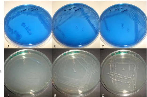
3.3.1. Selective medium for Streptococcus salivarius, Streptococcus mutans and Streptococcus sobrinus ... 34
3.3.2. Effect of a prebiotic compound on cariogenic bacteria ... 35
3.3.3. Effect of a prebiotic compound on bacteria present in saliva ... 42
3.4. Discussion ... 43
3.5. Conclusion ... 48
Chapter 4 – Conclusion and future work ... 51
Abbreviations
A. a.: Actinobacillus actinomycetemcomitans AER: aerobic conditions
ANA: anaerobic conditions BHI: Brain-Heart Infusion bp: base pair
C7: prebiotic compound CFU: colony-forming units CO2: carbon dioxide
DGGE: Denaturing Gradient Gel Electrophoresis DMF: Decayed, Missing, Filled
DMFS: Decayed, Missing, Filled Surfaces DMFT: Decayed, Missing, Filled Teeth DNA: deoxyribonucleic acid
dNTP: deoxynucleoside triphosphate EDTA: ethylenediaminetetraacetic Eh: redox potential
EMA: ethidium monoazide
EPS: Extracelular Polymeric Substances FAM: 6-carboxyfluorescein
FOS: fructo-oligosaccharides
GC content: guanine-cytosine content GCF: Gingival Crevicular Fluid GI tract: gastrointestinal tract GOS: galacto-oligosaccharides gtf: glucosyltransferase
IPS: Intracelular Polymeric Substances KCl: potassium chloride
L. fermentum: Lactobacillus fermentum L. reuteri: Lactobacillus reuteri
L. rhamnosus: Lactobacillus rhamnosus L. salivarius: Lactobacillus salivarius
MgCl2: magnesium chloride
MM: Miminal Medium MS: Mitis-Salivarius
MS-MUT: selective medium for Streptococcus mutans MS-SOB: selective medium for Streptococcus sobrinus OD: optical density
P. g.: Porphyromonas gingivalis PCR: Polymerase Chain Reaction
PCR-DGGE: Polymerase Chain Reaction – Denaturing Gradient Gel Electrophoresis PMA: propidium monoazide
qPCR: Quantitative Polymerase Chain Reaction R2: correlation coefficient
S. mitis: Streptococcus mitis S. mutans: Streptococcus mutans S. oralis: Streptococcus oralis
S. salivarius: Streptococcus salivarius S. sanguinis: Streptococcus sanguinis S. sobrinus: Streptococcus sobrinus SD: standard deviation
TAMRA: 6-carboxytetramethylrhodamine Tm: melting temperature
TYC: Trypticase, Yeast, Cysteine
TYCSB: Trypticase Yeast Cysteine Sucrose Bacitracin UV: ultraviolet
VBNC: Viable But Non-culturable Cells w/v: weight per volume
List of figures
Figure 1 - Oral biofilm formation. A. Pellicle formation. The acquired pellicle is a thin layer that consists of adsorbed organic molecules derived from the salivary glycoproteins attached to the tooth surface. B. Initial adhesion. Specific receptors allow the initial adhesion of bacteria to the pellicle. C. Maturation. Biofilm maturation results of interactions between later colonizers and early colonizers, previously attached in a cell-to-cell reaction (co-aggregation). D. Dispersion. Bacteria leave the biofilm and colonize a new site [2]. ... 4 Figure 2 - Ecological plaque hypothesis and prevention of periodontal diseases. Gingival Crevicular Fluid (GCF). Redox potential (Eh) [10]. ... 13 Figure 3 - Ecological plaque hypothesis and prevention of dental caries [10]. ... 14 Figure 4 - Extended caries ecological hypothesis [20]. ... 15 Figure 5 - PCR amplification for detection of the dextranase gene of oral streptococcal species by a PCR and a DNA probe (Ssal497T) with Ssal442F and Ssal615R primer pair: S. mutans (lane 1), S. sobrinus (lane 2), S. salivarius ATCC 7073 (lane 3), S. salivarius K12 (lane 4), S. salivarius clinical strain (lane 5), S. salivarius TOVE-R (lane 6). Negative control (lane 7) was made with physiological water. ... 25 Figure 6: Number of colony forming units (CFU) of Streptococcus salivarius (Y-axis) versus quantification of Streptococcus salivarius by quantitative Polymerase Chain Reaction (qPCR) (X-axis). Correlation coefficient: R2=0.9741. ... 26
Figure 7: Growth on Mitis-Salivarius agar plates (I) and on Trypticase Yeast Cysteine Sucrose Bacitracin (TYCSB) plates (II). (A): Streptococcus salivarius, (B): Streptococcus mutans and (C): Streptococcus sobrinus. ... 35 Figure 8: Number of colony forming units (CFU) of Streptococcus mutans in dual species model at 0 h, 24 h and 48 h: Growth of S. mutans testing the effect of C7 compound with (E) or without (D) the presence of S. salivarius and glucose with (C) or without (B) S. salivarius. The negative controls were made one with only the cariogenic bacterium (A) and another with the cariogenic bacterium and S. salivarius without addition of glucose (F). Standard errors of the mean (n=3)
are represented by error bars. Significance (p<0.05) between the control (only S. mutans) and test series was determined using Student’s t-test and are marked with *. ... 36 Figure 9: Number of Streptococcus mutans in dual species model at 0 h, 24 h and 48 h determined by quantitative Polymerase Chain Reaction (qPCR). The effect on S. mutans of C7 compound with (E) or without (D) the presence of S. salivarius and glucose with (C) or without (B) S. salivarius. The negative controls were made one with only the cariogenic bacterium (A) and another with the cariogenic bacterium and S. salivarius without addition of glucose (F). Standard errors of the mean (n=2 for 0 h and n=3 for 24 h and for 48 h) are represented by error bars. Statistically significant differences (p<0.05) between the control (only S. mutans) and test series were determined using Student’s t-test and are marked with *. ... 37 Figure 10: Number of Streptococcus salivarius in Streptococcus mutans dual species model experiment at 0 h, 24 h and 48 h determined by quantitative Polymerase Chain Reaction (qPCR): S. mutans conjugated with S. salivarius and glucose (C), S. mutans together with S. salivarius and the C7 compound (E). The control was made with the cariogenic bacterium and S. salivarius (F). Standard errors of the mean (n=3) are represented by error bars. Statistically significant differences (p<0.05) between the cariogenic bacterium (S. mutans) together with the probiotic bacterium (S. salivarius) with and without glucose are marked with * and was determined using Student’s t-test. ... 38 Figure 11: Number of colony forming units (CFU) of Streptococcus sobrinus in dual species model at 0 h, 24 h and 48 h: Growth of S. sobrinus testing the effect of C7 compound with (E) or without (D) the presence of S. salivarius and glucose with (C) or without (B) S. salivarius. The negative controls were made one with only the cariogenic bacterium (A) and another with the cariogenic bacterium and S. salivarius without addition of glucose (F). Standard errors of the mean (n=3) are represented by error bars. Significance (p<0.05) between the control (only S. sobrinus) and test series was determined using Student’s t-test and are marked with *. ... 39 Figure 12: Number of Streptococcus sobrinus in dual species model at 0 h, 24 h and 48 h determined by quantitative Polymerase Chain Reaction (qPCR): S. sobrinus culture testing the effect of C7 compound with (E) or without (D) the presence of S. salivarius and glucose with (C) or without (B) S. salivarius. The negative controls were made one with only the cariogenic bacterium (A) and another with the cariogenic bacterium and S. salivarius without addition of
glucose (F). Standard errors of the mean (n=3) are represented by error bars. Significance (p<0.05) between the control (only S. sobrinus) and test series was determined using Student’s t-test and are marked with *. ... 40 Figure 13: Growth of Streptococcus salivarius in Streptococcus sobrinus dual species model experiment at 0 h, 24 h and 48 h with quantitative Polymerase Chain Reaction (qPCR): S. sobrinus conjugated with S. salivarius and glucose (C), S. sobrinus together with S. salivarius and the C7 compound (E). The control was made with the cariogenic bacterium and S. salivarius (F). Standard errors of the mean (n=3) are represented by error bars. Statistically significant differences (p<0.05) between the cariogenic bacterium (S. sobrinus) together with the probiotic bacterium (S. salivarius) with and without C7 compound are marked with * and was determined using Student’s t-test. ... 41 Figure 14: PCR-DGGE analysis of effect of C7 compound in BHI and saliva after 24 h. BHI: Brain-Heart Infusion. C7: prebiotic compound. ANA: Anaerobic Conditions. AER: Aerobic Conditions. Black arrow: S. salivarius marker. Red rectangles: bands more clearly with the presence of C7 compound. Yellow rectangles: bands more clearly without the presence of C7 compound. ... 42
List of tables
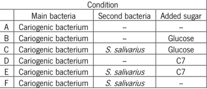
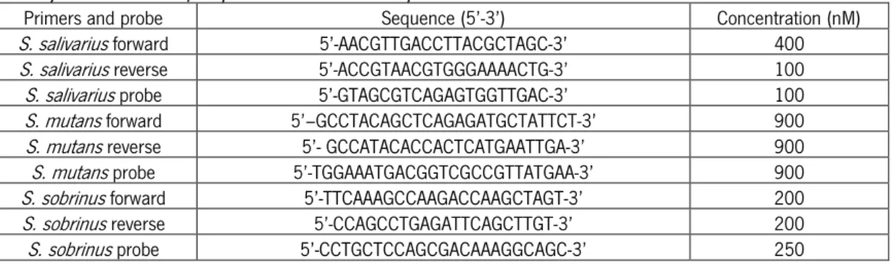
Table 1 – Comparison of the sequences of primers and probe designed for different strains of mutans streptococci ... 24 Table 2 – qPCR primers and probe: Guanine-Cytosine content (% GC), Melting temperature (Tm) 24 Table 3: Conditions used in dual-species model growth with each one of cariogenic bacteria (Streptococcus mutans or Streptococcus sobrinus). Prebiotic compound (C7) ... 31 Table 4: Sequence and final concentrations of primers and probe used in quantitative Polymerase Chain reaction (qPCR) assay for Streptococcus salivarius, Streptococcus mutans and Streptococcus sobrinus ... 32 Table 5: pH values for Streptococcus mutans in dual species model at 24 h and 48 h: Assessment of pH variation of S. mutans culture testing the effect of C7 compound with (E) or without (D) the presence of S. salivarius and glucose with (C) or without (B) S. salivarius. The negative controls were made one with only the cariogenic bacterium (A) and another with the cariogenic bacterium and S. salivarius without addition of glucose (F) ... 38 Table 6: pH values for Streptococcus sobrinus in dual species model at 24 h and 48 h: Assessment of pH variation of S. sobrinus culture testing the effect of C7 compound with (E) or without (D) the presence of S. salivarius and glucose with (C) or without (B) S. salivarius. The negative controls were made one with only the cariogenic bacterium (A) and another with the cariogenic bacterium and S. salivarius without addition of glucose (F) ... 41
C
HAPTER
1
1.1. Main objectives
The aim of this work is the evaluation of the effect of a prebiotic compound (C7) stimulating Streptococcus salivarius to a mixture of cariogenic bacteria (Streptococcus mutans and Streptococcus sobrinus). The purpose is to analyze and discover whether addition of this compound leads to a decrease in the concentration of these cariogenic bacteria.
Moreover, the effect of this compound in saliva is also aimed in this work in order to test which bacteria are stimulated by this prebiotic compound.
The discovery of a selective medium for each bacteria used (S. salivarius, S. mutans and S. sobrinus) is also aimed to quantify these strains by microbial culturing. The development of a quantitative Polymerase Chain Reaction (qPCR) for S. salivarius is another goal of this work to make possible its quantification by this molecular technique.
1.2. General Concepts
1.2.1. The oral cavityThe oral cavity is a complex microbial ecosystem consisting of several bacterial species that interact with each other competitively and cooperatively in a not isolated or confined compartment within the human body [1]–[5]. The mouth comprises teeth, supporting tissues and oral mucosa [6]. Bacteria colonize different structures such as teeth, tongue and oral mucosa, and some bacteria are associated to the maintenance of oral health and balance with the host and the environment, while others are related to biofilm formation and to oral diseases [1], [2]. The resident microflora differs in composition according to surface and consists not only by Gram-positive and Gram-negative bacteria, but also by other species as yeasts [7], [8]. Several oral bacteria are associated to systemic diseases such as cardiovascular diseases and bacterial endocarditis [9].
The mouth has permanently shedding forces and organisms need to be firmly attached to avoid being washed away [8]. Saliva is a complex fluid produced by salivary glands that helps the mouth to get the optimal conditions for the growth of numerous microorganisms [6], [10], [11].
Saliva has several protective functions such as the production of a digestive enzyme or antibodies, keeping the warm, clean and moist conditions (maintaining the normal pH of the oral microflora values around 6.75-7.25 and the temperature around 35-36 ºC) or helping the speech [6], [8], [10]–[12]. Saliva has buffering capacity to restore the pH and is a primary source of
carbohydrates, peptides and amino acids associated to clearance of fermentable sugars in mouth [7]. Saliva influences the ecology of the mouth with its ionic composition and organic components such as glycoproteins and proteins [10].
1.2.2. Oral biofilms
Oral biofilms are aggregates of microorganisms attached to a surface or to each other creating a dynamic and complex multispecies community called dental plaque leading to oral disorders such as periodontitis or dental caries [1], [2]. “Animalcules” were observed for the first time in gingival tooth scrapings by Antoine van Leeunwenhoek with a microscope [2], [13], [14].
Bacteria in biofilm form and act like a community and present some specific characteristics such as complex interspecies interactions, surface attachment, extracellular matrix of polymeric substances [1]. On the other hand, microorganisms that are free-floating and not attached are called planktonic cells and their characteristics differ from biofilms, which consist of with glycocalyx matrix and bacterial cells [2]. Planktonic cells can attach directly to surfaces of the oral cavity or to bacterial cells already colonized [1].
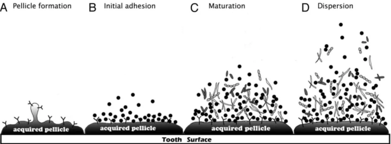
Figure 1 - Oral biofilm formation. A. Pellicle formation. The acquired pellicle is a thin layer that consists of adsorbed organic molecules derived from the salivary glycoproteins attached to the tooth surface. B. Initial adhesion. Specific receptors allow the initial adhesion of bacteria to the pellicle. C. Maturation. Biofilm maturation results of interactions between later colonizers and early colonizers, previously attached in a cell-to-cell reaction (co-aggregation). D. Dispersion. Bacteria leave the biofilm and colonize a new site [2].
Biofilm formation is a natural and highly dynamic process (Figure 1). It begins with the attachment of acquired pellicle by specific extracellular proteinaceous components (adhesins) crucial for the initiation of biofilm formation [2], [3], [15]. This pellicle is an acellular proteinaceous film, a thin layer of adsorbed organic molecules originates from the salivary glycoproteins, phosphoproteins and lipids attached to a clean tooth surface and consists of several components as glycoproteins, enzymes and other molecules [1]–[3], [8]. The mechanisms involved in acquired pellicle formation include long-range forces (Coloumb
interactions, van der Waals forces and dipole-dipole interactions), medium-range forces (hydrophobic interactions) and short range forces (covalent bonds, electrostatic interactions, hydrogen bonds, ionic interactions and Lewis acid-base interactions) and are based on Gibbs law of free enthalpy [2].
The second phase consists of initial adhesion of bacteria to the pellicle, which is important for oral bacteria interactions with host molecules allowing the connection between bacteria and receptors [1]–[3]. There are specific receptors in the pellicle on the tooth surface that allow bacterial binding based on a recognition system [8]. The mechanisms involved in the early attachments are the result of random bacterial movement and are based on electrostatic attractions or physical attachments and later on chemical forces, which include hydrogen bonds, hydrophobic interactions, calcium bridges, van der Waals forces, acid-base interactions and electrostatic interactions [2], [3], [8], [13]. This early attachment is weak and reversible and some bacteria previously attached may leave the tooth surface due to specific and non-specific molecular interactions involved on it. Early colonizers are pioneer species that attach the tooth surface mainly members of the genera Actinomyces and Streptococcus [2], [3], [7], [13].
The third phase comprises the attachment and biofilm maturation [2]. The attachment of colonizers is made through salivary glycoproteins by connection to early colonizers, previously attached to cells surfaces [1], [2]. Late colonizers include Gram-negative anaerobes, as for example Fusobacterium nucleatum, Porphyromonas gingivalis, Treponema spp. [2], [8]. The attachment of later colonizing bacteria is a cell-to-cell reaction mediated by adhesin-receptor interactions called co-aggregation [1], [2], [8]. This specific process is very complex because bacteria can only aggregate with specific bacteria according to polysaccharide recognition and not with any random bacteria. When bacteria attach to the pellicle by specific interactions, extracellular polymeric substances (EPS) excretion begins and attachment becomes stronger and irreversible [2], [8], [13]. EPS is the major component of biofilms surrounding bacteria present in biofilm establishing its structure, promoting bacterial accumulation to the tooth surface, providing a communication medium between bacteria, and promoting biochemical and physiological changes in the matrix of the biofilm [2], [13], [16]. EPS are largely insoluble and biosynthetic polymers such as polysaccharides, proteins, nucleic acids and phospholipids [13]. A mature biofilm has different microbial components from the initial biofilm [2]. The mature biofilm is a stable situation that can enhance the resistance to antibiotics due to its complex structure and multispecies composition, when compared to planktonic cells [1], [2].
The last phase in biofilm formation is the dispersion of biofilm cells and colonization because bacteria leave the biofilm by erosion, sloughing, and seeding and so, they can spread to colonize a new site [2]. This detachment is due to limited nutrients on biofilm requiring a new site with more nutrients to grow and due to limitation of biofilm development (bacteria are better protected against fluid shear force of saliva on rough surfaces) [2], [3].
Microorganisms in dental biofilm can cooperate and also compete with each other [1], [2]. The bacteria present in biofilm maintain equilibrium with the host through microbial cell-cell interactions helping the community dynamics [1], [3], [17]. From all oral bacteria, Streptococci are Gram-positive that have the strongest ability to produce several kinds of bacteriocins such as mutacins (lantibiotics and non-lantibiotics) [1], [2], [10]. Bacteriocins are non-specific proteins produced derived of ribosomal synthesis of cationic peptides with antimicrobial activity [1], [2], [4]. Quorum sensing system is a chemical communication process among bacteria characterized by the production of signal molecules, transport, sense and control of bacterial growth [1], [2].
Moreover, some commensal bacteria present in the dental plaque are able to exclude some pathogens and allochthonous bacteria by production of antimicrobial substances or competition for nutrients [1], [3], [17]. However, the accumulation of dental biofilms also modifies the bacterial composition leading to oral diseases such as periodontitis or dental caries [1]. This modification is characterized by a shift in oral biofilms from Gram-positive bacteria to Gram-negative anaerobic rods.
So, the biofilm results of the attachment of the bacterial cells to a clean surface (tooth surface) forming an acquired pellicle, followed by accumulation and multiplication of bacteria resulting in the colonization and maturation of biofilm [15].
1.3. Plaque-related diseases
1.3.1. Dental plaque and oral diseases
Dental plaque is a dynamic microbial ecosystem in which several Gram-positive and Gram-negative bacteria, that interact with each other through microbial interactions, grow as a biofilm maintaining a dynamic stability stage - microbial homeostasis [7]–[9], [18]–[20]. However, changes and imbalances in the oral microflora can occur and the microbial homeostasis breaks down allowing the development of plaque-related diseases due to an enrichment of pathogens and a reduction of beneficial bacteria within the microbial community [4], [7], [19]. Oral diseases are the result of a shift in the balance of the resident microbiota [21].
Periodontal diseases are infections of the supporting tissues of the teeth that result of an inflammatory response disturbing the harmonious relationship in the oral cavity [22]–[24]. Periodontal diseases are the most common infection diseases in the world and remain an important health problem associated to tooth loss in adults [4], [24]–[26]. These diseases are characterized by an increase of obligatory anaerobic bacteria as Gram-negative proteolytic species [10].
Gingivitis is a reversible infection of the soft tissue of the mouth that does not destroy the periodontal tissues [22]. Gingivitis results from accumulation of plaque triggering an inflammatory response and increasing the gingival crevicular fluid [7]. Periodontitis is a polymicrobial infection due to colonization of hard and soft surface tissues in the oral cavity that can result in attachment loss and destruction of alveolar bone and eventually tooth loss [24], [27]–[29]. So, periodontitis is a more severe stage of the infection that results of the evolution of gingivitis [24].
Dental caries is an infectious disease due to bacterial action resulting of a process of demineralization of enamel crystals and dentin by acids derived from interactions of specific bacteria with sugars of the dental plaque leading to tooth destruction [30]–[33].
Dental diseases are one of the most prevalent diseases nowadays with high treatment costs [10].
1.3.2. Periodontitis
The periodontium is a set of tissues that supports the tooth and can be divided in gingiva, cementum, alveolar bone and periodontal ligament [33], [34]. Gingiva is the soft tissue that covers the mouth. Cementum is a specialized calcified and hard tissue that covers the root of the tooth. The alveolar bone is the bone in the jaw that supports and protects the teeth, and the part of the maxilla that helps the resistance of mastication. The periodontal ligament is a group of soft and connective tissue fibers that makes the connection between the tooth attachment and the cementum [24], [33], [34]. The functions of periodontium are the support and the attachment of the tooth to the bone of the jaw, and the protection and the resistance of the tooth to the mastication forces [24], [33], [34].
The pathogenic microorganisms involved in oral biofilm originating periodontitis are principally gram-negative pathogens such as Porphyromonas gingivalis (P. g.), Prevotella intermedia, Fusobacterium nucleatum and Actinobacillus actinomycetemcomitans (A. a.) [23], [24], [28]. However, these microorganisms are not in high number in initial lesion, but the
number increase with the development of disease [35]. The oral biofilm with pathogenic microorganisms is the main etiological factor of periodontitis [24]. For instance, A. a. is a gram-negative bacterium that is indigenous of the oral cavity [25], [36]. A. a. can induce the periodontal disease by colonizing tooth surface with adhesins but it is also associated to systemic infection as endocarditis. On the other hand, P. g. is an anaerobic, motile and non-sporulating Gram-negative rod able to colonize the gingival sulcus and also the periodontal pocket [14], [37]. Virulence factors of P. g. help this species to survive in adverse conditions of growth as periodontal pocket.
The aim of therapy and treatment for patients with periodontitis is to halt the progression of the disease removing the inflammation and reducing the periodontopathogens from the subgingival area [23], [24], [27]. Traditional treatment involves scaling and root planning conjugated with antibiotics [23], [24], [26], [27].
Scaling and root planning is a surgical therapy that consists in removing supra- and subgingival plaque and the plaque from the root surfaces of the teeth preventing the progression of periodontitis [14], [24]. The most effective method in scaling and root planning used is called periodontal debridement and aims to stimulate the regeneration of lost and damaged tissues and to reduce deep probing depth [24], [38]. This technique has several benefits such as the reduction of clinical inflammation, disease progression and probing depth, gain of clinical attachment, and microbial shifts of oral cavity to an oral microflora with less pathogen. However, scaling and root planning is technically difficult to perform due to some mechanical limitations and time consuming [24], [38]. Moreover, this technique is disagreeable for patients and some bacteria present can persist and recolonize [24], [27], [38].
The use of antibiotics is an adjunctive therapy to treat periodontitis [24]. The application of antibiotics can be systemic or local depending on severity of periodontitis. The most commonly systemic antibiotics used are tetracycline, ciprofloxacin, metronidazole and penicillins [24]. This type of pharmacologic administration is easy to perform, but is also associated to resistance of bacterial species. On the other hand, local delivery gives site specific and therapeutic level at the site of infection allowing localized treatment areas [24]. Tetracyclines, metronidazole and chlorhexidine are drugs used for local delivery. The disadvantages are associated to their cost due to successive treatments and in the inconvenience by changing oral hygiene habits [26]. However, this is not a definitive solution because (re)colonization of the periodontal pockets and
resistance to antibiotics can occur, which is more difficult with bacteria within biofilm [4], [23], [24], [27], [29].
Periodontopathogens can also enter the blood stream triggering new infections [24]. For this reason, periodontal diseases have been investigated in order to verify their association to other diseases. Current research supposes periodontal diseases increase cardiovascular diseases [22]. Genetic factors such as age, hypertension and diabetes or environmental factors as diet, stress or cigarette smoking are tested risk factors associated to cardiovascular diseases. Periodontopathogens have been found in the association of etiology in cardiovascular diseases [22].
1.3.3. Dental caries
Dental caries is one of the most common diseases affecting humans and one of the most prevalent chronic diseases, characterized by a very slowly progression in the majority of individuals and becoming the most expensive part of the body to treat [18], [30], [39], [40]. Pain, localized destruction of the hard tissues, tooth destruction and tooth loss and impaired quality-of-life are some consequences of untreated caries [18], [41].
When a biofilm on tooth surface is able to attach, grow, develop and mature, the process of formation of dental caries can initiate and progress, leading to the loss of mineral from the tooth and the localized destruction of the tooth [8], [40], [42]. Although the presence of biofilm is necessary to develop dental caries, not all biofilms lead to dental caries, i.e., the amount is not enough to trigger this process, and in this case teeth can be healthy covered by biofilms [40].
Consumption of fermentable carbohydrates are the key environmental factors involved in initiation and development of dental caries [7], [10], [16]. Sucrose is the most cariogenic dietary carbohydrate because it is fermentable and used as a substrate for extracellular glucan synthesis by glucosyltransferases from mutans streptococci and the synthesis of extracellular (EPS) and intracellular (IPS) polyssacharides in the dental plaque [16]. Low pH environment promotes a change in the resident oral plaque, while EPS promote changes in the composition of matrix of the biofilm [16]. EPS such as glucosyltransferases and fructosyltransferases help bacterial adherence and accumulation on tooth surface and create biochemical and structural changes in the matrix of biofilm, such increase the porosity. IPS such as glycogen-like help to maintain a low pH in the matrix of dental plaque and to exposure organic acids to tooth surfaces. Then, sugar can easily diffuse into the biofilm and decreasing local pH by microbial catabolism [16]. Furthermore, sucrose is able to reduce the concentrations of the most important ions in
maintaining the mineral equilibrium between the tooth and the oral environment (calcium, inorganic phosphorus and fluoride) [16].
An increase of sucrose-rich diet and carbohydrates metabolism promotes acidification creating conditions for ‘low-pH’ non-mutans streptococci species grow, increasing the risk of dental caries [10], [16], [20]. Acid production exposure the dental plaque continuously under the critical pH for demineralization of tooth surfaces leading to net mineral loss and chemical dissolution [10], [16], [18], [20], [30]. This destruction can affect enamel, dentin and cementum [40], [42], [43]. Enamel and dentin are part of the tooth. Enamel is a hard, inert and acellular tissue that covers the crown and it is also the most highly mineralized tissue found in the body. Dentin is a hard, elastic, resilient, sensitive, connective and avascular tissue that supports the enamel [6]. Consequently, acid production by oral bacteria due to sugar metabolism decreases environmental pH. So, dental caries is an endogenous disease that consists of metabolic events in dental biofilms resulting in an imbalance in the equilibrium between biofilm fluid and tooth mineral [10], [18], [20], [42]. As a consequence, acid production increases, environmental pH decreases and chemical composition of dental plaque shifts to a predominantly Gram-negative bacteria such mutans streptococci and lactobacilli [10], [18]–[20].
The traditional detection of dental caries is made by visual examination (white opaque lesions as a consequence of enamel translucency) and radiographs [41], [43]. Recently, other methods have been developed, e.g. methods based on fiber-optics, fluorescence or electrical impedance [41].
It has been used the DMF index to quantify caries, where D is for decayed teeth, M is for teeth missing and F is for teeth previously filled [44]. This index can be applied to teeth entirety (DMFT) or to all surfaces of the teeth (DMFS) [44]. However, the application of DMF index has been decreasing nowadays, except in the quantification of treatment received, mainly due to modern preventive and restorative technology [44].
Several microorganisms are involved in the formation of dental caries. These microorganisms should be able to produce acid and to tolerate a low-pH environment [18].
Non-mutans streptococci and Actinomyces species are present in high levels at the initial stage of plaque formation, probably due to adhesins which facilitate their adhesion to proteins and sugar chains of acquired pellicle on tooth surface [18], [20], [35]. These species can acidify the environment through degradation of carbohydrates creating acidic and anaerobic conditions [35]. Non-mutans streptococci strains such as Streptococcus sanguinis, Streptococcus oralis and
Streptococcus mitis are the initial colonizers involved and mutans streptococci are present in low level [18], [20]. When dental caries is developed, the most common species present are Actinomyces and Streptococcus [20].
Mutans streptococci are a group of microorganisms (such as Streptococcus mutans and Streptococcus sobrinus) able to grow and develop at conditions of high sugar and low pH [7], [10], [18], [19]. Mutans streptococci are highly acidogenic and aciduric species able to produce water-insoluble extracellular glucan from sucrose by glucosyltransferase promoting bacterial adhesion to tooth surface and to other bacteria [18], [20], [35]. Although mutans streptococci are associated to dental caries, the disease can occur in the absence of these microorganisms [19]. Mutans streptococci are the major pathogens involved in dental caries formation (specially related with sucrose-rich diet) due their aciduric and acidogenic characteristics [18], [20]. S. mutans is the main etiological agent of human dental caries [17]. Additionally, S. sobrinus is also associated with the formation of dental caries [39]. The risk of transmission from mother to child is another etiological factor of dental caries [45].
An invasive intervention in clinical dentistry is an operative way involved on caries treatment [46]. Vaccination, gene therapy or antimicrobial treatment are several ways to control dental caries, however elimination of specific bacteria responsible for caries is not an effective method because these bacteria are essential for mouth equilibrium [20]. An alternative is the use of probiotics to treat caries infection through interfere on oral colonization of cariogenic bacteria [5].
S. mutans and S. sobrinus are also associated with non-oral infections [39]. Diabetes can also be related to dental caries due to salivary dysfunction [12].
1.3.4. Ecological plaque hypothesis
The mouth comprises surfaces such as mucosal surfaces and teeth (non-shedding surfaces) with different oral microflora. Some of these places, for example, teeth, allowing the attachment and growth of bacteria which leads to the dental plaque and later to disease, e.g., periodontitis or caries [10].
The resident oral microflora is characterized by a dynamic relationship with inter-microbial and host-inter-microbial interactions. This inter-microbial homeostasis leads to the stability of microflora [10]. Alterations between microbial ecosystem and host tissue are responsible for initiation of oral diseases [35].
The resident oral microflora has endogenous proteins and glycoproteins (mucins) as the main sources of carbon and nitrogen. Any change in the environment increasing oral pathogens within the microbial community will cause an imbalance in the microflora [10]. Changes in the nutrient status at the site, the diet, the dentition and radiation therapy are possible causes for alterations on oral microflora [10].
In a healthy situation, dental plaque results from a biofilm formation: conditioning film, early colonizers, attachment and colonization, later colonizers, co-aggregration and a microbial community [10]. In case of periodontitis, there is an increase of levels of obligatory anaerobic bacteria as a result of development of an inflammatory host response [10]. This increase will lead to inactivation of host proteins. In the case of dental caries, there is an increase on levels of acid-tolerating bacteria, principally mutans streptococci and lactobacilli, which will lead to demineralization of enamel as a result of development of an infection [10]. This increase in acidogenic and aciduric bacteria will lead to a decrease in pH by metabolizing dietary sugars to acid, creating an optimal growth conditions for these species.
There are two hypothesis to explain the different bacteria species present in diseased and healthy sites [10], [19]. The ‘specific plaque hypothesis’ proposes that only a small percentage of organisms present in dental plaque are actively involved in disease [7], [10], [19]. This hypothesis focuses the treatment only against microorganisms responsible for the disease [19]. However, caries can appear in the absence of typical etiological agents and these species can be present without caries lesion development [7], [10], [18].
On the other hand, the ‘non-specific plaque hypothesis’ proposes that interactions between bacteria present in dental plaque and the host can lead to a disease situation [7], [10], [19]. So, dental caries and processes associated to their development can be controlled [18].
The ‘ecological plaque hypothesis’ is an alternative explanation to define the relationship between dental plaque bacteria and the host in health and disease resulting from the combination of specific and non-specific plaque hypothesis [7], [10], [16], [18], [19]. This hypothesis is an ecological and dynamic model based on changes in microbial and environmental dynamics (key factors) in oral microflora leading to the development of oral diseases [7], [10], [35].
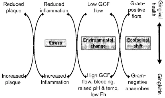
In case of periodontal diseases (Figure 2), putative periodontal pathogens exist on healthy sites in a small proportion, but when plaque biomass accumulates, an inflammatory host response is triggered [10]. This results on the increase of flow of gingival crevicular fluid (derived
from blood plasma and is rich in nitrogenous compounds as peptides, amino acids and proteins) and alteration of local nutrients status and pH will change to a neutral value [7], [10], [35]. The consequences are an increase in pH (to a neutral value), a decrease in redox potential (Eh) and an outgrowth in proteolytic, anaerobic and asaccharolytic Gram-negative bacteria [7], [10]. The microbial shift of periodontitis consists of an enrichment of anaerobic obligatory and proteolytic bacteria (such as Porphyromonas gingivalis) due to increase of gingival crevicular fluid. This results from host defense induced by colonization when bacteria promote a neutral pH environment and increase Gram-negative and unculturable bacteria in the mouth [21], [35].
Figure 2 - Ecological plaque hypothesis and prevention of periodontal diseases. Gingival Crevicular Fluid (GCF). Redox potential (Eh) [10].
In case of dental caries (Figure 3), potentially cariogenic bacteria exist at neutral pH in a low level (clinically insignificant) [10]. A low-sugar diet promotes a stable plaque microflora and demineralization and remineralization are in equilibrium [7], [10]. However, with an increase in the frequency of fermentable carbohydrates consumption, pH will decrease and acid-tolerating bacteria will proliferate, stimulating demineralization [10], [19]. This situation will shift the subgingival microflora from mainly Gram-positive bacteria to a more cariogenic resident plaque microflora with high levels of anaerobic, asaccharolytic and obligatory Gram-negative organisms, the best adapted to high sugar and low pH, such as mutans streptococci and lactobacilli, on oral cavity [7], [10], [16]. The microbial shift of dental caries are characterized by increase of population of aciduric bacteria (mutans streptococci and lactobacilli) and enrichment of cariogenic potential of supragingival plaque [21], [35].
Figure 3 - Ecological plaque hypothesis and prevention of dental caries [10].
Prevention strategies depend on type of dental disease but the main idea is interfering on factors responsible for diseases shifting to a health situation [7], [19].
In case of periodontitis, the site should be less anaerobic and the flow of gingival crevicular flow would be reduced in order to avoid growth of putative pathogens [7], [10]. Anti-inflammatory and antimicrobial agents reduce gingival crevivular fluid and growth of some periodontopathogens becomes restrict according their essential nutrients. Oxygenating and redox agents are an alternative to create an incompatible environment for growth of obligatory anaerobes when the redox potential of periodontal pocket can be raised to create these conditions [7]. Methylene blue is a redox dye able to prevent the growth of obligatory anaerobes by the increase of redox potential and decrease of gingival crevicular fluid [7], [10].
In the case of dental caries, the actions are based on inhibition of plaque acid production and consumption of non-fermentable sugar compounds [10]. Fluoride is able to enhance remineralization and acid resistance of enamel. Antimicrobial agents as chlorhexidine reduces the impact of pH changes and demineralization, promoting mechanical cleaning of plaque [7], [10]. Other alternative sweeteners, also known as sugar substitutes, act as promoter of remineralization of enamel and stimulate saliva flow when there is no significant acid production [7], [10].
An extended caries ecological hypothesis (Figure 4), based on the ecological hypothesis, proposes three reversible stages in the caries process to explain the relationship between the composition of the dental plaque and dental caries process, conjugating microbiological, biochemical, ecological and clinical perspectives [18], [20]. Dynamic stability stage is composed mainly by non-mutans bacteria (non-mutans streptococci and Actinomyces) promoting a natural
pH cycle for development, growth and equilibrium in the oral cavity [18], [20]. These bacteria can produce acids from sugary foods and these acids can demineralize enamel, but the equilibrium is easily achieved by changes between mineral net gain and mineral net loss helping demineralization and remineralization balance. This situation stimulates selection and increase of ‘low-pH’ non-mutans bacteria and microbial acid-induced adaptation creating an acidogenic stage [18], [20]. However, if acidification steps are rarely, balance will shift to net mineral gain tending to remineralization. The acidic environment increases aciduric bacteria allowing lesion development and net mineral loss creating an aciduric stage [18], [20].
Figure 4 - Extended caries ecological hypothesis [20].
1.4. New treatments
As current treatment options are not solving the problem of oral disorders, specially due to antibiotic resistance, other options have to be investigated [21], [47]. Included in these new options are probiotics and prebiotics, which are emerging in diverse fields, as oral health.
Probiotics are defined as live microorganisms, which when administered in certain quantities, have health benefits on the host (humans and animals) [4], [5], [21], [46], [48], [49]. First definition for probiotics was introduced by Lilly & Stillwell in 1965 and then several alterations were made [5], [50]. Probiotics are characterized by beneficial effects (immune stimulation, immune modulation of host defenses, anticarcinogenic effects, antidiabetic characteristics, cancer prevention, …), production of antimicrobial substances (depending on pH, catalase, proteolytic enzymes and temperature), adhesion to the mucosa, degradation of toxins,
improvement of colonization resistance, and competitive exclusion mechanisms [4], [46], [47], [49]–[51].
The main field of research of probiotics is the gastrointestinal (GI) tract and they are already being used in the GI tract (particularly the colon, a very heavily colonized site) to restore numbers of beneficial bacteria and decrease numbers of pathogenic bacteria [5], [21], [46]– [48], [51]. Bifidobacteria and lactobacilli are the most commonly probiotic species used such as Lactobacillus acidophilus, Lactobacillus casei, Lactobacillus rhamnosus, and Bifidobacterium bifidum or Bifidobacterium infantis [5], [21], [46]–[48], [51].
Probiotics require in vitro tests and substantiation of efficacy with human trials before their use on humans in order to guarantee their beneficial effects [4].
In recent years use of probiotics has also been demonstrated for urogenital infections, atopic disease, voice prostheses and in the dental field [4], [5], [21], [46], [47].
An ‘oral probiotic’ needs to able to adhere and colonize surfaces in the oral cavity such as hard non-shedding surfaces [4], [27], [46]. Oral probiotics should also not be able to ferment sugars, otherwise pH will decrease and caries will develop [4].
Some lactobacilli strains such as L. fermentum, L. salivarius and L. rhamnosus are used in dairy products (a way for probiotic administration), others are present in resident oral microflora [46]. L. rhamnosus GG has inhibitory activity against cariogenic streptococci [5], [47]. Lactobacillus species are also able to produce inhibiting substances avoiding and preventing adhesion and colonization of pathogenic bacteria [46]. For example, consumption of yogurt containing L. reuteri reduces S. mutans [46]. This reduction is verified during the period of yogurt consumption and some days after cessation of consumption, thereby studies are required to analyze caries-inhibiting effect after the probiotic administration [46], [47]. A reduction in mutans streptococci was also detected with Bifidobacterium DN-173 010 consumption [46]. L. reuteri is also able to reduce gingivitis [46]. Lactobacilli and bifidobacteria can inhibit growth and colonization of periodontopathogens to hard and soft tissues and can inhibit cariogenic streptococci [4], [47].
The best effect on the oral microbiota was seen when working with probiotic indigenous oral bacteria. Indigenous bacteria are already present in the oral microflora, do not need adaptation and do not change with intervention or disease [4], [50].
One example of such a bacterium is Streptococcus salivarius [46]. S. salivarius already demonstrated inhibitory effect on volatile sulfur compounds by competition for colonization sites
against other species [46]. This bacterium is one of the early colonizers of epithelial surfaces of the oral cavity [21].
An ideal way to stimulate the indigenous beneficial microbiota would be by prebiotics. Prebiotics are non-digestible food ingredients (substances or nutrients) that can beneficially affect, by stimulation, the growth and/or the activity of some bacterial species of the host [47]– [51].
To be considered as prebiotics, a dietary substrate needs to: be resistant and available in order to be used as fermentation substrate; be selective for beneficial bacteria; induce beneficial effects within the host by prebiotic fermentation [51], [52].
Prebiotics are non-digestible carbohydrates such as oligosaccharides [51]. Oligosaccharides are soluble short-chain polysaccharides with low degree of polymerization and can be found in fruits and vegetables and also produced by hydrolysis of polysaccharides. Lactulose, lactosucrose, fructo-oligosaccharides (FOS), galacto-oligosaccharides (GOS), gluco-oligosaccharides or xylo-gluco-oligosaccharides are some examples of gluco-oligosaccharides with prebiotic potential [21], [51], [52]. Prebiotics are often studied for application o GI tract, but other studies should be carried out to define their applicability into other areas [52].
Prebiotics produce protective metabolites, increase mineral absorption and reduce the risk of cancer, e.g., colon cancer [49], [51]. Prebiotics stimulate beneficial components proliferation in the microflora, specially components with probiotic characteristics such as lactobacilli and bifidobacteria [21], [49], [52]. The increase of these beneficial bacteria improve resistance to pathogenic bacteria due to production of natural antibiotics with inhibitory properties and antimicrobial effects [51].
Different techniques have been developed to enumerate bacteria such as Polymerase Chain Reaction (PCR) or Denaturing Gradient Gel Electrophoresis (DGGE) due to their applicability to culturable as well unculturable cells verifying that species present in disease are already on microflora in an healthy stage but at low numbers [21], [52].
The combination of pre and probiotics could be another point of interest for prevention and treatment to improve oral health [4]. A mixture that contains pre and probiotics and that beneficially affects the host is known as symbiotic [5], [50]. Previous research already demonstrated the inhibitory effect of pre and probiotics for head/neck, oral and respiratory tracts, pancreas and liver, and kidney, bladder and vagina [48]. Regarding to the oral cavity, the inhibitory effect of one specific prebiotic compound on the levels of several periodontopathogens
was already demonstrated however the effect of this prebiotic compound needs to be tested on cariogenic bacteria [48].
Further, some dairy food products have health benefits and have been investigated in order to use them properly [48]. It is already known that body’s microbiota can be controlled and modified according to use of pre and probiotics, but an evidence of probiotic therapy on oral diseases is required [46], [48].
C
HAPTER
2
2.1. Introduction
Quantification of oral streptococci is normally made by microbial culturing, which is laborious and time-consuming and has several disadvantages [53]. However, a quantitative analysis is always required to detect, count and control bacteria associated to dental caries [39]. So, several methods have been developed to quantify and identify different bacteria in the mouth such as biochemical, immunological and genetic tests [32], [39], [54]. The majority of these methods are, however qualitative and based on bacterial detection systems that are time-consuming, laborious and with possibility of contamination [39], [54]. Therefore, the use of molecular methods such as DNA-based methodologies would solve these problems and their use have been increasing [55].
Quantitative PCR (qPCR) assay is a method based on 5’-3’ exonuclease activity of Taq polymerase and DNA copy number [39]. qPCR enumerates the accumulation of reporter fluorescence as a result of the cleavage of the probe during PCR amplification [9], [39]. The probe consists of a specific sequence labeled with a fluorescent reporter dye and a quencher emitting fluorescence. The quencher dye avoids the extension of the probe by the polymerase and when it is cleaved it allows the accumulation of the reporter fluorescence [9]. The probe is normally marked with intercalating dyes (non-specific sequences of fluorescents dyes emitting a large fluorescence when intercalate into double-stranded DNA) [56].
This method requires only a small volume of sample and amplifies and quantifies the nucleic acid sequences at the same time and so there is no need to run a gel to see the product. Thus, qPCR is an accurate, sensitive, precise, specific, fast, reliable, powerful and useful method with a low possibility of contamination [9], [39], [56], [57].
qPCR assays are already developed for Streptococcus mutans and Streptococcus sobrinus, the main cariogenic bacteria [39]. However, the quantification of Streptococcus salivarius by a qPCR assay is also required due to the great interest in this microorganism as a probiotic strain. Nowadays, the interest on probiotics is growing due to their beneficial health effects [58]. S. salivarius is an important microorganism of the oral microbiota and it is the most abundant species of streptococcal present in the oral cavity and an early colonizer of oral surfaces with potential for use as an oral probiotic [59], [60].
Streptococcus species have a specific conserved locus on the dextranase gene. Dextranase gene is an enzyme that hydrolyses glucans in the plaque matrix and it could even be one of the responsible for the virulence of these strains [54].
So, in this stage a qPCR assay was developed for the culture-independent enumeration of S. salivarius and to maximize the specificity of the qPCR assay in environmental samples.
2.2. Materials and Methods
2.2.1. Bacterial strains and culturing conditions
In the present work Streptococcus mutans ATCC 25175, Streptococcus sobrinus ATCC 33478, Streptococcus salivarius TOVE-R, Streptococcus salivarius clinical strain, Streptococcus salivarius K12 and Streptococcus salivarius ATCC 7073 were the bacteria used. Bacteria were maintained on blood agar plates (Blood Agar Base II, Oxoid, Basingstoke, UK) supplemented with 5% sterile horse blood (Biotrading, Keerbergen, Belgium), 5 µg/ml hemin (Sigma Chemical Co, St. Louis, MO) and 1 µg/ml menadion. One day before each experiment, bacteria were collected from blood agar plates and incubated overnight in 10 ml Brain-Heart Infusion (BHI) broth (Becton, Dicksinson and Company, France) at 37 ºC in a 5% CO2 environment. Bacterial
concentration was adjusted by optical density measurements at a wavelength of 600 nm (Smartspec 3000, BioRad, USA).
2.2.2. Design of qPCR primers and probe
The forward primer (Ssal442F) was based on the dextranase gene from S. salivarius and was selected from Igarashi et al. (2001). The reverse primer (Ssal615R) and probe (Ssal497T) were also based on the dextranase gene from S. salivarius and were designed with primer 3 software. A search with BLAST (http://www.ncbi.nlm.nih.gov/BLAST/) and Probe Match [61] was carried out in order to assess, in silico, the homology of the selected primers and the probe with unrelated sequences. The primers-probe set for primer-dimers, melting temperature, hairpin configuration and GC content were checked with OligoAnalyzer (Integrated DNA Technologies, Coralville, IA, USA). Primers and probe were synthesized by Eurogentec (Seraing, Belgium). The probe was 5’ labeled with a fluorescent dye as a reporter, FAM (6-carboxyfluorescein), and another fluorescent dye as a quencher, 3’ TAMRA (6-carboxytetramethylrhodamine). A PCR assay against S. mutans, S. sobrinus and 4 different strains of S. salivarius was performed as a confirmation of the forward and reverse primer. The PCR steps consisted of an initial 1 min at 94 ºC, followed by 26 cycles of 94 ºC for 1 min, 60 ºC for 1 min and 72 ºC for 1 min, and an additional cycle of 5 min at 72 ºC. PCR fragments were checked in an electrophoresis on 1% agarose gel.
2.2.3. Construction of the qPCR plasmid standard
A fragment of 192 bp of the S. salivarius dextranase gene was used as a standard for the qPCR and was amplified with the forward primer Ssal442F (5´- AACGTTGACCTTACGCTAGC -3’) and lately designed reverse primer Ssal615R (5´- ACCGTAACGTGGGAAAACTG -3’). This fragment was purified with the QIAquick PCR purification kit (Qiagen, Hilden, Gemany), cloned with the pGEM-T easy plasmid vector system (Promega, Madison, WI, USA) and used to transform Escherichia coli DH5α. High Pure Plasmid Isolation Kit (Roche Diagnostics GmbH, Mannheim, Germany) was used to isolate the plasmids from the clones, according to the manufacturer’s instructions. The validation of the DNA sequence of the plasmid was made by sequencing with the BigDye Terminator v1.1 cycle sequencing kit (Applied Biosystems, Foster City, CA, USA). Dye ex sequencing purification kit (Qiagen) was used to purify the sequencing product and ABI 310 Genetic Analyzer (Applied Biosystems) was used to analyze it. Plasmid concentration and purity were determined with GeneQuant RNA/DNA calculator (Amersham Pharmacia Biotech). Each qPCR run was performed with a 10-fold dilution series of the plasmid to construct the standard curve.
2.2.4. qPCR optimization
A quantitative Polymerase Chain Reaction (qPCR) assay was performed with a CFX96 Real-Time System (BioRad, CA, USA). Taqman 5’ nuclease assay PCR method was used in order to detect and quantify bacterial DNA. Taqman reactions contained 12.5 µl mastermix (Eurogentec, Seraing, Belgium), 4.5 µl sterile water, 1 µl of each primer and probe, and 5 µl template DNA. Individual primer concentrations ranging from 100 to 900 nM and probe concentration ranging from 50 to 200 nM were tested. Assay conditions steps involved an initial step for 2 min at 50 ºC, followed by a denaturation step at 95 ºC for 10 min and 45 cycles of 95 ºC for 15 s and 60 ºC for 1 min.
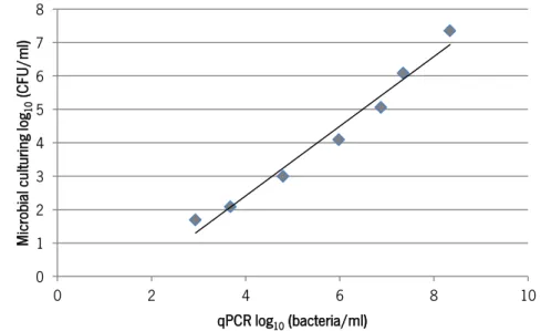
2.2.5. Comparison between qPCR and microbial culturing
An overnight culture of Streptococcus salivarius TOVE–R was used to compare qPCR and microbial culturing. For microbial culturing, 10-fold serial dilutions in sterile saline were used. For that, 50 µl of each dilution was plated on blood agar plates with a spiral platter (L.E.D. Techno). The plates were incubated in a 37 ºC and 5% CO2 environment for 3 days and the
colony-forming units (CFU) were determined considering plates with colony counts between 20 and 300. For qPCR, 100 µl aliquots of each dilution were used for DNA extraction with the QIAamp DNA mini kit according to the manufacturar’s instructions (QIAGEN). qPCR was
performed as described previously. The experiment was repeated five times and values were log-transformed to calculate an orthogonal regression.
2.3. Results and Discussion
2.3.1. Design of qPCR primers and probe
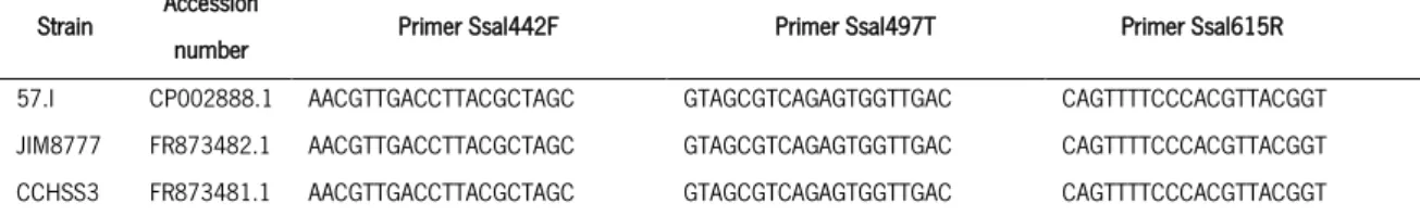
qPCR assay for S. salivarius could not be designed against 16S rRNA gene such as for other oral bacteria because this is not specific for every species of Streptococcus. So, a conserved locus of the dextranase gene of S. salivarius ranging from base number 442 to base number 615 was the base of the design of qPCR primers due to its specific and conserved sequences [54]. The design of the PCR primers was developed on the basis of the comparison of the nucleotide sequences of the dextranase gene of S. salivarius, S. mutans and S. sobrinus and the consensus sequence of the alignment served as template (Table 1).
Table 1 – Comparison of the sequences of primers and probe designed for different strains of mutans streptococci
Strain Accession
number Primer Ssal442F Primer Ssal497T Primer Ssal615R 57.I CP002888.1 AACGTTGACCTTACGCTAGC GTAGCGTCAGAGTGGTTGAC CAGTTTTCCCACGTTACGGT JIM8777 FR873482.1 AACGTTGACCTTACGCTAGC GTAGCGTCAGAGTGGTTGAC CAGTTTTCCCACGTTACGGT CCHSS3 FR873481.1 AACGTTGACCTTACGCTAGC GTAGCGTCAGAGTGGTTGAC CAGTTTTCCCACGTTACGGT
Primers for PCR were obtained by comparison of the nucleotide sequences of the dextranase genes of S. salivarius, S. mutans and S. sobrinus. The forward primer (Ssal442F) was kept from a PCR assay from Igarishi et al. (2001) and a new reverse primer (Ssal615R) was designed reducing the fragment length from 2271 base pairs (bp) to 192 bp. A qPCR probe (Ssal497T) was designed between the beginning of the forward primer and the end of the reverse primer (Table 2).
Table 2 – qPCR primers and probe: Guanine-Cytosine content (% GC), Melting temperature (Tm)
Oligonucleotide name Sequence (5’-3’) Position %GC Tm (ºC)
Forward primer: Ssal442F AACGTTGACCTTACGCTAGC 442 to 458 50 60 Reverse primer: Ssal615R ACCGTAACGTGGGAAAACTG 615 to 634 45 58 Taqman probe: Ssal497T GTAGCGTCAGAGTGGTTGAC 497 to 516 55 62
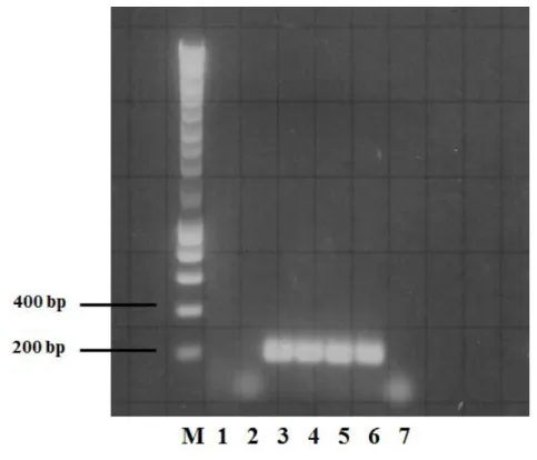
A PCR assay against S. mutans, S. sobrinus and four strains of S. salivarius was carried out to test the specificity of the primers (Figure 5).
Figure 5 - PCR amplification for detection of the dextranase gene of oral streptococcal species by a PCR and a DNA probe (Ssal497T) with Ssal442F and Ssal615R primer pair: S. mutans (lane 1), S. sobrinus (lane 2), S. salivarius ATCC 7073 (lane 3),
S. salivarius K12 (lane 4), S. salivarius clinical strain (lane 5), S. salivarius TOVE-R (lane 6). Negative control (lane 7) was made with physiological water.
A band at 200 bp was displayed for all strains of S. salivarius (lanes 3, 4, 5 and 6) and no amplification was observed for S. mutans (lane 1) and for S. sobrinus (lane 2). Looking at negative control, made with physiological water (lane 7), no amplification can be observed meaning no detection of contamination in this PCR assay. So, PCR amplification was verified only for S. salivarius strains. This indicates that the PCR primers and probe were specific for this species.
2.3.2. qPCR optimization
Data were collected during the annealing phase of qPCR assay. The optimal qPCR primers concentration was determined by titration assays. The best concentrations were obtained with 400 nM for the forward primer and 100 nM for the reverse primer. For the probe, the best result was obtained for a concentration of 100 nM. The reaction efficiency was on average 94.87% (± 2.36 SD). The reaction efficiency was calculated from the slope of the standard curve [= 10(-1/slope) – 1]. The lowest reproducible detection level of the qPCR was 4 plasmids per reaction,
![Figure 3 - Ecological plaque hypothesis and prevention of dental caries [10].](https://thumb-eu.123doks.com/thumbv2/123dok_br/17767676.836527/33.892.161.722.111.377/figure-ecological-plaque-hypothesis-prevention-dental-caries.webp)
![Figure 4 - Extended caries ecological hypothesis [20].](https://thumb-eu.123doks.com/thumbv2/123dok_br/17767676.836527/34.892.134.749.412.755/figure-extended-caries-ecological-hypothesis.webp)