Odete Sofia Lopes Gonçalves
Outubro de 2012
Development of DODAC/B:MO:DC-Chol
lipoplexes as a novel non-viral method for
transfection
UMinho|20 12 Ode te Sof ia Lopes Gonçalv es De velopment of DOD A C/B:MO:DC-Chol lipople xes as a no vel non-viral method for transfection
Odete Sofia Lopes Gonçalves
Outubro de 2012
Dissertação de Mestrado
Mestrado em Genética Molecular
Development of DODAC/B:MO:DC-Chol
lipoplexes as a novel non-viral method for
transfection
Escola de Ciências
Trabalho realizado sob a orientação da
Professora Doutora Andreia Ferreira
de Castro Gomes
E co-orientação da
Professora Doutora Maria Elisabete
Cunha Dias Real Oliveira
i
First of all, I would like to thank my supervisors, Dr. Andreia Gomes and Dr. Maria Elisabete Oliveira for their guidance, outstanding patience, remarkable support, kind advice and, most of all, for the trust they bestowed in me and my work throughout my Master research studies.
I would also like to thank João Neves, Ana Oliveira and Isabel Oliveira for all the knowledge they shared and for all the support and good advices in the laboratory. Additionally, I would like to thank my colleagues who worked with me in the laboratory. Thank you for all the support and for making it an unforgettable experience.
I would also like to thank my parents and my little brother for all the support, patience and for believing in me even when I didn’t.
And last, but not least, I would like to thank my friends for always being there for me, for better and for worse. You know who you are.
iii Abstract
One of the promising strategies concerning molecular therapy is gene therapy. By delivering genes for therapeutic purposes, we may treat both acquired and inherited genetic diseases. Non-viral vectors appear as a safer method of gene delivery into the cell, although not as efficient as viral vectors. Cationic lipids are an example of non-viral methods that have been thoroughly researched in the past few years as an effective tool to promote effective transfection of genetic material.
This project was focused on the development and characterization of lipoplexes based on dioctadecyldimethylammonium bromide/ chloride (DODAB/C): 1-monooleoyl-rac-glycerol (MO) liposomes with the inclusion of a cholesterol derivative 3β [N-(N’,N’- dimethylaminoethane)carbamoyl cholesterol (DC-Chol) for transfection. Monoolein (MO) is a neutral lipid that seems to function well as a helper lipid since it affects the physicochemical properties of the lipoplexes and interferes with lipoplex-cell interactions. The physicochemical characteristics of DODAB/MO cationic liposomes were previously studied and their transfection capacity evaluated (Neves Silva et al, 2012). The results show a successful mediation of in vitro cell transfection by the DODAB:MO formulations. DC-Chol is a synthetic cationic molecule derivate from cholesterol. It has been used before in lipoplex assemblies in order to enhance transfection efficiency.
The results in this project show that the counterion exchange (bromide and chloride) has a significant effect in terms of particle size and Zeta potential. Furthermore, the liposome preparation method affects the physico-chemical properties of the particles, as well as the citotoxixity and transfection efficiency. The inclusion of DC-Chol has different effects on transfection efficiency and citotoxicity when included in DODAB:MO or DODAC:MO lipoplexes, depending on the molar ratio and on the preparation method of the liposomes.
Overall, DODAC:MO:DC-Chol lipoplexes arise as promising non-viral vectors for transfection.
v Resumo
A terapia génica é uma das estratégias promissoras no que diz respeito à terapia molecular. Ao entregar às células e tecidos genes para fins terapêuticos, tanto doenças genéticas herdadas como adquiridas podem ser tratadas. Os vectores não virais aparecem como um método mais seguro de entrega de genes no interior das células, apesar de não serem tão eficazes como os vectores virais. Os lípidos catiónicos são um exemplo de métodos não virais que têm sido exaustivamente investigados nos últimos anos como meio capaz de realizar uma transfeção eficaz do material genético.
Neste projecto, focou-se no desenvolvimento e caracterização de lipoplexos baseados em lipossomas catiónicos de Brometo/Cloreto de Dioctadecildimetilamónio (DODAB/C): Monooleína (MO) e com a inclusão um derivado de colesterol (DC-Chol), a fim de testar a sua eficácia de transfecão. A monooleína (MO) é um lípido neutro que parece funcionar bem como lípido auxiliar, uma vez que afeta as propriedades físico-químicas dos lipoplexos e interfere com as interações lipoplexo-célula. As características físico-químicas dos lipossomas catiónicos DODAB:MO foram previamente estudadas e a capacidade de promover transfecção de lipoplexos preparados a partir destes lipossomas foi demonstrada (Neves Silva et al., 2012). Os resultados deste estudo revelaram sucesso na transfeção in vitro de células mediada pelas formulações testadas de DODAB:MO. O DC-Chol é uma molécula catiónica sintética derivada do colesterol. Esta molécula é muitas vezes utilizada em composições liposómicas utilizadas para produzir lipoplexos a fim de aumentar a eficiência de transfeção dos mesmos.
Os resultados mostram que a troca de contra ião (brometo e cloreto) tem um efeito significativo em termos de tamanho médio e do potêncial ζ das partículas. Além disso, o método de preparação dos lipossomas também afecta as propriedades físico-químicas das partículas, bem como a sua eficiência de transfeção e citotoxicidade. A inclusão de DC-Chol tem efeitos diferentes sobre a citotoxicidade e a eficiência de transfeção quando incluídos em lipoplexos DODAB:MO e DODAC:MO, dependendo da fração molar incluída e do método de preparação dos lipossomas.
Em geral, os lipoplexos DODAC:MO:DC-Chol surgem como promissores vetores não virais para transfeção in vitro.
Table of Contents Acknowledgments ... i Abstract ... iii Resumo ... v Abbreviations ... xi I. Introduction ... 1 1. Gene Therapy ... 1
1.1 Viral and non viral methods ... 1
1.2 DNA (deoxyribonucleic acid) ... 2
1.2.1 Plasmid DNA ... 3
2. Gene delivery ... 5
2.1 Transfection mediated by lipoplexes ... 5
2.1.1 Cellular binding... 5 2.1.2 Endocytosis ... 5 2.1.3 Endosomal escape ... 7 2.1.4 Nuclear Delivery ... 8 3. Lipids ... 9 4. Liposomes ... 13
4.1 Cationic lipids and liposomes ... 15
5. Lipoplexes ... 18
6. Outline of this study ... 21
II. Materials and Methods ... 23
A. Materials ... 23
B. Methods ... 23
1. Preparation of liposomes ... 23
1.1 Ethanolic injection ... 23
1.2 Film hydration method ... 24
1.3 Extrusion ... 24
2. Preparation of DNA solutions ... 25
2.1 Plasmid DNA ... 25
2.1.1 Transformation of competent cells ... 25
2.1.2 Purification of plasmid DNA ... 25
4. Dynamic Light Scattering and Zeta potential measurements ... 27
4.1 Dynamic Light Scattering (DLS) assays ... 27
4.2 Mean size of liposomes ... 28
4.3 Mean size of lipoplexes ... 28
4.4 Stability studies ... 29
4.4.1 Stability in serum ... 29
4.4.2 Stability in NaCl ... 29
5. Zeta (ζ) Potential assays ... 30
5.1 Zeta potential of the liposomes ... 31
5.2 Zeta potential of lipolplexes ... 31
5.3 Stability studies ... 32
5.3.1 Stability in NACl ... 32
6. Fluorescence Resonance Energy Transfer (FRET) assay ... 32
7. Cell Culture ... 34
7.1 MTT assay ... 34
7.2 LDH (lactate dehydrogenase) assay ... 35
7.3 Cytotoxicity assay ... 35
7.4 Transfection ... 36
7.5 Bradford protein quantification assay ... 36
III. Results and Discussion ... 39
1. Dynamic Light Scattering (DLS) assays ... 39
1.1 Mean size of liposomes ... 39
1.1.1 Effect of DC-Cholesterol ... 41
1.1.2 Stability in time of liposomes and lipoplexes ... 44
1.2 Mean size of lipoplexes ... 45
1.3 Effect of NaCl in liposome and lipoplex mean size and PDI ... 48
1.3.1 Liposomes ... 48
1.3.2 Lipoplexes ... 50
1.4 Effect of Serum in liposome and lipoplex mean size and PDI ... 52
1.4.1 Liposomes ... 54
1.4.2 Lipoplexes ... 55
2. ζ -potencial assays ... 59
2.1 ζ -potencial of liposomes ... 59
2.2 ζ-potencial of lipoplexes ... 61
2.3.2 Lipoplexes ... 64
3. Fluorescence Ressonant Energy Transfer (FRET) assays ... 65
4. Citotoxicity... 68
4.1 DODAB/C:MO:DC-Chol liposomes ... 68
4.2 DNA/DODAB/C:MO:DC-Chol lipoplexes ... 72
5. Transfection ... 75
IV. Conclusions and Future Perspectives ... 79
References ... 83
Appendix 1... 89
Appendix 2... 91
xi
A Adenine
C Cytocine
DC-Chol 3β [N-(N’,N’- dimethylaminoethane)carbamoyl cholesterol
DLS Dynamic Light Scattering
DMEM Dulbecco's Modified Eagle's medium
DNA Deoxyribonucleic acid
DODAB Dioctadecyldimethylammonium bromide DODAC Dioctadecyldimethylammonium chloride DOPC Dioleoyl-phosphatidyl-choline
DOPE Dioleyl-phosphatidyl-ethanolamine
DOTAP 1,2-dioleoyl-3-trimethylammonium-propane
DOTMA N [1-(2,3dioleyloxy) propyl]- N,N,N-trimethylammonium chloride
FBS Fetal Bovine Serum
FRET Fluorescence Resonance Energy Transfer
G Guanine
HCI Micellar hexagonal structure
HCII Hexagonal structure
LCα Lammelar structure
LDH Lactate dehydrogenase
LUV Large Unilamellar Vesicle Lα Lammelar disordered fluid state
xii
MO 1-monooleoyl-rac-glycerol (Monoolein)
MTT (3,(4,5-dimethylthiazol-2-yl)-2,5-diphenyl tetrazolium bromide)) NADH Nicotinamide adenine dinucleotide
PDI Polidispersity index
pDNA Plasmid DNA
PS Phosphatidylserine
Rho PE Rhodamine-DHPE
siRNA Short-interfering RNA SUV Small Unilamellar Vesicle
T Thymine
Tm Transition temperature
1
I. Introduction
1. Gene therapy
The concept of gene therapy has evolved in the last few years. At first, this new technology seemed a revolutionary promise to cure almost any disease to which the genetic or molecular basis was understood. Primarily, gene therapy’s main goal was replacing a deficient gene in a genetically inherited disease with a normal copy of that gene. This goal was soon extended to acquired diseases. A more actual approach offers a wider perspective by referring to the potential use of nucleic acids such as siRNAs, plasmid DNA or antisense oligonucleotides in order to modulate gene function for therapeutic purposes (Hoag, 2005; Wasungu, 2006).
1.1 Viral and non viral methods
Concerning the vectors used for nucleic acid transfer, we can roughly divide vectors into two categories: viral and non-viral methods.
Viral methods are based on the construction of viral particles containing the gene of interest within the viral genome. The viral vectors used include Retrovirus, Adenovirus and Lentivirus. These viral particles lack pathogenic functions and are incapable of self-replication. However, recombination events may occur and generate an infection-competent virus (Wasungu, 2006). Additional hazards include innate mutational issues, particularly with adenovirus, mutational insertion risks, mostly derived from retrovirus and potential appearance of oncogenicity (Thomas et al., 2003). These hazards have seriously limited the clinical usefulness of this gene-delivery method (Glover et al., 2005). Clinical trials like the one reported by Raper and colleagues (Raper et al., 2003) where a patient with partial ornithine transcarbmaylase (OTC) deficiency submitted to adenoviral gene transfer died of systemic inflammatory reaction confirm the safety issues inherent to viral methods.
Non-viral methods involve the delivery of DNA containing the gene of interest by means of physical or chemical techniques. These delivery systems rise as a safer alternative to viral methods as they are immunologically inert, easier to produce, and can accommodate larger molecules and wider variety of cargo then the viral systems,
2
although the efficiency of transfection is lower (Glover et al., 2005). There is a variety of nonviral delivery systems that have been developed for gene therapy in different clinical settings. As examples of conventional non-viral methods for gene delivery, we have the physical methods like DNA microinjection into cells, electroporation and ballistic delivery, which promote the entry of nucleic acid into the cells by disrupting the plasma membrane with electric pulses and bombardment of DNA-coated metal particles at high velocity respectively (Glover et al., 2005). There are also cationic polymer based methods, like cationic polymer-based gene delivery systems (polyplexes) and DNA/Cationic Liposomes complexes (lipoplexes) (Glover et al., 2005; Li and Huang, 2000; Niidome and Huang, 2002; Templeton, 2001). This study will focus on lipoplexes as a nonviral gene delivery system.
1.2 DNA (deoxyribonucleic acid)
The DNA molecule carries the genetic information on which human and other life forms are built. Therefore, it makes all sense that the DNA molecule is used in the development of a group of therapeutics modeled on its endogenous structure.
Briefly, DNA has a three-dimensional structure consisting in a double helix formed by two helical strands which are coiled around a common axis (Figure 1A). Each strand is composed by nucleotides (also called bases), monomers which consist of a purine or pyrimidine base, a sugar (Deoxyribose in the case of DNA) (Figure 1B) and a phosphate group. DNA is composed by four different bases: thymine (T), guanine (G), adenine (A) and cytosine (C) (Figure 1C) The bases on one strand unite to the bases of the opposite strand internally via hydrogen bonds between the purine and pyrimidine bases, linking the two strands together (Figure 1A)(Lodish et al., 1996).
3 A
B C
Figure 1. (A) DNA structure; (B) Deoxyribose present in DNA; (C) nucleotides that compose
DNA (Lodish et al., 1996).
1.2.1 Plasmid DNA
Plasmids are high molecular weight, circular, double stranded DNA molecules which are distinct from chromosomal DNA. They occur naturally in bacteria and in some lower eukaryotic cells, like yeasts, and are duplicated before every cell division,
4
the same as the host’s chromosomal DNA. Different types of plasmids can be constructed in vitro and often used as vectors for DNA cloning (Stryer, 1996).
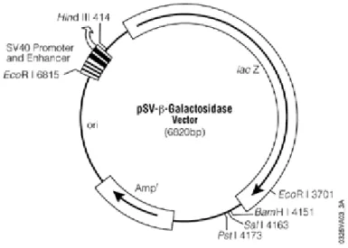
In a typical plasmid we can find a genome with promoter regions which are critical for the transcription process, a multiple cloning site where DNA fragments may be inserted to the plasmid and at least a gene expressing antibiotic resistance (Figure 2).
Figure 2. Map of the plasmid DNA used in this thesis (pSV-β-Galactosidase Vector).
Plasmids can easily be isolated and purified after being grown in bacteria. This may be done by transferring the plasmid into bacteria and allowing the bacteria to grow overnight, followed by bacteria lysis.
The application of plasmid DNA in gene therapy involves the introduction of a transgene into the cells in order to correct genetic errors inherent to the production of incompetent copies of a given protein or even to the lack of protein production altogether. The efficiency of this treatment depends on the access gained by the plasmid DNA to the nucleus through the nuclear pores, once it entries the cytoplasm. Besides disease treatment, plasmids may be applied to function as DNA vaccines or even in suicide gene therapy. So, on a molecular level, the plasmid DNA molecules may be considered as pro-drugs (Patil et al., 2005; Uherek and Wells, 2000).
5 2. Gene delivery
Some conditions are vital for a successful gene delivery. These conditions are the condensation of the DNA and its protection against intracellular nucleases, lipoplex adhesion onto the cell surface, lipoplex internalization followed by fusion with the endosome membrane, DNA escape from the endosome and, finally, DNA entry into the nucleus and its expression (Hui et al., 1996; Zhdanov et al., 2002).
2.1 Transfection mediated by lipoplexes
2.1.1 Cellular binding
Lipoplexes usually bind to the cellular surface mainly as a result of nonspecific ionic interactions between negative charges on the membrane surface and positive charges of the lipoplexes. There may also be specific targeting ligands interactions when these are incorporated in the system (Khalil et al., 2006). The presence of serum proteins, present in the culture medium, can interfere with the interaction between cells and lipoplexes, decreasing the efficiency of the transfection. It seems that the extent of interference of the serum components with the transfection efficiency is related to the cationic lipid chemical structure as well as the liposomal formulation and charge ratio (+/-) of the lipoplexes (Zelphati et al., 1998). Nevertheless there is still incomplete knowledge on the lipoplex-serum interaction.
2.1.2 Endocytosis
Given the general cellular membrane structure, the presence of a specific ligand as well as the vectors overall charge content will influence the way lipoplexes interact with the membrane (Chestnoy and Huang, 2000). There are two main pathways by which DNA-Lipid complex may enter the cell. One way is by endocytosis followed by the destruction of an endosome inside the cell and the other way is by direct fusion of the lipoplex with the cellular membrane (Figure 3). Endocytosis is the major way by which most complexes enter the cell and only a small percentage is internalized by direct fusion with the cell membrane (Hoekstra et al., 2007; Khalil et al., 2006). It
6
seems that lipoplexes may enter through clathrin-mediated endocytosis or caveolae-mediated endocytosis (Hoekstra et al., 2007). For example, Rejman (Rejman et
al.,2005) studied the effect of inhibitors of clathrin-mediated endocytosis
(chloropromazine and K+ depletion) and of caveolae-mediated endocytosis (filipin and genistein) on A549 pneumocytes and HeLa cells of FITC–poly-L-lysine-labeled DOTAP/DNA lipoplexes and on their transfection efficiency with the luciferase gene, concluding that the lipoplex uptake occurs only by clathrin-mediated endocytocis and transfection efficiency was entirely abolished by blocking clathrin-mediated endocytocis, whereas no effect was observed with the inhibition of the caveolae pathway. It seems important in these kinds of studies to establish a direct correlation between the pathway of entry and the transfection efficiency so that one can deduce how the pathway of entry may relatively contribute to a good transfection activity.
Figure 3. Proposed mechanisms of lipoplex entry into the cell (Zhdanov et al., 2002).
Once inside the endosome, it is important that plasmid DNA escapes to the cytosol before reaching the lysosomes to avoid DNA degradation (Hoekstra et al., 2007).
The size of the lipoplexes is an important aspect to consider in determining the nature of the entry pathway by endocytosis. The nature of the entry pathway and its efficiency can also be cell type dependent, according to the endocytic machinery of the
7
cell. For instance, Rejman (Rejman et al., 2004) studied the effect of cholesterol depletion and inhibitors on the internalization of fluorescent latex beads of defined various sizes, ranging from 50nm to 1000nm, by non-phagocytic B16 cells. The study revealed that the size of the particles strongly influenced the mechanism by which they were internalized as well as their subsequent intracellular route. Internalization of particles with sizes up to 200nm involved clathrin-coated pits. For particles with 500nm, the caveolae-mediated internalization becomes the predominant pathway of entry.
Still, the relative contribution of either pathway in lipoplex internalization and their relative contribution to the transfection efficiency of lipoplexes need a better insight (Zuhorn et al., 2007).
2.1.3 Endosomal escape
Escape from the (early) endosome into the cytosol is a critical step in order to avoid plasmid DNA degradation in the lysosome. Thus, it also constitutes one of the main criteria to successful transfection efficiency (Hoekstra et al., 2007). The escape into the cytosol by adenovirus involves the lysis of the endosomal membrane structure, and the hexagonal structure of the lipoplexes is thought to play a parallel role with the adenovirus mechanism to transfect cells (Wasungu and Hoekstra, 2006), although naturally, the lipolexes lack the protein machinery to promote the endosomal membrane destabilization.
A mechanism of endosomal membrane destabilization involving lipoplex-induced processes seems to be promoted by non-lamellar phases, including the hexagonal HII
and HI or a cubic phase. Apparently, lipoplexes that adopt these structures strongly
promote transfection, and this fact is consistent with this mechanism. The inclusion of PEGylated lipid derivatives, for example, in the DNA/Cationic lipid complexes, appears to have a stabilizing effect on the membrane bilayer structure and, at the same time, an inhibition effect on DNA dissociation. Moreover, the phospholipid phosphatidylserine (PS), present in the outer leaflet of the endosomal membrane seems to play an important role in this process as it facilitates the lamellar to
non-8
lamellar hexagonal-phase transition of the lipoplexes and thus, releasing the DNA from the lipoplex (Hoekstra et al., 2007; Wasungu and Hoekstra, 2006).
Then again, there may be a translocation of the cationic lipids into the endossomal membrane by the interaction of non-lamellar intermediates followed by a local increase which may give rise to pore formation of transient stability and membrane destabilization. The rate of lateral diffusion of the surrounding membrane lipids determines this. And so, the DNA may gain access to the cytosol (Hoekstra et al., 2007). It is still unclear if DNA dissociation occurs before or simultaneously with the perturbation of the endosomal membrane. Furthermore, it is also unclear if the complete lysis of the endosome is necessary for the DNA transfer or if it may occur across the perturbed membrane (Hoekstra et al., 2007).
New insights on molecular mechanisms underlying lipoplex induced endosomal membrane destabilization can be given by modeled endosomal membranes for instance. Berezhna and coleagues (Berezhna et al., 2005) performed a study using GUVs as model endosomal membranes in order to investigate what part some individual phospholipids play in interaction with lipoplexes, using laser scanning imaging in conjunction with fluorescence cross-correlation analysis.
Although there has been considerable progress in recent years in the study and knowledge of the barriers in lipoplex mediated transfection, there’s still a long way to go. Further studies are needed to understand the relative contribution of endocytosis in terms of effectiveness of internalization, efficiency of gene escape and, eventually, transfection efficiency, since this is the major pathway of entry of lipoplexes.
2.1.4 Nuclear Delivery
The final barrier to lipofection, and probably the most challenging too, is the nuclear membrane. Indeed, when plasmid DNA encoding β-galactosidase is injected directly into the nucleus, the gene expression was much higher when compared with when the same plasmid was injected into the cytosol. Furthermore, when DNA complexed with cationic lipids is directly injected into the nucleus, the complexed DNA is not expressed, suggesting that DNA has to be released from the lipoplex somewhere in the cytoplasm (Pollard et al., 1998).
9
During mitosis, the nuclear membrane is fragmented and this would allow the plasmid DNA into the nucleus. This seems to be the most widely accepted explanation as to how the DNA is delivered to the nucleus, so cell division plays an important role in the nuclear translocation of transgenes.This fact, nevertheless, cannot be the only explanation, since nondividing cells are targeted in in vivo transfection (Khalil et al., 2006).
3. Lipids
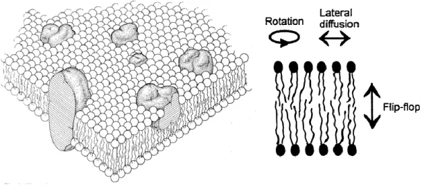
Lipids can have various usages in biological systems which make them important molecules for living beings. Some lipids assemble themselves into bilayer lipid membranes that separate cellular compartments from the external environment and participate in cellular regulation by mediating signal transduction processes. The lipid molecules found in the membrane are amphipathic molecules, which signifies they have a hydrophilic tail oriented to the inner part of the bilayer and a hydrophobic head oriented to the aqueous environment.
The model proposed by Singer and Nicholson (1972), called the “fluid mosaic” (Figure 4) proposes that biological membranes are composed of lipids, proteins and carbohydrates and describe that the nature of the lipid bilayer allows for considerable molecular motion in the lipid matrix and provides an appropriate milieu for the function of the proteins found in the membrane. (Alberts et al., 1994)
Figure 4. Cellular membrane according to the “fluid mosaic” model and phospholipid mobility
10
Depending on the structure and function, lipids can be classified into different groups. Among the lipids that constitute biological membranes we can find three groups: glycerophospholipids, sphingolipids, and sterols. (Chang, 2005)
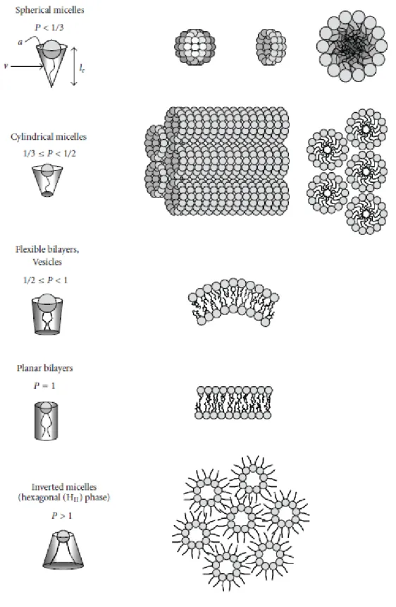
The surrounding environment is a very important factor in the structural behavior of lipids. Lipids may adopt different conformations when hydrated and the propensity of these molecules to adopt different phases as a response to some external variables like pH, presence of ions or temperature is referred to as lipid polymorphism. These phases include the micellar, the lamellar, the cubic and the inverted hexagonal phases (Figure 5).
11
Figure 5. Impact of packing parameter p on lipid assemblies formed in aqueous solutions
12
The packing parameter allows to predict which structure the molecules will adopt (Balazs and Godbey, 2011). This packing parameter is given by:
And is defined as the ratio of the hydrocarbon volume, v, and the product of the effective head group area, , and the critical length of the lipid tail, l . It is also related to the curvature by:
Where H is the mean curvature and K is the Gaussian curvature (Antonielli and Förste, 2003).
When p < 1/3 the molecules adopt preferentially a conical shape, forming conventional spherical micelles. When 1/3 < p < 1/2, the molecules adopt a geometry resembling a truncated cone, forming non-spherical micelles. The lipidic bilayer is formed when 1/2 < p < 1, and the molecules adopt a nearly cylindrical shape. At last, when p > 1,we can see the formationinverted structures with negative spontaneous curvature (Antonielli and Förster, 2003; Balazs and Godbey, 2011; Ryhänen, 2006).
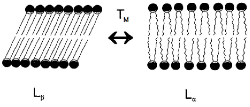
Lipids can also exist in “gel” state (Lβ) or in a fluid “liquid crystalline” state (Lα)
depending on the temperature (Figure 6). The transition temperature (Tm) is specific
for each lipid. The lipids arranging in a gel state, bellow the transition temperature, are rigid and in order. This is due to the carbon-carbon bonds of the acyl chains, which tend to extend into an all-trans conformation. When they reach the Lα state, the acyl
chains become shorter, reaching an all-gauche conformation and the packing parameter increases between individual lipid molecules (Mok, 1998), also increasing their diffusion. The lipid bylayer becomes more fluid and elastic in this state (Ryhänen, 2006).
The transition from gel to fluid state, affects the properties and structure of lipid bilayer at the level of lipid acyl chains, individual lipid molecules and at the supramolecular assembly as a whole (Ryhänen, 2006).
13
Figure 6. Lipid bilayer and its transition from gel” state (Lβ) into fluid “liquid crystalline” state
(Lα) (adapted from Mok, 1998).
4. Liposomes
Liposomes are good models of biological membrane systems to study physical properties and functional roles of lipid components. As non-viral vectors, they have unique advantages like having diverse morphologies and compositions, different release characteristics, lack of immunogenic response, and a low cost production (Balazs and Godbey, 2011). They may contain more than one lipid bilayers and so, we may classify them into three types: Multilamellar Vesicles (MLVs), Large Unilamellar Vesicles (LUVs)and Small Unilamellar Vesicles (SUVs) (Figure 7)
MLVs can easily be formed upon a lipid film hydration and are usually composed of concentric layers of bilayers. The size of this vesicles ranges from 0.5-10 nm, so they are very heterogeneous in size (Mayer et al., 1986). MLVs are not widely used for drug delivery since they show low trapping volumes. Furthermore their large sizes lead to a rapid clearance. However, they can be very useful for studying the structural and motional properties of lipids.
LUVs are vesicles possessing a single bilayer with sizes ranging from 50 to 200 nm. They can be formed by several methods but the most convenient is extrusion through polycarbonate filters with a well defined size pore (Mayer et al., 1986). They
14
show longer circulation lifetimes in vivo due to their smaller size relatively to de MLVs, so this makes them a frequently used model for drug delivery.
SUVs are vesicles composed of a single bilayer with sizes ranging from about 25 to 50 nm in diameter (Mok, 1998) and can be produced by submitting MLVs to sonication (Huang, 1969).
A method of liposome preparation proposed in order to optimize the properties of lipid-DNA complexes consists in both extrusion of the liposomes, so as to obtain LUVs, and controlled mixing of lipid and DNA. The results seem to show that the lipoplexes exhibit a narrow distribution and small sizes, which might be adequate for their in vivo application (Pedroso de Lima et al., 2001).
Figure 7. Multilamellar Vesicles (MLVs), Large Unilamellar Vesicles (LUVs) and Small Unilamellar Vesicles (SUVs) (adapted from Mok, 1998).
15 4.1 Cationic lipids and liposomes
There are three parts composing cationic lipids: a hydrophobic anchor, a linker, and a head group (Figure 8).
Figure 8. Scheme exemplifying a cationic lipid (Chestnoy and Huang, 2000).
The hydrophobic anchors can be grouped into two major types of hydrophobic moieties: aliphatic chains and cholesterol-based derivates. Traditionally, double-tailed cationic lipids are more efficient and less toxic than the single tailed cationic lipids (Hongtao et al., 2006). So, the majority of synthesized cationic lipids have double chain hydrocarbons. Commonly, they are capable of forming liposomes by themselves but it is usual to use another phospholipid as helper in the formulations for cationic lipid transfections (Chestnoy and Huang, 2000). The length of the alkyl chain in transfection activity can also influence the transfection activity (Felgner et al., 1994).
The linker, as the name states, represents any chemical part between the head group and the hydrophobic anchor. This link also plays an important role in gene transfer as stated, for example, by Liu (Liu et al., 1997), which concluded that less stable ester bonds are not as beneficial as stable ether bonds for in vivo gene transfer.
As to the head group, cationic lipids may be monovalent or multivalent, according to the number of charges. In monovalent lipids, we can find a head group consisting of either tertiary or quaternary ammonium groups (Chestnoy and Huang, 2000). Chemical modifications were also assessed by Felgner (Felgner et al., 1994).
The cationic nature of the lipid is a determinant factor to its toxicity (Hongtao et
16
transfer is not a solid one, since their efficiency for in vitro experiments in not necessarily the same as for in vivo experiments (Chestnoy and Huang, 2000), and further studies must be performed.
Cationic liposomes may be formed from a single cationic lipid or, as generally happens, from a combination of a cationic lipid and a neutral lipid. They were first introduced by Felgner (Felgner et al., 1987) as DNA transfection agentswhen Felgner and colleagues successfully transfected the COS-7 cell line with complexes between DNA and 2,3-dioleyloxypropyl-1-trimethyl ammonium chloride (DOTMA) cationic liposomes. Since then, new cationic liposome formulations have been reported to successfully transfect the cells and some of them have even been commercialized and engineered for a wide range of applications (Samad et al., 2007).
Transfection efficiency varies according to many factors, including the type of cationic lipid used or the cell type that is transfected. 1,2-dioleoyl-3-trimethylammonium-propane (DOTAP) , N [1-(2,3dioleyloxy) propyl]- N,N,N-trimethylammonium chloride (DOTMA) (Felgner et al., 1987), dioctadecyldimetylammonium bromide (DODAB), or synthesized derivatives from biologically active compounds including the cationic cholesterol derivatives such as 3β [N-(N’,N’- dimethylaminoethane)carbamoyl cholesterol (DC-CHOL) are some examples of the cationic lipids used for transfection. The addiction of a “helper” lipid seems to enhance transfection efficiency, although the mechanism responsible for such effect is not fully understood. The inclusion of neutral lipids in the liposome formulation facilitates the complex fusion to the cellular membrane because they tend to form nonbilayer structures which are related to membrane fusion intermediates (Hui et al., 1996). Dioleyl phosphatidylethanolamine (DOPE), cholesterol and dioleoyl phosphatidyl choline (DOPC) are three neutral lipids often incorporated in liposomal formulations. The DOPE- containing liposomes, as well as liposomes with some galactosydated cholesterol derivates seem to achieve high transfection efficiencies regarding human hepatoma cells, Hep 2. They also seem to exhibit low toxicity (Zhang
et al., 2004).
In this thesis, various formulations of liposomes composed of Dioctadecyldimethylammonium bromide (DODAB), Dioctadecyldimethylammonium
17
chloride (DODAC), 1-monooleoyl-rac-glycerol (MO) and 3β [N-(N’,N’- dimethylaminoethane)carbamoyl cholesterol (DC-Chol) are studied.
Dioctadecyldimethylammonium bromide (DODAB) was first synthesized by Kunitake and Okahata (Kunitake and Okahata, 1977), who prepared and characterized small unilamellar DODAB vesicles. Since then, Dioctadecyldimethylammonium bromide and chloride (DODAB/C) have been prepared through a series of methods in order to obtain size-controlled structures with long-term stability for different applications, namely as DNA carrier vehicles. They are synthetic lipids that tend to form LUV’s in the presence of excess water. These vesicles have their structural organization dependent on lipid concentration, the solvent composition, the methods of preparation and the temperature. They are also influenced by the presence of other substances (Feitosa et
al., 2009; Neves Silva et al., 2009).
Monoolein, 1-monooleoyl-rac-glycerol, is an amphiphilic neutral lipid of natural origin. This molecule has the particularity of, even in the presence of excess water, presenting two inverted bicontinuous cubic phases (Oliveira et al., 2012). MO has a fluidizing effect that seems to contribute to the complexation efficiency of pDNA in MO-based lipoplexes in a favorable way (Neves Silva et al., 2008; Neves Silva et al., 2011; Real Oliveira et al., 2010; Real Oliveira et al., 2011).
Cholesterol is a neutral lipid and a major component of biological membranes in most eukaryotic cells. Cholesterol may act as a “disordering agent” or as an “ordering agent”. At high concentration, it can form cholesterol rich domains within the lipid bilayer and also condensed complexes or cholesterol crystallites with phospholipids (Hungerford et al., 2005; Hungerford et al., 2006). It is frequently used in liposome formulations due to its biocompatibility and the stability it confers to lipid membranes (Balazs and Godbey, 2011). DC-Chol was first synthesized by Gao and Huang (Gao and Huang, 1991). It contains a cholesterol moiety attached by an ester bond to a hydrolysable dimethylethylenediamine. DC-Chol was found to have reduced toxicity when compared to Lipofectin in some cell lines (Gao and Huang, 1991). It is no surprise then, that DC-Chol and other cholesterol derivates have been incorporated in lipoplex assembly in order to increase transfection (Balazs and Godbey, 2011; Bennet et al., 1995).
18
DODAB DODAC MO DC-Chol
C38H80BrN C38H80ClN C21H40O4 C32H57ClN2O2
Table 1. Structure of the lipids relevant to this study
The DODAB:MO system has been previously produced and optimized in order to promote transfection in vitro of human cell lines (Neves Silva et al., 2011). The exchange of the counter ion in DODAC (Chloride instead of Bromide) might have an effect in the colloidal characteristics of the system and affect it’s stability in the presence of salt and serum. Also, the inclusion of DC-Chol in the system might have an effect on the membranes fluidity, molding it, which should influence the systems complexation with the plasmid DNA and, furthermore, its release inside the cell.
5. Lipoplexes
Both DNA and cationic liposomes suffer structural changes during the complexation process (Zhang et al., 2003).
Cationic lipids interact with negatively charged DNA molecules, which results in the formation of DNA/Cationic liposome complexes (lipoplexes). In the complex formation, DNA electrostatically binds to the liposome surface and the two components rearrange themselves into a new structure. The way lipoplexes interact
19
with the membrane will be influenced by aspects like the presence of specific ligands and the vectors overall charge (Chestnoy and Huang, 2000). But the main reaction source comes from the positive charge on lipoplex surface which reacts spontaneously with the cellular membrane (Zhdanov et al, 2002).
The study and characterization of the lipid-DNA complexes is very important since a better understanding of the assembly of these complexes might help to establish a correlation to their biological activity. It is known that the transfection efficiency is highly dependent on the structure and properties of a given lipid-DNA complex (Chestnoy and Huang, 2000). Indeed Sternberg and colleagues (Sternberg et
al., 1994), using freeze-fracture electron microscopy, observed different kinds of
structures of lipoplexes formed between plasmid DNA and cationic liposomes composed of DC-Chol and DOPE, depending on the DNA concentration and incubation time of the complexes. Experiments performed by Zhang (Zhang et al., 2003) using fluorescence resonance energy transfer (FRET) also suggested that the incubation time and charge ratio, among other experimental parameters, should be carefully controlled in order to achieve optimized transfection activity.
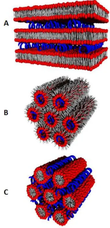
In the cationic lipoplex structures, DNA might be adsorbed onto the surface of the liposome or it might be surrounded by a lipid envelop (Ma et al., 2007). Usuallythe charge ratio (+/−) of the assembled complexes surpasses a value of 1. So we have complexes with a positive net charge which promotes an efficient binding with negative charges in the cell surface by electrostatic interactions (Hoekstra et al., 2007). The fact is, in different lipid mixtures, there has been observed several lipoplex morphologies. It has been proposed that some of these structures may correspond to metastable intermediates, spaghetti-like structure consisting of double-stranded DNA wrapped around the cationic and helper lipids. A lamellar structure, LCα, and hexagonal
structure, HCII, (Figure 9) are the two symmetries identified as equilibrium ordered
phases (May et al., 2000).
The LCα consists of DNA monolayers “sandwiched” between stacked lipid
monolayers and with intervening water gaps (Figure 9A). The electrostatic attraction between the cationic lipid bilayer and the negatively charged DNA confer stability to this morphology. Accordingly, the HCII phase may be considered as an ordinary
20
tubes (Figure 9B) (May et al., 2000). It has been reports as well of a micellar hexagonal phase, HCI (Figure 9C). In this phase the DNA can be found in the interstices of the lipid
micelle arrangement (Tresset, 2009).
The concept of lipoplex formation is relatively simple, however, this process can determine the morphology and transfection potential of these structures (Ma et al, 2007).
Figure 9. Lipoplex structures. (A) Lamellar phase (LCα); (B) Inverted hexagonal phase (HCII ); (C)
21 6. Outline of this study
Since their first introduction, cationic lipids have gained a widespread interest as a non-viral vector. But they are far from being perfect delivery vectors, and many issues are still to be overcome, hence the need for further investigation in this field.
Based on the DODAB:MO system, previously studied, this work aims to develop and characterize a new system, based on DODAC:MO:DC-Chol lipoplexes, suitable to perform efficient transfection.
On a first basis, the aim is to study how the the exchange of the Bromide counter ion for the Chloride affects the physical-chemical characteristics of the liposomes and the derived lipoplexes. It will be also studied the effect of inclusion of DC-Chol in the liposomal formulation. To do so, complexation studies using FRET technique, as well as measurements of size and zeta potential of the liposomes and lipoplexes were performed. The behavior of the system in the presence of serum and NaCl it was also investigated.
Secondly, the toxicity of the liposomes with different formulations and transfection efficiencies of this new system were assessed.
23
II. Materials and Methods
A) Materials
Dioctadecyldimethylammonium bromide (DODAB) and Dioctadecyldimethylammonium chloride (DODAC) were purchased from Tokyo Kase.
3ß-[N-(N',N'-dimethylaminoethane)-carbamoyl] cholesterol hydrochloride (DC-Chol) was purchased from Avanti Polaris Lipids.
Nucleopore Track-Etch Membranes were purchased from Whatman.
1-monooleoyl-rac-glycerol (MO), Dulbecco’s Modified Eagle’s Medium (DMEM), Antibiotic/Antimycotic solution, Bovine Serum Albumin (BSA), (NADH), The Wizard Plus
Midipreps DNA Purification System and Bradford reagent were purchased from
Sigma-Aldrich.
Heat-inactivated Fetal Bovine Serum (FBS), UltraPureTM Salmon Sperm DNA solution and rhodamine DHPE were purchased from Invitrogen (UK).
BOBO-1 was purchased from Molecular Probes (UK).
β-Galactosidase Enzyme Assay System with Reporter Lysis Buffer was purchased from Promega (USA).
Opti-MEM I Reduced Serum Medium was purchased from Gibco (UK).
pSV-β-gal plasmid DNA was kindly offered by the Hematopoietic Biology Unit, Faculty of Medicine, University of Lisbon.
Escherichia coli Xl1-Blue competent cells were kindly given by the Microbiology
laboratory in the Biology Department of the School of Sciences – University of Minho. EzWayTM Transfection Reagent was kindly offered by Koma Biotech.
B) Methods
1. Preparation of liposomes
1.1 Ethanolic injection
One of the methods used to produce liposomes was ethanolic injection. Defined volumes taken from stock solutions of DODAB/C, MO, and DC-Chol (20mM) in
24
ethanol were dissolved in a know amount of ethanol (organic solvent) and injected in a volume of distilled water, preheated to 50ºC, under vigorous vortexing. The organic solvent evaporates when it comes into touch with the distilled water due to the heat. So we obtained an aqueous solution of liposomes DODAB/C:MO:DC-Chol (ratios 6:3:1, 5:4:1, 4:1:1) and DODAB/C:MO (2:1) at a final concentration of 1mM , mostly composed of multivesicular liposomes.
1.2 Film hydration method
Again, defined volumes taken from the lipids stock solution of DODAB/C, MO, and DC-Chol (20mM) in ethanol were placed in a rotary evaporator (Heidolph VV Micro Rotary Evaporator). The organic solvent was then evaporated under vacuum and at 50ºC, safely above the lipids phase transition temperature, until all traces of organic solvent were gone and a lipidic film was formed. The resulting lipidic film was then dispersed in a given volume of ultra-pure water in order to obtain a solution with liposomes DODAB/C:MO:DC-Chol (ratios 6:3:1, 5:4:1, 4:1:1) and DODAB/C:MO (ratios 2:1) with a final concentration of 1mM.
1.3 Extrusion
The solution containing the liposomes obtained from the film hydration method was placed in the extruder (Northern Lipids Lipex Extruder). Under a 4-8bar pressure, the liposomes were forced to pass trough polycarbonate filters (Whatman) with a defined pore size. In this case, the liposomes were submitted to one passage through a filter with a pore size of 400nm and then four passages through a filter with a pore size of 100nm. The mean diameter of the obtained population reflects the diameter of the used filter pore, and unilamellar vesicles were obtained. This process was carried out at 50ºC so that the lipids were in a fluid state and preventing, in this way, as much retention of lipid in the filters as possible.
25 2. Preparation of DNA solutions
2.1 Plasmid DNA
2.1.1 Transformation of competent cells
Plasmid DNA (pSV-β-Galactosidase control vector) was added to a 200µL aliquot of Escherichia coli Xl1-Blue competent cells and the mixture swirled and incubated on ice for 30 minutes. The mixture was heat-pulsed with occasional agitation at 42 ºC for 90 seconds, and placed on ice for 10 min. Then 800µL of SOC medium (2% tryptone peptone, 0.5% yeast extract, 2.5mM KCl, 10mM NaCl , 10mM MgSO4, 10mM MgCl2, 20mM glucose) was added and the tubes were incubated at 37ºC
at 200 rpm, for 1 hour. The cells were then centrifuged at 14000 rpm for less than 10 seconds and the pellet resupended in 50µL of supernatant. The resuspended pellet was then pipetted to a petri dish with LB medium (1 % tryptone peptone, 0.5 % yeast extract, 1 % NaCl, 2 % agar) supplemented with ampicillin (100µg/µL) and spread gently and minimally using a bent (L-shaped) Pasteur pipette. A petri dish with competent cells not submitted to the transformation process was also used as control. These plates were incubated at 37ºC to establish colonies. Only transformed cells will form colonies since this strain is susceptible to ampicillin and the plasmid confers resistance to the cells. Once the colonies are grown, one of them is selected and transferred into 200mL of LB medium (1 % tryptone peptone, 0.5 % yeast extract, 1 % NaCl) at 37ºC at 200rpm overnight. This overnight recombinant E. coli culture is then harvested in order to isolate the plasmid DNA.
2.1.2 Purification of plasmid DNA
Wizard® Plus Midipreps DNA Purification System (Promega Madison, USA) was used to isolate the pSV-β-Galactosidase control vector from Echerichia coli Xl1-Blue. The isolated plasmid DNA was then ressuspended in ultra-pure water and measured with NanoDrop ND 100 Spectrophotometer in order to verify it’s purity by the determination of the ratio absorbance at 260/280nm.
26 2.2 Salmon Sperm DNA solution
Salmon sperm DNA solution was prepared in PBS buffer (10mM, pH 7.4) to a final concentration of 5×10⁻⁴ M from a stock solution (10mg/mL) of UltraPure™ Salmon Sperm DNA Solution (Invitrogen).
3. Preparation of lipoplexes
The DNA concentration used for cell transfection and lipoplex cytotoxicity assays was 1ug/ul and for the complexation and stability studies was 20ug/ul. The balance between charges is given by the charge ratio (+/-):
The positive charges are given by the concentration of ammonium groups present in DODAB/C and amine present in DC-Chol. The negative charges are given by the concentration of phosphate groups in DNA, which correspond to the nucleotide concentration.
For the mean size measurements, DODAC:MO:DC-Chol (ratios 6:3:1, 5:4:1, 4:1:1) and DODAB/C:MO (ratio 2:1) were added in a single step, at 25ºC, to sperm salmon DNA (20ug/ul), forming lipoplexes with charge ratios (+/-) of 0.5, 1.0, 1.5, 2.0, 3.0, 4.0. The addition was followed by a 5 minutes agitation period. Size measurements and Zeta Potential determination of the lipoplexes were performed after 1h of incubation.
For the FRET assay, liposomes were incubated with rhodamine DHPE (5×10⁻⁵ M) at a 1:200 ratio during their preparation by the film hydration method followed by esxtrusion, as previously described. The Salmon sperm DNA (20µg/µL) was also incubated with BOBO-1 probe (2×10⁻⁵ M). The lipoplex preparation method and the liposome formulations used were the same as the ones produced for the complexation studies forming lipoplexes with charge ratios (+/-) of 0.25, 0.5, 0.75, 1.0, 1.5, 2.0, 2.5, 3.0 and 4.0.
27
For stability studies, the lipoplex formation procedure and the used liposome formulations were the same as described before, except for the generated charge ratios (+/-), which was 4.0.
The liposomes used to form the DNA-liposome complexes for the complexation assays, FRET assay and stability studies were produced only by the film hydration method followed by extrusion.
For the transfection and cytotoxicity assays, lipoplexes were generated at a charge ratio (+/-) of 4.0 and plasmid DNA (1µg/µL) was used. The liposomes used in the production of these complexes were produced by the ethanolic injection and the film hydration method described previously and the used formulations were DODAB/C:MO:DC-Chol (ratios 6:3:1, 5:4:1, 4:1:1) and DODAB/C:MO (ratio 2:1).
4. Dynamic Light Scattering and Zeta potential measurements
4.1 Dynamic Light Scattering (DLS) assays
Dynamic Light Scatering (or PCS - Photon Correlation Spectroscopy) is a technique that measures the Brownian motion of the particles by illuminating them with a laser and analyzing the intensity fluctuations in the scattered light, relating these measurements to the size of the particles. It can also give us information about the homogeneity of the sample (Malvern, 2005). Particles suffer random movement in a liquid due to the surrounding molecules which bombard them and this random movement is defined as the Brownian motion. The speed of movement is used to determine the size of the particle. A laser beam is used to irradiate the randomly moving particles in order to record the intensity of the light scattered in a fixed or variable angle and in given time interval. Smaller particles move faster, so they lead to faster intensity fluctuations due to their high diffusion coefficient. Larger particles move slower, is manifested in slower intensity fluctuations. (Clark et al, 1970; Malvern, 2005).
The Stokes-Einstein equation defines the relationship between the particle size and its speed due to the Brownian motion:
28
Where (D) is the particle diffusion coefficient, (K) the Boltzmann constant, (T) the temperature, (R) the radius of the particle and (η) the medium viscosity
The Polydispersity index (PDI) is an indicative of the heterogeneity degree regarding to the sample size. The PDI values ranges between 0.0 and 1.0 and depend on the range of diameters found between the average diameter. The greater this range of diameters is, the higher the standard deviation, thus, the greater is the PDI (Malvern, 2005).
Measurements of particle size and PDI were performed using a Malvern zetasizer Nano SZ particle analyzer (Malvern Instruments).
4.2 Mean size of liposomes
1 mL solutions of DODAB/C:MO (2:1), DODAC:MO:DC-Chol (DODAC:MO (2:1) with 10%, 20%, 30%, 40% and 50% of DC-Chol) and DODAB/C:MO:DC-Chol (ratios 6:3:1, 5:4:1, 4:1:1) liposomes, produced by ethanolic injection and the film hydration method followed by extrution, were placed in disposable polystyrene cuvettes (Sarstedt, Germany) for DLS measurements. These measurements were performed at 25ºC in a Malver ZetaSizer Nano SZ particle analyser. The Malvern Dispersion Technology Software (DTS) was used and mean size (nm) average, polydispersity index and error values were taken in consideration.
4.3 Mean size of lipoplexes
1 mL solutions with DNA/DODAB/C:MO (2:1) and DNA/DODAC:MO:DC-Chol (ratios 6:3:1, 5:4:1, 4:1:1) lipoplexes at charge ratios (+/-) 1.0,1.5, 2.0,3.0 and 4.0 were placed in disposable polystyrene cuvettes (Sarstedt, Germany) for DLS measurements, which were executed in a Malver ZetaSizer Nano SZ particle analyser, at 25ºC. The mean size (nm) average, polydispersity index and error values were taken in consideration and data was analyzed with Malvern Dispersion Technology Software (DTS).
29 4.4 Stability studies
4.4.1 Stability in serum
Solutions of DODAC:MO:DC-Chol (ratios 6:3:1, 5:4:1, 4:1:1) and DODAB/C:MO (2:1) liposomes, prepared by the film hydration method, described previously, were incubated in 0, 30 and 80% of FBS (Fetal Bovine Serum) and were placed in disposable polystyrene cuvettes for DLS measurements. The same procedure was repeated for DNA/DODAC:MO:DC-Chol (ratios 6:3:1, 5:4:1, 4:1:1) and DNA/DODA(X):MO (2:1) lipoplex solutions at charge ratio (+/-) 4.0. The measurements were executed at different time points: 0h, 3h, 6h and 24h. The cuvettes were placed in a Malver ZetaSizer Nano SZ particle analyser and again, the Malvern Dispersion Technology Software (DTS) was used and mean size (nm) average, polydispersity index and error values were taken in consideration.
4.4.2 Stability in NaCl
Solutions of DODAC:MO:DC-Chol (ratios 6:3:1, 5:4:1, 4:1:1) and DODAB/C:MO (2:1) liposomes, prepared by the film hydration method followed by extrusion, as described above, were incubated with different volumes of a NaCl solution (3.84M) in order to obtain different final concentrations of this salt (0, 5, 10, 50, 100 and 150 mM). The same procedure was applied to the lipoplex solutions. The DNA/DODAC:MO:DC-Chol (ratios 6:3:1, 5:4:1, 4:1:1)/DNA complex and DNA/DODAB/C:MO (2:1)/DNA complex solutions at charge ratio (+/-) 4.0, were incubated with the same NaCl solution as the liposomes in order to obtain lipoplex solutions incubated with different concentrations of NaCl (0, 5, 10, 50, 100 and 150 mM). The samples were placed in disposable polystyrene cuvettes for DLS measurements which took place 1h after incubation. The cuvettes were placed in a Malver ZetaSizer Nano SZ particle analyser and again, the Malvern Dispersion Technology Software (DTS) was used and mean size (nm) average, polydispersity index and error values were taken in consideration.
30 5. Zeta (ζ) Potential assays
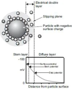
The zeta potential is a physical property found in particles in suspension. When in contact with a liquid, particles tend to acquire an electrical charge on their surface. There is an electrical double layer surrounding each particle in suspension. There are two parts of this layer: the stern layer, where the ions are strongly bound, and the diffuse layer where the ions are not so firmly attached (Figure 10). In this last layer, there is a boundary where the ions and particles form a stable entity. When the particle moves, only the ions within this boundary, the slipping plane, move with it. The potential in the slipping plane is known as the zeta potential (Malvern, 2005).
Figure 10. Schematic representation of the double layer surrounding a particle in suspension
(Malvern, 2005).
The zeta potential is not obtained by a direct measurement. It is calculated by the determination of the electrophoretic mobility, obtained by performing electrophoresis experiments on the samples and executing measurements of the particles velocity using Laser Dopller Velocimetry (LDV), and then applying the Henry equation:
31
where, μe is the electrophoretic mobility, the zeta (ζ) potential, ε is the dielectric constant, f (Ka) is the role of Henry and η is the viscosity of the medium (Malvern, 2005).
The electrophoretic mobility is measured by applying an electric field. The potential difference created by the opposite charges in the two electrodes makes the charged particles that exist in suspension move to the electrode of opposite charge. The particles will move with a constant velocity when equilibrium between these two forces is reached. The velocity at which the particles move in an electric field is known as electrophoretic mobility. This velocity is measured by Laser Dopller Velocimetry (LDV) (Malvern, 2005).
5.1 Zeta potential of the liposomes
1 mL solutions of DODAB/C:MO (2:1) and DODA(X):MO:DC-Chol (ratios 6:3:1, 5:4:1, 4:1:1) liposomes were placed in 0.7ml universal dip cells for ζ-potential measurements, which took place at 25ºC in a Malver ZetaSizer Nano SZ particle analyzer. The Malvern Dispersion Technology Software (DTS) was run with monomodal mode data processing and ζ-potential (mV) average and error values were taken into consideration.
5.2 Zeta potential of lipolplexes
1 mL solutions of DNA/DODAB/C:MO (2:1) and DNA/DODAB/C:MO:DC-Chol (ratios 6:3:1, 5:4:1, 4:1:1) lipoplexes at charge ratios (+/-) 0.5, 1.0, 1.5, 2.0, 3.0 and 4.0 were placed in 0.7ml universal dip cells (Malvern Instruments) for ζ-potential measurements, which were executed in a Malver ZetaSizer Nano SZ particle analyzer, at 25ºC. The Malvern Dispersion Technology Software (DTS) was run with monomodal mode data processing and ζ-potential (mV) average and error values considered.
32 5.3 Stability studies
5.3.1 Stability in NACl
Solutions of DODAC:MO:DC-Chol (ratios 6:3:1, 5:4:1, 4:1:1) and DODAB/C:MO (2:1) liposomes, prepared by the film hydration method followed by extrusion as described above, were incubated with different volumes of a NaCl solution (3.84M) in order to obtain different final concentrations: 0, 5, 10, 50, 100 and 150 mM. For DNA/DODAC:MO:DC-Chol (ratios 6:3:1, 5:4:1, 4:1:1) and DNA/DODAB/C:MO (2:1) lipoplex solutions at charge ratio (+/-)4.0, it was applied the procedure previously described for the liposome solutions. The samples were placed in 0.7ml universal dip cells (Malvern Instruments) for ζ-potential measurements which took place 1h after incubation. The cells were placed in a Malver ZetaSizer Nano SZ particle analyser and again, the Malvern Dispersion Technology Software (DTS) was used and and ζ-potential (mV) average and error values were taken in consideration
6. Fluorescence Resonance Energy Transfer (FRET) assay
FRET is a technique that can describe energy transfer between two chromophores. There is an energy transfer between a donor chromophore, initially in its electronic exited state, and an acceptor chromophore. The amount of energy transfer is dependent on the distance between the two chromophores.Also the absorption spectrum of the acceptor must overlap with the emission spectrum of the donor in order for the non-radiative transfer of excitation energy occurs (Figure 11). This way, several vibronic transitions in the donor and the acceptor have practically the same energy (Valeur, 2001). Other criterion that must be satisfied for FRET to occur are that the donor and the acceptor must have approximately parallel transition dipole orientations and that the donors fluorescence lifetime must have a sufficient duration in order to allow the FRET to occur (Piston et al., 1948; Valeur, 2001).
33
Figure 11. Donor (BOBO-1) and acceptor (rhodamine-DHPE) spectrums with the spectral
overlap integral between the emission spectrum of the donor and the absorption of the
acceptor. (adapted from:
http://www.invitrogen.com/site/us/en/home/support/ResearchTools/Fluorescence-SpectraViewer.html
FRET is widely used to measure distances between donors and acceptors in macromolecular systems. The efficiency process depends on the inverse sixth distance between the donor and acceptor.
E = Ro6/(Ro6 + r6)
Where Ro is the critical radius of Förster, which can be defined as the distance at which half the energy is transferred and r is the actual distance between donor and acceptor. So, it’s easy to see that the distance affects greatly the efficiency of energy transfer. The critical radius of Förster (R0) can be estimated by:
R0 (nm) = 979 (κ2 η4 φ0 ζ)1/6
Where η is the refractive index of the medium, φ0 is the fluorescence quantum
yield of the donor, ζ the spectral overlap integral and κ an orientation factor. The energy transfer rate (ΦFRET)can be determined by:
ΦFRET = 1/τd (R0/r)6
Where τd is the decay time of the donor fluorescence in the absence of an
34
This assay was executed by monitoring the decrease of fluorescence of BOBO-1 (donor) in the presence of rhodamine DHPE (acceptor) in a Horiba Jobin Yvon Spex Fluorolog spectrofluorimeter after the addition of the cationic liposomes to the DNA solutions, followed by a 5 minutes agitation period with a magnetic stirrer. The fluorescence intensities were determined at λexc = 460 nm with spectral bandwidths of
1 nm. All emission spectra were integrated, and the ratio of the areas for the dye solutions were determined. The fluorescence resonance energy transfer efficiency (ФFRET) was determined from the following expression:
(ФFRET)= (1- F(DA) / F(D))*100
Where F(DA) and F(D) are the fluorescence emission of donor in the presence and
absence of acceptor, respectively.
7. Cell Culture
The 293 T and L929 cell lines were cultured in DMEM medium supplemented with 10% (v/v) heat-inactivated FBS and 1% (v/v) of an antibiotic/antimycotic solution, with 5 % CO2 at 37 ºC.
7.1 MTT assay
This technique involves the use of MTT (3,(4,5-dimethylthiazol-2-yl)-2,5-diphenyl tetrazolium bromide)) which is a tetrazolium salt. This salt is metabolized by mitochondrial enzyme activity in living cells, forming an insoluble coloured formazan salt, thus being a rapid colorimetric method to assess cell viability (Masters, 2000).
Briefly, after an incubation period of 48h, the culture medium was replaced by fresh medium to which it was added 50µL of MTT 10x to each well. Cell cultures were then incubated for 2h in a humidified incubator at 37ºC, with 5% CO2. Then, the culture medium containing MTT was removed and it was added to each well 500µL of a DMSO/ethanol solution (1:1(v/v)), followed by a slight agitation of the plate in order to facilitate the dissolution of the crystals. It was taken from each well 150µL of the solution containing the dissolved crystals and it was placed in a 96 well reading plate in
35
order to read the optical density at 570nm in a microplate reader SpectraMax Plus (Molecular Devices, USA). The DMSO/ethanol solution (1:1(v/v)) was used as blank.
7.2 LDH (lactate dehydrogenase) assay
This technique allows us to make an indirect measurement of cell viability. LDH is a quantitative measurement of cell viability loss since it is released by dead or dying cells. LDH measurement basically follows the oxidation of NADH, which is initiated by the addition of pyruvate, by the change of absorbance at 340nm. These measurements are performed in a cell-free medium at a temperature of 30ºC (Masters, 2000).
So, after a 48h incubation period, the cell culture medium from each well was collected to individual eppendorfs in order to determine extracellular LDH. The eppendorfs were then centrifuged at 13000rpm for 1 minute in order avoid having cells or cellular fragments in suspension and the supernatant was collected to fresh eppendorfs. Then, 40 µl of supernatant were placed in a 96 well reading plate, along with 250ul of NADH solution (0.3mM in phosphate buffer 0.05M, pH 7.4) and 10 µl of pyruvate solution (0.05M in phosphate buffer 0.05M, pH 7.4) in each well. The pyruvate solution was added immediately before performing the optical density reading in a multiwall plate reader. The readings were executed at 30ºC in intervals of 10 seconds for 3 minutes at 340nm in a microplate reader SpectraMax Plus (Molecular Devices, USA).
7.3 Cytotoxicity assay
Cytotoxicity was assessed by MTT assay and by the measurement of extracellular Lactate Dehydrogenase (LDH), after a 48h hour period of incubation with the liposomes and lipoplexes. The 293T cell line was used to evaluate the cytotoxicity of the lipoplexes and the L929 cell line was used to evaluate liposome cytotoxicity.
Cells were cultured in 24-well culture plates (TPP, Trasadingen, Switzerland) at a density of 3×10⁴ cells per well and 1×10⁵ cells per well for liposomes and lipoplexes respectively. The cell culture medium was removed and replaced with fresh medium immediately before the liposomes and lipoplexes addition. To the plaque with the L929 cell line, it was added to each well the liposome solution of interest (buffered