K
NEE
O
STEOARTHRITIS AND
O
BESITY
:
E
FFECTIVENESS OF
PICO
AQUATIC
EXERCISE PROGRAM ON SYMPTOMS
,
PHYSICAL FITNESS AND QUALITY OF LIFE
Dissertação elaborada com vista à obtenção do grau de Doutor em Motricidade Humana na especialidade de Atividade Física e Saúde
Orientador: Professora Doutora Maria Margarida Espanha Coorientador: Professor Doutor Stephen Messier
Júri:
Presidente:
Reitor da Universidade de Lisboa
Vogais:
Doutor Jaime da Cunha Branco
Professor Catedrático da Faculdade de Ciências Médicas da Universidade Nova de Lisboa Doutor José Henrique Fuentes Gomes Pereira
Professor Catedrático da Faculdade de Motricidade Humana da Universidade de Lisboa Doutora Maria Joana Mesquita Cruz Barbosa de Carvalho
Professora Associada com Agregação Faculdade de Desporto da Universidade do Porto Doutora Maria Margarida Marques Rebelo Espanha
Professora Associada da Faculdade de Motricidade Humana da Universidade de Lisboa Doutora Maria Helena Santa Clara Pombo Rodrigues
Professora Auxiliar da Faculdade de Motricidade Humana da Universidade de Lisboa
FLÁVIAGIOVANETTIYÁZIGI
Julho de 2014
U
NIVERSIDADE DEL
ISBOAThe work presented in this dissertation was supported by the Portuguese Foundation for Science and Technology (scholarship: SFRH/BD/46425/2008).
Declaração de Reprodução da Tese
Nome: Flávia Giovanetti Yázigi
Endereço eletrónico.
[email protected]
Telefone: 214149159
Número do Cartão de Cidadão: 15918029
Título: Obesity and knee osteoarthritis : Effectiveness of PICO aquatic exercise
program on symptoms, physical fitness and quality of life.
Orientador: Maria Margarida Espanha
Coorientador: Stephen Messier
Ano de conclusão: 2014
Doutoramento: Motricidade Humana na especialidade de Atividade Física e Saúde
É autorizada a reprodução integral desta tese/trabalho apenas para efeitos de
investigação, mediante declaração escrita do interessado, que a tal se compromete.
Faculdade de Motricidade Humana – Universidade de Lisboa
Cruz Quebrada, 11/ 06 / 2014
Para as estrelas que me guiam, Roberto Yázigi e Sabina Gozzi Giovanetti
Para a minha mãe, Amalia Yázigi Mesmo distante, sempre presente Ensinaste-me a acreditar
És o meu eterno pilar A ti, minha filha Debora, que esta tese lhe
sirva de exemplo. Tudo tem seu tempo, nada é em vão Com trabalho, dedicação e resiliência
conseguiras tudo aquilo a que te propuseres, da melhor forma que puderes
When a person read this present document maybe everything will look very simple, clear and easy. Even for me, when I look backward the past became simple now. Only a person who lived similar process can understand the unimaginable conflict of feelings in which we are constantly confronted. The really true is that this thesis involved a very complex process which was not possible without the interaction of different labs and with and enormous human friendship.
In this way I would like to express my sincere acknowledge to the many person who had contributed in different ways to makes this thesis possible.
I cannot find words to express my gratitude to Professor Margarida Espanha. I would recognize her guidance, an everlasting encouragement and friendship, which supported me across this discovery and learning phase.
My special words to Prof Stephen Messier and all staff from Wake Forest University for having received me. Thank you for relevant insights provided to this investigation and also, for welcomed and supported my pregnancy stage.
I would also like to thank to Dr. Filomena Carnide for supervising my first study, the KOPS validation, to Dr Filomena Vieira, Dr Cristina Bento and Dr Antonio Veloso for their participation in this work supplying scientific knowledge.
I am very grateful to Dr Pedro Teixeira for giving me the opportunity to work with PESO Comunitario.
To PICO staff (André, Carlota, Joana e Ines), to aquatic exercise instructors from our faculty (Francisco, Susana, Joana e Iola) and to all volunteers, thank you, none of this would be possible without your commitment.
Thanks to my colleagues for any kind of motivation or support during this phase of my life. Espadinha, Adilson, Filomena, your availability to help is indescribable.
An special word to all my friends, you know how I respect and valorize your friendship, thank you to be in my way.
Finally, I would like to recognize the effort of my family to accompany me, giving me an enormous understanding and love in challenging times.
Institutional acknowledgements
Without institutional support, the PICO project would not be possible. In this way, I would like to thank to the Portuguese Institute of Rheumatology for the clinical diagnosis, to the Instituto Português do Desporto e Juventude, which provided the swimming pool, for the classes realization and also, to the laboratories of Faculdade de Motricidade Humana (Biomechanics and Functional Morphology lab, Health and Exercise lab and Biochemistry and Exercise Physiology lab), which supported the tests realization. Additionally, my thanks for Fundação da Ciência e Tecnologia, for my scholarship (SFRH/BD/46425/2008).
Abstract
Background: Aquatic exercise is a nonpharmacologic intervention recommended for
knee osteoarthritis (KOA) management. This study aimed to determine the effectiveness of 3-months of aquatic exercise program on KOA symptoms, physical fitness, and quality of life in overweight/obese adults with KOA. Methods: Eligibility criteria were 40 ≤ age ≤ 65 years; BMI ≥ 28 kg/m2; clinical and radiographic KOA.
Participants were randomized in aquatic exercise group (AEG) and control group (CG). Physical Fitness was assessed by Six Minutes Walking Test (6MWT), Chair Sit and Reach Test (CSR), Five-Repetition Sit-To-Stand Test (FRSTST)
,
Handgrip Strength Test (HST) and isokinetic and isometric knee strength tests. KOA symptoms and quality of life were assessed by self-reported questionnaires (Knee Injury and Osteoarthritis Outcome (KOOS), Brief Pain Inventory (BPI), Beck Depression Inventory (BDI), International Physical Activity Questionnaire (IPAQ), Weight and Lifestyle Inventory (WALLI). Body composition and morphology was measured by DXA scanner and waist, hip and thigh circumferences. Descriptive statistics and Pearson Correlation Coefficient were used for baseline analyses; Univariate Analyses of Covariance (ANCOVA) was used as primary analyses. Results: Final sample included 48 adults (BMI: 35.0±4.9 kg/m2; age: 55±7 years.), 23 in the CG and 25 in AEG. Regardingphysical function, significant group effect was found for 6MWT, FRSTST (p<.001) and Isokinetic flexion peak torque, in both knees (p<.05). Regarding KOOS; BPI and BDI, significant group effect was found in all dimensions. Conclusion: PICO aquatic program was effective in improving KOA symptoms, physical fitness and health-related quality of life of its practitioners. Trial Registration: NCT01832545
Resumo
Introdução: O exercício aquático é considerado uma opção não farmacológica no
tratamento e controlo dos sintomas da osteoartrose do joelho (OAJ). O principal objetivo deste estudo foi determinar a eficácia de um programa de 3 meses de exercício aquático nos sintomas, aptidão física e qualidade de vida de adultos com sobrepeso ou obesos com OAJ. Métodos: Os critérios de elegibilidade foram 40 ≤ idade ≤ 65 anos; IMC ≤ 28 Kg/m2 e diagnóstico clínico e radiológico de OAJ. Os
participantes foram randomizados em grupo de exercício aquático (GEA) e grupo controlo (GC). Aptidão física foi avaliada pelos testes de Seis Minutos Marcha (6MM), Sentar e levantar da Cadeira 5X (SLC), Sentar e alcançar, Alcançar atras das costas, força de preensão manual e avaliação isométrica e isocinética da força dos músculos do joelho. Os sintomas e qualidade de vida foram avaliados por questionários de autorrelato: Questionário KOOS sobre o Joelho, Inventário Breve da Dor (IBD), Inventario de Depressão de Beck (IDB), Questionário Internacional de Atividade Física (IPAQ), Inventário do Peso e estilo de vida (IPEV). A composição corporal e morfologia foram avaliados por DEXA scanner e medidas de circunferência. Estatística descritiva, coeficiente de correlação de Pearson, análise Univariada de covariância (ANCOVA) e regressão linear múltipla foram os métodos estatísticos utilizados. Resultados: 48 adultos (IMC: 35.0±4.9 Kg/m2, idade: 55±7 anos) completaram o estudo, 23 no GC e
25 no EAG. Quanto à função física, foi encontrado efeito de grupo significativo no 6MM, SLC (p<.001), no pico de torque da força de joelho na flexão, em ambos os joelhos (p<.05). Efeito de grupo significativo foi encontrado nas dimensões do KOOS; IBD e IDB. Conclusão: O programa aquático PICO foi eficaz para promover a melhoria dos sintomas da OAJ, da aptidão física, estado psicológico e qualidade de vida relacionada com a saúde de seus praticantes. Registro de Clinical Trial: NCT01832545
PALAVRAS-CHAVE: Exercícios aquáticos; Osteoartrose do joelho; Dor; Obesidade;
Table of Contents
Acknowledgements ... I Institutional acknowledgements ... II Abstract ... III Resumo ... IV Table of Contents ... V Tables ... VIII Figures ... IX Abbreviations ... X Chapter 1: Introduction ... 1 1.1 Overview ... 3 1.2 Dissertation structure ... 41.3 Publications that are related to the dissertation ... 5
Chapter 2: Literature Review ... 7
2.1 Knee Osteoarthritis ... 9
2.1.1. Symptoms and quality of life ... 10
2.1.2. Screening/Diagnosis ... 12
2.2 Obesity and Knee Osteoarthritis ... 13
2.3 Exercise on Knee Osteoarthritis ... 15
2.3.1. Aquatic Exercise ... 20
2.3.2. Aquatic Exercise an KOA... 22
Chapter 3: Aims of the study ... 25
3.1 Aims of the study ... 27
Chapter 4: Methods ... 29
4.1.1. Sample ... 32
4.1.2. Trial conduct ... 33
4.2 Intervention Programs ... 33
4.2.1. Aquatic Exercise (AE) ... 34
4.2.2. Educational program: Control Group (CG) ... 37
4.3 Knee osteoarthritis: screening and diagnosis ... 37
4.3.1. Outcomes and instruments ... 38
4.3.2. Statistical analysis... 43
CHAPTER 5: Results ... 47
5.1 Baseline results ... 49
5.1.1. Correlations between KOA symptoms, quality of life and physical fitness outcomes ... 52
5.1.2. Lower limb contralateral analysis: comparison between the most and least painful knee ... 53
5.2 Effectiveness of Aquatic exercise ... 54
5.2.1 KOA symptoms and quality of life ... 55
5.2.2 Physical fitness ... 58
5.3 Complementary analyses ... 61
5.3.3. Predictive factors of 6MWT in overweight and obese adults with KOA ... 63
CHAPTER 6: Discussion ... 67
6.1 Baseline outcomes ... 69
6.1.1. Correlations between KOA symptoms, quality of life and physical fitness outcomes ... 70
6.1.2. Lower limb contralateral analysis: comparison between the most and least painful knee ... 72
6.2 Effectiveness of PICO Aquatic exercise program ... 74
6.2.1. KOA symptoms and quality of life ... 75
6.3 Complementary analyses ... 82
6.3.1. BMI groups comparisons for KOA outcomes, physical fitness and quality of life 82 6.3.2. Predictive factors of 6MWT in overweight and obese adults with KOA ... 84
Chapter 7: Conclusions ... 87
7.1 Main research findings ... 89
7.2 Practical implications and future directions ... 91
References ... 93
Appendices ... 113
Appendix 1: Tests list of PICO Project ... 115
Appendix 2: Informed consent ... 119
Appendix 3: Aquatic Exercise Patterns ... 123
Appendix 4: Knee Osteoarthritis Pre- Screening Questionnaire (KOPS) ... 127
Appendix 5: Knee injury and Osteoarthritis Outcome Score (KOOS) ... 133
Appendix 6: Brief Pain Inventory (short version) ... 139
Appendix 7: Beck Depression Inventory II ... 143
Appendix 8: Physical Activity Questionnaire (IPAQ) ... 147
Appendix 9: The Weight and Lifestyle Inventory (WALI). ... 151
Appendix 10: Patients' Global Impression of Change (PGIC) ... 157
Appendix 11: Testimonials ... 161
Appendix 12: Yazigi F, Espanha M, Vieira F, Messier SP, Monteiro C, Veloso AP: The PICO project: aquatic exercise for knee osteoarthritis in overweight and obese individuals. BMC Musculoskelet Disord 2013, 14:320. ... 167
Appendix 13: Yázigi F, Carnide, F. and Espanha M. Development of the Knee OA
Pre-Screening Questionnaire (KOPS) (under review).Int J Rheum Dis 2013 ... 183
Appendix 14: Yázigi, F, Espanha, M., Marques, A., Vitorino, J., Silva, I., Sousa, M., &
Cunha, C. (2012). Predictive Factors of 6MW Test in Obese Individuals with Knee OA (abstract). Acta Reumatol Port, 37(Suppl), 66-67. ... 197
Tables
Table 1: Reference values of 6MWT for KOA, obese and health subjects. ... 17 Table 2: Reference of main published articles of knee strength and KOA. ... 18 Table 3: Reference of main published articles of exercise for KOA. ... 23 Table 4: The aquatic exercise protocol design for the 12 weeks of PICO program. .... 36 Table 5. Frequency analyses of demographic variables at baseline for Control Group
(CG), Aquatic Exercise group (AEG) and for total sample. ... 49
Table 6. Descriptive analyses for outcomes of total sample at baseline. ... 51 Table 7. Correlation analysis between physical fitness and KOA symptoms and quality
of life outcomes. ... 53
Table 8. Contralateral analyses of most painful and Least Painful Knee at baseline
T-tests for limb comparisons. ... 54
Table 9. Group effect analysis for questions scores of Symptoms (S1-S7) and Pain
(P1-P9) dimensions of KOOS questionnaire (0-4 where 4 is the worst condition). ANCOVA adjusted for sex BMI value no baseline. ... 56
Table 10. Group effect analysis of the physical function tests, controlling for baseline
BPI severity pain, gender, BMI, and baseline values for physical fitness tests (Mom1= Baseline and Mom2=After interventions). ... 58
Table 11. Group effect analysis of Isokinetic and Isometric normalized strength tests,
controlling for baseline BPI severity pain, gender and baseline values. ... 59
Table 12. Group effect size from univariate analysis of variance adjusted for sex, BMI,
Table 13. Means and standard deviations for the five dimensions of KOOS
questionnaire according to BMI classification (KOOS score range is 0-100 where nearest to zero is the worst condition). ... 62
Table 14. Means and standard deviations for Brief Pain Inventory (Severity and
Interference), Beck Depression Inventory (BDI) and International Physical Activity Questionnaire (IPAQ), according to BMI classification. ... 63
Table 15. Means and standard deviations for physical fitness tests, according to BMI
classification. ... 63
Table 16. Pearson product-moment correlations and 2-tailed p-value between Six
Minutes Walking Test (6MWT) and Knee injury and Osteoarthritis Outcome Score (KOOS), Brief Pain Inventory (BPI), Beck Depression Inventory (BDI), physical function, knee strength, International Physical Activity Questionnaire (IPAQ) and Body Composition ... 64
Table 17. Multiple regression models of predictive factors for 6MWT. ... 65
Figures
Fig. 1. Knee (http://en.wikipedia.org/wiki/Knee) and RX of a primary osteoarthritis of
the left knee. Note the osteophytes, narrowing of the joint space (arrow), and increased subchondral bone density (arrow). ... 10
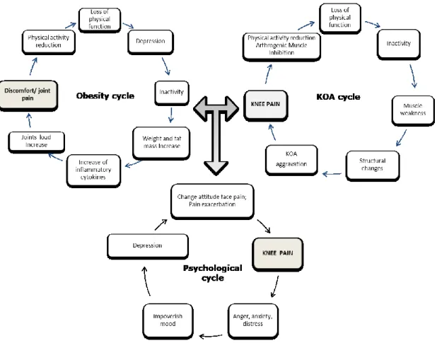
Fig 2. Bidirectional relationship between obesity and KOA cycle ... 14 Fig. 3. PICO project fluxogram. ... 31 Fig 4. Group effect analysis, mean differences with standard deviation error bars of
dimensions of Knee injury and Osteoarthritis Outcome Score (KOOS) for the Aquatic Exercise Group and Control Group. ... 55
Fig 5. Group effect analysis, mean differences with standard deviation error bars of the
of the Beck Depression Inventory (BDI) and both dimensions of Brief Pain Inventory (BPI): BPI severity and BPI interference for the Aquatic Exercise Group and Control Group. ... 57
Abbreviations
ADL Activities of Daily Living
AE Aquatic Exercise
AEG Aquatic Exercise Group
BDI Beck Depression Inventory
BM Body Mass
BMI Body Mass Index
BPI Brief Pain Inventory
CG Control Group
CSRT Chair Sit and Reach
DXA Dual Energy X-ray Absorptiometry
EXT Extension
FLEX Flexion
FM Fat Mass
FRSTST Five Repetitions Sit to Stand Test
HR Heart Rate
IPAQ International Physical Activity Questionnaire
ISOM Isometric
KOA Knee Osteoarthritis
KOOS Knee injury and Osteoarthritis Outcome Score
LPK Least Painful Knee
MET Metabolic Equivalent of Task
MOM 1 Baseline
MOM 2 After 12 weeks of intervention
MPK Most Painful Knee
NRS Numerical Rating Scale
OA Osteoarthritis
PAL Physical Activity Level
PGIC Patient Global Impression of Change Scale
PkTQ Peak Torque
6MWD Six Minutes Walking Distance
6MWT Six Minutes Walking Test
VO2max Maximal Oxygen Consumption
Chapter 1: Introduction
This chapters presents the dissertation structure and the list of abstracts and works related to the PICO project (Intervention Program Against Osteoarthritis).
1.1 Overview
Rheumatic diseases (RD) include over 150 disorders, and although death rates are low, they are one of the primary causes of compromised quality of life and absenteeism from work, with significant economic and social consequences[1].
In Portugal, RDs are responsible for 40 to 60% of cases of prolonged physical incapacity in daily activities, 43% of absenteeism from work, and 35 to 41% of early retirement due to illness[2]. The prevalence of rheumatic diseases, both in Portugal and
worldwide, is increasing, with significant repercussions for public health.
Following the establishment of the “Bone and Joint Decade” by the World Health Organization (WHO)[3], the Portuguese government established the National
Program Against Rheumatic Diseases (2004 – 2010), which aimed to improve the quality of life of patients with arthritis[2]. This was followed by the creation of the
ONDOR (Observatório Nacional contra as Doenças Reumáticas or National Observatory Against Rheumatic Diseases)[4] in 2003 by the Portuguese Society of
Rheumatology and the Faculty of Medicine of Porto. Since that time, the ONDOR has published two books with reports of their work, “O estado da reumatologia em Portugal”[5] and “Doenças remáticas em Portugal: da investigação às políticas de
saúde” [6]. The Portuguese Epidemiologic Study of Rheumatic Diseases (EpiReumaPt),
started in 2014, is an ongoing, national, population-based, cross-sectional, epidemiologic study to estimate the prevalence of rheumatic diseases and health-related quality of life in Portugal.
According to the WHO[1], rheumatoid arthritis, osteoarthritis, osteoporosis,
spinal disorders and severe limb trauma are the osteoarthritis (OA) conditions with the greatest impact on society and risk to an individuals’ quality of life, leading to loss of autonomy and incapacity, and are becoming a serious international public health problem. Because the knee is the most affected weight bearing joint[7], there is a high
prevalence (11.1%) of knee osteoarthritis (KOA) in the Portuguese population[8].
Obesity, prior knee injury, physical activity levels, lower limb strength and the extent of alignment/misalignment of body segments are the most often cited potential risk factors for KOA. In Portugal, more than 50% of the population is overweight or
obese[9], and this value is expected to increase as the rate of obesity[10] increases and
the population ages[11].
The guidelines for KOA management include recommendations for a combination of pharmacological and non-pharmacological approaches. Exercise is considered an effective non-pharmacological treatment and is recommended by the Osteoarthritis Research Society International (OARSI)[12-14], the American College of
Rheumatology (ACR)[15] and the European League Against Rheumatism (EULAR)[16].
Among exercise modalities, aquatic exercise can play an important role in reversing the framework of loss of functionality and in controlling OA symptoms [12-14]. However, there
are few published articles with detailed aquatic exercise protocols, and these studies have not been consistent with respect to the methodologies used and populations studied.
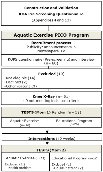
The present dissertation, entitled “Obesity and Knee Osteoarthritis: Effectiveness of PICO aquatic exercise program on symptoms, physical fitness and quality of life” is a result of the PICO project (Intervention Program Against Osteoarthritis / Programa de Exercício Contra a Osteoartrose) that aimed to develop an adequate aquatic exercise program for obese individuals with KOA.
1.2 Dissertation structure
The organization of this dissertation is intended to provide a fluid and continuous overview of the effects of an aquatic exercise program on pain and functional capacity of overweigh/obese individuals with KOA. Articles or abstracts related to this work that have been previously submitted or published can be found in the appendices. This dissertation is organized as follows:
Chapter 2 includes a literature review of the topic, highlighting general characteristics of KOA, its constraints and how exercise is related to KOA management. In addition, there is a review of the current literature regarding aquatic exercise programs, along with the main gaps that currently exist regarding the study of aquatic exercise effects on KOA.
Chapter 3 presents the aims of the study that supported this project.
4. Two articles included in the appendices describe specific methodologies.
In Chapter 5, the results of the study are organized into three parts. The first part presents sample characterization and data analyses for baseline outcomes. The second part has the analysis of the effectiveness of aquatic exercise and the third part includes all complementary analyses.
Chapter 6 contains a general discussion that provides a summary and integrated discussion of the main findings obtained in this study. This chapter is organized into three parts as described in Chapter 5 (results).
Chapter 7 contains the conclusion, research findings, practical implications and suggestions for future studies
The Bibliographic references are presented after chapter 7, at the end of document, in a numbered format.
Appendices are after the References section and include all material that is cited throughout the dissertation and that is essential to the integrity of the work presented. All submitted or published articles related to this project are also included.
1.3 Publications that are related to the dissertation
1. Yázigi F, F. C, Espanha M: Development of the Knee OA Pre-Screening
Questionnaire (KOPS) (under review). Int J Rheum Dis 2014.
2. Yazigi F, Espanha M, Vieira F, Messier SP, Monteiro C, Veloso AP: The PICO
project: aquatic exercise for knee osteoarthritis in overweight and obese individuals. BMC Musculoskelet Disord 2013, 14:320.1.
3. Yázig F, M. E, Marques A, Vitorino J, Silva I, Sousa M, Cunha C: Predictive
Factors of 6MW Test in Obese Individuals with Knee OA. Acta Reumatol
Port 2012, 37:66-67.Suppl.
4. Cunha C, Vieira F, Yázigi F, Espanha M, Carnide F: Influence of physical
activity and pain levels on lower limb morphology in obese adults with Knee OA . In International Convention on Science, Education and Medicine in
Chapter 2:
Literature Review
Topics in the literature review are presented from a general overview about KOA, highlighting general characteristics of KOA, its relation with obesity and quality of life. Thereafter, literature support about exercise in the KOA management and specificities about aquatic exercise are presented.
2.1 Knee Osteoarthritis
Although rheumatic diseases (RD) have low death rates, they are one of the primary causes of compromised quality of life and absenteeism from work, with attendant economic and social consequences[5, 17]. In Portugal, RD are responsible for 40 to 60%
of cases of prolonged physical incapacity in daily activities, 43% of cases of absenteeism from work, and 35 to 41% of early retirements due to illness[2].
Osteoarthritis (OA) is the most prevalent rheumatic disease and represents a great risk to the quality of life of the individual due to its effect on lower extremity based activities (such as walking up and down stairs, climbing and squatting)[17-19] and the
consequent loss of autonomy. OA is considered a disease of the whole joint that may result from multiple pathophysiological mechanisms[20] and involves joint degeneration,
including degradation of articular cartilage and subchondral bone[5, 21, 22], which can
lead to bone sclerosis and formation of bone cysts and marginal osteophytes (Fig. 1). The causes of OA are not completely understood but it is thought to be a complex, adaptive response of the joints to biomechanical, genetic and environmental events [23]. In OA, imbalances in subchondral bone turnover can lead to thickened,
low-quality bone, which contributes to cartilage loss, joint space narrowing and thinning of the trabecular bone[24].
OA signs include molecular, morphological, biochemical and biomechanical changes in both cells and the extracellular matrix, which lead to tissue softening, fibrillation, ulceration, loss of articular cartilage, sclerosis, and eburnation of subchondral bone, osteophytes and subchondral cysts. Recent studies have demonstrated that low-grade inflammation plays a pathophysiological role in OA. The severity and symptoms of OA, as well as its progression and the consequent impairment in physical fitness, may be mediated, in part, by the extent of chronic inflammation[25, 26]
In general OA affects knees, hips, hands and spine joints; however, the knees are the most commonly affected weight bearing joint[7]. Knee osteoarthritis (KOA) can
compromise an individual’s quality of life and exhaust considerable healthcare resources, making this rheumatic disease a stand out among the public health problems in many countries[27-29]. In Portugal, the National Observatory for the
Rheumatic Disease (ONDOR) reported a prevalence of KOA in the Portuguese population of 11.1% (IC95%: 9.4-13.1)[8], and this value is expected to increase due to
increases in obesity [10] and an aging population[11].
KOA can occur in both joints of the knee, patellofemoral and tibiofemoral joints, but it is more common in the medial tibiofemoral compartment, most likely because the medial compartment bears 60–80% of the compressive loads in the neutrally aligned knee during normal walking[30] and because of the higher frequency of varus
malalignment[31].
Fig. 1. Knee (http://en.wikipedia.org/wiki/Knee) and RX of a primary osteoarthritis of the left knee. Note
the osteophytes, narrowing of the joint space (arrow), and increased subchondral bone density (arrow).
Age, obesity, occupation, lower limb muscle weakness, previous knee injury and misalignment are the main KOA risk factors reported in the literature, however, only obesity, occupation and strength are considered modifiable factors[32-35]. In the
predictive model presented by Zhang and coworkers (2011)[36], six risk factors were
related to KOA incidence (age, sex, BMI, occupation, family history and knee injury) and four risk factors were related to KOA progression (age, sex, knee injury and sports). Inflammation and biomechanical factors, such as joint loading, are also recognized risk factors that can exacerbate the progression of KOA [37-39].
2.1.1. Symptoms and quality of life
When clinically evident, KOA causes joint pain, tenderness, functional impairment, morning joint stiffness, joint position stiffness, crepitus, occasional effusion and variable
degrees of inflammation without systemic effects[40]. In general, these symptoms
compromise both joint stability and the capacity to perform movements during daily activities.
Pain is the main symptom of KOA and can result from the wear of the subchondral bone, which is widely innervated[41], or from local factors, such as synovial
inflammation caused by by-products of cartilage degradation. The manifestation of this pain can have a mechanical or an inflammatory pattern[42]. Mechanical pain is related to
knee joint movements and weight-bearing activities, intensifying with increased knee joint strain and disappearing after short rest. Additionally, it can increase with prolonged periods of inactivity, and can disappear after some gentle movement of the joint.
The occurrence of inflammatory pain is less predictable, is often described as burning, may be accompanied by swelling and a sensation of warmth, and can be increased by different factors[42]. Synovitis may activate sensory nerves, leading to pain
symptoms and neurogenic inflammation[20]. Pain can inhibit muscle activity and,
therefore, contribute to the loss of strength, alterations in loading, and changes in gait velocity [43, 44]. Although arthrogenic muscle inhibition (AMI) has been well documented
after knee joint injury, pain and excessive intra-articular fluid, which is associated with the inhibition of alpha motor neurons due to abnormal afferent information from sensitized articular receptors, also compromise the ability to fully activate the quadriceps and hamstring muscles in KOA[45, 46].
The World Health Organization’s definition of Quality of Life (QOL) is a broad ranging concept that is affected in a complex way by the person's physical health, psychological state, level of independence, social relationships, personal beliefs and their relationship to salient features of their environment[47].
The symptoms of KOA can deeply affect a patient’s QOL; therefore, the implementation of interventions to control these symptoms and improve overall quality of life should be considered. In this dissertation, the dimensions of the patient’s life that were considered with respect to their level of health and QOL included physical well-being, functional ability, emotional well-well-being, and social well-being[48].
2.1.2. Screening/Diagnosis
The resources available for KOA diagnosis include clinical evaluations (specialist examination and questionnaires) and biochemical or imaging methods[49-51]. The
American College of Rheumatology (ACR) established three levels of diagnostic criteria for KOA: clinical only (92% sensitivity; 75% specificity), clinical and radiological (95% sensitivity; 69% specificity) or clinical and laboratorial (91% sensitivity; 86% specificity)[49].
Considering financial constraints, the clinical diagnostic criteria are the most viable option for primary care. According to the clinical criteria, KOA diagnosis should be based on the presence of knee pain in combination with at least three of the following variables: age>50, short-lived morning stiffness (<30 min), crepitus,
tenderness, bony enlargement and no palpable warmth[49]. More recent
recommendations from the European League Against Rheumatism (EULAR) for clinical KOA diagnosis are based on three symptoms (persistent knee pain, morning stiffness and functional impairment) and three clinical signs (crepitus, restricted movement and bony enlargement)[52]. A recent study concluded that crepitus could be an important
symptom for detection of KOA progression in the patellofemoral joint[53].
Laboratory and imaging methods are costly and the connection between the results and KOA symptoms are not clear, particularly in the initial stages (pre-radiographic KOA, Kellgren-Lawrence (pre-radiographic grade 1)[54]. Although x-rays alone
can provide a bone overview, the Kellgren-Lawrence (K-L) severity index is considered a useful method for KOA detection in epidemiological studies [55]. Magnetic resonance
imaging (MRI), despite being a very expensive technique, is a sensitive tool that can identify joint components and some cartilage degeneration in the early stages[51, 56, 57].
According to Schipohof and coworkers[58], the MRI definition for tibiofemoral
osteoarthritis (definite osteophyte and full-thickness cartilage loss or a combination of these factors with other MRI OA features) is a more sensitive method for detecting structural KOA than the Kellgren-Lawrence method[58]. However, more studies are
necessary to verify which MRI findings in early OA are clinically important[53, 59].
For public health purposes, self-report questionnaires are still considered a valid and accessible method for KOA screening, but it is necessary to improve diagnostic instruments to ensure that interventions occur in the early stages.
The available KOA-related questionnaires can be organized into two groups: patient outcomes and screening instruments. The first group includes questionnaires related to patient outcomes (functionality, signs, symptoms and quality of life)[60-65],
while the Western Ontario and McMaster Universities Arthritis Index (WOMAC)[60] and
the Knee Injury and Osteoarthritis Outcome Score (KOOS)[62, 66] are widely used
screening instruments.
The WOMAC has been validated with three types of scales: visual analog[60],
Likert[60] and a numerical rating scale (NRS)[67]. The NRS allows an immediate
evaluation and can be used on the phone or with a computerized touch screen (pain: Intraclass Correlation Coefficient (ICC)=0.915, rho=0.88; stiffness: ICC=0.745, rho=0.77, function: ICC=0.940, rho= 0.87).
The KOOS is considered an extension of the WOMAC and is a specific instrument developed to assess patients' perceptions about their knees, functional status and knee-related quality of life. The KOOS was validated with a sample of 21 participants with Anterior Cruciate Ligament (ACL) injuries. Its test-retest reliability after a 9-day interval showed an ICC of 0.75 for the Daily Living subscale (ADL), 0.81 for the Sport and Recreation subscale (Sport/Rec), 0.86 for the knee-related Quality of Life subscale (QOL), 0.85 for the Pain subscale and 0.93 for the Other Symptoms subscale[62] (O.Symptoms). The KOOS’s responsiveness over 6 months was verified by
assessing the effect size (QOL=1.65; Pain=0.84; ADL=0.94; O.Symptoms=0.87 and Sport/Rec=1.16)[62]. The construct validity of the KOOS was assessed in comparison to
the SF-36 questionnaire[66].
2.2 Obesity and Knee Osteoarthritis
Knee Osteoarthritis (KOA) is highly prevalent in obese individuals[32]. The analyses
published in 2010 by International Obesity Task Force (IOTF)[68], a part of the
International Association for the Study of Obesity (IASO), estimated that, worldwide, approximately 1.0 billion adults are currently overweight and a further 475 million are obese.
In Portugal, more than 50% of population are overweight (25<BMI≤29.5 kg/m2) or obese (BMI≥30 kg/m2), with a prevalence in adults (18-64yrs) of 66.6%
(males) and 74.7% (females)[9]. With increasing rates of obesity[10] and the aging of the
Portuguese population[11], the expectations are that the prevalence of KOA will
increase.
There is a bidirectional interaction between KOA and obesity (Fig. 2) where weight load exacerbates mechanical pain, a symptom that markedly affects the individual’s quality of life. Mechanical pain can cause irritability, sleeplessness, depression[69], and physical and psychological changes that may aggravate the
disease, providing a general loss of functionality and, thereafter, inactivity. The majority of patients with KOA do not achieve the recommended levels of physical activity[14, 70],
which can lead to weight gain and obesity[71]. The combination of obesity and KOA
creates a vicious cycle of pain, physical activity reduction, loss of functionality, and disease progression leading to more physical activity avoidance[72], weight gain and
increased pain. These cycles can be worsened with depressive symptoms, which are associated with obesity and KOA symptoms[69, 73, 74], where each can trigger and
influence the other, further compromising the quality of life.
In addition to local effects due to increased joint loading, obesity has systemic metabolic effects in KOA[75] caused by the higher concentrations of inflammatory
markers (such as TNF-α and leptin) that are predominantly secreted by adipose tissue in obese individuals. These, in turn, can induce the production of IL-6 and C-reactive protein (CRP)[76]. The pathogenesis of obesity is characterized by hypothalamic
inflammation and a subsequent, central resistance to leptin, which compromises the normal role of high leptin concentration to reduce food intake and increase energy expenditure. In addition, leptin increases the synthesis of TNF-β, a stimulator of osteophyte formation[77]. The resulting low-grade inflammation plays a
pathophysiological role in OA because it can affect muscle function, lower the individual’s pain threshold, and affect chondrocyte homeostasis, leading to degenerative changes in cartilage[25, 38, 78].
Weight loss in the prevention[34] and treatment of KOA is gaining increasing
importance[12] because it provides a reduction in the load exerted on the knee during
daily activities and can decrease the pro-inflammatory action of cytokines and adipokines, which are strongly activated by obesity[79-82]. Furthermore, in addition to
reductions in pain manifestation, obesity is a controllable risk factor and decreasing its occurrence should contribute to a reduction in KOA progression.
Walking is one of the most important functionalities of the human being and is essential for autonomy and an independent life[83]. The aerobic capacity and walking
ability in different populations have been evaluated by the Six Minutes Walking Test (6MWT), and reference values related to obesity and KOA have been published[36, 84-88].
Walking, as well other weight bearing movements, is the most common exercise pattern recommended for obese individuals when initiating their weight loss exercise program, however, the existence of knee pain and other KOA symptoms could be a constraint for exercise motivation and adherence, especially if symptoms of depression are also present[69, 89].
2.3 Exercise on Knee Osteoarthritis
The recommendations for KOA management include pharmacologic and non-pharmacologic approaches[12-15]. In general, and according to ACR[15], pharmacologic
modalities can include acetaminophen, oral and topical NSAIDs, tramadol, and intra-articular corticosteroid injections.
Exercise is an effective non-pharmacological treatment for the management of KOA and is recommended by the Osteoarthritis Research Society International (OARSI)[12-14], by the American College of Rheumatology (ACR)[15] and by the
European League Against Rheumatism (EULAR)[16]. Other non-pharmacologic
modalities include weight loss in overweight patients, medial wedge insoles for valgus KOA, subtalar strapped lateral insoles for varus KOA, medially directed patellar taping, manual therapy, walking aids, thermal agents, tai chi, self-management programs, and psychosocial interventions [12-15].
Aerobic, aquatic and resistance exercise have been shown to help interrupt the cycle of pain-physical activity reduction, control KOA symptoms, improve posture and physical fitness[15], and act to modify risk factors such as body mass index (BMI) and
muscle weakness[71]. However, the benefits of an exercise program depends on
participant adherence, which, according Bennel (2011)[90], is very difficult to achieve in
individuals with KOA symptoms.
Several reports indicate that an exercise program for KOA should be a broad intervention that includes cardiorespiratory training[15, 91, 92], lower limb strengthening [93-97], flexibility[83, 90], gait training[83, 98], and balance and posture training[99]. In addition,
weight reduction (in cases of obesity)[12, 35], patient education[13, 15] and a psychological
approach should be considered [73, 92]:
Cardiorespiratory training: Aerobic exercise is one of the main physical fitness components and is correlated with an improvement in cardiovascular and respiratory function, a reduction in cardio-metabolic disease risk factors and a reduction in morbidity and mortality[100]. In addition to providing individuals with
KOA the ability to perform daily living activities that require sustained aerobic metabolism[101], cardiorespiratory training has been shown to effect pain
control[15] and reduce disability[91]. The Six Minutes Walking test has been
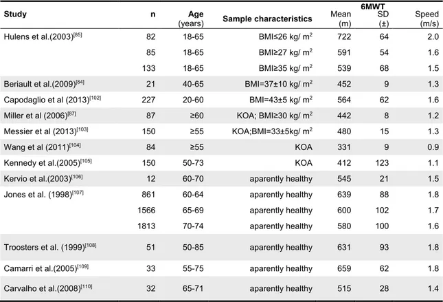
widely used for aerobic fitness assessment and reference values for obese adults, obese adults with KOA and healthy individuals are shown in table 1.
Table 1: Reference values of 6MWT for KOA, obese and health subjects.
6MWT
Study n Age Sample characteristics Mean
(m) SD (±) Speed (m/s) (years)
Hulens et al.(2003)[85] 82 18-65 BMI≤26 kg/ m2 722 64 2.0
85 18-65 BMI≥27 kg/ m2 591 54 1.6
133 18-65 BMI≥35 kg/ m2 539 68 1.5
Beriault et al.(2009)[84] 21 40-65 BMI=37±10 kg/ m2 452 9 1.3
Capodaglio et al (2013)[102] 227 20-60 BMI=43±5 kg/ m2 564 62 1.6
Miller et al (2006)[87] 87 ≥60 KOA; BMI≥30 kg/ m2 442 8 1.2
Messier et al (2013)[103] 150 ≥55 KOA;BMI=33±5kg/ m2 480 15 1.3
Wang et al (2011)[104] 84 ≥55 KOA 331 9 0.9
Kennedy et al.(2005)[105] 150 50-73 KOA 412 123 1.1
Kervio et al.(2003)[106] 12 60-70 aparently healthy 545 21 1.5
Jones et al. (1998)[107] 861 60-64 aparently healthy 639 88 1.8
1566 65-69 aparently healthy 600 102 1.7
1813 70-74 aparently healthy 580 100 1.6
Troosters et al. (1999)[108] 51 50-85 aparently healthy 631 93 1.8
Camarri et al.(2005)[109] 33 55-75 aparently healthy 659 62 1.8
Carvalho et al.(2008)[110] 32 65-71 aparently healthy 515 28 1.4
Lower limb strength training: Muscles constantly interact with the synovial joints, and this can influence the osteoarthritis process. The main goal of recommended strength training is to maintain basic muscle functions (movement, joint stability, the absorption of mechanical shock and proprioception[111], avoiding muscle weakness, improving body stability, and
posture and body weight support). In addition to being considered essential for the performance of daily activities[100], strength training has a positive effect on
the prevention of lower limb injury, one of the KOA risk factors[93]. Weakness of
the quadriceps is a frequent occurrence in KOA and previous clinical trials have shown that quadriceps strength training is effective for the improvement of pain and physical fitness[94, 95]. Obesity combined with sarcopenia, termed
sarcopenic obesity, is also closely associated with the prevalence of KOA[75].
Intensive strength training can change thigh composition and has shown promise in treating the underlying biomechanical (knee-joint loading) and inflammatory disease pathways[112]. However, in cases of varus malalignment,
its effects on pain and on the external Knee Adduction Moment (KAM) have been questionable[96]. In response to this and based on biomechanical
analyses, recent approaches suggest that the strengthening of hip abductor muscles can reduce medial knee loading[97]. Reference values for knee strength
in adults with KOA are cited in table 2.
Table 2: Reference of main published articles of knee strength and KOA.
Article Strength Protocol N BMI±SD Results
Authors Year Age (years±SD) (Kg/m²)
Sanchez-Ramirez et al.[113] 2013 Isokinetic extension (60º/s) N=284 (62±8)
29±5 Ext:0.8±0.4 Nm/kg
Bennell et al.[114] 2013 Isometric extension N=82
(62±7)
30±4 Ext:1.44 ±0.4Nm/kg
Diracoglu et al.[115] 2009 Isokinetic extension (60º/s) N=51 (56±10)
24±5 Ext:114±40 Nm
flexion(60º/s) Flex:49±18 Nm
Malas et al.[116] 2013 Isokinetic extension (60º/s) N=61 (59±7) - Ext:80±33 Nm
flexion(60º/s) Flex:72.2 Nm
Glass [117] 2013 Isokinetic extension (60º/s)
N=2404 31±6
F:(63±8) F: 67±25 Nm
M:(62±8) M:122±52 Nm
Kean et al.[118] 2010 Isokinetic extension (60º/s) N=20 (54± 9) 30±4 1.43±0.5Nm/Kg
Isometric extension 1.81±0.8 Nm
Lim et al.[119] 2009 Isometric extension N=184
(65±8)
29±5 1.33±0.5 Nm/kg
White et al.[120] 2012 Isokinetic extension(60º/s) N=1788 (67±8)
<25 1.17±0.4 Nm/kg 25-<30 1.1±0.4 Nm/kg 30-<35 1.0±0.4 Nm/kg ≥35 0.82±0.3 Nm/kg
Lund et al.[121] 2008 Isokinetic extension (60º/s) N=79 (≥50) Normal Ext:71±26 Nm
flexion (60º/s) Flex: 35±14 Nm Trans et al.[122] 2009 Isokinetic extension (60º/s) N=52 (60±10) 29±6 Ext:58.5±21 Nm flexion(60º/s) Flex: 39.5±12 Nm
Isometric extension Ext: 70±24 Nm
flexion Flex: 37±13 Nm
Flexibility: Patients with symptomatic KOA have poorer flexibility in both the affected and unaffected leg[83], mainly in the hamstrings muscles[123]. Inclusion
of stretching exercises is recommended in every exercise training program for adults[100] and can improve the range of motion and physical function in
individuals with KOA[90].
Gait training: In addition to being considered an aerobic exercise, walking is one of the most important functionalities of the human being and is essential for autonomy and an independent life[83]. Functional limitations imposed by KOA
symptoms and by obesity cause obese individuals with KOA to employ different gait patterns to find better balance and avoid pain. Hulens and his team (2003)[85] suggest that knee pain is an important predictor of the 6MWT in obese
individuals, based on their observation that the distance walked by this group in the study was significantly inferior to lean individuals (table 1). A correct walking pattern could generate some difficulty and joint discomfort during practice, therefore, a multicomponent training has increasingly been suggested. In addition to strength, flexibility and aerobic components of training, gait training should be considered[98].
Stability and posture training: KOA includes symptoms of instability[99]. Recent
evidence has suggested that changes in lower limb biomechanical factors during weight bearing activities may have substantial impact on the ability to maintain a neutral spine posture while moving the extremities in a manner that is independent of the trunk. Stability and posture training is therefore imperative for proper movement and function in all daily activities.
Weight reduction (in cases of obesity): The OARSI recommendations[12] for
weight loss in the treatment of KOA are gaining increasing importance [79-82]. For
a general weight loss program, the ACSM guidelines[100] recommend a
reduction of 5-10% of initial weight over 3-6 months by an intervention of moderate to intense aerobic exercise, resistance-training and behavior intervention. In cases of KOA, Messier and coworkers (2005)[80] reported that a
weight reduction of 1 kg was associated with a knee load reduction of 4 units per step, a clinically meaningful reduction when considered over the many steps performed each day.
Education: This intervention should include general information for a healthy lifestyle and specific information about KOA, its implications and alternatives for managing and living with it. Likewise, posture strategies and gentle movements
can help in pain control. The routine of pain self-assessment is essential for understanding its response to exercise and for management of exercise intensity. The essence of an educational program should be to help patients learn to live with the disease while improving their quality of life and reaching a feeling of wellness [13, 15, 124].
Psychological approach: Factors such as fear, anxiety, and depression have adverse effects on the disability in people with KOA[69, 73, 92], and like pain,
depression is considered a major obstacle for KOA management[69, 89]. An
individual’s perception of pain severity can be influenced by centrally mediated factors and behavioral components[125]. A structured exercise group class can
be an effective way to increase motivation by providing a social support for exercise adherence and, thereby, promoting lifestyle changes[100]. In addition,
exercise and physical activity improve factors related to psychological distress and these changes could lead to improved pain and function in people with KOA.
2.3.1.
Aquatic Exercise
Aquatic exercise (AE) is an exercise modality that is a group of exercises performed in the water, mainly in the vertical position. AE has been used for rehabilitation (hydrotherapy)[126-130], for athletic training[131-135] and for general fitness[136-141]. According
the Aquatic Exercise Association[142], AE can be practiced in shallow or deep water
pools and, for both, can be performed with or without music and with or without accessory equipment. In general, positive effects of AE have been reported for aerobic fitness[143-146], strength[147-150], flexibility[151-153]; body composition[154-156], balance[149, 157-161], functional capacity for daily living activities[153, 162, 163], relief of symptoms in some
musculoskeletal disorders[127, 164], quality of life[165], and psychological factors such as
depression, state of anxiety, and self-esteem[166].
The main characteristics of AE are the utilization of the hydrostatic and hydrodynamics properties of water. The specific properties of water such as hydrostatic pressure, buoyancy and the hydrodynamic resistance are factors that explain the reported chronic adaptations associated with AE programs[167, 168].
AE has become popular as a modality for improving cardiorespiratory fitness while avoiding excessive joint loading[167, 169]. There are some AE programs that are
used worldwide and recognized by international aquatic exercise associations[170, 171],
including Jogging (JO), Cross Country Ski (SK), Jumping Jacks (JJ), Kicks (KC), Rocking Horse (RH), Water Walking (WW), Alternating Leg Curl (LC) and Twist (TW). Most AE classes, which are not physiotherapy, use music to provide motivation, exercise rhythm and acceleration to help reach the desired intensity. The guidelines of the Aquatic Exercise Association (AEA)[171] recommend that aerobic aquatic exercise
classes be performed in water between 28-30º C so as not to compromise the endocrine responses. Water temperatures above 32º C are recommended for passive work, relaxation techniques or for individuals with low movement levels, but are not advisable temperatures for aerobic or strength based exercise[172]. In cases of patients
with low aquatic abilities, the Arthritis Foundation Guidelines suggest a superior limit range of water temperature of 31º C[170].
According to AEA guidelines, beyond the variation in music cadence, exercises in water can be performed in three different cadences (land tempo, water tempo and half-water tempo), and this should be considered when planning research protocols[171].
Barbosa and coworkers.[173] studied the effect of music cadence on heart rate (HR) and
lactate when performing the “rocking horse” in water tempo and found a significant correlation between HR and music cadence (r=0.61; p<.01) and between lactate and music cadence (r=0.54; p<.01), providing important information for instructors who intend to use music during classes. Alberton and coworkers[134] studied the water
running exercise in comparison with maximal land tests and found greater cardiorespiratory responses with increases in music cadence at land tempo AE. With respect to cardiorespiratory training, Raffaelli and coworkers[169] compared
cardiorespiratory responses across several AE programs and they concluded that the Alternated Front Kick had higher VO2 and calorie expenditure than Side Kick, Jogging
and Jumping Jacks, when performed at the same music cadences.
With respect to biomechanics it’s well known that buoyancy force is responsible for softening the impact of vertical movements by reducing the vertical ground reaction forces (V-GRF), which are lower in water than on land for the same exercise[140, 174-176].
Therefore, buoyancy is considered the main mechanism for reduction of the intra-articular compression forces in AE[177, 178]. The magnitude of the impact generated by
practitioner and the technical standards chosen[167, 178, 179]. In addition, some exercises
are made easier, even with muscle weakness, when performed toward the surface of the pool (and are thus assisted by buoyancy force), while others are made harder when performed toward the pool bottom (and are thus resisted by buoyancy force)[180].
With respect to the main AE programs, studies reveal that stationary exercises performed with vertical propulsion of the body have a greater impact than those obtained in exercises with horizontal displacement of the body, such as water walking[174]. Moreover, they show that, among stationary movements, Cross Country
Ski had a lower V-GRF peak than alternated Front Kicks and Water Running[174]. The
authors of this study explained that, due to the buoyancy and drag forces created by the contralateral movement, it is possible to reach high intensities without an increase in V-GRF, mainly because of the absence of heel strike and the accentuated propulsive support phase in maximal velocities[175, 181]. With respect to the V-GRF, another study
assessed mechanical loading from body weight by using plantar pressures and found, for the same water depth and exercise cadence, that the alternated Knee Lift provided higher impact than Cross Country Ski, and that Cross Country Ski had a higher impact than Jumping Jacks[179].
Concerning neuromuscular aspects, an important difference between AE and land exercise is that, in AE, joints are surrounded by water, thus providing multiplanar movements that allow agonist and antagonist muscles to work in the same movement. For example, the neuromuscular function of the quadriceps and hamstring muscles, acting as agonists and antagonists in the repeated knee flexion-extension exercises, was modified by the flowing properties of water, where an early reduction of agonist activity occurred concurrently with a high level of activity of the antagonists while the level of antagonistic activity was low throughout the range of motion[182]. On the other
hand, these same authors suggested that, in a single trial exercise, the level of agonist activity was higher during the final phase of the range of motion when compared with the repeated exercises.
2.3.2. Aquatic Exercise an KOA
Considering the aquatic exercises characteristics and the exercise components suggested in the present dissertation to be included in all KOA intervention programs (cardiorespiratory training[15, 91, 92], strength training[93-97], flexibility training[83, 90], gait
training[83, 98], posture and balance training[99],patient education[13, 15] and psychological
approaches)[73, 92], the analyses of literature previously published (Table 3) indicates
that aquatic exercise, when correctly structured, can be a very complete and embracing exercise type for KOA[168, 171, 183]. Another important aspect of aquatic
exercise is that a person in pain has difficulty with weight bearing exercises, and due to buoyancy, aquatic exercise allows aerobic and resistance exercise to be accomplished with less joint overload[151].
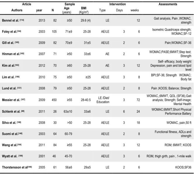
Table 3: Reference of main published articles of exercise for KOA.
Article Sample Intervention Assessments
Authors year N (years) Age (Kg/m²) BMI Type Days weeks
Bennel et al. [114] 2013 82 ≥50 29.6 (4) LE 12 Gait analysis, Pain ,WOMAC,
strength
Foley et al.[184] 2003 105 71±9 25-28 AE/LE 3 6 Isometric Quadriceps strength;
WOMAC;SF-12
Gill et al. [185] 2009 82 70±9 31±5 AE/LE 2 6 Pain;WOMAC;SF-36
Hinman et al.[165] 2007 71 ≥50 33±6 AE 2 6 WOMAC;PASE;6MWT;Step test;
Strength
Kim at al.[183] 2012 70 ≥60 25-28 AE 3 12
Self- efficacy, body weight Depression, pain and blood lipid level
Lim et al. [186] 2010 75 ≥50 ≥25 AE/LE 3 8 BPI;SF-36; Strength WOMAC;
Body fat
Lund et al. [121] 2008 79 ≥50 25-28 AE/LE 2 8 Pain ;KOOS; Balance; Strength
Messier et al. [187] 2009 450 ≥55 28-40.5 LE /Diet/
Education 3 72
WOMAC,;6MWT, QOL (SF36),Gait analysis; Strength; Self-image; Mental Health
Schlenk et al. [88] 2011 26 63±10 33±6 LE 6 24 WOMAC;6MWT;Short Physical
Performance Battery
Silva et al. [188] 2008 30 >50 25-28 AE/LE 3 18 WOMAC, pain,50 ft
Suomi et al.[148] 2003 64 60-79 AE/LE 2 8 Functional fitness, ADLs and
strength
Wang et al.[104] 2011 84 ≥55 25-28 AE/LE 3 12 ROM; 6MWT; KOOS
Wyatt et al.[189] 2001 46 45-70 AE/LE 3 6 ROM, thigh girth, pain , 1-mile walk
Thorstensson et al[190] 2005 61 56±6 29±5 LE 2 6 KOOS;SF36
Abbreviations: AE= Aquatic exercise; LE= Land exercise; 6MWT= Six Minutes Walking Test; WOMAC=; Western Ontario and McMaster Universities Osteoarthritis Index; BPI=Brief Pain Inventory; KOOS= Knee injury and Osteoarthritis Outcome Score; ROM= Knee range of motion.
Several studies have examined the effects of water exercise (hydrotherapy, aquatic exercise) on KOA outcomes[104, 121, 139, 151, 153, 157, 161, 165, 183-186, 188, 189, 191-194],
mainly on symptoms, functional capacity, knee strength and quality of life. However, there is no consistency among most of these aquatic exercise studies[101, 195, 196]
regarding sample characteristics and methodology used, such as exercise quality and volume, intensity management, water temperature, water depth and cueing strategies used[101, 195, 196]. The present literature review indicates that the best results for aquatic
exercise programs were obtained in trials longer than 11 weeks, or at least with a total of 24 sessions. However, many studies used protocols shorter than this.
The primary articles on land or aquatic exercise for KOA, that involved samples similar to the present dissertation study or had detailed methodologies that were considered relevant, are referenced in table 3.
Lim and coworkers[186] and Wang and coworkers[104] compared land and aquatic
exercise and provided detailed information about exercise protocols. Regarding sample characteristics, the majority of KOA studies have worked with age groups above 55 years old and elderly groups, and with normal weight to obesity grade 1. There are few study protocols focusing on obese adults with KOA. However, Hinman and coworkers[165] studied the effect of aquatic exercise on KOA symptoms and physical
fitness of obese adults with KOA, and despite their use of a twice weekly program that lasted only 6 weeks, they had significant improvements. In the study of Kim and coworkers[183], a solid exercise methodology was presented that used music for
motivation and the basic aquatic exercise patterns were applied according the AEA guidelines[171]. Although the study of Messier[187] only applied a land protocol, the
detailed methodology, the sample size, and the sample characteristics were similar to those of this dissertation study and they included a strong educational component.
Based on this literature review, it is clear that there is a need to develop and validate detailed and controlled exercise protocols for obese adults with KOA symptoms, with the intent to disrupt the KOA/Obesity cycles. Thus, specific goals for the PICO aquatic exercise program were established and methodologies were designed.
Chapter 3:
Aims of the study
This chapter presents the general and specific aims stipulated for this dissertation .