Faculdade de Farmácia
Research Institute for Medicines (iMed.ULisboa) Neuron Glia Biology in Health and Disease Group
Alterations at the blood-brain barrier and brain
parenchyma along brain metastasization of
breast cancer
Tânia Custódio Santos
Dissertação de Mestrado para obtenção do grau de Mestre em Ciências Biofarmacêuticas
Orientadora: Prof.a Doutrora Maria Alexandra Brito
Co-orientadora: Prof.a Doutrora Mafalda Videira
The studies presented on this master thesis were performed in the research group “Neuron Glia Biology in Health & Disease”, from Research Institute for Medicines (iMed.ULisboa), Faculty of Pharmacy, Universidade de Lisboa, under the supervision of Maria Alexandra Brito, Ph.D. and Mafalda Videira, Ph.D.
This study was supported by Fundação para a Ciência e Tecnologia (FCT – UID/DTP/04138/2013), Portugal, and by National Research, Development and Innovation/Hungarian Scientific Research Fund (NKFIH/OTKA – K-100807 and K-116158), Hungary.
Publications in international scientific periodicals with referees
Custódio-Santos T, Videira M, Brito MA (2016). Review: Brain metastasization of breast cancer (Submitted).
Custódio-Santos T, Haskó J, Malhó R, Wilhelm I, Krizbai IA, Videira M, Brito MA (2016). Capturing changes in the blood-brain barrier and brain parenchyma during brain metastasization of breast cancer (In preparation).
Publication in scientific meeting abstract book
Custódio-Santos T, Haskó J, Malhó R, Wilhelm I, Krizbai IA, Videira M, Brito MA (2016). Insights into brain metastasization of breast cancer. 50ª Reunião da Sociedade Anatómica Portuguesa (SAP) e 3ª Reunião Científica da Associação Anatómica Portuguesa (AAP). Universidade do Minho, Braga, April 30th, 2016, Portugal.
Poster
Custódio-Santos T, Haskó J, Malhó R, Wilhelm I, Krizbai IA, Videira M, Brito MA (2016). Insights into brain metastasization of breast cancer. 50ª Reunião da Sociedade Anatómica Portuguesa (SAP) e 3ª Reunião Científica da Associação Anatómica Portuguesa (AAP). Universidade do Minho, Braga, April 30th, 2016, Portugal.
Despite the restricted permeability of the blood-brain barrier (BBB), the brain is a privileged organ regarding the appearance of metastases, particularly from breast cancer. Patients with brain metastases from breast cancer have a severe prognosis, rendering this issue a serious oncologic problem that deserves further attention. Therefore, additional studies are required to establish when breast cancer cells cross the brain endothelium and what are the routes used for the transendothelial migration, to understand what is their precise phenotype along the processes of transmigration and establishment of brain metastases, to determine the alterations occurring in brain endothelium, to study how endothelial cells communicate with malignant ones to promote the attraction of malignant cells into the brain vasculature and tumour-associated vascular development. Based on this, we aimed at establishing the temporal profile of breast cancer metastasization to the brain and characterize the metastasizing cells phenotype, as well as, to investigate the vascular events and BBB properties along the process of metastasization to this target organ. In addition, we aimed to assess signalling mechanisms involved in attraction of carcinoma cells into the brain and proliferation in the nervous tissue. To establish the temporal evolution of the players involved in such processes, we used cerebella, cranial hippocampi, and striata of female mice inoculated with 4T1 breast cancer cells sacrificed at 5 hours, 3 days, 7 days or 10 days, and of female mice injected with vehicle (control) sacrificed at 5 hours. Our results showed the presence of brain metastasis of breast cancer at 7-days after inoculation, which increased thereafter. The malignant cells crossed the BBB as mesenchymal cells and, once inside the brain, these cells underwent a complete or partial mesenchymal-epithelial transition to acquire the epithelial characteristics that allow the establishment of new tumours. In addition, the process of brain metastasization of BC contributed to the downregulation of the tight junction protein claudin-5 of brain microvascular endothelial cells, as well as to the entrance of the blood-borne component thrombin in brain parenchyma. On the other hand, hypervascularization in cranial hippocampus appeared to be associated to the process of brain colonization by breast cancer cells. Regarding the role of platelet-derived growth factor B signalling along the process of brain metastasization, we found that this growth factor was expressed by tumour cells and its expression increased during the formation of brain metastasis. Interestingly, a continuous entrance of cysteine-X amino acid-cysteine receptor 4 (CXCR4)-positive cells into the brain parenchyma appeared to occur
to clarify the time-course andinterdependence of the signalling events, BBB breach and phenotypic transition of malignant cells along endothelial transposition and brain metastases establishment by breast cancer cells. Moreover, the demonstration of early cellular and molecular events points to novel targets for modulation in order to prevent metastasis formation and development.
Keywords: Blood-brain barrier; brain metastasis; breast cancer; phenotypic transition;
Apesar da barreira hematoencefálica (BHE) ter uma permeabilidade restrita, o cérebro é um órgão preferencialmente afetado pelo aparecimento de metástases, particularmente de cancro mama. Pacientes com metástases cerebrais provenientes do cancro da mama têm um prognóstico severo, tornando a metastização num sério problema oncológico que merece toda a atenção. Por este motivo, novos estudos são necessários para estabelecer quando é que as células cancerígenas da mama atravessam o endotélio cerebral e quais são as vias que utilizam para migrarem através do endotélio, para se perceber qual o fenótipo que têm ao longo dos processos de migração para dentro do encéfalo e durante o estabelecimento de metástases, para determinar as alterações que ocorrem no endotélio cerebral, para estudar como as células endoteliais comunicam com as células malignas para promover a atracão das células cancerígenas da mama para a vasculatura do encéfalo e o desenvolvimento vascular associado ao tumor. Com base nisto, tivemos com objetivos estabelecer o perfil temporal da metastização do cancro da mama para o encéfalo e caracterizar o fenótipo destas mesmas células, assim como, estudar as alterações vasculares e as propriedades da BHE ao longo do processo de metastização para este órgão secundário. Para além disso, também pretendíamos avaliar os mecanismos de sinalização envolvidos na atracão das células tumorais para o encéfalo e na proliferação no tecido nervoso. Para estabelecer a evolução temporal dos intervenientes envolvidos em tais processos, utilizámos cerebelos, hipocampos craniais e estriados de ratinhos fêmea inoculados com células cancerígenas da mama 4T1 sacrificados às 5 horas, 3 dias, 7 dias, ou 10 dias, e de ratinhos fêmea injetados com veículo (controlo) sacrificados às 5 horas. Os nossos resultados mostraram a presença de metástases cerebrais do cancro mama 7 dias após a inoculação, aumentando ao longo do tempo. As células malignas atravessaram a BHE como células mesenquimais e, uma vez dentro do encéfalo, estas células sofreram uma transição completa ou parcial de fenótipo mesenquimal para epitelial para adquirirem as características epiteliais necessárias para o estabelecimento de novos tumores no encéfalo. Além disso, o processo de metastização cerebral do cancro da mama contribuiu para a diminuição da expressão da proteína das junções de oclusão claudina-5 nas células endoteliais microvasculares cerebrais, assim como para a entrada do componente sanguíneo trombina no parênquima cerebral. Por outro lado, o aumento de vascularização no hipocampo cranial aparentou estar associado ao processo de colonização do encéfalo pelas células cancerígenas da mama.
ao longo do processo de metastização cerebral, descobrimos que as células tumorais expressavam este fator de crescimento e que a sua expressão aumentou durante a formação de metástases no encéfalo. Curiosamente, a entrada contínua de células que expressam o recetor CXCR4 para dentro do parênquima cerebral aparentou ocorrer ao longo do processo de metastização cerebral do cancro da mama. Deste modo, este estudo contribui para clarificar o curso temporal e a interdependência de vias de sinalização, a quebra da BHE e a transição fenotípica das células malignas ao longo na transposição do endotélio e estabelecimento de metástases cerebrais pelas células cancerígenas da mama. Além disso, a demonstração dos eventos celulares e moleculares iniciais aponta para novos alvos para modulação de modo a prevenir a formação e desenvolvimento de metástases.
Palavras-chave: Barreira hematoencefálica; cancro da mama; metástase cerebral;
Gostaria primeiramente de agradecer à Professora Alexandra Brito, orientadora deste trabalho. Sem a sua persistência, este projeto nem teria ganho pernas para andar. Não posso deixar de mencionar todo o seu espírito crítico e a sua exigência que me fez sempre tentar dar o meu melhor. Obrigada por toda a ajuda e orientação a delinear o passo seguinte.
Agradeço à Professora Mafalda Videira por todo o suporte e ajuda que deu ao longo deste ano e por me ter integrado tão bem no seu grupo de investigação.
To the Hungarian research group that collaborate with us, especially to Doctors Imola Wilhelm and István A. Krizbai, I would like to thank you for the opportunity to work with you. I hope that this collaboration continues to yield results.
Agradeço ao Professor Rui Malhó da Faculdade de Ciência pela disponibilidade, pelo apoio e pela flexibilidade demonstrados ao longo do ano. Sem as idas ao microscópio, este estudo não seria nada. Gostaria também de agradecer ao físico Telmo Nunes por toda a paciência e compreensão, principalmente na reta final deste projeto.
Margarida e Sara, o que teria sido de mim sem vocês? Margarida, a ti agradeço pela amizade genuína, por todo o bom ambiente que proporcionaste e por todos os momentos em que precisava daquele abraço e tu estavas lá para mo dar. Obrigada do fundo do meu coração. Sara, a ti agradeço por todas as “baboseiras” que dizes e por toda a tua “boa” música e os teus(/do Luís) vídeos que animavam qualquer dia. Obrigada por seres a amiga incrível que és!
Alicia, obrigada por seres um ser fora do comum, tu és a pessoa que mais me inspira a fazer mais e melhor. Inês, apesar da distância, estiveste/estarás sempre por perto. Mel, a ti agradeço por seres a pessoa especial que és. Nuno, obrigada pela tua espontaneidade e iniciativa para me vires chatear de vez em quando. Tides, a ti agradeço pela amizade inalterável e incondicional que te “obrigou” a ouvir-me tantas e tantas vezes. E Vinte, obrigada por todos os sermões, por todas as tentativas para largar o computador e sair de casa. Gosto imenso de cada um vocês!
Pessoal de casa, obrigada por tudo. Em especial, Alina agradeço-te por toda a tua curiosidade e por todo o teu interesse em saber mais e mais; ao Daniel e ao Nuno por todas as noites que me obrigaram a ir para sala, por todas as noitadas na cozinha, por todas as horas de sono perdidas à vossa pala e por se preocuparem comigo. Vocês são os maiores!
participar na organização dos eventos deste ano. Sem vocês, acreditem que teria batido muitas mais vezes com a cabeça na parede. Ter trabalho convosco, especialmente integrada na equipa da Publicidade, foi incrível e foi muito bom ver que o espírito de equipa continua bem vivo neste Núcleo.
Aos meus amigos de longa data, Inês, Tiago e Zé, agradeço-vos pela amizade que continua a perdurar. Como a Inês e eu costumamos dizer, agora que já nos aturamos há mais de oito anos, já não temos volta a dar!
Por último, mas nunca menos importante, agradeço por tudo à minha família e, em especial, a ti mãe. Tu que foste incondicional ao longo deste ano, estiveste sempre lá para me ouvir, desde as histórias mais hilariantes até às lamúrias e às queixas. Obrigada por todos os esforços que fizeste e continuas a fazer por mim. Se não fosses tu, esta futura cientista (espero) nem teria saído do Algarve. Adoro-te, mãe! E desculpa as longas semanas de ausências, mas quero que saibas que não há um dia em que não pense em ti, no pai e no “godinho” e que não sinta saudades vossas.
i
Figure Index ... iii
Table Index ... iv Abbreviations ... v Chapter I ... 1 Abstract ... 3 Resumo ... 4 1. Breast cancer ... 5
2. From primary tumour to secondary organ ... 9
2.1. Epithelial-mesenchymal transition ... 12
2.2. Local invasion ... 15
2.3. Intravasation ... 18
2.4. Survival in circulation ... 22
3. Brain metastasization ... 24
3.1. Extravasation and role of the blood-brain barrier ... 25
3.2. Proliferation in brain microenvironment ... 31
3.3. Mesenchymal-epithelial transition and brain colonization ... 36
4. Conclusion ... 39 5. Aims ... 40 6. References ... 41 Chapter II ... 57 Abstract ... 59 Resumo ... 60 1. Introduction ... 61
2. Materials and methods ... 63
2.1. Cell culture and animals ... 63
2.2. Histology ... 63
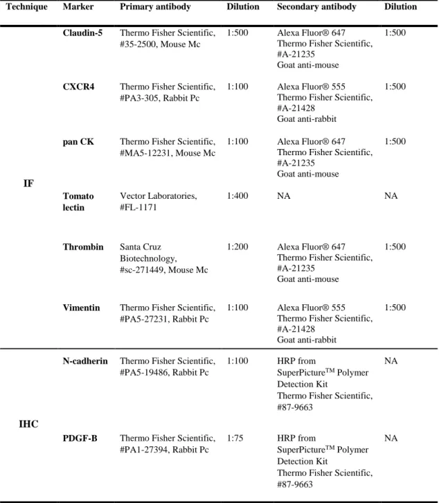
2.3. Reagents and antibodies ... 64
2.4. Haematoxylin and eosin staining ... 65
2.5. Immunofluorescence and immunohistochemistry ... 65
2.6. Data analysis ... 67
2.7. Statistical analysis ... 68
3. Results ... 69
3.1. Brain metastases occur from 7-days onwards after inoculation ... 69
ii
3.4. Formation of brain metastasis requires increased vascularization ... 81
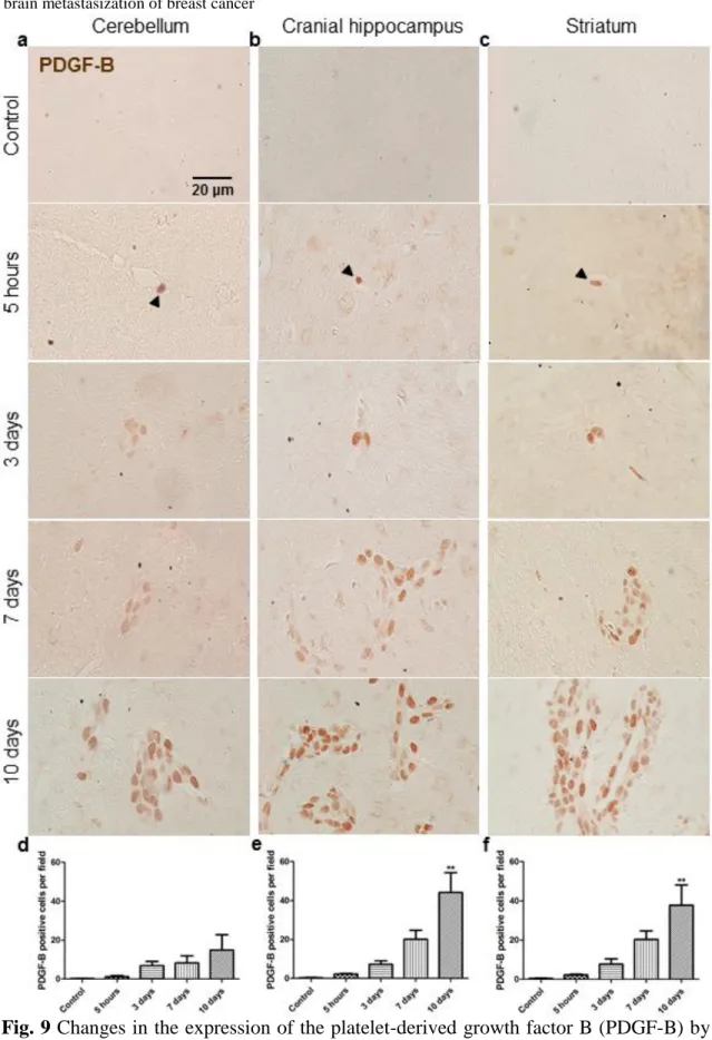
3.5. Malignant cells express PDGF-B ... 81
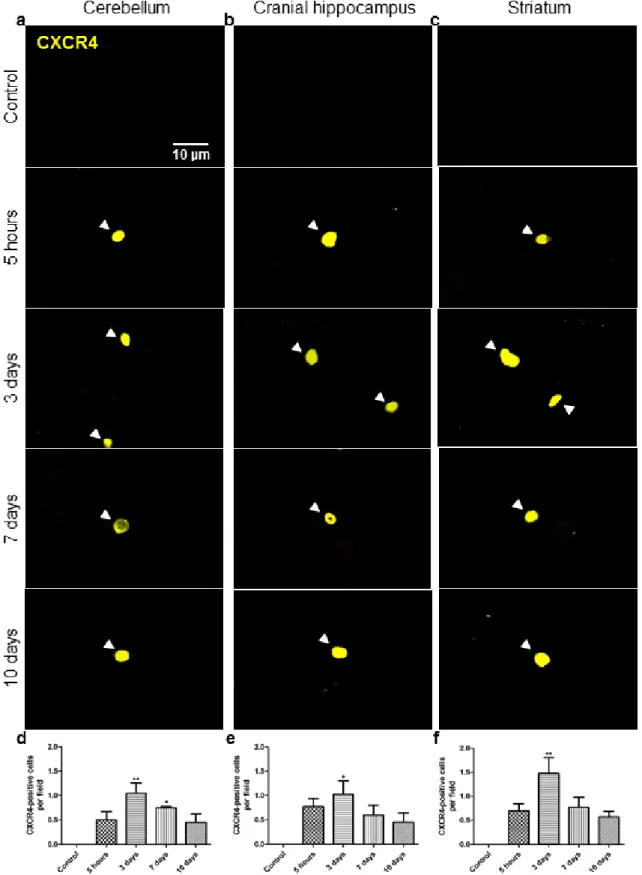
3.6. Malignant cells express CXCR4... 83
4. Discussion ... 85
Acknowledgements ... 91
5. References ... 91
Chapter III ... 95
iii
Chapter I
Introduction
Figure 1 Schematic representation of the mammary duct and of the alterations occurring during the progression of breast cancer (BC) ….……….10 Figure 2 Schematic representation of epithelial-mesenchymal transition (EMT), a process by which epithelial breast cancer cells acquire mesenchymal characteristics ....13 Figure 3 Schematic representation of the transendothelial migration (TEM) pathways during the intravasation process …….……….………...…...….…….20 Figure 4 Schematic representation of key steps of extravasation of a breast cancer cell (BCC) …...………….…………...………...……….……...….27 Figure 5 Schematic representation of key interactions between breast cancer cell (BCC) and neuron, astrocyte, microglia, and pericyte in brain microenvironment ….…34
Chapter II
Results
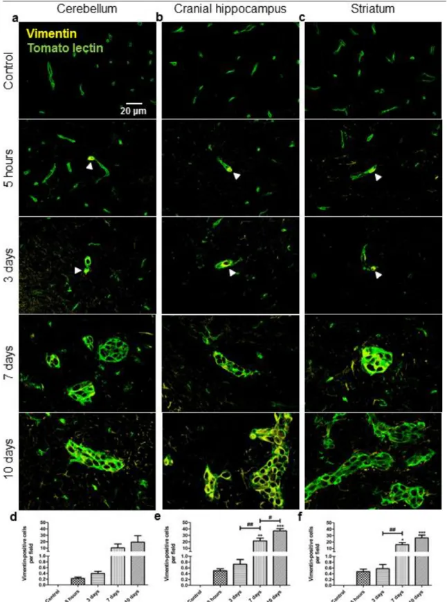
Figure 1 Tumour area occupied by breast cancer cells along the process of brain metastasization ………...….70 Figure 2 Changes in the expression of the mesenchymal marker N-cadherin by breast cancer cells along the process of brain metastasization ………....….71 Figure 3 Entrance of vimentin-positive breast cancer cells and establishment of tomato lectin-positive macrometastases along the process of brain metastasization ...72 Figure 4 Phenotypic changes associated to mesenchymal-epithelial transition of breast cancer cells along the process of brain metastasization ………....……….74 Figure 5 Changes in the expression of the mesenchymal marker vimentin and the epithelial marker pan Cytokeratin by breast cancer cells associated to mesenchymal-epithelial transition along the process of brain metastasization .….….….75
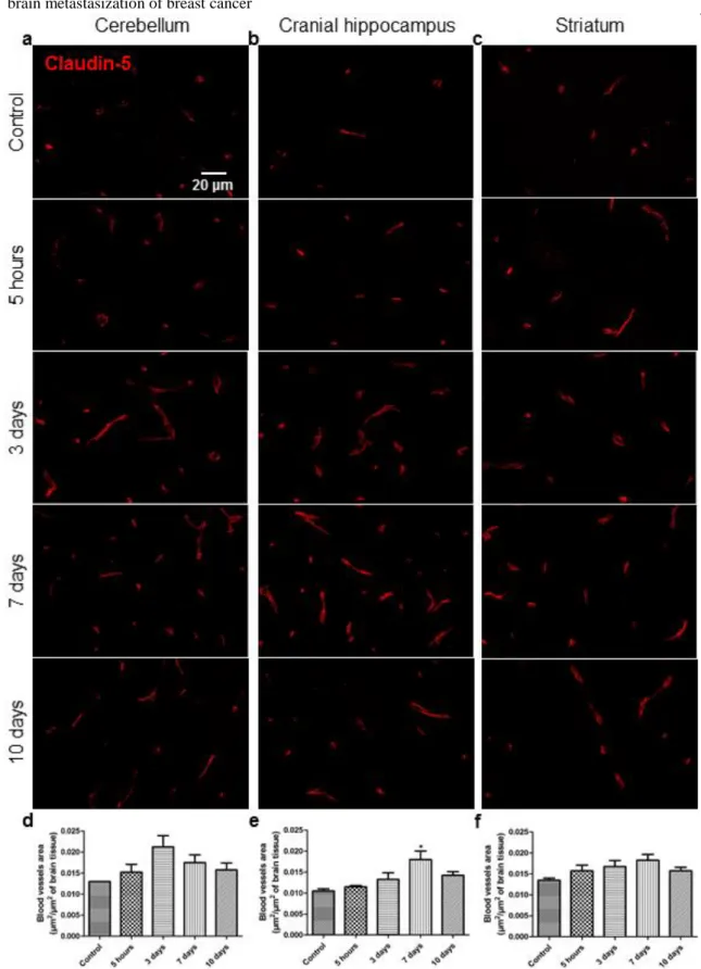
Figure 6 Endothelial tight junction protein claudin-5 changes along the process of brain metastasization of breast cancer ...……….….………...………….77 Figure 7 Entrance of the blood-borne component thrombin into the brain parenchyma along the process of brain metastasization of breast cancer ....……….79
iv
of breast cancer ...……….…….………...80 Figure 9 Changes in the expression of the platelet-derived growth factor B (PDGF-B) by breast cancer cells along the process of brain metastasization …..……….82
Figure 10 Changes in the expression of cysteine-X amino acid-cysteine receptor 4 (CXCR4) by breast cancer cells along the process of brain metastasization ………….84
Table Index
Chapter I
Introduction
Table 1 Tumour, node, and metastasis (TNM) staging according to the American Joint Committee on Cancer (AJCC) staging system ……….……….6
Chapter II
Materials and methods
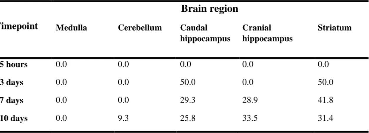
Table 1 Percent distribution of metastases in each brain region of mice injected with 4T1 cells along time ………...……….….64 Table 2 Summary of the antibodies and experimental conditions used for immunofluorescence and immunohistochemical analysis ....………...……...66
v
12(S)-HETE 12(S)-hydroxyeicosatetraenoic acid
ADAM A disintegrin and metalloproteinase
AJ Adherens junction
AJCC American Joint Committee on Cancer
BBB Blood-brain barrier
BC Breast cancer
BCC Breast cancer cell
BHE Barreira hematoencefálica
BM Basement membrane
BMVEC Brain microvascular endothelial cell
BSA Bovine serum albumin
bTNM Biologic tumour, node, and metastasis
CA California
CD Cluster of differentiation
CK Cytokeratin
CNS Central nervous system
CTC Circulating tumour cell
Cx Connexin
CXCL12 Cysteine-X amino acid-cysteine ligand 12
CXCR4 Cysteine-X amino acid-cysteine receptor 4
DAB 3,3’-diaminobenzidine tetrahydrochloride
DAPI 4’,6-diamidino-2-phenylindole
EC Endothelial cell
E-cadherin Epithelial cadherin
ECM Extracellular matrix
EGF Epidermal growth factor
EMT Epithelial-mesenchymal transition
EndMT Endothelial-mesenchymal transition
ER Oestrogen receptor
E-selectin Endothelial selectin
FCT Fundação para a Ciência e Tecnologia
GABA γ-aminobutyric acid
GJ Gap junction
H&E Haematoxylin and eosin
HER2 Human epidermal growth factor receptor 2
HGFR Hepatocyte growth factor receptor
HGF/SF Hepatocyte growth factor/scatter factor
HRP Horseradish peroxidase
vi
IHC Immunohistochemistry
JAM Junctional adhesion molecule
Lhx2 LIM homeobox gene 2
MA Massachusetts
Mc Monoclonal
MET Mesenchymal-epithelial transition
MLC Myosin light chain
MLCK Myosin light chain kinase
MMP Matrix metalloproteinase
MO Missouri
MUC1 Mucin 1
MW Microwave
NA Not applicable
N-cadherin Neuronal cadherin
NKFIH National Research, Development and Innovation
PBS Phosphate-buffered saline
Pc Polyclonal
PDGF Platelet-derived growth factor
PDGFR-β Platelet-derived growth factor receptor beta
PECAM-1 Platelet endothelial cell adhesion molecule-1
PR Progesterone receptor
PSGL-1 Platelet selectin glycoprotein ligand-1
OTKA Hungarian Scientific Research Fund
ROS Reactive oxygen species
SEM Standard error of mean
sLex Sialyl Lewis x
Snail Zinc finger protein snail 1
TEM Transendothelial migration
TGF-β Transforming growth factor beta
TJ Tight junction
TNM Tumour, node, and metastasis
TX Texas
UK United Kingdom
USA United States of America
VCAM-1 Vascular cell adhesion molecule-1
VE Vascular endothelial
VEGF Vascular endothelial growth factor
ZEB Zinc finger E-box-binding homeobox
1
Chapter I
2
The work presented in this chapter corresponds to the following manuscript submitted for publication:
Brain metastasization of breast cancer Custódio-Santos T, Videira M, Brito MA
3
Abstract
Breast cancer can lead to formation of metastases in distant organs, such as the brain, which is commonly associated with diminished quality of life. In addition, central nervous system metastases have been reported in 12-31% of breast cancer patients, and the incidence is increasing with the improvement of primary tumours treatment by therapeutic agents that do not cross the blood-brain barrier, thus rendering the brain a vulnerable organ. Moreover, the survival of patients with breast cancer metastatic to the central nervous system is generally poor, with reports of a 1-year survival rate of 20%. Therefore, a better knowledge of the determinants of brain metastasization is essential for the identification of patients at risk of brain metastases formation, and for the development of novel preventive therapeutic strategies. Here, we summarize and discuss the current data about the multistep process of brain metastasization, ranging from the output of cancer cells from the primary tumour to their colonization in the brain, which involves the epithelial-mesenchymal transition that facilitates the invasion of mammary tissue, the intravasation into circulation, the survival through the vasculature that allows the arrest of the most metastatic cancer cells at brain microvasculature, and the extravasation towards this secondary organ. The several phases of extravasation are also dissected, namely the rolling, adhesion, and transendothelial migration across brain microvascular endothelial cells, and the transcellular passage pathways are addressed. Furthermore, the proliferation of metastatic cancer cells, and their colonization in the brain, the change in malignant cells phenotype via mesenchymal-epithelial transition, and the importance of the microenvironment in the formation of brain metastases are inspected. Finally, the role of angiogenesis along the process of brain metastasization is also address, playing a crucial role not only in primary tumour growth, but also in spread and proliferation of cancer cells. Such detailed cellular and molecular characterization of brain metastasization process contributes to an in-depth understanding of the malignant behaviour.
Keywords: Brain metastasis; breast cancer; epithelial-mesenchymal transition;
4
Resumo
O cancro da mama pode levar à formação de metástases em órgãos distantes, como por exemplo o cérebro, e esta formação de tumores secundários está normalmente associada à diminuição da qualidade de vida dos pacientes. Para além disso, metástases no sistema nervoso central têm sido reportadas em 12-31% dos pacientes e a incidência está a aumentar com o melhoramento do tratamento dos tumores primários através de agentes terapêuticos que não atravessam a barreira hematoencefálica, tornando assim o cérebro um órgão vulnerável à formação de tumores secundários. A taxa de sobrevivência de pacientes com cancro da mama metastático para o sistema nervoso central é geralmente reduzida, com estudos a reportarem uma taxa de sobrevivência de um ano de 20%. Portanto, um melhor conhecimento dos intervenientes envolvidos na metastização cerebral é essencial para a identificação de pacientes em riscos de desenvolverem metástases cerebrais e para o desenvolvimento de novas estratégias terapêuticas preventivas. Deste modo, nós sumariamos e discutimos a informação atual acerca do processo de metastização do cérebro que compreende um conjunto de passos desde a saída das células cancerígenas do tumor primário até à sua colonização no cérebro, envolvendo a transição epitelial-mesenquimal que facilita a invasão do tecido mamário, a entrada no sistema circulatório, a sobrevivência através da vasculatura que permite a chegada das células cancerígenas com maior potencial metastático à micro-vasculatura do cérebro, e a extravasação para dentro deste órgão secundário. As três fases da extravasação também são examinadas, nomeadamente o rolamento, a adesão e a migração por entre as células endoteliais micro-vasculares do cérebro, e as vias da migração trans-celular são abordadas. Para além disso, a proliferação das células cancerígenas metastáticas e a sua colonização no cérebro, a mudança de fenótipo por parte das células malignas através da transição mesenquimal-epitelial e a importância do micro-ambiente na formação de metástases cerebrais são discutidas. Por fim, o papel da angiogénese ao longo do processo de metastização do cérebro também é abordado, tendo este processo um papel crucial não só no crescimento do tumor primário, como também na disseminação e proliferação das células cancerígenas. Assim, a caracterização celular e molecular detalhada do processo de metastização cerebral contribui para uma compreensão cada vez mais aprofundada do comportamento de uma célula maligna.
Palavras-chave: Cancro da mama; metástase cerebral; microambiente tumoral; migração
5
1. Breast cancer
Breast cancer (BC) is a malignant tumour that usually starts in the epithelial cells of the mammary ducts, which are structures responsible for drainage of milk from the lobules secretory acini to the nipple during lactation (Videira et al. 2014). It is considered the most frequently diagnosed cancer in women, with estimated 1.7 million new cases worldwide and nearly 521,900 related deaths in 2012 (Torre et al. 2015). The early detection of this cancer through a mammography screening increases the chances for successful treatment and consequently decreases the mortality from BC (Heinävaara et al. 2016). For early-stage BC, the typical treatment procedure involves either mastectomy (total removal of the breast) or lumpectomy (removal of breast tumour and some of the normal surrounding tissue followed by radiation therapy) plus adjuvant treatment (Zujewski 2016). Early surgical intervention has made an impact in preventing the recurrence of BC. However, this solid cancer is not always timely diagnosed, and in more aggressive and advanced stages the recurrence at distant organs of the body is overwhelming.
All cancers are classified at diagnosis due to its importance for prognosis and responsiveness to therapy. The most widely used BC classification is the tumour, node, and metastasis (TNM) staging system that is based on tumour size (T), regional nodal involvement (N), and distant metastasis (M), and divides BC in four stages (Whitman et al. 2006), as summarized in Table 1. Another classification considers non-anatomical factors such as biomarkers, and distributes BC patients according to the expression of receptors in three groups: hormone receptor-positive when patient presents either oestrogen receptor (ER) or progesterone receptor (PR); human epidermal growth factor receptor 2 (HER2)-positive when HER2 is overexpressed; and triple-negative when the patient is hormone receptors-negative and HER2-negative (Mouttet et al. 2016). The hormone receptors and HER2 are cell surface receptors present in normal breast cell, and their cross-talk results in a positive feedback to cell cycle, survival, and proliferation (Chung et al. 2002, Caldon 2014). However, in BC cells (BCCs), the overexpression of HER2 together with DNA damage induced by high levels of oestrogen lead to deregulated proliferation and growth of these cells, contributing to the initiation and progression of BC. Among the three groups, the triple-negative phenotype is associated with worse survival as compared with patients with the expression of the receptors (Ovcaricek et al. 2011). So, understanding of each group of patients at a molecular level allows not only
6
Table 1 Tumour, node, and metastasis (TNM) staging according to the American Joint
Committee on Cancer (AJCC) staging system.
deciding the most effective treatment for each patient, but also to study and develop more specific therapeutic drugs. Since these two classification systems are routinely used in clinical practice, Bagaria et al. proposed the biologic TNM (bTNM) as a better prognostic indicator (Bagaria et al. 2014). The bTNM incorporates the biomarker profile, defined solely by the triple-negative phenotype, in the TNM staging system, whereby the stage is first determined by TNM, and the addition of the triple-negative phenotype upstages the cancer to the next stage.
The development of primary tumour of breast is a step-wise process that stars with a proliferative growth of epithelial cells in a mammary duct, and culminates in invasion of surrounding tissue, as explained below. After the initial growth of cells,
tumour-Cancer stage Tumour size (T) Regional nodal involvement (N) Distant metastasis (M) Description
Stage I T1 N0 M0 Lesions measure 2 cm or less without metastases
Stage IIA T0 or T1 N1 M0 Lesions measure 2 cm or less with involvement of the axillary lymph node
T2 N0 M0 Lesions measure from 2 cm to 5 cm without
involvement of the axillary lymph nodes
Stage IIB T2 N1 M0 Lesions measure from 2 cm to 5 cm with involvement of the axillary lymph node
T3 N0 M0 Lesions measure more than 5 cm
Stage IIIA T0, T1 or T2 N2 M0 Lesions of any size with metastases in axillary lymph nodes (absence of clinically evident axillary lymph node metastases)
T3 N1 or N2 M0 Lesions measure more than 5 cm with or
without metastases in axillary lymph nodes
Stage IIIB T4 N0, N1 or N2 M0 Tumour of any size which directly extend to the chest wall, skin or lymph nodes
Stage IIIC Any T N3 M0 Tumour of any size with involvement of the axillary and/or internal mammary lymph nodes
7 associated angiogenesis must occur if a malignant mass is to exceed 1 mm in diameter (Fidler 2002). The association between angiogenesis and cancer progression was first described by Judah Folkman et al., who stated that tumour growing was directly dependent of blood vessel network development (Folkman et al. 1971). Angiogenesis is stimulated when tumour tissues require nutrients and oxygen, being essential for cell viability. In fact, the absence of vascular support can cause the tumour to become necrotic or even apoptotic (Nishida et al. 2006). Angiogenesis involves highly regulated paracrine signalling between vascular growth factors released by cancer cells and host cells, and their respective receptors expressed on endothelial cells (ECs) of preexisting vessels. Among the proangiogenic factors expressed by BCCs are vascular endothelial growth factors (VEGFs) and angiopoietins, which can be induced through hypoxia inducible factor 1α and integrins (Felcht et al. 2012, Raja et al. 2014). On the other hand, ECs begin to release specific proteases, such as matrix metalloproteinases (MMPs), to degrade the basement membrane (BM) and extracellular matrix (ECM) that surround a nearby capillary, thereby facilitating their migration into the malignant mass (Papetti and Herman 2002). After ECs proliferation, the newly formed capillary is generally leaky due to the weak cell-cell junctions between ECs and the vulnerable reconstituted BM that surrounds these cells (Shenoy and Lu 2014). Therefore, angiogenesis plays an important role not only in BC growth and progression in primary tumour site, but also during the metastatic stage of BC. Thence, one of the therapies for solid cancers may involve the use of angiogenesis inhibitors, which would consequently block angiogenesis and tumour cell growth and may prevent metastasization to distant organs (Brave et al. 2011).
According to the TNM staging system, when the cancer reaches its most advanced or metastatic stage, tumour cells have the ability to spread and form new tumours in distant visceral organs such as lungs, liver, and brain, and/or in non-visceral organs that include bone, and skin (Berman et al. 2013). The arrest and growth of malignant cells in ‘target organs’ present a preferential distribution and location, a process called organotropism (Lu and Kang 2009). The organotropism depends on the following factors: the receptor status of BCCs; the circulatory pattern, despite the fact that the most frequently organs metastasized by BCCs do not have an immediate direct vascular connection with the primary tissue; genetic signatures present in tumour cells that orchestrate and control the metastatic tropism; and the microenvironment of the organ that will be metastasized (Minn et al. 2005, Spano and Zollo 2012, St Romain et al. 2012). Regarding BC, this cancer represents the second most frequent cause of central nervous
8
system (CNS) metastases, after lung cancer, with metastases occurring in 12-31% of patients (Vuong et al. 2011, Saha et al. 2013).
Brain metastasis is commonly associated with poor prognosis and diminished quality of life, being normally a catastrophic life-threat outcome for patients with solid cancers, such as BC (Cruz-Muñoz and Kerbel 2011, Jaboin et al. 2013). In fact, the 1-year survival rate of patients with BC metastatic to the CNS was reported as only 20% (Altundag et al. 2007). Moreover, there are no targeted therapies specific for this secondary tumour formation, and it is expected that the incidence of brain metastasis continues to increase (Clayton et al. 2004, Steeg et al. 2011). In addition, the incidence of brain metastasis from the primary breast tumour also increases among patients who received chemotherapy or targeted molecular therapeutic such as trastuzumab (Yau et al. 2006, Tonyali et al. 2016). The causes of this increased incidence of brain metastases are unknown, despite the several theories that have been posited. Since the treatment of BC improves the patient’s quality of life and survival, the probability of cancer progression and hence formation of metastases, particularly in the brain, are also enhanced. On the other hand, the brain represents a ‘sanctuary’ organ at risk for disease relapse due to the presence of the blood-brain barrier (BBB), a highly impermeable structure that prevents CNS penetration of most of the conventional and new chemotherapeutic agents (Tonyali et al. 2016). Moreover, glial cells that compose the cerebral microenvironment have a profound impact on the efficacy of chemotherapy by upregulating drug resistance, antiapoptosis, and survival genes in cancer cells, thus rendering malignant cells resistant to therapy (Lee et al. 2016). So, both vascular ECs and brain resident cells protect BCCs from chemotherapy. Another hypothesis postulates that the incidence of brain metastases only seems to have increased since their detection is facilitated by the increased use of refined imaging and the greater attention paid to neurological signs or symptoms (Fink and Fink 2013). Therefore, a better knowledge of the determinants of brain metastasization is essential for the identification of patients at risk of formation of secondary tumour and for the development of novel therapeutic approaches able to prevent such effect from occurring and, thus, to improve the clinical outcomes. In this review, we present the processes that occur during the course of BCCs from the primary tumour site to brain. Furthermore, we explore the still intriguing and unclear mechanisms by which these cells are able to cross the BBB and form brain metastasis, which may constitute potential therapeutic targets.
9
2. From primary tumour to secondary organ
Metastasization is a multistep process ranging from the output of cancer cells from the primary tumour to their colonization in the ‘target organ’ (Nguyen et al. 2009). Among these steps are epithelial-mesenchymal transition (EMT) that facilitates invasion of mammary tissue and intravasation into the circulatory system, survival through the vasculature, arrest at distant organ sites, extravasation within the secondary organ, and finally proliferation and formation of metastasis. When the secondary organ is the brain, the extravasation of BCCs requires the interaction between the tumour cell and the brain microvascular endothelium that forms the BBB (Wilhelm et al. 2014). Once inside the brain, the cells that compose the microenvironment of this secondary organ play a key role in promoting proliferation and colonization by metastatic cells, providing crucial support for cell survival and tumour growth (Lorger 2012). To complete the invasive-metastatic cascade, BCCs reacquire their epithelial phenotype via mesenchymal-epithelial transition (MET), and form well-established brain metastases (Gunasinghe et al. 2012). Identifying and understanding all the mechanisms behind the BC metastasization into the brain may lead to limiting tumour progression, making all steps of the metastatic process potential targets for therapeutic intervention.
Mammary ducts are lined by columnar or cuboidal epithelial cells, surrounded by a discontinuous layer of contractile myoepithelial cells, encircled by the BM, and embedded by the ECM (Muschler and Streuli 2010, Owens et al. 2013) that separates epithelial and stromal compartments (Fig. 1). So, the morphogenesis and architecture of normal mammary epithelium are ensured by cell-cell and cell-BM interactions. Epithelial cells establish interactions with neighbouring cells through intercellular junctions that include tight junctions (TJs), adherens junctions (AJs), desmosomes, and gap junctions (GJs). TJs are located at the apical membrane of epithelial cells and their functions are the restraining of paracellular transport, and establishment and maintenance of apical-basal epithelial polarity (Giepmans and van Ijzendoorn 2009, Owens et al. 2013). These junctions are formed by transmembrane proteins occludin, claudins, and junctional adhesion molecules, and the cytosolic proteins of the zonula occludens (ZO) family that are responsible for the attachment to the cytoskeleton protein actin. AJs are located subjacent to TJs, and have a crucial role in providing adherent strength and attaching the actin cytoskeleton to the plasma membrane (Giepmans and van Ijzendoorn 2009, Owens et al. 2013). These junctions are formed by the transmembrane cadherins, namely
10
epithelial cadherin (E-cadherin), and the cytosolic catenins. Basally to AJs are desmosomes that provide mechanical stability by anchoring cytoskeleton keratinous intermediate filaments to the plasma membrane and facilitate cell-to-cell communication
Fig. 1 Schematic representation of the mammary duct and of the alterations occurring
during the progression of breast cancer (BC). In normal breast, the mammary duct is lined by polarized epithelial cells that are surrounded by a discontinuous layer of myoepithelial cells, and encircled by a basement membrane (BM); mammary epithelial cells establish intercellular junctions between neighbouring cells and with myoepithelial cells, and attach to the BM through hemidesmosomes (a); the intercellular junctions are composed by tight junction (TJ), adherens junction (AJ), desmosome, and gap junction (GJ), and each junction possesses a specific composition and assembly (a1). The duct is embedded in the extracellular matrix of the mammary parenchyma. The development of breast primary tumour occurs through a step-wise progression from benign flat epithelial atypia to atypical ductal hyperplasia, evolves into malignant ductal carcinoma in situ, and finally to invasive ductal carcinoma; both flat epithelial atypia and atypical ductal hyperplasia are characterized by intraductal proliferation of epithelial cells, resulting in multi-layering of ductal epithelium (b and c); in ductal carcinoma in situ, there are still tumour cells remaining connected by intercellular junctions between neighbouring ones, and this type of BC consists of malignant cell masses that lack ductal organization, but remain restricted to breast ducts, and are surrounded by both myoepithelial cells and an intact BM (d); finally, invasive ductal carcinoma is characterized by an extensive growth of the malignant cells beyond the ductal structure and the BM, complete loss of cell-cell adhesion between cancer cells, with the exception of the tumour cells that invade the surrounding tissue as clusters, reduced number of myoepithelial cells, and breaching of BM, which consequently lead to infiltration of the breast ducts by tumour cells and grow into the surrounding tissue (e).
11 through signal transmission (Brooke et al. 2012, Owens et al. 2013). The proteins of this type of intercellular junctions include transmembrane adhesion proteins (desmoglein and desmocollin) that belong to the cadherin family, and cytosolic proteins (desmoplakin, plakoglobin, and plakophilin) that form the desmosomal plaque. This cytoplasmic plaque is responsible for connecting the cytoskeleton to the transmembrane adhesion proteins (Brooke et al. 2012). Desmosomes are also responsible for the attachment of epithelial cells to myoepithelial cells (Gudjonsson et al. 2005). Finally, less regularly organized, GJs are unique intercellular channels that allow diffusion of small molecules, being essential for the communication between neighbouring cells (Giepmans and van Ijzendoorn 2009). These channels are composed by the transmembrane proteins connexins (Cx), wherein the Cx43 is the most abundantly expressed in humans. Similarly to other intercellular junctions, GJs also establish interactions with the cytoskeleton, namely through microtubules and actin, which increase GJ stability (Giepmans 2006, Giepmans and van Ijzendoorn 2009). Regarding cell-BM interactions, they are established by junctional complexes like hemidesmosomes, which anchor the cells to the underlying BM, and maintain tissue polarity (Borradori and Sonnenberg 1999). The hemidesmosomes are composed by three classes of proteins: the cytoplasmic plaque proteins (bullous pemphigoid antigen 1 isoform e and plectin) responsible for the linkage of cytoskeleton to the cell surface; the transmembrane proteins [integrin α6β4, bullous pemphigoid antigen 2, and cluster of differentiation (CD)151] that act as cell receptors connecting the cell and the ECM, and the BM-associated proteins (laminins, such as laminin-332) of the ECM (Borradori and Sonnenberg 1999, Walko et al. 2015). In mammary duct, hemidesmosomes bind myoepithelial cells to BM (Gudjonsson et al. 2005). Since the myoepithelial cells form a discontinuous layer, breast epithelial cells can also interact with the BM through hemidesmosomes and proteoglycan adhesion complexes, whose proteins are coupled to the cytoskeleton and associated to signalling pathways that control cell fate (Bergstraesser et al. 1995, Muschler and Streuli 2010). Thus, the regulation of cell-cell junction complexes and epithelium polarity ensures structural and functional differentiation of the epithelial mammary tissue and their deregulation is associated with cancer. In fact, alterations in desmosomes, GJs, and hemidesmosomes have been increasingly associated with BC progression (Bergstraesser et al. 1995, Brooke et al. 2012, Owens et al. 2013), whereas TJs and AJs are involved in the migration of malignant cells across the endothelium (Martin et al. 2002, Arvanitis et al. 2014).
12
It is consensual that the development of a primary breast tumour is a multistep process, although no consensus has still been reached on the steps and features of each stage (Ellis 2010, Bombonati and Sgroi 2011). For the ductal type BC progression, the classical ductal model proposes that evolution initiates in normal epithelium, progresses from benign flat epithelial atypia to atypical ductal hyperplasia, evolves into malignant ductal carcinoma in situ, and finally to invasive ductal carcinoma (Ellis 2010, Bombonati and Sgroi 2011), as schematically depicted in Figure 1. In addition to the mentioned phases, an alternative model proposes an intermediate step before the progression to flat epithelial atypia, a phase called usual ductal hyperplasia (Bombonati and Sgroi 2011). However, immunohistochemical and molecular biological evidences strongly support that this alternative model of ductal BC progression is likely invalid (Ellis 2010, Bombonati and Sgroi 2011). Thus, according to classic ductal model, both epithelial atypia and hyperplasia are precursor lesions characterized by intraductal proliferation of epithelial cells, resulting in multi-layering of ductal epithelium. Ductal carcinoma in situ is the most common type of non-invasive BC. This type of BC is another intraductal proliferative lesion characterized by lacking ductal organization, but remains restricted to breast ducts without invasion through the BM into the surrounding breast stromal compartment (Ellis 2010, Pinder 2010, Bane 2013). The tumour cells that compose the primary tumour still express E-cadherin that is progressively lost with disease progression, and are surrounded by both a layer of myoepithelial cells and an intact BM. Lastly, the invasive ductal carcinoma consists in an extensive growth of the malignant cells beyond the ductal structure and the BM (Sgroi 2010). Although still under debate, increasing evidence suggests that the progression of carcinoma in situ to the invasive type is related to activation of the EMT that allows malignant cells to migrate and escape from the primary tumour (Knudsen et al. 2012). So, invasive ductal carcinoma is characterized by complete loss of cell-cell adhesion, reduction in myoepithelial cell number, and breaching of BM, which consequently lead to infiltration of the breast ducts by tumour cells and migration into the surrounding tissue (Gusterson et al. 1982, Debnath et al. 2003, Kominsky et al. 2003). Therefore, invasive cells have the potential to spread into lymphatic and/or blood vessels and finally form metastases in other organs.
2.1. Epithelial-mesenchymal transition
To date, the EMT has been the phenomenon that better explains distant metastases formation by epithelial cancers, such as BC. EMT is a reversible process whereby an
13 epithelial cell acquires mesenchymal features (Yang and Weinberg 2008), as depicted in Figure 2. In contrast to epithelial cells, mesenchymal cells usually are not involved in cell-cell interactions and lack the apical-basal polarity (Greenburg and Hay 1982). Thanks to these features, mesenchymal cells have a higher invasiveness than epithelial cells. Initially, carcinoma cells lose the junctions that join them to other cells and/or to BM. Hence, BCCs downregulate the expression of epithelial marks, such as the transmembranar E-cadherin and the intermediate filament pan cytokeratin, followed by enhanced production of mesenchymal proteins, including neuronal cadherin (N-cadherin) and vimentin (transmembrane adhesion molecule and intermediate filament protein, respectively) (Onder et al. 2008, Lv et al. 2013). The switch between E-cadherin and N-cadherin induces the resistance to anoikis, a programmed cell death induced by loss of cell adhesion, through the modulation of pro- and anti-apoptotic genes (Onder et al. 2008, Paoli et al. 2013). Thus, the ability to overcome anoikis is correlated with the acquisition of the mesenchymal phenotype, allowing the survival and proliferation of cancer cells without cell-cell interactions. In addition, activation of EMT is accompanied by a breakdown of the BM due to increase of protease secretion, such as MMPs, and alterations in the production of BM proteins (Tester et al. 2001, Ota et al. 2009, Espinosa
Fig. 2 Schematic representation of epithelial-mesenchymal transition (EMT), a process by
which epithelial breast cancer cells acquire mesenchymal characteristics. Mammary epithelial cells are connected to each other through intercellular junctions (tight junctions, adherens junctions, desmosomes, and gap junctions), as well as to the basement membrane (BM) by hemidesmosomes. During EMT, mammary epithelial cells lose cell adhesion and polarity, and consequently stability, acquiring invasive ability. Along this process, the expression of epithelial proteins is decreased, whereas that of mesenchymal proteins is increased, the morphology changes from cuboidal/cylindrical to fusiform. In parallel with the changes in the morphogenesis and architecture of the mammary ducts, the BM undergoes disruptive changes.
14
Neira and Salazar 2012). These alterations lead to a loss of polarity, a reorganization of cytoskeletal constituents, and an acquisition of mesenchymal-like phenotype, essential for their entry into blood or lymphatic stream.
Although EMT allows malignant cells to change their shape and motility, not all BCCs complete this process, retaining certain epithelial characteristics. This fact is supported by the co-expression of mesenchymal and epithelial markers in BCCs, allowing them to have simultaneously adhesion and migratory properties (Yu et al. 2013). In addition, oestrogen binding to ER also promotes intercellular adhesion via upregulation of the desmosomal proteins and enhancement of formation of desmosomes (Maynadier et al. 2012), increasing the cell-cell attachments that allow tumour cells to move and invade collectively. Thus, some malignant cells can form multicellular clusters even in primary tumour mass. Collective migration of grouped cells that maintain their cell-cell interactions has been implicated in cancer metastasis, and may favour the survival of BCC clusters in circulation. Once in the circulatory system, cancer cells are called circulating tumour cells (CTCs) and can be detected as single cells and as multicellular clusters termed as tumour emboli (Bidard et al. 2010, Tsoi et al. 2010). Both individual and clusters of CTCs can interact with platelets, which protect the malignant cells against the immune response (Gay and Felding-Habermann 2011, Roop et al. 2013). Such interaction with platelets also enhances chemotaxis and chemoinvasion, and posteriorly facilitates extravasation. However, since CTCs are probably targets of a selective process responsible for eliminating carcinoma cells with a lower metastatic potential, the tumour embolus presents a higher ability to form new tumours compared to an individual cell (Aceto et al. 2014). Due to their higher dimensions and resistance to apoptosis compared to an individual cell, multicellular clusters are more likely not only to survive during the way up to the organ that these cells will metastasize, but also to be trapped in small capillaries of secondary sites.
Regardless of the type of EMT, many genes are transcriptionally altered in order to coordinate the repression of epithelial proteins. This repression is orchestrated by regulators such as the transcription factors Twist, Snail (zinc finger protein snail 1), Slug, and ZEB (zinc finger E-box-binding homeobox) family (Samatov et al. 2013). Among the EMT transcription factors, Twist has the ability to promote the formation of invadopodia, specialized actin-based membrane protrusions found in malignant cells that facilitate the local invasion through focal ECM degradation (Eckert et al. 2011). In addition, the regulators of EMT mediate several signal transduction pathways, including
15 transforming growth factor beta (TGF-β) and Notch1 signalling, which in turn promote metastasization.
So, during the transition from in situ to invasive BC, the primary tumour cells exhibit mesenchymal characteristics that allow them to survive without cell-cell interactions and to degrade the BM, resulting in migration and local invasion of mammary tissue.
2.2. Local invasion
Invasion is one of the main processes of metastatic cascade, being not only a fundamental step in tumour progression, but also a driving force for metastasis. In ductal carcinoma in situ, the cancer cells reside within a primary tumour site well-confined by myoepithelial cells and a BM that oppose tumour growth and invasion (Sternlicht et al. 1997, Hu et al. 2008). However, when the tumour progresses to an invasive form, the number of myoepithelial cells reduces at sites of invasion, becoming the minor component of invasive ductal carcinomas (Gusterson et al. 1982). In addition, malignant cells undergoing EMT induce the secretion of proteases that are responsible for degrading the BM. This component of mammary gland plays an important role in invasion of BCCs, since the BM is a reservoir of growth factors that are released during its degradation by tumour cell-secreted proteases (Vukicevic et al. 1992, Hu et al. 2008). Moreover, the BM also plays pivotal roles due to its involvement in pathways initiated by integrin-mediated cell-matrix adhesions that induce intracellular signals and lead to alterations in cell polarity, survival, proliferation, and migration (Nisticò et al. 2014). In addition to myoepithelial cells and BM, even normal mammary epithelial cells promote the invasion of neighbouring tumour cells by secreting soluble laminin, a BM protein, as recently suggested (Lee et al. 2015). The continuous production of laminin induces long membrane cellular protrusions in BCCs, which in turn allow the contraction and subsequently invasion of the surrounding matrix by malignant cells. So, although the precisely controlled tissue architecture of normal epithelium serves as an intrinsic barrier to invasiveness, this may be not enough to stop the cascade that will culminate in metastasization of a secondary site.
Tumour cells can have a diversity of invasive strategies, which are closely related to EMT. When a cancer cell undergoes a complete EMT and acquires the mesenchymal phenotype, this cell invades singly into the surrounding tissue. However, as mentioned above, some malignant cells maintain epithelial characteristics, preserving cell-cell
16
junctions, and this strategy allows the BCC cluster to invade and migrate collectively. These two different strategies of invasion can also coexist within the same tumour (Friedl et al. 1995). Curiously, cancer cells can also undergo transition between invasion modes through the switch between activation and blockade of TGF-β signalling associated to single cell movement and collective cell invasion, respectively (Giampieri et al. 2009). Regardless of the chosen strategy of invasion, the proteolytic destruction of the BM is essential for migration and invasion of tumour cells. Once motile BCCs have dissolved the BM, they enter in mammary stroma where they interact with stromal cells that facilitate the tumour progression.
In normal morphogenesis, mammary ducts are surrounded by the stroma formed by the ECM and a wide variety of cell types, including adipocytes, fibroblasts, and macrophages (Sternlicht et al. 2006). The ECM is the major component of mammary gland microenvironment and, similarly to the BM, regulates epithelial architecture and function (Muschler and Streuli 2010). The cancer progression is dependent on the remodelling and stiffening of the ECM surrounding the tumour cells (Levental et al. 2009, Ko et al. 2016). The remodelling of this matrix is associated to a crosslinking of collagen (the most abundant ECM scaffolding protein), which in turn stiffens the ECM, promotes focal adhesion, and induces invasion and tumour progression (Levental et al. 2009). In addition, aggressive malignant cells are able to alter the mechanical environment of the matrix through the secretion of bioactive lipids that increase ECM stiffness (Ko et al. 2016). Moreover, carcinoma cells release MMPs, such as MMP-2 and MMP-9, that degrade the ECM and augment the bioavailability of ECM-derived growth factors (Tang et al. 2005). One of these growth factors is VEGF that attracts ECs to migrate into areas of active tumour cell proliferation, where cancer cell-induced cleavage of ECM components results in increased vascularization (Lee et al. 1998, Tang et al. 2005). In addition, the degradation of ECM components by invasive tumour cells can lead to an extensive endothelial anoikis, and to attraction and migration of metastatic cells through apoptotic endothelium (Peyri et al. 2009). Thus, deregulation of ECM stiffness and dynamics enhances cancer cell survival, proliferation, migration, and invasion, playing an essential role in BC progression. Besides ECM, stromal cells also have an important role in invasion. Regarding adipocytes, the crosstalk and interaction between these stromal cells and malignant cells lead to an activated phenotype in adipocytes, named cancer-associated adipocytes (Dirat et al. 2011). In turn, this activation results in overexpression of proteases and proinflammatory cytokines by adipocytes that stimulate
17 the invasiveness of tumour cells. Curiously, adipose cells can also promote the resistance of BCCs to cellular cytotoxicity by therapies such as trastuzumab (Duong et al. 2015), thus ensuring that the malignant cells continue to invade the breast tissue. Local invasion leads to the proximity of cancer cells and adipocytes, which respond to this proximity by phenotypical changes that generate fibroblast-like cells (Bochet et al. 2013). The fibroblasts are stromal cells whose functions are maintaining the mammary ECM intact through the synthesis of ECM components, such as collagens, proteoglycans and fibronectin, and regulating the differentiation and homeostasis of adjacent epithelia (Kalluri and Zeisberg 2006, Inman et al. 2015). In tumour stroma, BCCs recruit and activate the majority of fibroblasts, termed as cancer-associated fibroblasts, which result in ECM remodelling to create tracks for tumour cell migration (Gaggioli et al. 2007, Avgustinova et al. 2016). The generation of cancer-associated fibroblasts depends on TGF-β signalling in response to Wtn7a, a factor secreted by aggressive malignant cells that drives the acquisition of a desmoplastic response (Avgustinova et al. 2016). Host macrophages are phagocytic cells that have an important role in host defence against pathogens (Paape et al. 2000) and, like the other stromal cells, also participate in local invasion. This initial step of BC progression depends on both paracrine signalling with macrophages as well as autocrine signalling involving the tumour cells themselves (Patsialou et al. 2009). On one hand, macrophages secrete epidermal growth factor (EGF), which promotes the formation of elongated protrusions that guide malignant cells toward blood vessels, this way favouring the invasion process (Goswami et al. 2005, Wyckoff et al. 2007). On the other hand, tumour cells secrete the cytokine colony-stimulating factor, and sense and stimulate EGF secretion by macrophages. Thus, all stromal components contribute, directly or indirectly, to the movement of BCCs toward lymph nodes and blood vessels, as will be addressed below.
Dynamic interaction between cancer cells and the surrounding microenvironment of mammary ducts is essential to potentiate the malignant behaviour of carcinoma cells. Since the influence exerted by the stromal components remains largely unknown, detailed characterization of these components will provide further evidence of their critical roles in BC progression. However, it is unquestionable that the interactions between cancer cells and the surrounding microenvironment are essential for their entry into the stroma of mammary gland and move directly towards the systemic circulation. Once near a blood or lymphatic vessel, intravasation of tumour cells can occur and, if they survive in circulation, dissemination to distant sites may take place.
18
2.3. Intravasation
As mentioned above, malignant cells detach from the primary tumour mass by undergoing EMT and then invade the surrounding microenvironment of mammary duct; once in the stroma of the mammary gland, the stromal components, such as fibroblasts and macrophages, guide the cancer cells towards vessel wall. The subsequent entry of cancer cells into the circulatory system is called intravasation (Wyckoff et al. 2000). Cancer cells can escape from the primary tumour via two main routes of dissemination: haematogenous or lymphatic (Markiewicz et al. 2013). The decision to intravasate into either blood or lymphatic vessels may depend on the following factors: anatomical differences of the blood and lymphatic vessels; invasion strategy utilized by tumour cells (individual movement versus collective migration); movement promoted by stromal cells; active mechanisms that attract cells to a specific type of vasculature; induction of angiogenesis and/or lymphangiogenesis; and the effect of fluid shear stress on cell survival in circulation (Wong and Hynes 2006, Bockhorn et al. 2007).
Although both blood and lymphatic vessel walls are lined by ECs, there are anatomical differences between them that may influence the intravasation of the malignant cell. Similarly to epithelial cells, the ECs of blood vessels are also connected by cell-cell junctions although lacking desmosomes (Wallez and Huber 2008). These ECs are embraced by some pericytes, perivascular cells that wrap around blood capillaries and reinforce vascular structure, and both ECs and pericytes are embedded within the BM (Fujiwara and Uehara 1984, Davis and Senger 2005). Thence, the anatomical barriers of blood vessels appear to be more hardly penetrated by individual cancer cells. Thus, when the intravasation occurs through the blood vessels, invasive cells must induce cellular and molecular changes to promote their cross through BM, pericyte, and EC barriers. In contrast, ECs that compose the lymphatic capillaries lack tight intercellular junctions, are not embraced by pericytes, and are surrounded by a discontinuous BM (Tanis et al. 2001). Indeed, the higher permeability of lymphatic vessels may favour the passage of individual and mainly clusters of tumour cells (Byers et al. 1995). Despite the migration of cancer cell clusters through blood vessels has not been described, the observation of tumour emboli in bloodstream of BC patients (Tsoi et al. 2010) raises two hypotheses: whether cancer cells intravasate under the form of clusters or, once in bloodstream, aggregate with components and cells present in blood circulation. In addition, stromal cells of surrounding microenvironment can promote a preferential movement of carcinoma cells to blood vessels, since the interaction between cancer cell and stromal ones to promote
19 lymphatic intravasation has never been reported. Like local invasion, the haematogenous intravasation is facilitated by macrophages. Macrophages occupy a perivascular localization in vessel surface, and tumour cells are attracted by the macrophage-derived EGF (Wyckoff et al. 2007). This attraction probably explains why malignant cells are more likely to migrate toward blood vessels than lymphatic vessels in mammary tumours. In addition, macrophage-derived VEGF-A, a growth factor that induces EC proliferation and promotes cell migration, causes local disruption of EC junctions, which hence leads to transient vascular permeability of the blood vessel (Harney et al. 2015). Thus, the interaction between perivascular macrophages and BCCs forms chemoattractive gradients that augment the recruitment of malignant cells towards the blood vessel wall, and facilitate the transendothelial crossing through vascular endothelium. The availability of blood and/or lymphatic vessels in the surrounding area of tumour cells may also influence the pathway taken for dissemination. Both angiogenesis and lymphangiogenesis are induced by the same growth factors, such as VEGFs (Alitalo et al. 2005, Shibuya 2006). Hence, tumours might concomitantly induce angiogenesis and lymphangiogenesis; however, lymphangiogenesis has been less documented in BC compared to angiogenesis (Wong and Hynes 2006). Finally, the fluid shear stress plays a pivotal role in cell survival within the circulation and differs between bloodstream and lymphatic stream. Whereas tumour cell survival may benefit from the low-shear system of fluid transport characteristic of lymphatic vessels, the increased haemodynamic flow rate may also help individual cells to reach distant organs (Byers et al. 1995). Thus, the dissemination via the haematogenous circulation appears to represent the major mechanism by which invasive BCCs are able to spread and form secondary tumours.
The intravasation through blood vessels occurs in the abluminal to luminal direction and involves important interactions between the invasive cancer cell and the EC that composes vessel walls, as schematically depicted in Figure 3. Although the intravasation is considered one of the key steps of the metastatic cascade, this interaction between tumour cells and ECs remains poorly characterized, not only because the transendothelial migration (TEM) is a localized and transient process, but also because the in vitro models used to study this process are only useful to understand and identify molecular components and signalling pathways involved in intravasation (Khuon et al. 2010). Prior to TEM, adhesion of cancer cell to EC must occur to approach these two cellular types and facilitate the translocation of the tumour cell into the circulation.
20
Curiously, the cell surface adhesion receptors expressed by vascular and lymphatic ECs
Fig. 3 Schematic representation of the transendothelial migration (TEM) pathways during
the intravasation process. Breast cancer cell (BCC) can cross endothelial cells (ECs) to enter into the blood circulation by paracellular or transcellular routes. This is a multistep process that involves interactions between BCC and ECs through the binding of specific ligands to the corresponding receptors. Vascular ECs express membrane receptors such as cluster of differentiation (CD)31, endothelial selectin (E-selectin), and integrin β1 that interact with the respective ligand integrin αvβ3, CD44, and integrin β1 expressed by BCC. The adhesion step approaches the cancer cell to the vascular endothelium and facilitates the TEM. During paracellular migration, there is a disruption and reorganization of intercellular junctions and also a reorganization of EC cortical actin cytoskeleton around the invasion site. The ectodomain shedding, a mechanism that compromises the integrity of cell-cell junctions, of the adherens junction protein vascular endothelial (VE)-cadherin is promoted by a disintegrin and metalloproteinase (ADAM)12, and the phosphorylation of this protein is induced by both adhesion step via integrin β1 and reactive oxygen species (ROS). In addition, adhesion via integrin β1 promotes the dissociation of β-catenin from the VE-cadherin/catenin complex. Regarding tight junctions (TJs), the hepatocyte growth factor/scatter factor (HGF/SF), a cytokine secreted by stromal cells, interacts with its receptor, HGFR, expressed in ECs surface. The interaction between HGF/SF and HGFR reduces the expression of the TJ proteins claudin-1, occludin, and zonula occludens (ZO)-1, but not of junctional adhesion molecules (JAMs) and claudin-5. On the other hand, during transcellular migration, the border of the EC remains intact since the migration of BCC occurs when the EC envelopes the malignant one. Firstly, there is a reorganization of the EC actin filaments that circumscribe the invasion pore. Then, EC myosin organizes into a ring-like invasion array via activation of endothelial myosin light chain kinase (MLCK). This enzyme phosphorylates its myosin light chain (MLC), leading to myosin contraction. Thus, the rapid cytoskeletal and membrane remodelling creates a transitory pore-like structure that encapsulates the BCC and facilitates its migration across the EC.