U
NIVERSIDADE DEL
ISBOAF
ACULDADE DEC
IÊNCIASD
EPARTAMENTO DEB
IOLOGIAV
EGETALBiogeography of Xanthophyllomyces dendrorhous,
a yeast with biotechnological potential – population
mapping and host associations
Márcia Cristina David Palma
M
ESTRADO EMM
ICROBIOLOGIAA
PLICADAU
NIVERSIDADE DEL
ISBOAF
ACULDADE DEC
IÊNCIASD
EPARTAMENTO DEB
IOLOGIAV
EGETALBiogeography of Xanthophyllomyces dendrorhous,
a yeast with biotechnological potential – population
mapping and host associations
Dissertação orientada por Prof. Dr. José Paulo Sampaio (CREM, FCT-UNL) e Profª. Drª. Margarida Barata (FCUL)
Márcia Cristina David Palma
M
ESTRADO EMM
ICROBIOLOGIAA
PLICADABiogeography of Xanthophyllomyces dendrorhous,
a yeast with biotechnological potential – population
mapping and host associations
Márcia Cristina David Palma
M
ASTERT
HESIS2011
This thesis was fully performed at CREM (Centro de Recursos Microbiológicos), Departamento de Ciências da Vida of Universidade Nova de Lisboa under the direct
supervision of Prof. Dr. José Paulo Sampaio.
Prof. Dr. Margarida Barata was the internal designated supervisor in the scope of the Master in Applied Microbiology of the Faculty of Sciences of the University of Lisbon.
I To my father, the best of teachers
II
Acknowledgments
I wish to express my gratitude to my supervisor, Prof. Dr. José Paulo Sampaio first, for allowing me to integrate his team as a research fellow and for giving me the opportunity to learn and grow as a scientist. Secondly, I would like to thank him for accepting me as a master student, for giving me time to write this thesis and for all the scientific knowledge passed on to me during this period.
I would like to convey my acknowledgement to the host laboratory CREM (Centro de Recursos Microbiológicos) and FCT-UNL, for giving me the necessary conditions in order to perform my work. I also thank FCT (Fundação para a Ciência e Tecnologia) for providing financial support to the project in which this work was included (PTDC/BIA-BDE/73566/2006).
A special thanks to Dr. Diego Libkind for all the strains that he conveyed for this study, for all the information regarding the genome of X. dendrorhous and for all his collaboration in so many aspects of this work.
I would like to thank Prof. Dr. Rogério Tenreiro and Prof. Dr. Margarida Barata for their help, answering promptly to all my questions during the time of preparation of this thesis.
I would like to thank all my lab colleagues whose knowledge, encouragement and cheer kept me up when science got me down, especially Marco Coelho, Dr. Elisabete Valério, Carla Gonçalves and Jorge Santos.
To my best friend, my love and my life, thank you for being all this and more.
My parents deserve special mention for their limitless support and for all the things that they taught me. Words fail me to express my never ending gratitude and love for my mother whose support has allowed me to pursue my dream of becoming a scientist. No words are necessary to thank my father for wherever he may be, he knows that my inquisitive spirit was born from his own and my unwearyingly willpower and work ethics is but a fragment of his.
III
Resumo
Xanthophyllomyces dendrorhous é uma levedura basidiomiceta cuja grande relevância biotecnológica advém do facto de ser a única espécie cujo principal pigmento produzido é astaxantina. Este carotenoide possui várias atividades biológicas relevantes e inúmeras aplicações industriais, sendo o segundo carotenoide mais rentável do mercado. Apesar do interesse nesta levedura devido às suas aplicações biotecnológicas, ainda são limitados os conhecimentos sobre a sua biologia e biogeografia.
X. dendrorhous foi isolada pela primeira vez durante a década de 60 do século passado a partir de exsudados de árvores de folha caduca em diversas localizações no Hemisfério Norte, tendo-se considerado durante muito tempo restrita a estes habitats e latitudes. Inicialmente descrita como uma levedura assexuada designada Phaffia rhodozyma, em 1995 o seu estado sexuado, Xanthophyllomyces dendrorhous, foi observado e descrito pela primeira vez. O seu ciclo sexuado é caracterizado pela conjugação entre a célula-mãe e a gémula, após a qual ocorre a formação de um holobasídio aéreo que subsequentemente dá origem a basidiósporos na sua extremidade apical. Este estado sexuado é despoletado pela presença de polióis no meio de cultura. Taxonomicamente esta levedura encontra-se classificada na ordem Cystofilobasidiales, classe Tremellomycetes e subfilo Agaricomycotina. A descoberta de um novo habitat de X. dendrorhous em 2007 na Argentina (Patagónia), veio mostrar que esta levedura possui uma distribuição geográfica mais diversificada do que aquela considerada inicialmente. Nas florestas da Patagónia, X. dendrorhous encontra-se associada aos corpos frutíferos de um fungo ascomiceta, parasita obrigatório de árvores Nothofagus, denominado Cyttaria. À semelhança dos exsudados de árvores, os corpos frutíferos de Cyttaria são ricos em açúcares simples, estando presentes nos troncos de Nothofagus uma vez por ano. Após a descoberta de novas populações de X. dendrorhous no Hemisfério Sul foi realizado um estudo filogenético da região ITS (internal transcribed spacer) do rDNA das estirpes de X. dendrorhous e das diferentes árvores hospedeiras tendo-se verificado concordância entre ambas as filogenias. Este facto levou à formulação da hipótese de que a variabilidade intraespecífica observada em X. dendrorhous, poderia dever-se ao facto de cada linhagem se encontrar associada a um hospedeiro diferente. Após a descoberta de X. dendrorhous no Hemisfério Sul, a exploração de outras regiões geográficas onde, tanto Nothofagus como Cyttaria estão presentes, foi levada a cabo em 2009 por elementos do laboratório de acolhimento. As amostras biológicas recolhidas na Austrália e Nova Zelândia resultaram na obtenção de novos isolados de Xanthophyllomyces.
Face ao número crescente de isolados de Xanthophyllomyces existentes, o presente trabalho teve como principal objetivo o estudo das relações filogenéticas entre as diferentes
IV linhagens existentes assim como, a exploração de possíveis relações entre a variabilidade genética encontrada em Xanthophyllomyces e os hospedeiros aos quais esta levedura se encontra associada. De forma a atingir este objetivo, duas abordagens distintas foram exploradas: (i) amplificação, sequenciação e estudo filogenético de sete fragmentos de genes de Xanthophyllomyces e (ii) a cariotipagem de isolados representativos das diferentes linhagens encontradas. Os dados obtidos foram analisados sob o ponto de vista da associação aos hospedeiros e localização geográfica dos isolados. Os resultados mostraram a existência de quatro populações distintas em X. dendrorhous. A estrutura populacional encontrada sugere que as populações naturais se propagam preferencialmente de forma clonal. Cada uma das diferentes populações apresenta-se associada a diferentes plantas hospedeiras, sendo esse o fator que mais parece influenciar a estrutura populacional. No entanto verificou-se também alguma diferenciação geográfica apesar da sua influência parecer menos relevante. Na filogenia concatenada dos sete fragmentos de genes estudados foi observado que cada grupo filogenético principal agrupa estirpes isoladas a partir de um mesmo hospedeiro, sendo igualmente possível verificar a existência de subdivisões dentro destes mesmos grupos, que estão por sua vez associadas à origem geográfica dos isolados. Este facto foi corroborado pelas networks filogenéticas construídas no software Splistree4 e pela análise da estrutura populacional usando uma abordagem bayesiana implementada no software Structure. A determinação dos cariótipos de estirpes representativas de cada uma das populações encontradas em X. dendrorhous revelou padrões únicos de cromossomas para cada estirpe. O número de cromossomas das onze estirpes estudadas varia entre seis e 11, sendo o seu tamanho entre 0,8 Mb e 3,5 Mb. Não foi observada uma relação entre a estrutura populacional de X. dendrorhous e os cariótipos observados para as diferentes estirpes.
Verificou-se que um dos isolados em estudo, possui três genes heterozigóticos. Os alelos de cada gene foram separados por clonagem e posteriormente sequenciados, tendo-se verificado a existência de dois alelos distintos para cada gene heterozigótico. A análitendo-se filogenética permitiu observar que, para os três genes heterozigóticos, os alelos encontrados pertencem a duas populações distintas originárias respectivamente da Australásia (em associação com Nothofagus) e Japão (em associação com Betula). Este facto sugere a existência de contacto entre as duas populações.
Diversos estudos biogeográficos têm sido realizados sobre os géneros Nothofagus, Betula e Cornus. Enquanto que a biogeografia de Nothofagus parece ter sido moldada por fenómenos como a fragmentação do super continente Gondwana e eventos de dispersão a longas distâncias, as histórias biogeográficas de Betula e Cornus parecem ter sido influenciadas por migrações intercontinentais através das pontes terrestres de Bering e do Oceano Atlântico durante o período geológico Terciário. A análise da atual distribuição de X.
V dendrorhous levando em consideração as histórias biogeográfias dos seus hospedeiros parece sugerir uma correlação entre ambas. É possível que a distribuição de X. dendrorhous no Hemisfério Sul esteja relacionada com a biogeografia de Nothofagus, assim como a sua distribuição no hemisfério Norte parece ser coerente com as histórias biogeográficas de Betula e Cornus. A coexistência de Nothofagaceae, Fagaceae e Betulaceae na região do arquipélago indo-australiano poderá ter permitido um salto de hospedeiro por parte de X. dendrorhous que assim colonizou outras áreas no Hemisfério Norte.
Entre os isolados recolhidos na Australásia foram encontradas duas putativas novas espécies de Xanthophyllomyces. É possível observar que a divergência destas novas espécies relativamente a X. dendrorhous se verifica ao nível molecular tanto na região ITS como nas sete regiões genómicas estudadas. As duas espécies, Xanthophyllomyces sp. I e Xanthophyllomyces sp. II apresentam o mesmo estado sexuado que X. dendrorhous, assim como produção de astaxantina. O presente estudo constitui o primeiro registo da existência de mais do que uma espécie no género Xanthophyllomyces. Com este estudo iniciou-se a exploração da recém descoberta biodiversidade existente na Australásia, identificando-a como um possível hotspot de diversidade e possivelmente como ponto de origem do género Xanthophyllomyces.
Palavras chave: Xanthophyllomyces; Biogeografia; Associação a hospedeiros; Espécies novas;
VI
Abstract
The basidiomycetous yeast Xanthophyllomyces dendrorhous has biotechnological relevance due to the production of astaxanthin, a carotenoid with remarkable biological properties. First isolated during the 1960s from exudates of deciduous trees throughout the Northern Hemisphere, this yeast was for long considered to be restricted to these habitats and latitudes. The discovery of a new Xanthophyllomyces habitat in the Southern Hemisphere in 2007 brought a better understanding of the geographic distribution and ecology of this yeast. In Patagonian forests, X. dendrorhous was discovered to be associated with the fruiting bodies of Cyttaria, an ascomycetous parasite of Nothofagus trees. Given that the Cyttaria-Nothofagus host system is also present in Oceania, sampling of selected localities in Australasia was carried out in 2009 yielding a considerable number of new isolates of Xanthophyllomyces.
Benefiting from the growing collection of Xanthophyllomyces strains available, that encompasses now novel Australasian lineages, the major intend of this work was to elucidate the phylogenetic relationships between the different lineages, and to further explore the genetic variation within Xanthophyllomyces. To achieve this goal a representative group of isolates was studied using multilocus sequence analysis and karyotyping assays within an ecological and geographic framework.
The results showed that X. dendrorhous is composed by four well structured populations, which suggests that natural populations of this yeast are mostly clonal. The existence of an isolate bearing alleles from two distinct populations suggests contact between populations from Australasia and Japan. Xanthophyllomyces dendrorhous populations have different ecological associations and appear to have diverged first according to host tree and secondly by geography. Different karyotypes were presented by each of the tested strains and no correlation with the revealed population structure was observed. The Australasian population harbors two putative new species that appear to be endemic to the area. This study represents the first record of the existence of more than one species in the Xanthophyllomyces genus. The extant distribution of the X. dendrorhous in the Southern Hemisphere may be related with the biogeographic history of Nothofagus trees, while its Northern Hemisphere distribution appears to be coherent with the biogeographic histories of Betula and Cornus trees.
VII
Table of contents
Page
Figure Index VIII
Table Index IX
Symbol and Abbreviation List X
_________________________________________________________________________________
1. Introduction 1
2. Material and Methods 5
2.1. Studied strains 5
2.2. DNA extraction, MLSA primers and PCR conditions 6
2.3. Cloning assays 6
2.4. Phylogenetic analysis 7
2.5. Phylogenetic Networks analysis 7
2.6. Analysis of X. dendrorhous population structure by a quantitative clustering
method 8
2.7. Karyotyping assays 8
3. Results and discussion 11
3.1. Xanthophyllomyces multilocus phylogeny. 11
3.2. Xanthophyllomyces novel species. 14
3.3. Exploring the population structure of X. dendrorhous. 16
3.4. X. dendrorhous karyotype. 20
3.5. X. dendrorhous population structure, host system and geographical distribution. 22
4. Final considerations and future perspectives 27
5. References 28
VIII
Figure Index
Page
Figure 1 - Xanthophyllomyces dendrorhous sexual state. 2
Figure 2 - Presently known habitats for Xanthophyllomyces. 3
Figure 3 - Nothofagus world distribution. 3
Figure 4 - Xanthophyllomyces multilocus phylogeny and distribution. 12 Figure 5 - ITS phylogeny of Xanthophyllomyces and Cystofilobasidium. 15
Figure 6 - Splits network of Xanthophyllomyces. 17
Figure 7 - Ancestry of X. dendrorhous estimated with Structure software. 19
Figure 8 - X. dendrorhous karyotypes. 21
Figure 9 - X. dendrorhous population structure and host tree phylogeny. 23 Figure 10 - Xanthophyllomyces extant distribution and hypothetical routes for the colonization
of the Northern Hemisphere. 25
________________________________________________________________________________ Figure S1 - Individual phylogenies of Xanthophyllomyces by ML method. 33 Figure S2 - Multilocus phylogeny of Xanthophyllomyces by ML method. 34 Figure S3 - Sexual state of Xanthophyllomyces strains from Australasia. 35
IX
Table Index
Page Table 1 - List of strains used in the study and relevant information regarding them. 5
Table 2 - Summary of the PFGE running parameters used. 9
________________________________________________________________________________ Table S1 - Sequences used for the construction of the primers for the multilocus sequence
analysis. 32
Table S2 - List genes and genetic regions used in this study and relevant information
regarding them. 32
X
Symbol and abbreviations list
AIC − Akaike information criterion
ATCC - American Type Culture Collection bp − Base pairs
CBS − Centraalbureau voor Schimmelcultures, Netherlands CHEF − Contour-clamped homogeneous electric field CLP − Chromosome length polymorphism
CN − Chromosome number
CRUB - Centro Regional Universitario Bariloche
Cy. − Cystofilobasidium
E. coli − Escherichia coli
ITS − Internal transcribed spacer k − Population
MCMC − Markov Chain Monte Carlo ML − Maximum likelihood
MLSA − Multilocus sequence analysis MP − Maximum parsimony
MYA − Million years ago
PALM − Phylogenetic reconstruction by automated likelihood model selector PCR − Polymerase chain reaction
PFGE − Pulse field gel electrophoreses rDNA − Ribosomal deoxyribonucleic acid RN − Reticulate network
SDS − Sodium dodecyl sulfate SN − Splits network
Tris − 2-amino-2-hydroxymethyl-propane-1,3-diol w/v − Weight / Volume
Δk − Delta k (determined by the rate of change in the log probability of data between
1
1. Introduction
Xanthophyllomyces dendrorhous (asexual state Phaffia rhodozyma) is the only basidiomycetous yeast that produces astaxanthin, a high-value carotenoid, as its main pigment (Johnson 2003). Astaxanthin has remarkable antioxidant, neuroprotective, anti-inflammatory and antitumor properties (Naguib 2000, Kurihara et al. 2002, Frengova and Beshkova 2009, Lee et al. 2010) and is the second most important carotenoid in the market (Schmidt et al. 2010). The ability to ferment sugars, a rare attribute for basidiomycetous yeasts, makes X. dendrorhous a promising candidate for a cost-competitive biotechnological production of astaxanthin (Rodriguez-Saiz et al. 2010).
This yeast was first isolated by Herman Phaff in 1967 from exudates of deciduous trees in Japan and Alaska (Miller et al. 1976, Golubev 1995) and was described as an asexual species, Phaffia rhodozyma, in 1976 (Miller et al. 1976). On the following years, other isolates were obtained also from exudates of deciduous trees in Finland, Germany and Russia (Golubev 1995, Weber et al. 2006). Through phylogenetic analysis using rDNA sequences, it was observed that the species of genus Cystofilobasidium were the closest relatives of Phaffia rhodozyma. The molecular phylogenies placed this yeast in the order Cystofilobasidiales, class Tremellomycetes and subphylum Agaricomycotina (Scorzetti et al. 2002). In 1995 the sexual state was observed for the first time after inoculation of strains in an agar medium containing polyols. The sexual state was described as Xanthophyllomyces dendrorhous and is characterized by the conjugation between cell and bud (pedogamic sexual process), after which a slender cylindrical holobasidium forms. Basidiospores, usually four, are produced in the terminal apex of the basidium (Golubev 1995) (Figure 1). This sexual state is not yet fully understood since the real ploidy of the species remains uncertain. Several studies have provided evidence that the vegetative stage is diploid but suggested also the existence of different levels of ploidy among the few tested strains, including possible aneuploids (Kucsera et al. 1998, Medwid 1998, Hermosilla et al. 2003). Another interesting aspect of Xanthophyllomyces is the existence of chromosome length polymorphisms (CLP) between strains. Despite the fact that the number of strains used was small no two strains presented the same chromosomal pattern in the karyotyping studies performed to date (Nagy et al. 1994, Adrio et al. 1995, Cifuentes et al. 1997, Nagy et al. 1997). Besides polyploidy and CLP, determination of genetic diversity using different regions of the rDNA repeat, revealed that Xanthophyllomyces has a high intra-specific variability (Fell et al. 2007). Because so little was known regarding the ecology and biogeography of this yeast, there were no factors that could be directly related to such differences.
2
Figure 1 – Xanthophyllomyces dendrorhous sexual state. (a) Scanning electron microscopy photos showing
two conjugated cells [1] originating a non-septate basidium [2]; (b) aerial basidium [2] with apical basidiospores [3]; (c) detail of the basidium apex [2] and basidiospores [3]. Image adapted from the website of the American Society for Microbiology, http://202.195.144.50/ASM/106-Introduce.htm.
Since all Xanthophyllomyces isolates were obtained from tree exudates belonging to genera Betula, Alnus, Fagus and Cornus it was assumed that the distribution of X. dendrorhous was limited to these host trees. Moreover, considering the biogeography of the plants (Chen et al. 1999, Chen and Li 2004, Manchester et al. 2007), it was assumed that Xanthophyllomyces was restricted to the Northern Hemisphere (Golubev 1995, Fell and Blatt 1999). However, the discovery of a new Xanthophyllomyces habitat in the Southern Hemisphere brought novel insights regarding the geographic distribution and ecology of this yeast. In Patagonia (Argentina), X. dendrorhous was found to be associated with the fruiting bodies of Cyttaria, an obligate biotrophic ascomycete fungus restricted to Nothofagus trees (Rawlings 1956) (Figure 2). The mycelium of Cyttaria develops inside the tree and persistent tumors are formed in the tree branches (Rawlings 1956). On these tumors, fruiting bodies rich in simple sugars are annually produced, and from them X. dendrorhous is consistently isolated (Libkind et al. 2007). In the same study a collection of strains from diverse geographical origins and also from different host systems comprising four different plant families, Betulaceae, Cornaceae, Fagaceae, and Nothofagaceae, was compared using the internal transcribed spacer (ITS) sequences of both the yeast and the host trees. Since a general concordance was observed between the two ITS phylogenies it was hypothesized that the genetic polymorphisms that segregated the strains into different lineages could represent intraspecific variation resulting from host-specific association. It was also hypothesized that the association of X. dendrorhous with Cyttaria derived from an earlier association of the yeast with Nothofagus trees. The genus Nothofagus forms a monophyletic group, sister to the rest of the Fagales that has been instrumental in understanding the biogeography of the Southern Hemisphere (Cook and Crisp 2005, Peterson et al. 2010).
3
Figure 2 – Presently known habitats for Xanthophyllomyces. (a) Betula tree trunk with an orange colored
exudate in North America (enlarged detail on the right); (b) branch of Nothofagus cunninghamii with Cyttaria
gunnii fruiting bodies (Tasmania).
The extensive fossil record of Nothofagus pollen found throughout what was once the Gondwana supercontinent, has allowed tracking major shifts in geology and ecology that have taken place in the last 80 million years (Cook and Crisp 2005).
Figure 3 – Nothofagus world distribution. Map depicting in dark gray the present geographic distribution of
Nothofagus. The distribution of several species is also indicated. Adapted from Knapp et al. (2005) and Swenson
et al. (2001).
The extant disjunctive distribution of Nothofagus in the Southern Hemisphere, both in South America and Australasia has been correlated with the Gondwana break up and the continental drift theory, as well as more recent (in geological terms 44.6 – 28.5 million years ago) long-distance dispersal events within Australasia (Swenson et al. 2001, Mathiasen and Premoli 2010, Peterson et al. 2010). After the discovery of the association of X. dendrorhous with Nothofagus in Patagonia, the existence of unknown populations of this yeast in other regions colonized by Nothofagus had to be considered. In 2009 a collecting trip to Nothofagus forests in New Zealand and Australia by members of the host laboratory, yielded
4 a considerable number of new Xanthophyllomyces isolates confirming the existence of additional populations associated with Nothofagus trees. As the number of Xanthophyllomyces strains increased, also did the possibilities of better understanding the biology, ecology and world distribution of this microorganism.
With the increase of sequence information available, a broad application of phylogenetic methods has taken hold of fungal taxonomy and of biogeography studies. The multilocus sequence analysis (MLSA) or complete genome comparisons are being widely applied in detriment of the single genetic marker approach, given the extensively grater information yielded (Taylor et al. 1999). These techniques allow not only the recognition of species (Taylor et al. 2000) but also the study of distinct populations among a species (Taylor and Fisher, 2003). The statistical phylogeographic approach connects phylogeography methodologies and classical population genetics methods, providing insights on the evolutionary history of organisms (Knowles, 2009). Bioinformatics tools available today allow the inference of parameters such as recombination, population structure and to assign individuals to a population based on their ancestry composition from MLSA datasets (Crandall et al. 2000, Posada and Crandall 2001, Excoffier and Heckel 2006, Huson and Bryant 2006, Falush et al. 2007, Perez-Losada et al. 2007, Chen et al. 2009)
Taking advantage of the growing collection of Xanthophyllomyces strains that encompasses novel Australasian lineages, the major intend of this work was to shed light on the phylogenetic relationships between the different lineages, and to explore in more detail the genetic variation previously observed. To achieve this goal a representative group of isolates was investigated by MLSA and karyotype analysis within an ecological and geographic framework.
5
2. Material and methods
2.1. Studied strains
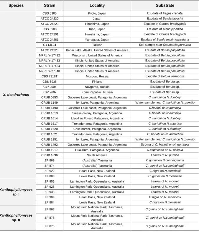
Forty strains from nine different geographical localizations and two distinct host systems were studied (Table 1).
Table 1 – List of strains used in the study and relevant information regarding them.
Species Strain Locality Substrate
X. dendrorhous
CBS 5905 Kyoto, Japan Exudate of Fagus crenata
ATCC 24230 Japan Exudate of Betula tauschii
ATCC 24229 Hiroshima, Japan Exudate of Cornus brachypoda
CBS 5908 Kiso, Japan Exudate of Alnus japonica
ATCC 24201 Hiroshima, Japan Exudate of Cornus brachypoda ATCC 24261 Yamagata, Japan Exudate of Betula maximowicziana
GY13L04 Taiwan Soil sample near Stauntonia purpurea
ATCC 24228 Kenai Lake, Alaska, United States of America Exudate of Betula papyritosa NRRL Y-17432 Wisconsin, United States of America Exudate of Betula populifolia NRRL Y-17433 Illinois, United States of America Exudate of Betula populifolia NRRL Y-17434 Illinois, United States of America Exudate of Betula populifolia NRRL Y-27348 Illinois, United States of America Exudate of Betula populifolia CBS 7918T Moscow, Russia Exudate of Betula verrucosa
CBS 6938 Finland Exudate of Betula sp.
KBP 2604 Novgorod, Russia Exudate of Betula sp.
KBP 2607 Komi Republic, Russia Exudate of Betula sp. CRUB 0853 Gutierrez Lake coast, Patagonia, Argentina C. hariotii on N.dombeyi
CRUB 1149 Ilón Lake, Patagonia, Argentina Water sample near C. hariotii on N. pumilio CRUB 1490 Gutierrez Lake coast, Patagonia, Argentina C.hariotii on N.dombeyi
CRUB 1613 Suisse colony, Patagonia, Argentina C.hariotii on N.dombeyi
CRUB 1614 Llao-llao Forest, Patagonia, Argentina C. hariotii on N.dombeyi
CRUB 1617 Tronador area, Patagonia, Argentina C. hariotii on N.antartica
CRUB 1620 Chile border, Patagonia, Argentina C. hariotii on N.dombeyi
CRUB 1621 Tronador area, Patagonia, Argentina C. hariotii on N. antarctica
CRUB 1151 Ilón Lake, Patagonia, Argentina Water sample near C. hariotii on N. pumilio CRUB 1492 Gutierrez Lake coast, Patagonia, Argentina Stroma of C. hariotii on N. dombeyi CRUB 1917 Hua-Hum, Patagonia, Argentina C.espinosae on N. obliqua
CRUB 1896 South America Leaves of N. pumilio
ZP 869 (Australia ) Tasmania C.gunnii on N.cunninghamii
ZP 874 (Australia ) Tasmania C. gunnii on N.cunninghamii
ZP 922 Haast Pass, New Zealand C.nigra on N.menziesii
ZP 888 Lewis Pass, New Zealand C. gunnii on N.menziesii
Xanthophyllomyces
sp. I
ZP 955 Lamington Park, Queensland, Australia Leaves of N. mooreii ZP 928 Lamington Park, Queensland, Australia Leaves of N. mooreii ZP 938 Lamington Park, Queensland, Australia Leaves of N. mooreii ZP 909 Haast Pass, New Zealand C.nigra on N. menziesii
ZP 884 Lewis Pass, New Zealand C.nigra on N.menziesii
Xanthophyllomyces
sp. II
ZP 863 Mount Field National Park, Tasmania,
Australia C.gunnii on N. cunninghamii ZP 878 Mount Field National Park, Tasmania,
Australia C. gunnii on N.cunninghamii ZP 875 Mount Field National Park, Tasmania,
6 All the strains from Australasia (referred to as ZP) were isolated (Libkind et al. 2007) during the research project in which the present study is included. Reference cultures were obtained from the following culture collections: Centraalbureau voor Schimmelcultures, Netherlands (CBS); Department of Soil Biology, Moscow State University (KBP); American Type Culture Collection (ATCC), Agricultural Research Service Culture Collection (also referred to as NRRL Collection) and from Centro Regional Universitario Bariloche (CRUB) in Argentina. Strain GY13L04 was provided by Dr. Ching-Fu Lee of the National Hsinchu University of Education in Taiwan.
2.2. DNA extraction, MLSA primers and PCR conditions
Genomic DNA of all strains studied was obtained as previously described (Gonçalves et al. 2011) and used to amplify different genomic regions. Primers for the MLSA were designed using sequences publicly available on GenBank (Table S1). The ITS region of the rDNA comprising the ITS1, 5.8S gene and the ITS2 region was amplified and sequenced using universal primers (Sampaio et al. 2001) for all Xanthophyllomyces isolates from Australasia and Taiwan (Table S2). For MLSA a total of seven nuclear genes with distinct biological functions were selected and fragments were amplified by PCR using the conditions presented in Table S2. The evaluation of the chemical properties of each of the pair of primers was performed with software Primer Premier 5.0 (PREMIER Biosoft International, Palo Alto, California) and also with AutoDimer v1 software (Vallone and Butler 2004). After the PCR reaction, all products were purified either with NucleoSpin® kit from Macherey-Nagel Company or the GFXTM PCR DNA and Gel Band Purification Kit from GE Healthcare Company according to the manufacturers protocols. All PCR products were sequenced by STAB VIDA. All sequences presented in this study will became available on Genbank once the results of this thesis are published in a scientific paper.
2.3. Cloning assays
The detection of heterozygous sequences of CRTS, IDI and EPH1 genes in CBS 5905, made necessary the separation of the different haplotypes through cloning. The genes fragments were amplified and the products of the PCR reaction were purified as previously mentioned. Those products were then inserted into the vector pJET1/blunt and the cloning protocol of the CloneJET™ PCR Cloning Kit from Fermentas® was carried out. Competent DH5-α E. coli cells were prepared according to previously described method (Sambrook J. 1989) and then used for cloning assays. Due to specific characteristics of the cloning vector used, only transformed cells are able to grow and no additional screening method was
7 necessary. Colony PCR was performed using gene-specific primers (Table S2) in the obtained clones and the resulting products were sequenced as previously mentioned.
2.4. Phylogenetic analysis
All sequences produced in this study were aligned using ClustalW Multiple alignment tool (Thompson et al. 1994), present in the BioEdit software (Hall 1999). The MLSA phylogenetic analysis was performed using two different inference methods: Maximum Parsimony (MP) and Maximum Likelihood (ML). All MP phylogenies were performed in MEGA5 software (Tamura et al. 2011) using 1000 replicates (bootstrap analysis) for each phylogeny. The ML phylogenetic trees were performed in PALM server (phylogenetic reconstruction by automated likelihood model selector), which performs automated evolution model selection (Chen et al. 2009). The best fitting model was determined by Akaike Information Criterion (AIC) (Akaike 1987) for all alignments (Table S3). The nucleotide alignments were submitted in PHYLIP format (Felsenstein 1981), the number of substitution rate category was four (Chen et al. 2009). Individual phylogenetic trees were constructed for each of the seven partially sequenced genes (CRTS; CRTI; IDI; L41; INV; EPH1 and GDHA) using both MP and ML. The concatenated alignment was composed by the seven genes fragments and 41 sequences (CBS 5905 is represented by two sequences consisting of the two different haplotypes resolved by cloning). The multilocus alignment was constituted by a total of 3187 positions (1287 informative positions). ML phylogenies are presented as supplementary data.
The phylogenetic analysis of Xanthophyllomyces ITS sequences was performed using sequences produced in this study together with sequences already available in GenBank. Moreover, 17 representative sequences of Cystofilobasidium available in GenBank were included in the ITS phylogenetic analysis, having the resulting alignment a total of 45 sequences and 369 positions. The phylogenetic analysis of the ITS region of several X. dendrorhous host trees was also performed. For both ITS phylogenies, the MP method was employed as detailed before and in these two phylogenies the complete deletion option was used.
2.5. Phylogenetic Networks analysis
The multilocus alignment was also used to construct phylogenetic networks. This method allows a more accurate representation of reticulate evolutionary events and also to evaluate the strength of the phylogenetic signal of a dataset. Two different types of networks were constructed: (i) Splits Networks (SN) using the Neighbor-net model, and (ii) Reticulate networks (RN) using the Recombination Network model (Huson and Bryant 2006). In a SN
8 the length of an edge is proportional to the weight of the associated split analogously to the length of a branch in a phylogenetic tree. A set of incompatible splits is represented in a network using bands of parallel edges. In a RN the internal nodes represent ancestral species and nodes with more than one parental group correspond to reticulated events. Additionally the Phi-test was also performed. This test uses the pairwise homoplasy index, Phi statistic, to detect refined incompatibility indicating recombination (Bruen et al. 2006). Both the construction of networks and the Phi-test were conducted in SplitsTree4 software (Huson and Bryant 2006).
2.6. Analysis of X. dendrorhous population structure by a quantitative clustering method
In order to investigate the population structure of X. dendrorhous, the multi-locus sequence dataset was analyzed in Structure software (Falush et al. 2007, Hubisz et al. 2009). This model-based clustering method (Pritchard et al. 2000) consists on a Bayesian approach using Markov Chain Monte Carlo (MCMC) (Hastings 1970). It detects the underlying genetic population structure among a set of individuals genotyped at multiple markers and also computes the proportion of the genetic information of an individual originating from each inferred population using a quantitative clustering method. The main characteristics of the computation process were a burning length of 100.000 and a MCMC after burning of 1.000.000. The ancestry model used was admixture and the number of populations (K) tested was from 1 up to 10 with 20 runs per each K value. The assumed ploidy of the genome was diploid. The results obtained by Structure were treated according to a mathematical method developed in order to perform a more formal analysis of the output (Evanno et al. 2005). This method uses an ad hoc statistic (ΔK) based on the rate of change in the log probability of data between successive K values thus allowing accurately detecting the uppermost hierarchical level of population structure in a dataset.
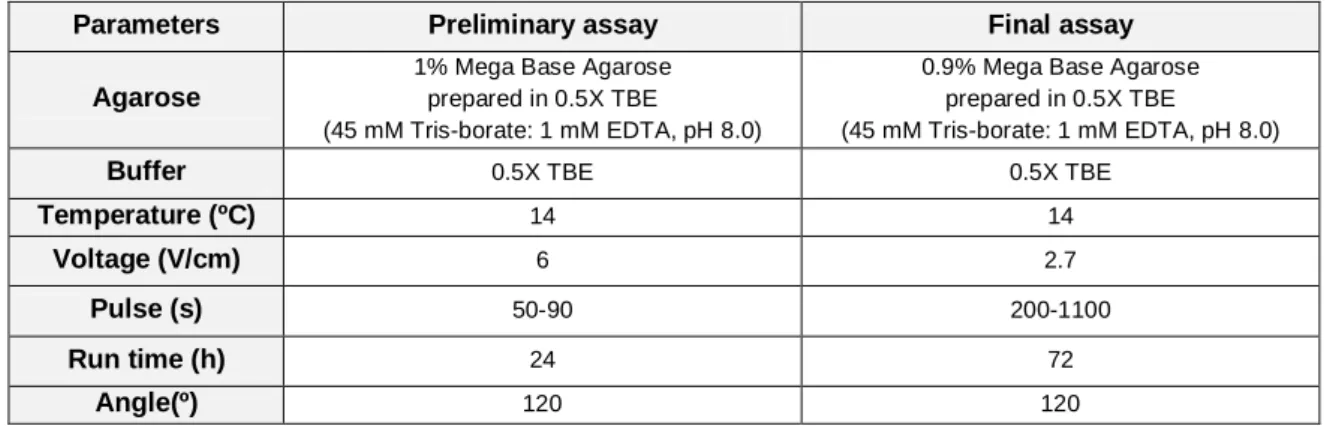
2.7. Karyotyping assays
With the aim of characterizing the chromosomal profile of strains representative of each of the X. dendrorhous lineages, an optimized protocol for PFGE-CHEF based on previous works was developed (Nagy et al. 1994, Adrio et al. 1995, Santopietro et al. 1995). Briefly, a pre-inoculum of each strain was prepared in a complete medium [Yeast extract, 0.3% (w/v); peptone, 0.5% (w/v); malt extract, 0.3% (w/v); glucose, 1.0% (w/v)], and incubated at 20ºC and 150 rpm for 24 h. A portion of that culture was then used to inoculate (DO600 nm= 0.2) 100
ml of new liquid culture medium. After 24 h, cells were collected by centrifugation, washed with sterile water and quantified on the hemocytometer. A pellet containing 4x10⁸ cells was
9 resuspended in 8 mL of a pre-treatment solution (0.1 M β-mercaptoethanol, 0.01 M EDTA, 0.1 M Tris-HCl, pH 7.4) and incubated for 30 minutes at 25°C, followed by a washing step and gentle centrifugation. The pellet was subsequently resuspended in an enzyme solution containing 10 mg/ml of lysing enzymes from Trichoderma harzianum from Sigma prepared with 1.2 M sorbitol in sodium citrate buffer. Incubation was done in a 50 ml Erlenmeyer at 25°C and 30 rpm. Because the incubation time is strain-specific, periodic microscopic observations were done and the cells were only collected when at least 90% had formed protoplasts. The protoplasts were washed with the osmotic stabilizer solution (1.2 M sorbitol) and then added to 1 ml of low melting point agarose [1.3% (w/v)] also prepared with the osmotic stabilizer solution. Using Bio-Rad plug molds, protoplast plugs are prepared and subsequently treated with Proteinase K enzyme solution (1 mg/ml Proteinase K from Sigma; 0.01 M Tris-HCl; 1% SDS; 0.5 M EDTA) for 24 h at 50°C. The plugs were then washed with 0.05 M EDTA and stored at 4ºC until used. For strains that did not form protoplasts conveniently an extra step was introduced, consisting in the addition of MnCl2 to the liquid
culture medium 12 h after inoculation (to a final concentration of 4.0 mM), which resulted in a weakening of the cell wall (Pera et al. 1999). The preparation of protoplasts in sufficient quantity to obtain distinguishable bands without degraded genetic material represents one limiting step of this technique. Saccharomyces cerevisiae CBS 8803 was used as a standard for the PFGE-CHEF assays, and its plugs were prepared with the same protocol but the incubation time with the enzyme solution was only 20 minutes. Two different sets of conditions were used for the PFGE-CHEF assays. First a shorter preliminary assay, in order to access the absence/presence of degraded genetic material. If good quality plugs were obtained, another PFGE run would be done under adequate conditions for the separation of the X. dendrorhous chromosomes (Table 2). All PFGE-CHEF assays were performed in a CHEF-DR III system from Bio-Rad.
Table 2 – Summary of the PFGE running parameters used.
Parameters Preliminary assay Final assay Agarose
1% Mega Base Agarose prepared in 0.5X TBE
(45 mM Tris-borate: 1 mM EDTA, pH 8.0)
0.9% Mega Base Agarose prepared in 0.5X TBE
(45 mM Tris-borate: 1 mM EDTA, pH 8.0)
Buffer 0.5X TBE 0.5X TBE
Temperature (ºC) 14 14
Voltage (V/cm) 6 2.7
Pulse (s) 50-90 200-1100
Run time (h) 24 72
10 The gel was stained with an ethidium bromide solution (0.5 µg/ml) for 30 min and then unstained for 30 min in sterile water. Images of the gel were collected with a GelDoc (Bio-Rad) image acquisition system and analyzed in Gelcompar v4.1 from Applied Maths.
11
3. Results and discussion
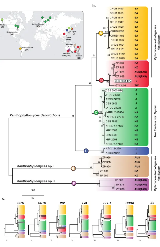
3.1. Xanthophyllomyces multilocus phylogeny.
With the aim of assessing the phylogenetic relationships between the different Xanthophyllomyces lineages a MLSA approach was devised using seven different genomic regions encoding proteins involved in several metabolic processes (Table S2). The biotechnological interest in the production of astaxanthin has driven the sequencing and characterization of the genes associated with its biosynthesis. Three of those genes were chosen for MLSA due to the fact that each of them had been sequenced for more than one strain, enabling the selection of regions comprising a larger number of single nucleotide polymorphisms (SNP). Unfortunately for genes not related to astaxanthin biosynthesis only one sequence was usually available therefore, the four other genes were chosen for the multi-locus scheme based on the information of one single sequence each. The expanded collection of isolates studied made it necessary to construct several sets of primers for four of the seven genes (Table S2). Previously, it was determined that CRTI and CRTS genes are not in the same chromosome (Niklitschek et al. 2008) but regarding the rest of the 5 genes used for the MLSA no such information is available. However the genes chosen codify proteins involved in very distinct roles in the yeast metabolism, lowering the probability of being genetically linked. Moreover, unpublished data regarding the genome draft of X. dendrorhous type strain (CBS 7918T) indicated that all seven genes are present in separate contigs, being the smallest ~3.0 kb long (personal communication by Dr. Diego Libkind). Although this does not prove that they are genetically unlinked, it points in favor of this hypothesis.
Each of the seven genes was amplified for the collection of 40 strains and the resulting alignments were used to construct MP (Figure 4c) and ML (Figure S1) based phylogenies. The individual phylogenies yielded similar results in both methods and were found to be generally concordant. The multilocus alignment of the seven gene fragments was used to construct a phylogeny using MP (Figure 4b) and ML (Figure S2). The concatenated dataset increased the resolution of the analysis in comparison to the individual ones. On every individual phylogeny a total of six major clades were identified whatever the inference method used and the same number of clades was resolved in the multilocus phylogeny (Figure 4b). Before this study, a phylogenetic analysis using the ITS region resolved Xanthophyllomyces into 3 major clades/lineages (Libkind et al. 2007) that in the multilocus phylogeny (Figure 4b) would partially correspond to clade A, C and D. The phylogeny also had a more divergent branch corresponding to strain CBS 5905 that was phylogenetically isolated from the other lineages (Libkind et al. 2007).
12
13
Figure 4 - Xanthophyllomyces multilocus phylogeny and distribution. (a) World map pinpointing the
geographical origins of the strains belonging to each of the main phylogenetic clades presented in the multilocus phylogeny. Colored pins correspond to the phylogenetic clade to which the isolate belongs. (b) MP multilocus phylogeny identifying six major clades shown in distinct colors and represented with the letters A - F. Next to each strain the geographical origin is indicated and also the host system from which it was isolated. Bootstraps values from 1000 replicates are shown next to the branches. There were a total of 3187 positions in the final dataset (1287 informative positions). The scale is in the units of the number of changes over the whole sequence. Strain CBS 5905 presents two different haplotypes (HA and HB) that are highlighted by two white rounded boxes. (c) Individual MP phylogenies of the seven genes used in MLSA are depicted. Clades are presented in the same colors has shown above. Each phylogeny is identified by the abbreviated gene names. Full information regarding the genes is present in Table S2.
In the multilocus phylogeny the clades A, C and D include not only the strains used in the study by Libkind et al. (Libkind et al. 2007), but also additional isolates comprising a more diverse sampling both in geography and host trees. From the six resolved clades, three represent novel Xanthophyllomyces clades (B, E and F), comprising strains from Australasia and also including one strain (GY13L04) from Taiwan. Within the cluster formed by clades A, B, C and D small differences in the topology exist between the individual gene phylogenies, coherent with what is expected under the Phylogenetic Species Recognition model proposed by Taylor and co-authors (Taylor et al. 2000). In this case, the species cluster of X. dendrorhous encompasses four different lineages (Figure 4b) where clade B represents a novel clade. Inside each of the four clades of X. dendrorhous, subgroups of strains are also resolved and well supported by high bootstrap values. For example, in clade A two subgroups exist and in clade C, whereas all the strains from Japan group together with the strain from Alaska, the rest of the strains cluster separately (strains from NA and NE) (Figure 4a and b).The clades E and F always appear in a most divergent position in relation to the other 4 clades. The fact that the concatenated phylogeny, which has a greater resolution power than any of the individual ones, is not able to present one of the referred clades as being the most divergent is probably related to the fact that an appropriate outgroup does not exist for this multilocus phylogeny.
Regarding strain CBS 5905 it was found that three of the seven gene fragments sequenced were heterozygotic (Figure 4c; genes CRTS, EPH1 and IDI). The haplotypes were separated by cloning and then sequenced. It was found that each heterozygotic gene presented two different haplotypes that always grouped in clades B and C in the respective gene phylogeny. The other 4 genes were found to be homozygotic for strain CBS 5905 and presented haplotypes that grouped either in clade B (INV gene) or in clade C (CRTI, L41 and GDHA genes) (Figure 4c), which further confirms the results obtained for the heterozygotic genes. These results seem to indicate that this strain is a result of a cross between members of two lineages represented by clades B and C, which originate respectively from
14 Australasia/Taiwan and Japan. This suggests that a contact area between lineages B and C might have existed allowing gene flow between them.
Concerning the host system, it is possible to observe that the strains from the Cyttaria - Nothofagus host system are dispersed along the phylogeny in opposition with the isolates collected from tree exudates that are confined to clades C and D (Figure 4b).
3.2. Xanthophyllomyces novel species.
The recently found Australasian populations of Xanthophyllomyces encompass the largest genetic diversity ever observed in this genus. The Australasian genotypes are divided into three distinct lineages, one (clade B) somewhat similar to X. dendrorhous found in Southern America and two others (clades E and F) that are distinct from any other population ever reported (Figure 4b). Although the isolates from the latter lineages present the same life cycle as X. dendrorhous (Figure S3) and also produce astaxanthin (personal communication by Dr. Diego Libkind), they have a considerable genetic divergence in comparison to all known populations of X. dendrorhous (Figure 4b and Figure 5). The relevant divergence existing between clade E and F and the remaining clades suggests that these novel Australasian clades might in fact represent two new distinct species within the genus Xanthophyllomyces (Figure 4b). The separation of Xanthophyllomyces into three species is not only supported by the multilocus phylogeny but also by the ITS phylogeny (Figure 5). The ITS phylogeny presented in Figure 5 includes the genera Xanthophyllomyces and Cystofilobasidium and allowed comparing the genetic divergence of the different species within both genera. The degree of genetic divergence observed in the ITS sequences within X. dendrorhous species is somewhat similar to that observed within Cy. capitatum or Cy. macerans (Figure 5). However, the number of different genotypes included in X. dendrorhous appears to be higher than that found in each Cystofilobasidium species, which seem to be much more homogenous. The two putative novel species of Xanthophyllomyces also exhibit genetic diversity. Within clades, E and F, two different lineages appear to exist both in ITS (Figure 5) and in the multilocus phylogeny (Figure 4b). The study of more strains from these two new species might elucidate the biogeographic reasons that are accountable for this apparent lineage differentiation within each species. On clade E, strains ZP 938 and ZP 928 were collected from leafs of N. mooreii in Australia, while strains ZP 909 and ZP 884 were collected from fruiting bodies of C. nigra on N. menziesii in New Zealand (Table 1). Therefore geography and host are different and might justify the observed genetic diversity of Xanthophyllomyces sp. I.
15
Figure 5 - ITS phylogeny of Xanthophyllomyces and Cystofilobasidium. The phylogeny depicts the
relationships of the two new species of Xanthophyllomyces with X. dendrorhous and Cystofilobasidium species. A total of 45 sequences of representative strains of different species from both genera Xanthophyllomyces and
Cystofilobasidium were included. The phylogeny was constructed using the MP method and the bootstrap values,
from 1000 replicates, are presented next to the branches. All positions containing gaps and missing data were eliminated. There were a total of 369 positions in the final dataset. Strains that have ITS sequences already available in GenBank have the corresponding access number in front of the strain name. Clades E and F are identified in the phylogeny and correspond to the homonymous clades in the multilocus phylogeny. Filobasidiella
16 On the other hand in clade F all Xanthophyllomyces sp. II isolates share the same geographical origin and host system (C. gunnii on N. cunninghamii of Tasmania) but present clearly distinguishable genotypes. Although studied strains belonging to this new species are few, it seems that the factors influencing their population structure may differ from those influencing the population structure of X. dendrorhous. While X. dendrorhous has a vast geographic distribution (Figure 4c, orange, green, red and blue pins), these new putative species appear to be endemic to Australasia. Whereas X. dendrorhous apparently adapted to different host trees as new geographic areas where colonized and therefore formed different lineages, the Australasian species remained in the same host (clade E and F) and in the same geographical area (Clade F). Both, sympatric speciation or a phenomenon of secondary contact may be related to the formation or present existence of the different species encountered in Tasmanian samples (X. dendrorhous and Xanthophyllomyces sp. II). A more extensive study of isolates of the new species and of the Australasian habitats is needed in order to draw further insights on their biogeography and evolutionary history.
3.3. Exploring the population structure of X. dendrorhous.
The multilocus sequence analysis provided new information regarding the phylogenetic relationships of the different lineages that had previously been identified in X. dendrorhous by sequencing the ITS region. In light of the data provided by the MLSA, X. dendrorhous seems to be composed by four different lineages represented in the multilocus phylogeny by four distinct clades (A, B, C and D in Figure 4b). The assessment of evolutionary relationships using the phylogenetic tree model is done under the assumption that the evolution process is dominated by two types of events, mutation and speciation (Huson and Scornavacca 2011). Under more complex models of evolution that might involve events as complex as horizontal gene transfer, hybridization and recombination, phylogenetic trees may not provide an adequate representation of the phylogenetic history of the organism. Phylogenetic networks comprise more realistic models which are able to clearly represent events such as recombination (Huson 1998, Huson and Bryant 2006). The utilization of phylogenetic networks also allows determining if the dataset under analysis has a strong phylogenetic signal or not. In order to assess the strength of the phylogenetic signal (Figure 6) and if reticulate events as recombination played a relevant role in the evolution of this species (Figure S4), phylogenetic networks were constructed using the multilocus dataset. Two types of networks were tested: (i) Splits Networks (SN), which constitutes a combinatorial generalization of phylogenetic trees therefore enabling the visualization of incompatibilities within and between datasets (Huson and Scornavacca 2011) and (ii) Reticulate networks (RN) that provide a explicit representation of the evolutionary history in
17 the presence of reticulate events such as recombination (Huson and Bryant 2006, Huson and Scornavacca 2011).
Figure 6 - Splits network of Xanthophyllomyces. (a) Neighbor-net network constructed with the multilocus
dataset, depicting all Xanthophyllomyces strains except CBS 5905 (39 strains). The main edges of the network correspond to the clade of the multilocus phylogeny of Figure 4b. (b) Neighbor joining tree constructed with the multilocus dataset, depicting all Xanthophyllomyces strains except CBS 5905 (39 strains). (c) Phi-test results between and within populations. Analysis was performed using the multilocus dataset with all X. dendrorhous strains (d) Neighbor-net network depicting all 40 strains of Xanthophyllomyces.
In order to determine if the phylogenetic signals obtained from the seven different genomic regions were strong and coherent, individual alignments of each genetic marker were introduce and concatenated in SplitsTree4 to build Neighbor-net networks (a category of SN) that implicitly represents such differences. Three different multilocus sub datasets were
18 tested in network analysis: (i) one comprising all Xanthophyllomyces species (40 strains) (Figure 6d); (ii) another comprising all Xanthophyllomyces strains except CBS 5905 (39 strains) (Figure 6a); (iii) and finally one compose only by the strains belonging to X. dendrorhous but without CBS 5905 (31 strains) (Figure S4). The Neighbor-net network on Figure 6a presents a group of incompatible splits regarding the relation of clade D with the other 3 clades that compose X. dendrorhous. This, and the small topological differences observed when comparing the individual phylogenetic trees (Figures 4c and 5) suggests more clearly that the limit of the species X. dendrorhous lies in the transition from concordance among branches to incongruity among branches observed within clades A to D (Taylor et al. 2000). On Figure 6d the network differs slightly from the tree-like form (Figure 6b) because it includes the two haplotypes of the CBS 5905 heterozygotic genes. The network clearly shows that alleles in CBS 5905 most likely derived from representatives of clade B and from a subgroup of Japanese strains in clade C. The remaining strains present a position in the network similar to that in the phylogenetic tree. A RN was constructed for the X. dendrorhous multilocus dataset but did not yield any additional information regarding the occurrence of reticulate events since the obtained topology overlapped that of the SN (Figure S4). Because all SN have generally a tree-like form (Figure 6a, 6b and 6d), it is possible to assess that reticulate events were not dominant in the evolution of X. dendrorhous and also that the phylogenetic signal in the multilocus dataset is strong (Wagele and Mayer 2007).
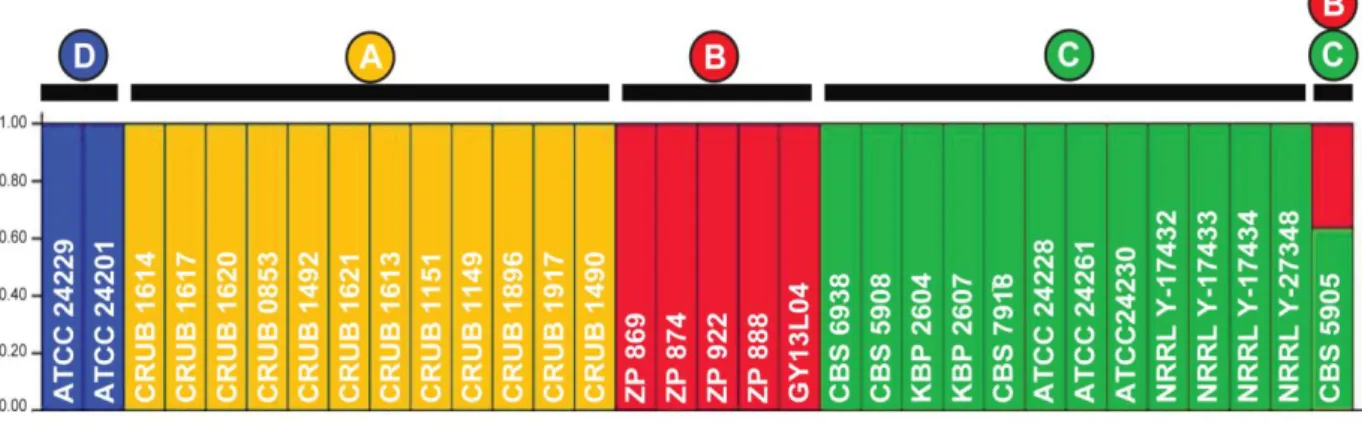
The pairwise homoplasy index (Phi) test (Bruen et al. 2006) was applied in order to determine if signs of recombination within and between the four distinct populations in X. dendrorhous could be detected. This test detects refined incompatibility in sequence data (Bruen et al. 2006) assuming the infinite sites model of evolution (Nei 1973) in which the detection of incompatibility for a pair of sites indicates recombination. The test was performed in a two by two comparison as represented in Figure 6c using the multilocus dataset with all the strains from X. dendrorhous. Since at least three sequences are necessary to perform the test, it was not possible to perform Phi-test within clade D. Recombination was detected only between clades B and C. When the same test was performed using the multilocus dataset composed by all X. dendrorhous strains with the exception of CBS 5905 strain, no statistically significant values were retrieved from the analysis, therefore no recombination was detected. Recombination was already demonstrated in the laboratory between strains CBS 6932 and CBS 5908 (both belonging to clade C) by crossing auxotrophic mutants and analyzing of the progeny (Kucsera et al. 1998). However no recombination was detected by Phi-test within that clade. This might imply that although the strains are able to cross and undergo meiosis in the laboratory, in natural populations of X. dendrorhous inbreeding appears to be dominant. The association to
19 very specific ecological niches and the peculiar mating process between parental cell and bud might also be contributing to the well structured populations detected in X. dendrorhous. The analysis of the multilocus alignment of X. dendrorhous was also performed using the Structure software (Figure 7). Given a number of populations (K) and assuming Hardy– Weinberg and linkage equilibrium within populations, Structure estimates allele frequencies in each cluster and population memberships for every individual strain. It is able to detect the underlying genetic population structure among a set of individuals genotyped at multiple markers and also of computing the proportion of the genetic information of an individual originating from each inferred population (quantitative clustering method). Therefore, in order to determine the ancestry of the X. dendrorhous strains the respective multilocus dataset was tested (Figure 7). The graphic output of Structure clearly shows the existence of four distinct populations that coincide with the clades defined by the phylogenetic analysis of the same dataset (Figure 7). Strain CBS 5905 shows a mixed ancestry with more than half belonging to the population identified in green and corresponding to clade C.
Figure 7 - Ancestry of X. dendrorhous estimated with Structure software. Each column represents a different
strain and the different colors represent distinct populations inferred by Structure. The vertical axis represents the amount of ancestry that each strains has from each of the inferred populations. The phylogenetic clades where each strain appears in the phylogenetic tree are identified in the upper part. The graph corresponds to the highest probability run for the number of populations inferred by Structure.
The complex ancestry of CBS 5905 detected in Structure is congruent with the two disjoint haplotypes depicted in the phylogenetic tree (Figure 4) in clade B (haplotype HA) and in clade C (haplotype HB) and on the splits network (Figure 6d). Therefore this confirms that this strain is indeed the result of a cross between members of populations originating from Australasia and Japan.
20
3.4. X. dendrorhous karyotype.
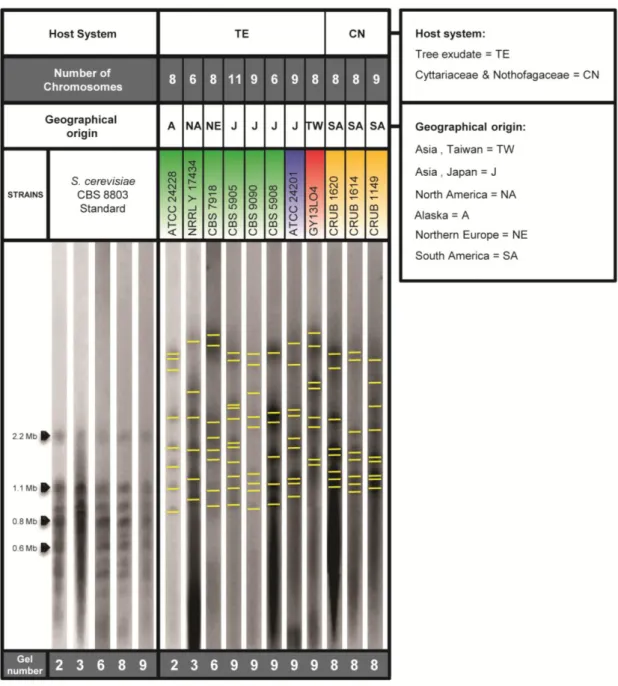
The use of Pulsed-field gel electrophoresis (PFGE) coupled with contour-clamped homogeneous electric fields (CHEF) apparatus allows the determination of karyotypes as well as chromosome-length polymorphisms (CLP), thus facilitating evolutionary and population studies (Lin et al. 2007). Aiming to elucidate the chromosomal composition of strains from different lineages of X. dendrorhous and to assess if the population structure observed by phylogenetic methods had a correlation with the chromosomal profile of the strains, the PFGE-CHEF technique was employed. Approximately 25 strains were tested by this technique although only 11 yielded satisfactory results due to the fact that each strain presented a unique response to the experimental conditions used (Figure 8). The bands observed in the PFGE gels are considered to represent chromosomes. In this study all the chromosomes were identified by visual scrutiny of the gels and by the analysis of the correspondent intensity graphics. On Figure 8 the image corresponding to the lane of each strain is presented with indication of the observed bands. All the strains were tested in duplicate and the images presented in Figure 8 were all normalized with GelCompar software using the S. cerevisiae standards as reference for the normalization. Observing the first column of Figure 8, were the S. cerevisiae standards are represented, it is possible to see that there exists reproducibility among all performed assays (referred to as gel number in Figure 8). Observing the PFGE results obtained for the X. dendrorhous strains it can be observed that no two strains present the same chromosomal profile. Nevertheless some chromosomal bands of similar size were observed in strains CRUB 1620, CRUB 1614 and CRUB 1149 (Figure 8). It is possible that some of the differences observed in the chromosome number (CN) of some strains might be due to the limited ability that PFGE technique has for the resolution of co-migrating chromosomes with equal size, a fact that may lead to the underestimation of CN. The obtained karyotypes are in the size range of 0.8 Mb to 3.5 Mb, which corroborates the results from previous studies [i.e. chromosomes sizes ranging from 0.83 Mb to 3.1 Mb (Nagy et al. 1994, Adrio et al. 1995, Cifuentes et al. 1997, Nagy et al. 1997)]. Furthermore, in these studies the CN of X. dendrorhous was determined to vary from seven to 17 but, because the different studies used different experimental conditions and different assumptions regarding the possibility of co-migration of the bands, no direct comparison of the CN is possible (Nagy et al. 1994, Adrio et al. 1995, Cifuentes et al. 1997, Nagy et al. 1997). In the current study, CN varies from six to 11, but most of the strains have between six to nine chromosomal bands, being CBS 5905 the only strain with 11 chromosomes.
21
Figure 8 - X. dendrorhous karyotypes. Chromosomal patterns of eleven strains and information regarding host
system, geographic localization, and CN are also presented. The color associated to the strain number identifies the population (A, B, C or D) to which each strain was assigned.
Among the group of 11 strains analyzed by PFGE, a strain that is a copy of CBS 5905 (named CBS 9090) was included. It is presently known that these two strains have differences in the ITS sequence (Figure 5). It is assumed that the repeated transfers over decades, a procedure commonly used in culture collections, might have influenced the accumulation of these differences (Libkind et al. 2007). The chromosomal pattern of these two strains was also observed to be different in both the number of chromosomes and in their sizes (Figure 8). From the preliminary results presented in Figure 8, there seems to be
22 no direct relation between the chromosomal profiles and the population structure of X. dendrorhous.
Although X. dendrorhous karyotypes show a considerable variability in chromosomal patterns, this species does not represent a unique situation among Fungi, since many other species present CLP olan P re - rt n et al. 2002, Giraud et al. 2008).The existence of intraspecific CLP can arise mainly by rearrangements of linked groups of genes or gains and losses of non coding sequences (Zolan 1995, Poláková et al. 2009). It was shown for the basidiomycetous Coprinopsis cinerea (Coprinus cinereus) that two strains differing in the length of most of their chromosomes are interfertile, and that new CLP appear after meiosis (Zolan 1995). This means that the progeny of such cross presents a variety of karyotypes, indicating that the changes in length observed in the chromosomes represent gains and losses of nonessential sequences (Zolan 1995). A similar situation has been observed when crossing strains of X. dendrorhous. The progeny showed not only composed karyotypes from both parental strains, but also novel CLP what could indicate that the genomic regions altered in the karyotypes of the progeny might not be essential (Kucsera et al. 1998). Nevertheless in order to assess the importance of the CLP observed in X. dendrorhous, an understanding of how the genome is arranged is necessary (Zolan 1995).
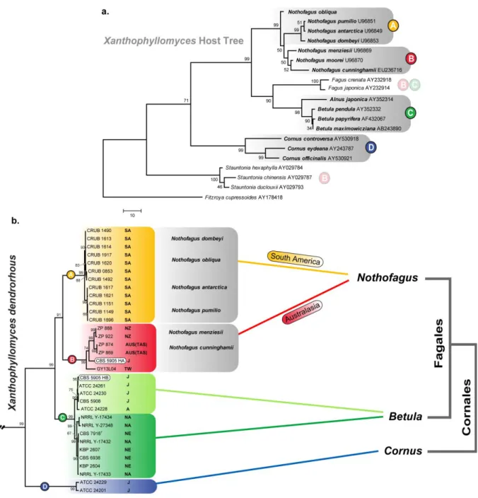
3.5. X. dendrorhous population structure, host system and geographical
distribution.
In the present study X. dendrorhous was found to have a well defined population structure with four distinct populations. Considering both host and geographic origin, it seems that the populations that colonize the same host tree are more similar among themselves than those sharing the same geographical origin but a different host tree (Figure 4). The ITS phylogenies of yeast and host trees were compared by Libkind et al. in 2007 and a correlation between both was proposed. In light of the broader sampling of Xanthophyllomyces isolates presently available, this analysis was updated with greater detail. In Figure 9a the ITS phylogeny of the host trees shows that all the strains isolated from Nothofagus are present either in clade A or clade B of the multilocus phylogeny (Figure 9b). Moreover, strains isolated from Betula grouped in clade C and those isolated from Cornus grouped in clade D (of the multilocus phylogeny) (Figure 9). Two strains, CBS 5905 and GY13L04, present a unique situation regarding the yeast-host association. Strain CBS 5905 was isolated from an exudate of Fagus crenata and due to its mixed ancestry presents two different haplotypes, one in clade B and another in clade C of the multilocus phylogeny (Figure 9b). Strain GY13L04 was collected in Taiwan from Stauntonia purpurea (Figure 9a). Unfortunately the lack of more isolates from the same host tree or same geographic location
![Figure 1 – Xanthophyllomyces dendrorhous sexual state. (a) Scanning electron microscopy photos showing two conjugated cells [1] originating a non-septate basidium [2]; (b) aerial basidium [2] with apical basidiospores [3]; (c) detail of the basidium](https://thumb-eu.123doks.com/thumbv2/123dok_br/18272141.880748/15.892.114.787.107.308/xanthophyllomyces-dendrorhous-scanning-electron-microscopy-conjugated-originating-basidiospores.webp)