Universidade de Lisboa
Faculdade de Farmácia
Biophysical Characterization of Sarcosine Oxidase - Effect of
Physical and Chemical Parameters on Sarcosine Oxidase Activity
Cátia Sofia Santos Damião
Mestrado Integrado em Ciências Farmacêuticas
Universidade de Lisboa
Faculdade de Farmácia
Biophysical Characterization of Sarcosine Oxidase - Effect of
Physical and Chemical Parameters on Sarcosine Oxidase Activity
Cátia Sofia Santos Damião
Monografia de Mestrado Integrado em Ciências Farmacêuticas
apresentada à Universidade de Lisboa através da Faculdade de Farmácia
Orientador: Professor Doutor René Kizek
Co-Orientadora: Doutora Ana Paula Costa dos Santos Peralta Leandro,
Professora Auxiliar
The laboratory work was developed in:
Prevention Medicals Laboratory with collaboration of Veterinární
a Farmaceutická Univerzita Brno
Brno, Czeck Republic
Advisor: Professor Doutor René Kizek
ABSTRACT
Flavoproteins remain key players in enzymology and biochemistry corresponding approximately to 1% of all documented proteins. The members of this enzyme family are involved in several biological processes from cell DNA repair to drug and xenobiotic metabolism. Among the several flavoenzymes identified to date, sarcosine oxidase (SOX) is a two domain monomeric oxidoreductase protein of 43 kDa with many clinical applications. Monomeric sarcosine oxidase (MSOX) can be used as a diagnostic tool to evaluate the renal function since it has the capacity, when in combination with cretinase and creatininase, for assaying creatinine levels in human serum. Apart this, SOX has the potential to be used for the measurement of other analytes, such as sarcosine, by employing an enzyme-coupled colorimetric assay.
The main aim of this work was to characterize some biochemical and physical properties of MSOX, namely its optimum catalytic conditions, in order to improve its redox activity. For this purpose, a coupled colorimetric method was utilized based on hydrogen peroxide (H2O2) formation via SOX. The produced H2O2 reacts further with 4-aminoantipyrine (4-AAP) in the presence of horseradish peroxidase (HRP) to produce a violet product, quinoneimine, which can be measured spectrophotometrically.
Apart from the spectral properties, the influence of reactional temperature and the presence of urea upon the catalysis of the enzyme and the kinetics parameters of the MSOX were investigated. The effect of pH on the electrophoretic mobility of the native enzyme was also studied. The obtained data showed a good tolerance of the enzyme at pH 8.0 to high temperatures which infers a potential application in various biotechnological industries. The experimental results also showed no significant interference from urea.
This study provides a rapid and simple tool to determine sarcosine levels which can be useful for prostate cancer diagnosis. Therefore, this method is promising to distinguish prostate cancer patients from healthy or benign prostatic hyperplasia subjects according to urinary sarcosine level.
Key words: sarcosine oxidase, enzyme-coupled colorimetric assay, sarcosine, temperature, prostate cancer
RESUMO
As flavoproteínas integram um grupo de proteínas fundamentais nas áreas da bioquímica e enzimologia correspondendo aproximadamente a 1% de todas as proteínas documentadas. Esta família de enzimas participa em diversos processos biológicos desde a reparação do DNA celular ao metabolismo de fármacos. De entre as inúmeras flavoenzimas já identificadas, destaca-se a sarcosina oxidase (SOX), uma oxidoredutase monomérica de 43 kDa com diversas aplicações clínicas. A SOX é considerada um agente de diagnóstico fundamental para a determinação dos níveis de creatinina possibilitando a avaliação da função renal quando utilizada em combinação com a creatinase e creatininase. Para além disso, a SOX pode ser potencialmente utilizada para determinar outras moléculas como por exemplo a sarcosina, sendo a sua quantificação possível graças a um método colorimétrico enzimático acoplado.
Neste trabalho pretendeu-se obter uma melhor compreensão das propriedades físicas e químicas desta flavoproteína de forma a otimizar o seu desempenho na reação catalítica. A caracterização enzimática da SOX foi realizada através de uma reação específica baseada na formação de peróxido de hidrogénio, a partir de sarcosina, reação mediada por esta enzima. O peróxido de hidrogénio resultante reage por sua vez com a 4-aminoantipirina na presença de fenol e de peroxidase originando um produto final de cor violeta, a quinoneimina, que pode ser detetada espectrofotometricamente.
Para além das propriedades espectrais, a influência da temperatura reacional sobre a reação catalítica assim como os parâmetros cinéticos foram investigados. Foi também estudado o efeito do pH e tempo de reação na atividade da enzima. Os resultados mostraram uma boa tolerância da enzima, a pH 8.0, a temperaturas elevadas refletindo uma potencial aplicação em indústrias biotecnológicas. Os ensaios efetuados também demonstraram que a ureia não apresenta uma interferência significativa na atividade da SOX.
Este trabalho abre perspetivas para a quantificação da sarcosina através da utilização da SOX, sendo útil para o diagnóstico do cancro da próstata. Deste modo, a técnica proposta poderá ser uma ferramenta promissora para diferenciar doentes com cancro da próstata de indivíduos saudáveis ou mesmo de doentes com hiperplasia benigna da próstata de acordo com os níveis urinários de sarcosina.
Palavras-chave: Sarcosina oxidase, ensaio colorimétrico enzimático acoplado, sarcosina, temperatura, cancro da próstata
ACKNOWLEDGMENTS
I would like to thank the Erasmus+ programme, particularly to Dr. René Kizek for the opportunity to develop this project abroad and for his continued support. Also, I would like to express my sincere thanks the laboratory members, particularly, Dagmar Uhlirova and Zuzana Tothova for their helpful advice.
I owe my deep gratitude to Dr. Ana Paula Leandro for her availability and for giving me the support and guidance during the course regarding many concepts on enzymology and biochemistry.
To my family, particularly Sofia Damião, Paulo Damião and Beatriz Damião I wish to express my sincere gratitude for all the encouragement and counselling along this journey. It is because of them this work became possible.
I want to thank all my friends from the faculty that somehow became this journey easier, particularly, Ana Nunes, a friend with whom I shared unforgettable moments. Also, I want to thank Inês, Carolina, Andreia, Patricia, Catarina, Marta and Diana for their friendship and support during these last 5 years.
To my friends from the primary school, particularly, Mariana, Rita, Madalena, Vasco, Miguel and Bernardo I want to express my thanks for all the support and motivation along this journey.
ACRONYMS
4-AAP 4- Aminoantipyrine
5, 10-CH2-H4-folate 5,10-methylenetetrahydrofolate
AgNO3 Silver nitrate
AgNPs Silver nanoparticles AMP Adenosine monophosphate APS Ammonium persulfate BPH Benign prostate hyperplasia CPG N-cyclopropylglycine
DMGDH Dimethylglycine dehydrogenase DRE Digital rectal examination
EDAC N-ethyl-N- (3-dimethylaminopropyl) carbodiimide ETF Electron-transferring flavoprotein
FAD Flavin adenine dinucleotide FAOD Fructosyl amino acid oxidase FMN Flavin mononucleotide
GC-MS Gas chromatography coupled with mass spectroscopy GCE Glassy carbon electrode
GMC Glucose-methanol-choline oxidoreductase GNMT Glycine-N-methyltransferase
H2O2 Hydrogen peroxide
HEPES 4-(2-hydroxyethyl)-1-piperazineethanesulfonic acid HRP Horseradish peroxidase
IVD In vitro diagnostics
LC-MS Liquid chromatography coupled with mass spectroscopy MSOX Monomeric sarcosine oxidase
MTSOX N-methyltrytophan oxidase NAD Nicotinamide adenine dinucleotide NHS N-hydroxysuccinimide
NMDA N-methyl-D-aspartate PCA Pyrrole-2-carboxylate PCa Prostate cancer PIPOX Pipecolate oxidase PSA Prostate specific antigen RT Room temperature
SAR Sarcosine
SARDH Sarcosine dehydrogenase SET Single electron transfer SOX Sarcosine oxidase
TEMED Tetramethylethylenediamine TMAO trimethylamine N-oxide
TOPS Sodium N- Ethyl-N-(3-sulfopropyl)-m-toluidine TSOX Tetrameric sarcosine oxidase
INDEX
1. Introduction ... 14
1.1 Flavin-Dependent Enzymes ... 14
1.2 Sarcosine Oxidase... 17
1.2.1 Flavin Environment in MSOX ... 20
1.2.2 MSOX Active Site Ligands ... 22
1.2.3 A Brief Overview of MSOX Reductive Mechanisms ... 23
1.3 Clinical Applications of Sarcosine Oxidase and Role of Sarcosine as a Potential Marker of Early-Stage Prostate Cancer ... 25
1.4 Methods For Sarcosine Quantification Using Sarcosine Oxidase ... 28
2. Objectives ... 31
3. Materials and Methods ... 31
3.1 Materials ... 31
3.2 Methods ... 32
3.2.1 Analysis of Sarcosine Oxidase at Different pH using Native Polyacrylamide Gel Electrophoresis ... 32
3.2.2 Spectrophotometric Assay of Sarcosine Oxidase - UV-Visible Spectroscopy Measurements ... 32
3.2.3 Enzyme Coupled Colorimetric Assay – Kinetics and Thermal Properties ... 34
3.3 Interference Study - Effect of Urea on Sarcosine Oxidase Activity ... 35
4. Results and Discussion ... 36
4.1 Analysis of Sarcosine Oxidase at Different pH using Native Polyacrylamide Gel Electrophoresis ... 36
4.2 Spectrophotometric Assay of Sarcosine Oxidase - UV-Visible Spectroscopy Measurements ... 38
4.3 Enzyme Coupled Colorimetric Assay – Thermostability ... 40
4.3.1 Kinetics Properties of SOX and Catalytic Activity ... 45
4.4 Interference Study - Effect of Urea on Sarcosine Oxidase Activity ... 49
5. Conclusions and Future Perspectives ... 52
FIGURE INDEX
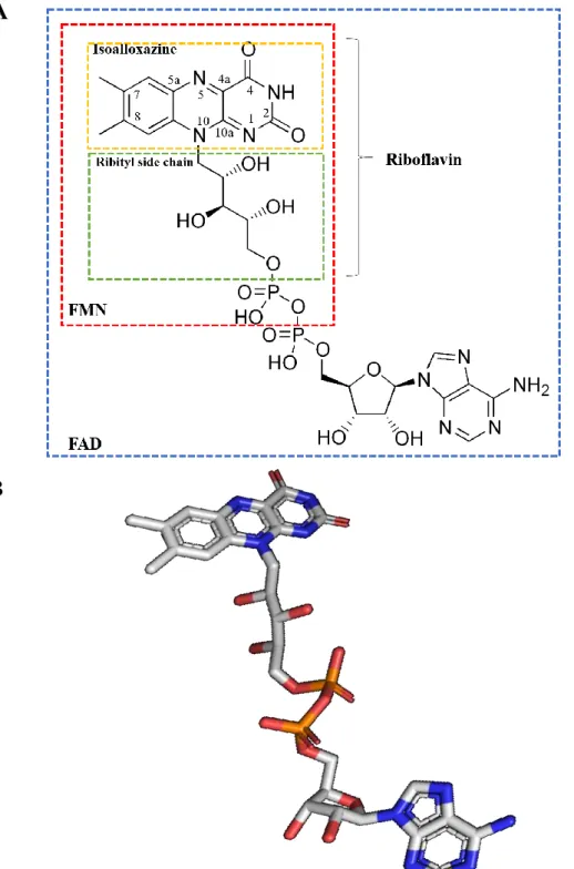
Figure 1. Flavin adenine dinucleotide (FAD). (A) Molecular structure of FAD based on reference (1). Chemical structure of the isoalloxazine ring and riboflavin are also shown. Its N5, C4a, C10a and N1 atoms are the reactive positions. (B) Stereoview of FAD. Carbon atoms are in white, oxygen atoms are in red, nitrogen atoms are in blue and the phosphorus atoms are in orange. The figure was generated using the program PYMOL. ... 16
Figure 2. Monomeric sarcosine oxidase (MSOX). (A) Stereoview of the structural model of covalently flavinylated amine oxidising enzyme (PDB code 2GBO). (B) Amino acid composition of MSOX. Glycine (Gly), glutamate (Glu) and leucine (Leu) are the most abundant amino acids in MSOX composition representing 9.5%, 8.5% and 8.2% respectively. Adapted from Expasy database. ... 17
Figure 3. Schematic representation of redox reaction with TSOX. (A) Oxidation of sarcosine by FAD. (B) Reaction with imine intermediate formed in A in the presence and absence of H4-folate resulting in different final products. ... 19
Figure 4. Schematic representation of the reaction catalysed by monomeric sarcosine oxidase (equation 1). Equation 2 shows de reaction catalysed by peroxidase using as substrate the hydrogen peroxide (H2O2) produced by SOX reaction. ... 20 Figure 5. Stereoview of MSOX (PDB code: 2GBO) showing the region above the si-face of the flavin ring. Adopted from (9). Residues Thr48 and Lys265 and five water molecules making up a proton relay system are also indicated. Carbons atoms are in white, oxygen atoms are in red, nitrogens atoms are in blue and the sulfur is yellow. Active sites waters are shown as balls. Hydrogen bonds are indicated by dotted lines. Water molecule 1 is hydrogen bonded to flavin N(5), the carbonyl oxygen of Thr48 and the amino group of Lys265. The other water molecules (2, 3 and 4) are accessible to bulk solvent (22). ... 22
Figure 6. Postulated mechanisms for the oxidation of sarcosine by MSOX. (A) Single electron transfer (SET) mechanism; (B) Hydride transfer mechanism; (C) Carbanion mechanism and (D) Polar mechanism (27). ... 24
Figure 7. Schematic representation of SOX-coupled assay to monitor the effect of temperature on SOX activity. (4-AAP) 4-aminoantipyrene; (HRP) horseradish peroxidase; (SAR) sarcosine; (SOX) sarcosine oxidase; (TOPS) sodium N- ethyl-N-(3-sulfopropyl)-m-toluidine. Assayed temperatures: 20, 25, 30, 35, 40 ºC. See text for details... 33
Figure 8. Schematic representation of SOX-coupled colorimetric assay to monitor the effect of substrate concentration on SOX activity previously incubated at different temperatures. (4-AAP) 4-aminoantipyrene; (HRP) horseradish peroxidase; (SAR) sarcosine; (SOX) sarcosine oxidase; (TOPS) sodium N- ethyl-N-(3-sulfopropyl)-m-toluidine. Assayed temperatures: 5, 15, 20, 30, 35, 40, 45, 50 and 60 ºC. See text for details. ... 35
Figure 9. Analysis of SOX electrophoresis in native conditions after silver staining (A) Native – PAGE of SOX pre-incubated for 2 hours at different pH at room temperature (25ºC) (B) Native – PAGE of SOX previously incubated for 2 hours at different pH at 37 ºC. (C) Electrophoretogram obtained from densitometric analysis of the gel in (A). (D) Electrophoretogram obtained from densitometric analysis of the gel in (B). Graphs (C) and (D) were done using the Colortest programme. Three main bands with high (H), intermediate (I) and low (L) molecular mass were observed and identified with black arrows. High molecular mass protein probably to aggregated forms. ... 37
Figure 10. (A) Spectral course of MSOX reduction with 125 μM sarcosine when incubation temperature is 20ºC, (B) 25ºC, (C) 30ºC (D) 35ºC and (E) 40ºC. (F) Absorption values at λmax= 546nm at different reactional temperatures (20, 25, 30, 35, 40ºC). Error bars represent standard deviation of three independent experiments. ... 39
Figure 11. Reductive reaction of SOX previously incubated at temperature 5ºC (A), 15ºC (B), 20ºC (C). Enzymatic reaction was followed for 60 minutes at room temperature (RT) using a range of sarcosine concentrations (15.7-216.7 µM) and monitored at 546nm. The six wells contain decreasing concentrations of sarcosine. The last well corresponds to the negative control (without sarcosine). ... 41
Figure 12. Reductive reaction of SOX previously incubated at temperature 30ºC (A), 35ºC (B), 40ºC (C). Enzymatic reaction was followed for 60 minutes at room temperature (RT)
using a range of sarcosine concentrations (15.7-216.7 µM) and monitored at 546 nm. The six wells contain decreasing concentrations of sarcosine. The last well corresponds to the negative control (without sarcosine). ... 42
Figure 13. Reductive reaction of SOX previously incubated at temperature 45ºC (A), 50ºC (B), 60ºC (C). Enzymatic reaction was followed for 60 minutes at room temperature (RT) using a range of sarcosine concentrations (15.7-216.7 µM) and monitored at 546 nm. The six wells contain decreasing concentrations of sarcosine. The last well corresponds to the negative control (without sarcosine). ... 43
Figure 14. Effect of incubation temperature of SOX (30, 35 and 50ºC) on the absorbance of the enzymatic charge transfer complex at various concentrations of sarcosine. Absorbance values correspond to reaction time 60 (min). Reactions were conducted under aerobic conditions at room temperature in buffered solution containing 0,025mg/mL SOX as detailed in section 3.2.3. Measurements were performed in triplicate and correspond to mean of three values with standard deviation. (For clarity, other measurements at additional temperatures are not shown). ... 45
Figure 15. Effect of pre-incubation temperature on SOX activity. Error bars are the result of standard deviation of three experiments. ... 48
Figure 16. Effect of urea on sarcosine oxidase activity. 60 minutes-long absorbance of all samples at 546 nm. ... 50
Figure 17. Effect of urea on sarcosine oxidase activity. Absorbance at 546 nm with increasing urea concentrations after 60 minutes of SOX reduction. Error bars represent standard deviation of three experiments. ... 51
TABLE INDEX:
Table 1. Spectral properties of SOX catalysis at different temperatures with 125μM of SAR after 60 minutes of reaction ... 40
Table 2. Catalytic parameters for the reduction of SOX with different sarcosine concentration at different temperatures. The kinetics values are represented with the corresponding standard deviation of three experiments. ... 47
14
1.
Introduction
1.1 Flavin-Dependent Enzymes
A high number of flavoenzymes have been identified and characterized. It is currently estimated that, on average, about 1–3% of the genes in bacterial and eukaryotic genomes encode flavin-binding proteins. There have been motivating developments in the field of flavoenzymes with the discovery of new proteins of this family that are unexpectedly connected with several diversified functions (1). In fact, flavin-dependent enzymes have arisen as pervasive catalysts in biological process such as energy production, nucleotide biosynthesis, biodegradation, cell signalling, chromatin remodelling, cell DNA repair, apoptosis, protein folding, drug and xenobiotic detoxification, immune defence and neural development. Consequently, flavoenzyme malfunctions can compromise these biological processes resulting in pathological conditions (2).
Flavoenzyme is a common term used to describe enzymes that contain the heterocyclic isoalloxazine chromophore (3) as shown in Figure 1. This ring system provides a powerful catalyst function in terms of redox reactions; this redox mediation arises from the flavin itself, in the form of flavin mononucleotide (FMN) or flavin adenine dinucleotide (FAD), the two most common active flavins acting as cofactors in enzymatic catalysis (3,4).
The versatility of these redox molecules is due to the active isoalloxazine ring, the catalytic core of the cofactors mentioned above, which allows to accept one or two electrons, protons, or form a covalent bond via a flavin adduct (5,6). Nevertheless, other cofactors such as pyridine nucleotides are limited to one of these transfer process shifting exclusively two-electron transfers (5). The flavocoenzymes can exist in three different states: oxidized, one-electron-reduced and two-one-electron-reduced which can consequently induce remarkable absorbance changes in the UV-visible spectral region (3,6).
The protein environment can modulate these different states (redox/ionic/electronic) of flavin, each one of them with different chemical properties. This modulation allows the protein-bound flavin to perform an extensive variety of enzymatic reactions (3). In other words, the nature of reaction performed by each flavoenzyme is determined eventually by the protein matrix and hence by the interactions between the flavin and the substrate (2).
15
The flavin-dependent enzymes family include, among others, the amino oxidases such as vanillyl alcohol oxidase (VAO), glucose-methanol-choline oxidoreductase (GMC) and sarcosine oxidase (SOX) which are composed of an FAD-binding domain, apart from the substrate-binding domain. As the name suggests, the majority of amino oxidases are merely active on amines. All members of this family have a similar domain in the N-terminal half of the protein responsible for FAD binding.
Although a few examples exists of amino oxidases not dependent of cofactor , it has been shown that most amino oxidases depend on a closely bound cofactor playing a vital role in the stability of the native structure and in enzyme activity. This tight connection is due to the fact that amino acids do not have the capacity to act as intermediates in redox reactions, thus needing redox cofactors to provide enzymes an oxidising power (7). Usually, the loss of natural cofactors causes misfolding and inactivation of the respective enzymes (8).
Typically, flavins are yellow chromophores generated from riboflavin (vitamin B2) and consist of tricyclic isoalloxazine moiety condensed with a ribityl chain at N10 as shown in Figure 1 (2). These flavin cofactors can be present as FAD resulting from the condensation of FMN and adenosine monophosphate (AMP) or, less often, as FMN consisting of riboflavin phosphorylated at the 5’-OH of the ribytil chain (1,6,7). These molecules can exist as quinone, semiquinone or hydroquinone depending on whether the cofactor is in oxidized, one electron-reduced or two-electron reduced state, respectively (6). Vitamin B2 can be synthetized de novo by plants, many bacteria and fungi, but not by animals, which must therefore obtain it from dietary sources (3).
Therefore, under most circumstances, the flavin can be thought of as part of the enzyme. In contrast to the usual noncovalent complexes, there are many cases where the flavin is covalently attached to the protein (1). The covalent linkage helps to modulate the properties of the cofactor as a redox mediator catalyst and may also play an important structural role in some enzymes. This covalent flavin attachment occurs at either the 8-α (methyl) or the 6-position of the isoalloxazine, and sometimes at both sites, through the sulphur of an active site cysteine residue. The same binding is also found in numerous other enzymes (e.g., monoamine oxidase). Covalent binding through a cysteinyl bond on the 8-α position of FAD is the most frequently observed, but the N-atom of the imidazole ring of histidine, or the phenolic oxygen of tyrosine may replace cysteine as the site of attachment to the enzyme (9).
16
A dual role has been proposed for covalent flavin attachment; on one hand it prevents loss of the weakly bound oxidized flavin and on the other hand it modulates the redox potential (1).
Figure 1. Flavin adenine dinucleotide (FAD). (A) Molecular structure of FAD based on reference (1). Chemical
structure of the isoalloxazine ring and riboflavin are also shown. Its N5, C4a, C10a and N1 atoms are the reactive positions. (B) Stereoview of FAD. Carbon atoms are in white, oxygen atoms are in red, nitrogen atoms are in blue and the phosphorus atoms are in orange. The figure was generated using the program PYMOL.
A
17 6,7 3,64,1 5,1 0,8 2,1 8,5 9,5 3,8 5,9 8,2 5,6 2,1 5,6 4,6 6,9 5,6 0,8 3,8 6,7 0 1 2 3 4 5 6 7 8 9 10 A R N D C Q E G H I L K M F P S T W Y V Q u a n tity (% ) Amino acid
1.2 Sarcosine Oxidase
In this work, the SOX from Bacillus sp., a monomeric stable, readily expressed 43 kDa (390 amino acids) two domain protein containing 1 mol of FAD, was used as a model for stabilizing enzymes (Figure 2) (10).
Figure 2. Monomeric sarcosine oxidase (MSOX). (A) Stereoview of the structural model of covalently
flavinylated amine oxidising enzyme (PDB code 2GBO). (B) Amino acid composition of MSOX. Glycine (Gly), glutamate (Glu) and leucine (Leu) are the most abundant amino acids in MSOX composition representing 9.5%, 8.5% and 8.2% respectively. Adapted from Expasy database.
Presently, several SOXs from different microorganisms have been studied. This enzyme can be produced by different bacteria including Bacillus, Corynebacterium, Streptomyces,
Arthrobacter and Pseudomonas (8). The bacterial SOX enzymes are induced in these
micoorganisms when grown on the presence of sarcosine, a common soil metabolite, which is used as the sole source of carbon and energy (11). SOX enzymes can be grouped into three subclasses according to their quaternary structure: monomers, heterodimers and heterotetramers (7). Generally, the most widely enzymes used in clinical assays are the ones with monomeric structure containing just covalently bound flavin (12).
A B
Amino acids symbols: Ala (A), Arg (R), Asn (N), Asp (D), Cys (C), Gln (Q), Glu (E), Gly (G), His (H), Ile (I), Leu (L), Lys (K), Met (M), Phe (F), Pro (P), Ser (S), Thr (T), Trp (W), Tyr (Y), Val (V)
18
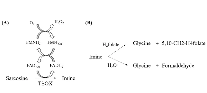
Among the three subclasses, heterotetrameric SOX (TSOX) is a complex, multimeric enzyme that catalyses both sarcosine oxidation and the synthesis of 5, 10-methylenetetrahydrofolate (5,10-CH2-H4-folate).
TSOX contains four different subunits (α, β, γ, δ) that range in molecular mass from about 10 to 100 kDa (103, 44, 21, 11 kDa, respectively). This enzyme contains three coenzymes: noncovalently bound FAD, nicotinamide adenine dinucleotide (NAD+) and covalently bound FMN. It also presents a binding site for the coenzyme, tetrahydrofolate (THF), which in this case acts as the substrate of the enzymatic reaction (13). Therefore, contrarily to monomeric SOX (MSOX), TSOX does not depend only on FAD as cofactor. Although sarcosine dehydrogenation takes place at the noncovalent flavin, the oxidase activity in TSOX is associated with the covalently bound flavin (11). The α-subunit of this tetrameric enzyme binds NAD+ but this cofactor has no described catalytic function (7). It has been proved that sulphite inhibits the oxidase activity but, unlike MSOX, it does not prevent enzyme reduction with sarcosine by establishing a reversible covalent complex with the 8-α-(N-hystidyl)-FMN (11). The β-subunit resembles monomeric sarcosine oxidase but contains both FAD and covalently bound FMN. In this subunit, sarcosine is oxidised to the corresponding imine by FAD (7). Sarcosine oxidation is accompanied by the reduction of FAD to FADH2. One at a time, electrons from FADH2 are transferred to the covalently bound FMN, inducing the reduction of oxygen which results in the formation of hydrogen peroxide (H2O2) (13) as shown in Figure 3A.
Through γ and δ subunits, TSOX can interact with 5,10- CH2-H4-folate synthase, forming a channel, which prevents hydrolysis of the imine (7). In the presence of THF, the methylene group of the imine intermediate is incorporated into THF, producing 5,10-CH2-THF and glycine as final products. In the absence of THF, the enzyme produces formaldehyde and glycine by imine intermediate hydrolysis (14) as shown in Figure 3B.
Unlike TSOX, other known members of this family of amino acid oxidases are simple monomeric proteins that contain covalently bound FAD as their only prosthetic group and do not use THF as substrate (13). MSOX is a prototypical member of this family which also includes N-methyltrytophan oxidase (MTOX), pipecolate oxidase (PIPOX) and fructosyl amino acid oxidase (FAOD). Although mammalian sarcosine dehydrogenase (SARDH) and dimethylglycine dehydrogenase (DMGDH) catalyse similar oxidative demethylation reactions
19
of secondary and tertiary amino acids they do not belong to the same enzyme family (11,13– 15).
Figure 3. Schematic representation of redox reaction with TSOX. (A) Oxidation of sarcosine by FAD. (B)
Reaction with imine intermediate formed in A in the presence and absence of H4-folate resulting in different
final products.
Monomeric sarcosine oxidase (EC 1.5.3.1; sarcosine: oxygen oxidoreductase) (Figure 3A) catalyses the oxidative demethylation of sarcosine (N-methylglycine) and produces equimolar amounts of formaldehyde, glycine and H2O2 as shown in Figure 4 (equation 1).
Taking into account the difficulty found in direct quantification of sarcosine, indirect determination can be more easily accomplished by an indirect method using the enzymatic reaction shown in Figure 4 (equation 2) (16). The amount of H2O2 produced by SOX (directly proportional to sarcosine concentration) can be determined using horseradish peroxidase (HRP) and the substrates 4-aminoantipyrene (4- AAP) and phenol (12,17,18). The compound produced in this reaction is quinoneimine which has a maximum of absorbance at λ510 nm, allowing its measurement by spectrophotometry. According to equations in Figure 4, one unit of activity is defined as the amount of enzyme that catalyses the oxidation of one µmol of substrate per minute (one unit leads to the formation of one µmol of hydrogen peroxide and ½ µmol of quinoneimine per minute (17,19,20).
20
Figure 4. Schematic representation of the reaction catalysed by monomeric sarcosine oxidase (equation 1).
Equation 2 shows de reaction catalysed by peroxidase using as substrate the hydrogen peroxide (H2O2) produced
by SOX reaction.
According to these reactions, formaldehyde formation is not inhibited in the presence of THF, unlike TSOX, SDH and DMGDH, which can catalyse the production of 5, 10-methylenetetrahydrofolate (11).
Along with sarcosine, MSOX can also oxidize other secondary amino acids such as N-methyl-L-alanine, N-ethylglycine and L-proline (1,15).
MSOX exhibits 23% amino acid sequence identity and structural similarity (RMSD = 1,37Å) with the β-subunit of TSOX described above(14). Thus, amino acid residues of MSOX which have been implicated in sarcosine binding and oxidation are conserved in the β subunit of TSOX. MSOX and TSOX contain similar sites for sarcosine oxidation above the re face of FAD. However, remarkable differences are found above the si-face of FAD such as Arg49 and Lys265, two basic amino acids which are present in MSOX but absent in TSOX (21).
1.2.1 Flavin Environment in MSOX
The cofactor, FAD, binds to the enzyme through a set of several interactions including one covalent bond, 28 hydrogen bonds, of which eight are to solvent molecules, and two helix dipole interactions (22). According to Xin et al. and based on the results of structural analysis, FAD phosphate and adenine groups might play essential functions in maintenance of the enzyme structure (8). Other amino acid residues also play important roles, not just in catalysis, but also in maintenance of MSOX conformation, such as Arg49 and Lys265, among others. The side chain of Arg49 establishes van der Waals interactions with the si face of the flavin ring, important for covalent flavin binding while the substrate binds in van der Waals contact to the re face (15). There is no direct interaction between Arg49 and sarcosine since
21
they are separated by the flavin ring. According to Hassan-Abdallah et al., an exchange of Arg49 with a neutral residue can block covalent flavinylation. This amino acid possesses an essential role in sarcosine oxidation due to its electrostatic effect on the active site environment.
Furthermore, knowing that MSOX binds the unreactive zwitterionic form of sarcosine and consequently substrate activation is reached through a large decrease in the pKa of the zwitterion bound to enzyme, it is suggested that the electrostatic interaction between the positive charge of Arg49 side chain and the anionic substrate, the reactive form, might have some influence in pKa decrease of the bound zwitterionic sarcosine (15,23,24).
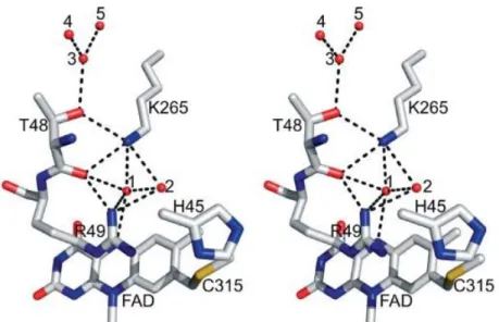
Lys265 is oriented over the si face of FAD with a hydrogen bonded at position N5 via a bridging water 1, as shown in Figure 5. The N5 atom of the flavin ring is also hydrogen bonded to a second nearby water 2 (25). It is known that this amino acid (Lys265) may play an important role in the reaction of MSOX with oxygen (21). Besides these interactions, FAD is covalently bound to the protein through a cysteinyl binding (8α –S) to Cys315 of the catalytic domain, in other words, a tioether linkage with 8-α–methyl group in the isoalloxazine ring of the cofactor makes possible the interaction with the side chain of the cysteine residue (9,25). Substrate binding is likely stabilized by hydrogen bonds between carboxylate group of substrate and two basic side chains such as Arg52 and Lys348, found above the re face of the flavin ring (22).
Moreover, there are no acidic residues in the area nearby the flavin ring, a region which includes a number of basic residues (Lys348, Arg52, Arg49 and Lys265). It is thought that this electropositive environment in the active site might play a role in MSOX catalysis by turning the cofactor a more powerful oxidant and by stabilizing the reactive anionic form of amino acid sarcosine (24).
22
Figure 5. Stereoview of MSOX (PDB code: 2GBO) showing the region above the si-face of the flavin ring.
Adopted from (9). Residues Thr48 and Lys265 and five water molecules making up a proton relay system are also indicated. Carbons atoms are in white, oxygen atoms are in red, nitrogens atoms are in blue and the sulfur is yellow. Active sites waters are shown as balls. Hydrogen bonds are indicated by dotted lines. Water molecule 1 is hydrogen bonded to flavin N(5), the carbonyl oxygen of Thr48 and the amino group of Lys265. The other water molecules (2, 3 and 4) are accessible to bulk solvent (22).
1.2.2 MSOX Active Site Ligands
Recently, many compounds able to bound to the MSOX active site have been identified due to the capacity of these ligands to perturb the flavin spectrum (data not shown). Some of them can induce a distinctive spectral change and may act as enzyme inhibitors. Previous steady state kinetic studies with pyrrole-2-carboxylate (PCA) indicated that this ligand acts as a competitive inhibitor of MSOX with respect to sarcosine (26). PCA binds in an active site cavity located above the re face of the flavin ring establishing hydrogen bounds to the side chains of basic residues (Lys348 and Arg52) through its carboxylate group (15). Also, N-cyclopropylglycine (CPG) acts as inhibitor that can quickly rearrange and inactivate the enzyme by covalently modifying the flavin with a single electron transfer. Besides this aromatic heterocyclic carboxylate, the tertiary amine, dimethylglycine also acts as competitive inhibitor of MSOX (1,24).
Regarding substrate binding determinants, it is important to emphasize that the carboxylate group (C-terminal) of the sarcosine substrate presents an important role on enzyme-substrate binding, while the amino group, generally, does not (1). The methyl group of sarcosine is not
23
essential, however, it may have some contribution in binding energy since unmethylated substrate derivatives establish weaker bindings (26).
1.2.3 A Brief Overview of MSOX Reductive Mechanisms
Many possible mechanisms of MSOX reduction can be anticipated. In the single electron transfer (SET) mechanism, a single electron is transferred from the sarcosine amino group to the flavin, forming a radical pair. The catalytic reaction of this flavoprotein continues with the proton abstraction from the amine radical cation in the sarcosine to the flavin anion radical (7). In addition, another electron is transferred resulting in the formation of the imine product and 1,5-dihydroflavin (Figure 6A) (27).
Moreover, the reaction can occur through hydride (H-) transfer from the methyl group of sarcosine (N-methyl-glycine) to the N5 atom of the Flavin (Figure 6B). Reaction can also involve the carbanion mechanism in which the reduction of flavin occurs via proton abstraction from the methyl group of sarcosine by an active side base (Figure 6C). This is followed by the formation of a covalent bond between the carbanion and the N5 atom of the cofactor (11,27). The polar mechanism involves nucleophilic attack of the sarcosine amino group on the C4a flavin atom to yield a covalent flavin-sarcosine intermediate. This is followed by the loss of a proton from the methyl group of substrate and the elimination of the imine derivative as shown in Figure 6D.
According to all these possible mechanisms for MSOX catalysis, it is settled that the unprotonated amino group is generally, the reactive species in flavin-dependent amine oxidation reactions, in other words, amino acid substrates are activated by the respective enzyme by a process involving stabilization of the reactive anionic form. According to Zhao et al., it is thought that this partial stabilization may be due to Tyr317 (24).
In spite of the several studies that have been already performed to investigate these postulated mechanisms, till present it is not completely known if the mechanism of enzyme reduction proceeds via hydride transfer to N5 of flavin or single electron transfer mechanism or even according to the carbanion mechanism (1).
24
Figure 6. Postulated mechanisms for the oxidation of sarcosine by MSOX. (A) Single electron transfer (SET)
25
1.3 Clinical Applications of Sarcosine Oxidase and Role of Sarcosine
as a Potential Marker of Early-Stage Prostate Cancer
Sarcosine oxidase is considered an essential diagnostic enzyme for quantification of creatinine level in human serum allowing assessment of renal function. Besides this capacity of SOX to estimate the function of kidney when used in combination with creatininase and creatinase (12,20,28), SOX is also able to detect sarcosine, an amino acid which has been investigated as a new biomarker for prostate cancer diagnosis.
Several biological matrixes such as plasma, urine, saliva, among others, are used to find out tumor markers which can consequently be useful for an early detection of tumors and evaluation of the efficiency of treatment (29,30). In vitro diagnostics (IVDs) have been developed in order to guide earlier diagnosis and more effective clinical intervention with improved cost effectiveness. Cancer is not yet well represented in IVD diagnostics but the need for early detection and triage is unquestionable (31).
Prostate cancer (PCa) is a common cancer in men with an increasing incidence with age. Presently it represents the second most frequent cause of death from cancer among men, with a higher incidence in developed geographic areas such as North America, Western and Northern Europe and Australia (32). This cancer tends to be asymptomatic not being detected until the age of 40-50 years. The conventional methods for prostate cancer screening suggested by the American Cancer Society, are the serum prostate specific antigen (PSA) testing and digital rectal examination (DRE) (33). Nevertheless, in spite of being widely used, PSA should not be used for routine screening of PCa. In fact, due to its poor sensitivity, specificity and accuracy, this molecule is not considered a reliable biomarker of PCa. Detection of higher levels of PSA identifies prostate disease but not necessarily PCa. In diagnosed patients, it is a good marker for disease progression but not for cancer diagnosis (31). Consequently, the methods mentioned above might cause misdiagnosis leading to delayed treatment or even overtreatment (16). Thus, more sensitive and specific biomarkers of prostate cancer, particularly in early-stage, are still needed (34).
Diagnostic techniques may involve monitoring the presence of metabolites. As intermediates or products of metabolic exchanges, metabolites can reflect the progression of certain cancers by their analysis and quantification in appropriate media (35). Many efforts have been made
26
in order to identify potential biomarkers not just for a more precise diagnosis but also for a better selection of the therapeutic options and timely response to therapy in PCa (16).
As stated above, PSA is an organ specific biomarker that does not specify the stage and the type of disease. In fact, it is unable to distinguish benign prostatic hypertrophy (BPH) from malignant PCa in patients presenting 4-10 ng/mL PSA levels, thus leading to many false-positive and false-negative results (36,37). These facts are now guiding the search for better biomarkers and therefore for improved in vitro diagnostics. Thereby, biomarkers (metabolite, gene and protein based) are being recognised as a new approach in combination with PSA testing (31). In this context, the amino acid sarcosine (N-methylglycine) assumes as a potential molecule for inclusion into a multiplex biomarker panel for PCa detection (38). Sarcosine is a non-proteinogenic amino acid that can be found in muscle and prostate. It is expressed at low levels in healthy individuals or patients with BPH, whereas high levels of urinary sarcosine are found in men with localised or metastatic prostate cancer (34). This amino acid is involved in the one-carbon metabolism (methionine metabolism) and thus it presents an important role as a methyl donor contributing to the cellular methylation homeostasis. Sarcosine also presents other biological functions such as an involvement with cell division, accelerating the growth and development of the foetus (39).
Sarcosine can be synthesized from dimethylglycine by DMGHD action and also from glycine due to oxidative methylation by glycine-N-methyltransferase enzyme (GNMT) (29). Sarcosine can also be provided from dietary intake of choline. Sarcosine can be converted by the mitochondrial SARDH flavoenzyme into glycine, the precursor of several essential metabolites such as creatine, heme, serine, glutathione and purines. Therefore, it might help indirectly in nucleic acid synthesis. SARDH is associated with to the respiratory chain by the electron-transferring flavoprotein (ETF) (40).
The enzyme GNMT, expressed at high levels in mammalian liver, exocrine pancreas and prostate, catalyses the conversion of glycine to sarcosine, whereas SARDH and PIPOX catalyse the oxidative demethylation of sarcosine converting it back to glycine (38). Therefore, increased sarcosine concentration might be associated with an elevated GNMT activity and a decrease of SARDH and PIPOX enzymes activities (29). According to Khan et al. knockdown of SARDH leads to increased sarcosine levels, resulting in an increased proliferation of cancer cells (38). Thereby, low levels of SARDH and PIPOX are detected in PCa progression and metastatic process (29). Therefore, it is postulated that sarcosine and the
27
associated enzymes that regulates its metabolism have an essential role in neoplastic progression modulating cell invasion and metastatic process (41).
The role of sarcosine as a biomarker of early-stage PCa has been intensively studied. The obtained data suggest that sarcosine exhibits significant stimulatory effects on growth in malignant/metastatic prostate cells, probably due to accumulation in the tumor of sarcosine metabolites such as serine and glycine, therefore providing tumor growth promoters (31). In face of these results, multiple cross institutional validation studies to evaluate the discriminatory power of urinary sarcosine have been carried out. In addition, it has been reported significantly elevated levels of sarcosine in presurgical urine specimens of PCa patients who developed early biochemical recurrence (38).
Sarcosine is not only expressively increased in patients with malignant PCa but also it can be excreted to urine (42). The concentration of sarcosine in blood is 1.4 ± 0.6 µM under normal conditions and 20 nmol L-1 in urine. Nevertheless, for patients urine values can reach micromolar levels (29,43).
Elevated sarcosine concentrations are found not just in PCa but also in other diseases such as colorectal cancer, stomach cancer, and sarcosinemia (39). Sarcosinemia is an inborn error of sarcosine metabolism caused by folate deficiency, which is necessary for conversion of sarcosine to glycine by SARDH. Consequently, this will lead to an increased sarcosine concentration (43). Also, eosinophilic esophagitis and Lewy Body disease have been associated with increased levels of sarcosine.
Additionally, interesting studies has been done suggesting that plasma sarcosine can be used as a metabolic biomarker of HIV/AIDS infection (44). There are studies proposing that glycine and, sarcosine, among other amino acids, are down regulated in acute aortic dissection (AD) patients (40,45).
Research during the last years led to discovery of clinical benefits of sarcosine in treating mental health disorders such as schizophrenia, Alzheimer’s disease, depression, obsessive-compulsive disorder and stroke (39). Dysfunction of N-methyl-D-aspartate (NMDA) receptors is likely the potential contributory factor in these psychiatric disorders. NMDA receptors need agonists and co-agonists such as glycine for their function. Since glycine transporters (GlyT1 and GlyT2) regulate glycine concentration, it is expected that GlyT inhibitors increase glycine levels and consequently potentiate NMDA receptor function and moderate the symptoms of schizophrenia (46). Sarcosine, as an endogenous antagonist of
28
GlyT1, potentiate glycine’s action on NMDA glycine site, thereby, it has been shown to be beneficial as adjuvant therapy improving the symptoms in patients with both chronically stable and acutely schizophrenia who are under stable doses of typical or atypical (risperidone) antipsychotics (47–50). Also, it has been demonstrated that mediation of NMDA receptors through GlyT1 inhibitors (sarcosine) can bring advantages in Alzheimer’s disease treatment (46).
From the above, it is possible to understand the importance of sarcosine study in clinical trials and, consequently the significance of SOX for its implications in sarcosine detection and quantification. Furthermore, this quantitative determination of sarcosine is essential not only in clinical chemistry, but also in food and fermentation industries. Biosensors and probes based on SOX have been employed in the detection of organic acids and glycerol in wine fermentation (8).
In this work, SOX has been studied in order to identify the best conditions for its optimized performance.
1.4 Methods For Sarcosine Quantification Using Sarcosine Oxidase
The deficiency or deregulation in sarcosine metabolism may induce multiple chronic conditions leading to strong research efforts in order to develop various analytical methods/devices to detect sarcosine (39). Several assays are available for determination of sarcosine. For direct determination of this metabolite it is common a combination of liquid and gas chromatography coupled with mass spectroscopy (LC-MS and GC-MS, respectively). Other techniques often used are enzymatic colorimetric and electrochemical methods (35,43). However, LC-MS and GC-MS require sophisticated and very expensive equipment which often are not available in clinics. In addition, there are interferences from other chemicals; in this case, alanine has the same mass as sarcosine and consequently co-elutes with it which makes more difficult the analysis and quantification of this amino acid. To overcome these disadvantages, indirect determination of sarcosine is an alternative method which can be achieved through the oxidative demethylation of sarcosine using SOX as catalyst of the reaction (35).
29
Currently, available methods for sarcosine quantification mainly use a classical oxidase linked assay and although there are reports of amperometric sarcosine biosensors with SOX, most of the techniques available are colorimetric or fluorimetic versions of this classical oxidase assay (31). In this work, the effect of chemical and physical parameters on SOX activity was studied through a colorimetric method. Although this method was not exactly used for the quantification of sarcosine it was used for the analysis of the effect of physical and chemical parameters on SOX performance through different environmental conditions. Here, the amount of H2O2 produced from oxidative demethylation of sarcosine is measured based on the amount of quinoneimine dye formed by the reaction with 4-AAP and phenol by H2O2 in presence of HRP (17). However, this colorimetric assay can be distorted in complexes matrixes such as urine or blood whereby the interferences can induce erroneous outcomes. In order to reduce this interferences 3,3’,5,5’ tetramethylbenzidine with palladium nanoparticles as catalyst have been used alternatively to HRP (31). Another alternative method to the colorimetric assay is the fluorimetric determination of sarcosine using fluorescein derivatives. This method is based on the fact that non fluorescent fluorescein derivatives will fluoresce after oxidation by H2O2 and other ROS whereby the fluorescence is directly proportional to the concentration of ROS produced. Therefore, the coupling of an enzyme and fluorescein add some improvements as the high specificity for the substrate, sarcosine, and a relatively cost-effective alternative to ongoing techniques such as colorimetric assays or chromatographic assays (LC-MS and GC-MS) where alanine is thought to be the main interfering molecule (35).
Nevertheless, all the previous methods have some major drawbacks such as high-cost instrumental setup, low sensitivity, expertise in operating the instruments, reagent and sample volumes consumption and time-consuming sample preparation since these techniques demand a lot of laboratory based processing in the analytical pathway. In order to overcome these shortcomings a few sensing methods have been reported (39). One of them remains on the sarcosine biosensor based on SOX/screen printed electrode (SPE) where the carbon screen printed electrodes are modified by a covalent immobilization of SOX on the electrode surface through the formation of N-hydroxysuccinimide (NHS) / N-ethyl-N-(3-dimethylaminopropyl) carbodiimide (EDAC) layer to connect the enzyme to the electrode (19,43). Indeed, a diversity of enzymatic immobilization methods has been described since these procedures are increasingly necessary to the biosensors function. The main difficulty here is that they are
30
usually not tested under conditions that exists under real-life conditions such as room temperature or at 37ºC when in contact with biological fluids (blood, urine) (18).
Also, analytical performance and electron kinetics of electro-chemical biosensors have been enhanced since the research in the last years uncovered the advantages of using nanomaterials in the designing process of biosensors (39). Therefore, another biosensor device equally used for the quantification of sarcosine is the biosensor based on SOX/graphene/ chitosan/ silver/ nanoparticles (AgNPs)/ glassy carbon electrode (GCE); Graphene has been widely used in enzyme-based biosensors and its important role in this biosystems is due to the capacity to promote the electron transfer rate of redox enzymes and excellent ability to preserve their bioactivity. Furthermore, chitosan is used in these biosensors in order to overcome the limitation of graphene insolubilization in certain solvents. Consequently, the decrease of graphene conductivity due to chitosan demands the incorporation of graphene and metal nanoparticles such as graphene-Pt, graphene-Au or graphene-Ag composites. Increasingly, silver nanoparticles have been used due to their good conductivity and good capacity to retain the bioactivity of redox enzymes like SOX (51).
Indeed, in recent years, a variety of nanomaterials have been applied in the development of biosensors in order to enhance their analytical performance. Since nanoparticles are surrounded by oxygen lattice, it is possible to increase the catalytic activity by immobilization of enzymes onto nanocomposite (43). Immobilization of SOX has been widely applied on different solid carriers such as magnetic nanoparticles, chitosan microspheres, mesoporous material and other materials as mentioned above. These immobilized processes are of great interest since they have the advantage of simple operating and high efficiency (17). In spite of these advantageous evidences, it is important to know which interferences have more significance in these quantification methods of sarcosine. Creatinine and urea, for example, are two species that coexist with sarcosine in the biological fluids and consequently may interfere in the detection of this molecule (19). Thus, a better comprehension of these possible interferences on quantification methods is needed to improve the outcomes and make them trustworthy quantification methods.
31
2.
Objectives
The applicability of enzymes in clinical assays is restricted by their stability since many fluids analysers operate at 37ºC which further limits enzyme stability.
In this work, it is described the structure-function-environment relationships that are required to use SOX as a reliable diagnostic reagent. Our aim was to study the effect of physical and chemical agents on SOX catalytic activity. Thus, the effect of temperature on its catalytic function was studied using an enzyme-coupled colorimetric assay. This method was used in order to understand some spectral and kinetics properties of SOX from Bacillus sp., an enzyme with the property of high thermal stability that might have advantages in transportation, storage and clinical application.
3.
Materials and Methods
3.1 Materials
Sarcosine (SAR) was obtained from Merck (USA). The enzyme MSOX from Bacillus sp. was the monomeric enzyme used in this work and from now will be identified as SOX.
SOX (yellowish amorphous powder lyophilized), HRP, 4-AAP, sodium N- ethyl-N-(3-sulfopropyl)-m-toluidine (TOPS) and phenol were purchased from Sigma–Aldrich (USA). All these reagents were stored at 4 ºC.
All chemicals used for gel electrophoresis, including acrylamide, bis-acrylamide, tetramethylethylenediamine (TEMED), ammonium persulfate (APS), glycerol, bromophenol blue, 4-(2-hydroxyethyl)-1-piperazineethanesulfonic acid (HEPES), imidazole and silver nitrate (AgNO3) were purchased from Merck (USA). Basic laboratory plastic material (tubes, tips, etc) was purchased from Eppendorf (Germany). Stock solutions of sarcosine were prepared with ultra-pure water (18 MΩ·cm conductivity) from ELGA system (USA). When necessary, diluted standards solutions were prepared in ultra-pure water. Stock solutions of SOX were prepared in phosphate buffer (pH 8.0). The pH adjustment of the solutions was performed on the Inolab multimeter (Germany). Also, BOECO Analytical Weight (Germany) was used in this study.
32
3.2 Methods
3.2.1 Analysis of Sarcosine Oxidase at Different pH using Native
Polyacrylamide Gel Electrophoresis
For the analysis of pH enzyme stability, a native polyacrylamide gel electrophoresis (Native – PAGE) with 6% gel (6% acrylamide/bis-acrylamide, 0.16% TEMED and 44 µM APS) was prepared, and using 60 mM HEPES and 40 mM imidazole as running buffer. Sarcosine oxidase solutions previously diluted in 0.2 M phosphate buffer were prepared in Britton-Robinson buffer (0.04 M phosphoric acid, 0.04 M acetic acid and 0.04 M boric acid) at different pH. Solutions were maintained at different pH (5.0, 5.5, 6.0, 6.5, 7.0, 7.5, 8.0, 8.5, 9.0, 9.5, 10.0 and 10.5) for 2 hours at room temperature (25 ºC) and at 37 ºC. Different pH stock solutions were obtained by adding different volumes of NaOH into Britton-Robinson buffer previously prepared at pH 2.0. Samples with SOX at different pH were mixed with the loading buffer (0.01% of bromophenol blue (0.01 g in 5 mL of mili-Q water) and 50% of glycerol) in a ratio 2:1 and applied to the gel. The electrophoresis was performed for 1 hour at 150 volts. Gels were stained with silver nitrate solution (silver staining) in order to visualize SOX electrophoretic mobility as well as the bands intensity. Densitometric analysis of the gels was performed using the programme Colortest.
3.2.2 Spectrophotometric Assay of Sarcosine Oxidase - UV-Visible
Spectroscopy Measurements
A spectrophotometric assay was performed to determine the wavelength of maximal absorbance change of sarcosine-SOX complex. Additionally, the effect of temperature and the influence of reaction time on charge-transfer absorbance at a specific wavelength were also studied. Although the aim of this study was not to quantify sarcosine, indirect quantification of this amino acid can be accomplished using the sequential reaction of SOX and HRP with 4-AAP as a chromogen.
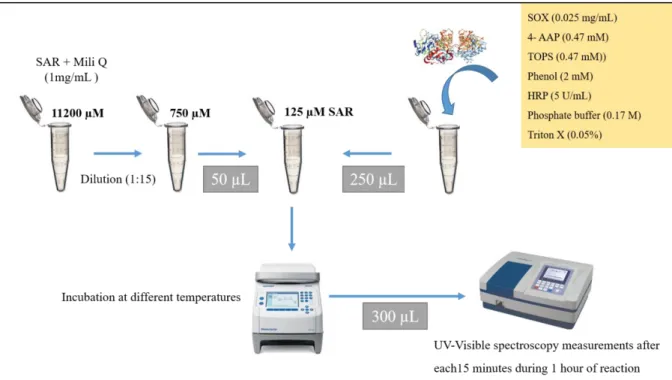
Firstly, a SAR solution at 750 µM was prepared. The reaction mixture contained 0.47 mM 4-AAP, 0.47 mMTOPS, 2 mM phenol, 0.05 % triton-X, 0.173 M phosphate buffer at pH 8.0, 5
33
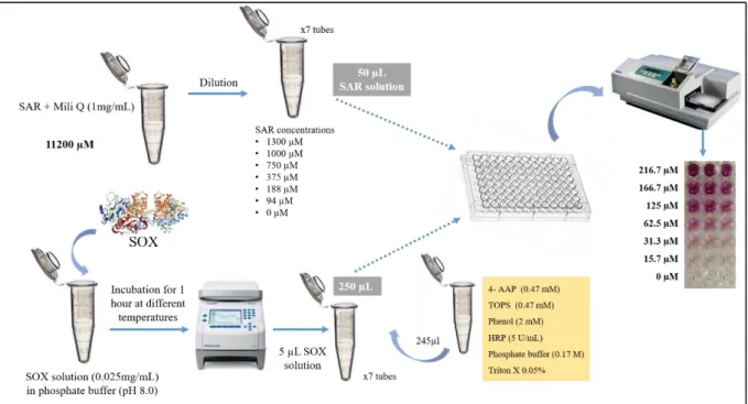
U/mL HRP and 0.025 mg/mL of SOX. Then, in each tube, the reactions were initiated by rapidly adding 50 µl of SAR solution (final concentration of 125 µM) into 250 µl of buffered solution containing all the reagents mentioned above, using the Thermomixer F1.5. The reactions were performed at 5 different temperatures (20, 25, 30, 35 and 40 ºC). In this method, all the reaction mixture including both enzymes and all the other reagents were incubated at the same temperature. For the absorbance measurement of final colored product (quinoneimine dye), a UV-3100PC spectrophotometer was used to record the visible (VIS) spectrum. After each 15 minutes, the VIS spectrum was obtained and the absorbance was recorded from 350-750 nm (0.5 nm increments), in plastic cuvettes (300 µl) with an optical path of 1 cm (Figure 7). All of the assays were carried out in triplicate.
Figure 7. Schematic representation of SOX-coupled assay to monitor the effect of temperature on SOX activity.
(4-AAP) 4-aminoantipyrene; (HRP) horseradish peroxidase; (SAR) sarcosine; (SOX) sarcosine oxidase; (TOPS) sodium N- ethyl-N-(3-sulfopropyl)-m-toluidine. Assayed temperatures: 20, 25, 30, 35, 40 ºC. See text for details.
34
3.2.3 Enzyme Coupled Colorimetric Assay – Kinetics and Thermal
Properties
To measure the SOX activity, 1 mg of enzyme (lyophilized powder) was added into 100 µl of phosphate buffer at pH 8.0. SOX solutions were placed in Eppendorf tubes and sealed in a thermoblock (Mastercycler Nexus, Eppendorf, Germany). The enzyme solution was incubated at 9 different temperatures (5, 15, 20, 30, 35, 40, 45, 50, 60 ºC) for 1 hour. The absorbance was measured at time 0 and then every minute for 1 hour using an Infinite reader F50 (Tecan, Switzerland) on a polystyrene plate (Gama group a.s, Czech Republic). The plates were previously washed with Hydroflex ELISA 8/2 (Tecan, Switzerland). Standard solutions of SAR were prepared by dilution of a stock solution (1 mg/mL) in the ultra-pure water obtaining different concentrations (94, 188, 375, 750, 1000 and 1300 µM). A reaction mixture (2 mL) containing 0.17 M phosphate buffer at pH 8.0, 0.47 mM 4-AAP, 0.47 mM TOPS, 2 mM phenol, 0.025 mg/mL SOX, 5 U/mL HRP (500 U/mL stock solution) and 0.05% triton-X (25% stock solution) was prepared. The production of H2O2 was measured by using the 4-AAP-peroxidase system as described above. The final violet coloured product was formed through the reaction between 50 µl SAR, 245 µl of the above reaction mixture and 5µl SOX solution (previously incubated at different temperatures) in a final volume of 300 µl. The reactions were performed in 96-well polystyrene microtiter plate and the absorbance was measured by photometric detector. The rate of formation of coloured product was immediately measured at 546 nm at room temperature (RT) every minute for 60 minutes. One unit of enzyme activity was defined as the amount of the enzyme that catalysed the oxidation of 1 µmol substrate per minute under conditions described above (Figure 8). All assays were performed in triplicate.
35
Figure 8. Schematic representation of SOX-coupled colorimetric assay to monitor the effect of substrate
concentration on SOX activity previously incubated at different temperatures. (4-AAP) 4-aminoantipyrene; (HRP) horseradish peroxidase; (SAR) sarcosine; (SOX) sarcosine oxidase; (TOPS) sodium N- ethyl-N-(3-sulfopropyl)-m-toluidine. Assayed temperatures: 5, 15, 20, 30, 35, 40, 45, 50 and 60 ºC. See text for details.
3.3 Interference Study - Effect of Urea on Sarcosine Oxidase Activity
The effect of urea concentration on SOX activity was determined also using the enzyme linked assay previously described (Figure 4; Equation 1 and 2). However, in this assay SOX was not pre-incubated at different temperatures.
An initial urea solution at 0.4 g/mL was prepared. Six different concentrations in a range from 0.3 g/mL to 0.0125 g/mL were obtained by dilution of initial solution of urea. The same volume of sarcosine solution 67 µL was added to those different urea concentrations making up a total volume of 1 mL. A reaction mixture containing 0.17 M phosphate buffer at pH 8.0, 0.47 mM 4-AAP, 0.47 mM TOPS, 2 mM phenol, 0.05% triton-X, 5 U/mL HRP and 0.025 mg/mL from lyophilized powder of SOX was prepared. A microplate reader (Infinite reader F50) was used in order to measure the absorbance and consequently the effect of urea on SOX activity. The reactions were performed in 96-well microtiter plates. To each well, containing 250 µL of reaction mixture (SOX and HPR included) 50 µL of the solutions initially prepared with increasing urea concentrations and fixed concentration of sarcosine (125 µM) was added.
36
Urea concentration on the wells of microtiter plates ranged from 34 µM to 832 µM. Furthermore, a positive control (sample without urea) and a negative control (sample without urea and sarcosine) were performed. The absorbance was measured every minute for 60 minutes at RT. The assay was performed in triplicate.
4.
Results and Discussion
4.1 Analysis of Sarcosine Oxidase at Different pH using Native
Polyacrylamide Gel Electrophoresis
Gel electrophoresis is a crucial method for analysis of macromolecules such as proteins, polypeptides and nucleic acids fragments. In this case, SOX, was analysed by polyacrylamide gel electrophoresis (PAGE) in the absence of sodium dodecyl sulphate (non-denaturing electrophoresis). The proteins, under native conditions, are not denatured which means that electrophoretic mobility is exclusively dependent of their intrinsic charge and hydrodynamic size (52,53).
This technique was used to investigate the influence of pH conditions in SOX native structure following its electrophoretic mobility and band intensity. In this case, an electrophoresis using non-denaturing conditions is of interest, since we are evaluating only one type of protein (same molecular weight), in the same concentration, but at different pH in a range from 5.0 to 10.5 (Figure 9).
Through the analysis of the gels obtained at two different pre-incubation temperatures, it is possible to visualize three main bands corresponding to protein aggregates with different molecular mass. The first band corresponding to high molecular mass protein complexes is more visible in the pH range from 5.0 to 6.5, however, this band intensity tends to decrease with increasing pH. This fact suggests those protein aggregates might be found in combination with other molecules of low molecular weight and hence, pH conditions are able to influence the native structure of proteins as well as its electrophoretic mobility.
37 0 0,2 0,4 0,6 0,8 1 1,2 1,4 0 100 200 300 B a n d d en sity Migration distance pH 5,0 pH 5,5 pH 6,0 pH 6,5 pH 7,0 pH 7,5 pH 8,0 pH 8,5 pH 9,0 pH 9,5 pH 10,0 pH 10,5 0 0,2 0,4 0,6 0,8 1 1,2 1,4 0 100 200 300 B a n d d en sity Migration distance pH 5,0 pH 5,5 pH 6,0 pH 6,5 pH 7,0 pH 7,5 pH 8,0 pH 8,5 pH 9,0 pH 9,5 pH 10,0 pH 10,5
Figure 9.Analysis of SOX electrophoresis in native conditions after silver staining (A) Native – PAGE of SOX pre-incubated for 2 hours at different pH at room temperature (25ºC) (B) Native – PAGE of SOX previously incubated for 2 hours at different pH at 37 ºC. (C) Electrophoretogram obtained from densitometric analysis of the gel in (A). (D) Electrophoretogram obtained from densitometric analysis of the gel in (B). Graphs (C) and (D) were done using the Colortest programme. Three main bands with high (H), intermediate (I) and low (L) molecular mass were observed and identified with black arrows. High molecular mass protein probably to aggregated forms.
At the studied temperatures (25 and 37 ºC), no significant difference was observed on SOX electrophoretic mobility as similar peaks (intensity and migration) were observed on the obtained electrophoretograms (Figure 9). However, at temperature 37 ºC it is possible to visualize that the enzyme in acidic conditions (pH 5.0 – 6.0) demonstrate very low density whereas in basic conditions (pH 8.0 – 9.0) in spite of the migration distance is slightly lower than that observed in basic conditions, its density is much higher as shown in Figure 9 (D).
5,0 5,5 6,0 6,5 7,0 7,5 8,0 8,5 9,0 9,5 10,0 10,5 5,0 5,5 6,0 6,5 7,0 7,5 8,0 8,5 9,0 9,5 10,0 10,5 A B C D I H L I L H H H L L I I
38
According to some authors, the low density at pH 5.0-6-0 can be due to the conformational change in the sarcosine oxidase structure or even structural denaturation (12).
In fact, this assay does not allow to conclude the optimal pH for SOX reaction in order to have its performance in clinical assays optimized. Nevertheless, through the electrophoretic performance of sarcosine oxidase on the native conditions, at temperature 37ºC, it is possible to estimate the optimum pH range suggesting that the enzyme is more stable and consequently might have its activity improved at the pH range from 6.5-10.5 as shown in Figure 9 (B). Although the effect of pH on SOX activity has not been studied in this work, the results obtained from the electrophoresis and according to other authors, the optimal reaction pH is thought to be between 7.0 and 8.0 (23,28). After optimal pH conditions have been investigated, it is predictable that at pH 8.0 SOX might have its activity enhanced. Therefore, a phosphate buffer 0,2 M at pH 8.0 was used in the following spectrophotometric assays in order to study the effect of temperature on SOX activity in its optimized pH conditions.
4.2 Spectrophotometric Assay of Sarcosine Oxidase - UV-Visible
Spectroscopy Measurements
The spectroscopic properties of SOX from Bacillus sp. were investigated in this study.
The principle of this method is based on the fact that SOX produces H2O2 in the presence of sarcosine. H2O2 oxidatively couples with 4-AAP and phenol in the presence of HRP to yield a chromogen, quinoneimine dye (31). This allows 4-AAP to be used as a reagent in the determination of molecules whose method of detection, in this case, is coupled through the peroxidase.
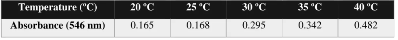
Absorption spectra were recorded using a spectrophotometer UV-3100PC in the range 350 to 750 nm as observed in Figure 10.
These reactions under such conditions (at pH 8.0) show that SOX forms a complex with sarcosine that is characterized by a large charge-transfer absorbance at an intense long-wavelenght absorption band of 546 nm.