BJRS
RADIATION SCIENCES
06-02-A (2018) 01-05Acepted: 2018-04-30
Patient-specific phantoms versus reference phantoms: a
preliminary comparison on organs dose distribution
I. V. B. Lacerda
a; J. W. Vieira
b; M. L. Oliveira
c; F. R. A. Lima
ca Universidade Federal de Pernambuco, 50670-901, Recife, Pernambuco, Brazil b Instituto Federal de Pernambuco, 50740-540, Recife, Pernambuco, Brazil
c Centro Regional de Ciências Nucleares do Nordeste, 50730-120, Recife, Pernambuco, Brazil
ABSTRACT
Discrepancies between ICRP phantoms and real patients lead to disparities on patient-dose estimations. This paper aims to compare distribution of dose in organs of male/female individualized-phantoms and ICRP reference phantoms. The absorbed dose estimation was performed using the EGSnrc Monte Carlo code and a parallel source algorithm. In this work were used an individualized-phantom for a man (1.73m/70.3kg) and another for a woman (1.63m/60.3kg) and the male and female ICRP reference phantoms. The comparison of the absorbed dose from each phantom gender was per-formed using the relative error. The results were expressed in terms of conversion coefficients to brain, lungs, liver and kidneys. The greatest absolute relative error between the organs of the patient-specific and the reference phantom was 22.92% in the liver and 62.84% in the kidneys, respectively for man and woman. There are errors that cannot be disre-garded. This paper shows the need for a specific study for each patient or for the population of each country, since there are different body types, which affects the distribution of the organ doses.
1. INTRODUCTION
The absorbed dose received by an exposed individual cannot be directly measured. Thus, it is necessary to use models that determine coefficients between the interest and measurable quantities. The computational dosimetry helps to determine the risk of exposure to ionizing radiation through Exposure Computational Models (ECM). These ECM are composed of an anthropomorphic phan-tom coupled to radiation transport codes that use radioactive source algorithms [1].
The International Commission on Radiological Protection (ICRP) recommends that the phan-tom used in computational simulations should be voxel-based and also obtained from computed tomography and/or magnetic resonance images of real patients, since they faithfully represent hu-man anatomy. In the Publication 110, the ICRP adopted the ICRP Adult Male (ICRP-AM) and ICRP Adult Female (ICRP-AF) phantoms in order to update the dose conversion factors to estimate the equivalent dose in organs and tissues for occupational, medical or environmental radiation pro-tection [2]. These phantoms are based on medical image data of the Eastern European and North American population and also are consistent with the information provided by ICRP Publication 89 [3] about the reference anatomical and physiological parameters for male/female human bodies. However, patients rarely fit to these parameters, which can lead to a disparity of the real dose re-ceived during the examination, mainly in obese and pediatric patients. This paper aims to compare the dose distribution in organs of male/female patient-specific phantoms and ICRP reference phan-toms.
2. MATERIALS AND METHODS
In this work, the Male SUPine (MSUP) and Female SUPine (FSUP) phantoms, based in the ICRP reference phantoms and available in caldose.org, were compared to male and female patient-specific phantoms. These were built from CT images of a man (1.73m/70.3kg) and another of a woman (1.63m/60.3kg), which have height and weight similar to those of the ICRP reference phan-toms. The tomographic images were obtained in DICOM format (* .dcm) and later converted into a
binary file containing a stack of images (SGI format). The original dimensions of the stacks were 512 x 512 x 635 and 512 x 390 x 571 for the male and female patient, respectively. The transfor-mation of these medical images into patient-specific phantoms for use in computational simulation was performed by means of organs segmentation using the Digital Image Processing software [4]. Thus, each organ was labeled with an identifier number (ID), which corresponds to a grayscale in-tensity. These data were attached to one of the six groups of materials belonging to the human body (air, lung, fat, water, bone and muscle) [5].
The simulation was performed for brain, lungs, liver and kidneys using the EGSnrc Monte Car-lo code [6] and a parallel source algorithm [1]. The choice of the number of stories more suitable to obtain satisfactory dosimetric results was based on the statistical uncertainty using the coefficient of variance function. The results were expressed in terms of conversion coefficients (CC) (Absorbed dose/ Air KERMA) and the comparison of these results from each phantom gender was performed using the relative error (Equation 1).
(1)
Where:
Δrel = Relative error; Xs = Simulated value; Xr = Reference value.
3. RESULTS AND DISCUSSION
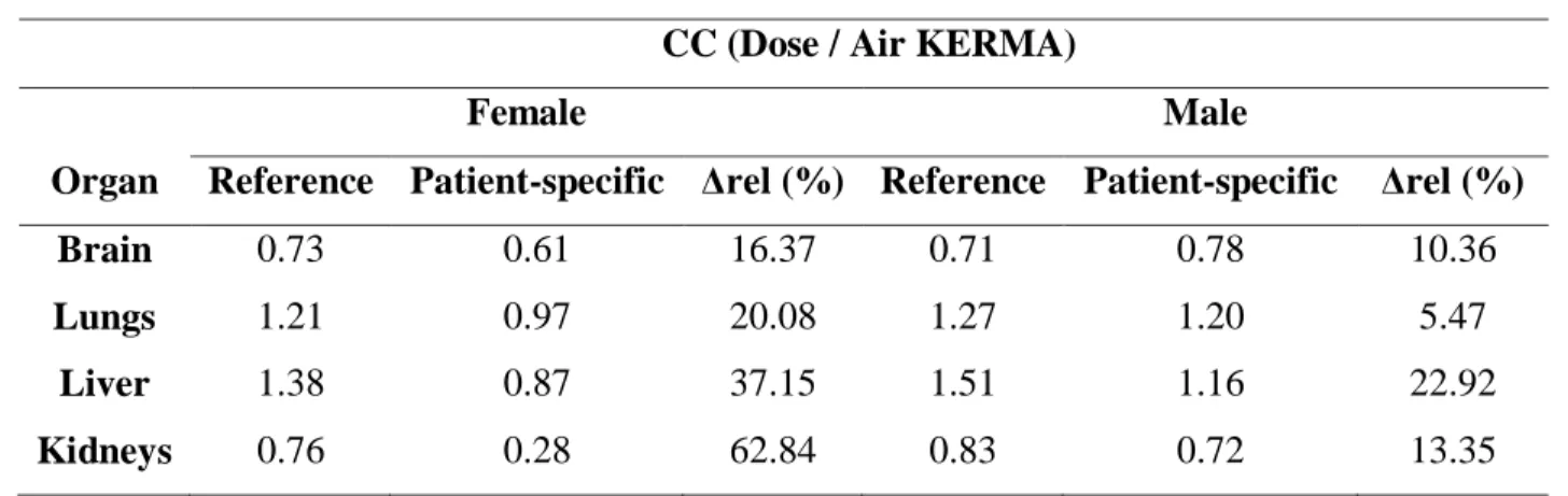
This study obtained results for brain, lungs, liver and kidneys. As presented in Table 1, the greatest absolute relative error between the organs of the patient-specific phantom and the reference phantom was 22.92% in the liver and 62.84% in the kidneys, respectively for man and woman.
Table 1: Conversion coefficients obtained for organs of patient-specific and reference
phan-toms and their respective relative errors.
CC (Dose / Air KERMA)
Female Male
Organ Reference Patient-specific Δrel (%) Reference Patient-specific Δrel (%) Brain 0.73 0.61 16.37 0.71 0.78 10.36
Lungs 1.21 0.97 20.08 1.27 1.20 5.47
Liver 1.38 0.87 37.15 1.51 1.16 22.92
Kidneys 0.76 0.28 62.84 0.83 0.72 13.35
These errors cannot be disregarded. This can occur because the CCs obtained in the computa-tional simulation are based on the interaction of the photons with the matter and consequently in the particle fluence per unit of area of the material. Despite the patients chosen in this study have height and weight similar to those of the reference phantoms, it does not guarantee that their organs have the total cross section or attenuation coefficient. The difference between the CCs presented in Table 1 confirms the importance of the use of patient-specific phantoms in the individualized dosimetry, since reference phantoms only allow obtaining average values of doses in organs.
4. CONCLUSION
In this paper, patients which have weight and height values similar to those of the ICRP reference phantoms were chosen. Despite this, the estimation using patients-specific phantoms results in different doses of those obtained using the ICRP reference phantoms. Thus, this paper shows the need for a specific study for each patient or for the population of other countries, since there are different body types, which affects the distribution of the dose in their organs.
5. ACKNOWLEDGMENT
Authors would like to thanks Fundação de Amparo à Ciência e Tecnologia de Pernambuco (FACEPE), Universidade Federal de Pernambuco (UFPE), Instituto Federal de Pernambuco – Campus Recife (IFPE), Centro Regional de Ciências Nucleares do Nordeste (CRCN-NE), Comis-são Nacional de Energia Nuclear (CNEN) and Instituto de Medicina Integral Professor Fernando Figueira (IMIP).
REFERENCES
[1] VIEIRA, J. W. Construção de um Modelo Computacional de exposição para cálculos
do-simétricos utilizando o código Monte Carlo EGS4 e fantomas de voxels. Tese de
Doutora-do, PROTEN, UFPE, Recife, Pernambuco, 2004, 101 p.
[2] INTERNATIONAL COMMISSION ON RADIOLOGICAL PROTECTION. Adult Reference Computational Phantoms. ICRP Publication 110. Oxford, 2009.
[3] INTERNATIONAL COMMISSION ON RADIOLOGICAL PROTECTION. Basic Anatomical and Physiological Data for Use in Radiological Protection Reference Values. ICRP
Publica-tion 89. Oxford, 2002.
[4] VIEIRA, J. W.; LIMA, F. R. A. A software to digital image processing to be used in the voxel phantom development. Cellular and Molecular Biology, v. 15, n. 55(3), p. 16-22, 2009. [5] REYNAERT, N.; SCHAART, D. R.; Van der ZEE, W.; Van VLIETVROEGINDEWEIJ, C.;
TOMSEJ, M.; JANSEN, J.; HEIJMEN, B.; COGHE, M.; de WAGTER, C. Monte Carlo Treatment Planning for Photon and Electron Beams. Radiation Physics and Chemistry, v. 76, 2007.
[6] KAWRAKOW, I.; ROGERS, D. W. O.; TESSIER, F.; WALTERS, B. R. B. The EGSnrc Code System: Monte Carlo Simulation of Electron and Photon Transport. NRCC Report PIRS-701.
National Research Council of Canada, Ottawa, K1A OR6, 2013, 311 p.
[7] LAMART, S.; BOUVILLE, A.; SIMON, S. L.; ECKERMAN, K. F.; MELO, D.; LEE, C. Com-parison of Internal Dosimetry Factors for Three Classes of Adult Computational Phantoms with Emphasis on I-131 in the Thyroid. Physics in Medicine and Biology, v. 56, n. 22, p. 7317-35, 2011,