Biofilm forming ability and exoproteomic analysis of Listeria monocytogenes
121
0
0
Texto
(2) À memória de meus Avós.
(3) AGRADECIMENTOS ACKNOWLEDGMENTS. A realização deste trabalho contou com ajuda pessoal e/ou institucional que não posso deixar de referir e agradecer. À Professora Luisa Brito, enquanto minha orientadora, agradeço toda a confiança que depositou na minha formação, toda a disponibilidade que sempre teve e o constante incentivo ao longo de todo o trabalho. Após vários anos de intensos e rigorosos ensinamentos científicos, guardo ainda com mais apreço todos os ensinamentos de vida e a amizade com a qual, estou certo, poderei contar para a vida. To Professor Joseph F. Frank for accepting to be my co-supervisor, having received me in his laboratory and all his guidance during the thesis work. I thank all his teachings that helped me grow scientifically and broaden my horizons, allowing me to meet different people and different approaches to science. To Professor Sixue Chen, Marjorie Chow and Carolyn Diaz I thank the kind welcome in the ICBR laboratory and all the availability for teaching me. To Professor Vasquez-Boland and Mariela Scortti for the teachings and interest in my work. Ao Professor Santos Oliveira (FCT/UNL), com quem tive ainda a sorte de poder privar, pelas suas sugestões tão relevantes para este trabalho. Agradeço aos meus colegas de laboratório no ISA, com quem ao longo dos últimos anos tive a sorte de partilhar a bancada. A boa disposição e espirito de partilha em muito contribuíram para o bom decorrer deste trabalho. Não posso deixar de realçar a Ana Carla Silva, com quem muito aprendi e que pacientemente sempre esteve disponível para resolver os problemas do dia-a-dia. O seu profissionalismo e desempenho de excelência é a pedra angular deste laboratório. À Paula Cabrita pela ajuda que sempre me deu e por ser um exemplo de organização ao qual sempre aspirei. Ao meu colega de bancada, agora já Doutor, André Barata, pelo companheirismo e pela boa disposição sempre presente com quem as trocas de ideias sempre foram proveitosas. To Maesh Chandra, I thank the kind words present whenever necessary. À Ana Abrunhosa por toda a ajuda laboratorial e por ser. i.
(4) sempre uma fonte de motivação e boa disposição. À Patrícia Vidigal pela amizade e incentivos que souberam dar-me ânimo em momentos de maior desalento. Um especial agradecimento também pela ajuda com os tratamentos gráficos da tese. À Mara Pereira por um apoio e amizade sempre presentes. À D.ª Helena Nunes e D.ª Manuela Rodrigues pela ajuda essencial que prestaram na preparação de material e pela boa disposição com que sempre encararam os meus pedidos. To may lab mates from University of Georgia, Shawn Lyons, Chi Ching Lee, Tang yanjie, Belle Piansay, Rowaida Khalil, Ana Lucia Rodriguez, Dvijal Patel and Amudhan Ponrajan I thank the warm welcome in a foreign country, easygoing coexistence in the lab and their friendship. To Julia Vastola for our long and pleasant talks at Walker’s and to Solandre Perez, for having enriched my life. A Rosa Raudales y Cecilia Sánchez de la University of Florida, gracias por su compañía, que me hizo sentir como en casa lejos de casa. To Aitor de las Heras, Vasanthakrishnan Balasubramaniam, Jesus Navas, John Bell, Iain MacArthur, Rob Cain, Patricia Gonzalez, Sonsiray Alvarez, Alexia Hapeshi, Elisa Anastasi Ana Serrano and Victoria Avila from University of Edinburgh. I am very grateful for their help and friendship. Muchas gracias. Aos meus familiares que sempre me apoiaram nesta caminhada, em especial as minhas irmas Clara, Sonia e Ana. Ao meu Pai, a quem tudo devo, não posso deixar de pôr por escrito o meu mais profundo agradecimento e admiração.. To State and Hatch funds allocated to the Georgia Agricultural Experiment Stations. À Fundação para a Ciência e Tecnologia (FCT) (Grant SFRH/BD/46996/2008).. ii.
(5) THESIS TITLE: Biofilm forming ability and exoproteomic analysis of Listeria monocytogenes. ABSTRACT. The eradication from the food industry environment of the foodborne pathogenic bacterium. Listeria monocytogenes requires a better understanding of its biofilm state. The susceptibility of biofilms of L. monocytogenes, in pure and in co-culture with Pseudomonas aeruginosa, to commercial sanitizers was determined using the Calgary Biofilm Device (CBD). Biofilms produced at 12 ºC were generally less susceptible to sanitizers than at 37 ºC. In co-culture biofilms, L. monocytogenes although representing only 1% of the co-culture, was in the same order of magnitude as the one obtained when in pure culture (5.5 log CFU/cm2). In addition to the CBD, three other methods to assess biofilm formation (crystal violet assay, epifluorescence microscopy observation and cell enumeration on stainless steel) were compared. A significant correlation was found between methods using stainless steel, evidence that data is dependent of the surface material. These results also allowed the selection of two good biofilm producing strains to be used for exoproteome investigation. For both strains, 16 identified exoproteins were more abundantly detected in the biofilm exoproteomes compared to the planktonic counterparts. One of these proteins was the putative cell wall binding protein, Lmo2504. The construction of a deletion mutant on its coding gene, confirmed the positive role of this protein in the biofilm forming ability of the wild type strain. This is the first report on the role of the exoproteome in biofilm formation.. Keywords: Listeria monocytogenes, biofilm, sanitizer susceptibility, exoproteome, Lmo2504 iii.
(6) TÍTULO DA TESE: Capacidade de formação de biofilme e análise do exoproteoma de Listeria. monocytogenes. RESUMO. A erradicação da bactéria patogénica Listeria monocytogenes, dos ambientes relacionados com as indústrias alimentares, requer uma melhor compreensão do seu estado de biofilme. A susceptibilidade, a desinfectantes comerciais, de biofilmes de L. monocytogenes, em cultura pura e em co-cultura com Pseudomonas aeruginosa, foi avaliada utilizando o Calgary. Biofilm Device (CBD). Biofilmes produzidos a 12 ºC foram, em geral, menos susceptíveis a desinfectantes do que quando formados a 37 ºC. Em co-cultura, L. monocytogenes, embora representando apenas 1 % da população total, apresentou a mesma ordem de grandeza do que a obtida quando em cultura pura (5,5 log UFC/cm2). Além do CBD, foram comparados três outros métodos para avaliar a formação de biofilme (cristal violeta, observação através de microscopia de epifluorescência e enumeração de células em superfícies de aço inoxidável). Foi encontrada uma correlação significativa entre os métodos que utilizam aço inoxidável, evidência de uma dependência dos resultados obtidos do material da superfície de crescimento do biofilme. Esta análise permitiu também a selecção de duas estirpes boas produtoras de biofilme para serem utilizadas no estudo do exoproteoma. Para ambas as estirpes, foram identificadas 16 exoproteínas com maior abundância no exoproteoma do biofilme, quando comparado com o respectivo estado planctónico. Uma dessas proteínas foi uma proteína putativa de ligação à parede celular, Lmo2504. A construção de um mutante de deleção do gene que a codifica confirmou o papel positivo desta proteína na capacidade de formação de biofilme da estirpe selvagem. Este é o primeiro relato sobre o papel do exoproteoma na formação de biofilme.. Palavras-chave:. Listeria. monocytogenes,. biofilme,. susceptibilibade. a. sanificantes,. exoproteoma, Lmo2504 iv.
(7) PREAMBLE For the food industries, food safety is the main concern. In order to avoid contamination of food products, the food industries relay on preventive measures such as effective hygiene to eradicate pathogens. Listeria monocytogenes is a high concern to food industry, especially in the case of ready to eat food producers. Previous work carried out from our group, has shown that, when planktonic cells were tested, neither the adaptation nor the resistance to disinfectants explained the existence of resident strains in particular food facilities. The biofilm state of L. monocytogenes has been regarded as a key factor for strain persistence and for unsuccessful disinfection treatments. The assessment of L. monocytogenes biofilm forming ability and susceptibility, in conditions similar to the industry environment, is therefore of relevance. The knowledge of the factors influencing biofilm formation is important to help target preventive solutions. In the process of biofilm development, the extracellular proteins or exoproteins, a fraction of the bacterial total proteome, are considered to be highly dynamic and responsive to the existing environmental conditions. For the above-mentioned, the following objectives were defined for this work: . To evaluate biofilm forming ability of a set of L. monocytogenes isolates with diverse origin and genetic diversity (from humans, from food and from food related environments, including persistent and sporadic isolates).. . To elucidate the role of the temperature of biofilm formation on the susceptibility of the biofilms to commercially available sanitizers (minimum biofilm eradication concentration – MBEC determination) by using pure cultures and co-cultures with a strain from a strong biofilm producing species (Pseudomonas aeruginosa).. . To compare methods frequently used in laboratory setting for biofilm formation and therefore, to identify the best methodologies for strain categorization in industrial setting.. . To establish a protocol for obtaining exoproteins from biofilms, minimizing the contamination with proteins from planktonic cells.. v.
(8) . To evaluate the differences in exoproteins from biofilm cells versus their planktonic counterparts by using two-dimensional difference gel electrophoresis (2D DIGE) and to perform identification of selected protein spots by electrospray mass spectrometry (MS/MS).. . To confirm, the involvement of one of the proteins detected in higher abundance in the biofilm exoproteome, by constructing a deletion mutant on the corresponding gene and to access the biofilm forming ability of the mutant strain versus the wild type.. vi.
(9) TABLE OF CONTENTS. Chapter I - Literature Review 1. Listeria monocytogenes. 2. 1.1.. General Characteristics. 2. 1.1.. Listeriosis: human and animal infection. 5. 1.2.. Impact of Listeria monocytogenes to the food industry. 7. 1.3.. Legal parameters. 9. 2. Biofilms 2.1.. 11. Biofilm Composition. 12. 2.1.1.. Exopolysaccharides. 14. 2.1.2.. Extracellular Nucleic Acids and Proteins. 17. Biofilm development and structural organization. 18. 2.2.. 2.2.1.. Biofilm formation and dispersal. 2.2.1.1. 2.2.2. 2.3.. 18. Molecular determinants of biofilm formation. 23. Biofilm structural organization. 30. Methods to access biofilm formation. 31. 3. References. 34. Chapter II - Biofilms of Listeria monocytogenes produced at 12 º C either in pure culture or in co-culture with Pseudomonas aeruginosa showed reduced susceptibility to sanitizers 1. Introduction. 46. 2. Materials and methods. 47. 2.1.. Characterization of the isolates. 47. 2.2.. Preparation of the inocula. 48. 2.3.. Disinfectant agents used. 49. 2.4.. Evaluation of biofilm forming ability using the Calgary Biofilm Device (CBD). 51. 2.5.. Determination of the Minimum Biofilm Eradication Concentration (MBEC). 52. 2.6.. Interpretation of the results and data analysis. 52. ®. 3. Results and discussion. 53 ®. 3.1.. Evaluation of biofilm forming ability using the Calgary Biofilm Device (CBD). 53. 3.2.. Co-cultures of L. monocytogenes and P. aeruginosa at 12 ºC. 54. 3.3.. Determination of the Minimum Biofilm Eradication Concentration (MBEC). 56. 4. Conclusion. 60. vii.
(10) 5. Acknowledgments. 61. 6. References. 61. Chapter III - Evaluation of methods to access the biofilm forming ability of Listeria. monocytogenes 1. Introduction. 66. 2. Materials and methods. 67. 2.1.. Characterization of isolates. 67. 2.2.. Stainless steel coupons. 68. 2.3.. Evaluation of biofilm forming ability on stainless steel by cell enumeration (SSC). 70. 2.4.. Evaluation of biofilm forming ability on stainless steel by microscopy (SSM). 70. 2.5.. Evaluation of biofilm forming ability by crystal violet. 71 ®. 2.6.. Evaluation of biofilm forming ability using the Calgary Biofilm Device (CBD). 72. 2.7.. Data analysis. 72. 3. Results. 73. 4. Discussion. 77. 5. Acknowledgments. 79. 6. References. 79. Chapter IV - Comparison of Listeria monocytogenes exoproteomes from biofilm and planktonic state: Lmo2504 a protein associated with biofilms 1. Introduction. 85. 2. Materials and methods. 86. 2.1.. Strains. 86. 2.2.. Growth of biofilm and planktonic cultures and protein secretion. 86. 2.3.. Protein precipitation and quantification. 87. 2.4.. Two-dimensional Difference Gel Electrophoresis (2-D DIGE). 87. 2.1.. Gel analysis. 88. 2.2.. Peptide sequencing by ESI-MS/MS. 89. 2.3.. Protein Search Algorithm. 90. 2.4.. Construction of deletion mutant strain ( L. monocytogenes 3119Δlmo2504). 90. 2.5.. Evaluation of biofilm forming ability: crystal violet, ruthenium red and scanning electron microscopy (SEM). 3. Results. 91 92 viii.
(11) 4. Discussion. 99. 5. Acknowledgments. 103. 6. References. 103. Chapter V - Final conclusions and future perspectives 1. Final conclusions and future perspectives. 108. ix.
(12) Chapter I Literature Review.
(13) 1. Listeria monocytogenes. 1.1. General Characteristics Listeria monocytogenes is a species belonging to the kingdom Bacteria, division Firmicutes class Bacilli, order Bacillales, family Listeriaceae, genus Listeria. Up to recently, the genus Listeria contained six species: L. monocytogenes, L. innocua, L.. welshimeri, L. seeligeri, L. grayi subsp. grayi, L. grayi subsp. murrayi and L. ivanovii subsp. ivanovii and L. ivanovii subsp. londoniensis (Rocourt & Buchrieser, 2007). A new species was then added in 2009, Listeria rocourtiae (Leclercq et al., 2009) and another in 2010 Listeria. marthii (Graves et al., 2010). In 2012, two more species have been proposed: Listeria fleischmannii (Bertsch et al., 2013) and Listeria weihenstephanensis (Halter et al., 2013). In March 2014, five new species have been proposed L. floridensis, L. aquatica, L. cornellensis, L.. riparia and L. grandensis. (Bakker et al., 2014). The genus Listeria is characterized by small Gram positive rods with 0.5 µm in diameter and 1 to 2 µm in length. Cells are not encapsulated and are not sporulated. Listeria spp. are catalase positive and oxidase negative. The optimum growth temperature for Listeria spp. is between 30 °C and 37 °C however it presents a wide range of growth temperatures, from 1-2 ⁰C to 45 ⁰C (Mclauchlin.& Rees, 2009). When grown at 20 to 28 ⁰C it presents mobility by 1 to 5 peritrichous flagella (Allerberger, 2003). Bertsch et al. (2013) reported no mobility for L. fleischmannii. The most common phenotypic characteristics of L. monocytogenes are presented in Table I.1 The first description of this specie was published in 1926 by E.G.D. Murray, R.A. Webb and M.R.B. Swann in an article entitled “A disease of rabbits characterized by a large mononuclear. leukocytosis,. caused. by. a. hitherto. undescribed. bacillus. Bacterium. monocytogenes (n.sp.)”. Earlier reports, such as the one by G. Hülpher in 1911, might also have described the isolation of L. monocytogenes nevertheless the isolates were not deposited in any culture collection and for this reason the comparison with other strains was never possible (Rocourt & Buchrieser, 2007).. 2.
(14) Until its current designation the name of the species has undergone several changes. In 1927 Pirie isolated this microorganism from gerbils and designated it as Listerella hepatolitica. The evidence that it was the same microorganism isolated by Murray, led them to rename it as. Listerella monocytogenes. The name changed to its current designation in 1940 due to previous use of the generic name Listerella for different microorganisms.. Table I.1 - Phenotypic characteristics of Listeria species. 1 - L. monocytogenes, 2 - L. innocua, 3 - L. welshimeri, 4 - L. seeligeri, 5 - L. grayi subsp. grayi, 6 - L. grayi subsp. murrayi, 7 - L. ivanovii subsp. ivanovii, 8 - L. ivanovii subsp. londoniensis, 9 - L. rocourtiae, 10 - L. marthii, 11 - L. fleischmannii, 12 - L. weihenstephanensis, ++ Strong positive reaction, + positive reaction, - Negative reaction, (+) Weak or delayed reaction, * Reported positive by Graves et al., (2010). Characteristics. 1. 2. 3. 4. 5. 6. 7. 8. 9. 10. 11. 12. Hemolysis CAMP test: Staphylococcus. + –. – –. – –. + –. – –. – –. ++ –. ++ –. – –. – –. – –. – –. +. –. –. (+). –. –. –. –. –. –. –. –. Acid production from: amidon D-galactose D-lactose D-lyxose D-mannitol D-melezitose D-melibiose D-ribose D-tagatose D-turanose D-xylose glucose-1phosphate glycerol inositol L-rhamnose methyl-αDglucopyranoside sucrose xylitol Arylamidase (DIM test) α-mannosidase Nitrate reduction H2S production. –. –. –. +. –. –. +. +. –. –. –. –. – – + – – + – – – – –. + – + – – + – – – + –. – – + – – + – – + – +. – – + – – + – – + – +. + + + + + + – + – – –. + + + + + – – + – – –. – – – – – – – + – – +. – – + – – + – – – – +. – – + – – – + + – – +. – – + – – – – – – – –. – – + – + + – + – + +. + – + – + – – + – – +. +. –. –. –. –. +. –. +. –. –. –. –. + – +. + – +. – – –. – – –. + – –. + – +. – – –. – – –. – – +. – – –. – – +. + + +. +. +. +. +. +. +. –. –. –. +. +. –. – + – + – –. – + + + – –. – + + + – –. – + + – – –. – + + + – –. – + + + – –. – + + – – –. – + + – – –. – – – – + –. – + – + – –*. (+) + – – + (+). – + – – + –. aureus Rhodococcus equi. (Adapted from Bertsch et al., 2013 and Halter et al., 2013). 3.
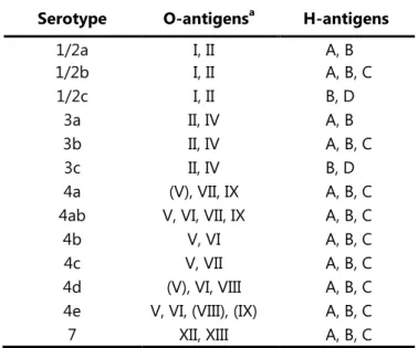
(15) L. monocytogenes is a facultative anaerobic bacterium that tolerates pH 4.5 to 9.2 with an optimum of 7, and is able to grow in 10 % (w/v) NaCl and aw bellow 0.93 (Petran & Zottola., 1989). Chemical defined medium with the minimum requirements for L. monocytogenes growth has been described. It includes cysteine, leucine, isoleucine and arginine, (Premaratne et al., 1991; Tsai & Hodgson, 2003) This microorganism when grown for 24 hours on nutritive agar produces smooth, punctiform colonies of 0.2 to 0.8 mm in diameter, lightly raised with fine texture and entire margin. The colonies are translucent of a bluish gray color, and when observed under an oblique light exhibit a blue-green iridescence (Rocourt & Buchrieser, 2007). The circular genome of L. monocytogenes has a low percentage of G and C ranging between 36.4% and 41.5% and a size varying from 2.7 and 3.0 Mb in length (Hain et al., 2006). The differentiation at a sub-species level was often achieved by an agglutination method developed by Seeliger & Höhne (1979) based on the reactions of somatic (O) and flagellar (H) antigens. This method has gradually been replaced by several other methods less costly and time-consuming. Palumbo et al. (2003) developed a high-throughput testing method of Enzyme-Linked Immunosorbent Assay (ELISA) which improves efficiency when compared with traditional slide agglutination. In 2004, Doumith et al. proposed a multiplex PCR assay targeting five genes: lmo0737,. lmo1118, ORF2819, ORF2110 and prs to differentiate the major serogroups into five phylogenetic groups. A more recent work by Vitullo et al. (2013) describes the use of a real-time PCR assay to allow the serogrouping of L. monocytogenes and the differentiation from other Listeria species. It combines the results from two triplex PCR’s, the first targeting ORF2110, ORF2819, and lmo1118, and the second targeting lmo0737, plcA, and prs. Other methods such as the one proposed by Rebuffo-Scheer et al., (2007) based on Fourier transform infrared (FTIR) spectroscopy combined with artificial neural network analysis have been used to assign the serovar of L. monocytogenes isolates. Currently, there are described 13 serotypes for L. monocytogenes: 1/2a, 1/2b, 1/2c, 3a, 3b, 3c, 4a, 4ab, 4b, 4c, 4d, 4e and 7 (Table I.2).. 4.
(16) Subspecies differentiation has been achieved by methods based on DNA fingerprinting by DNA macrorestriction and analysis by pulsed-field gel electrophoresis, by DNA amplification using specific or random primers and ribotyping (Louie et al., 1996; Liu, 2006).. Table I.2. - Antigen components of each L. monocytogenes serotype.. a. Antigens in parentheses may not be present in all isolates. a. Serotype. O-antigens. 1/2a 1/2b 1/2c 3a 3b 3c 4a 4ab 4b 4c 4d 4e 7. I, II I, II I, II II, IV II, IV II, IV (V), VII, IX V, VI, VII, IX V, VI V, VII (V), VI, VIII V, VI, (VIII), (IX) XII, XIII. H-antigens A, B A, B, C B, D A, B A, B, C B, D A, B, C A, B, C A, B, C A, B, C A, B, C A, B, C A, B, C. (adapted from Palumbo et al., 2003 and Seeliger & Langer, 1989). L. monocytogenes natural habitat is considered to be decaying plant material, where they live as saprophytes (Vazquez-Boland et al., 2001). Nevertheless, it seems to have a widespread distribution and has been isolated from a wide range of environments such as soil, water, vegetation, sewage, animal feeds, farm and food processing environments (reviewed by Sauders & Wiedman, 2007). This bacterium is able to switch from a saprophyte lifestyle to parasitic.. 1.1.. Listeriosis: human and animal infection. The disease caused by pathogenic Listeria spp. is named listeriosis. L. monocytogenes and L.. ivanovii are until now the two species considered pathogenic. L. monocytogenes is usually associated with human and animal listoriosis (with special incidence on ruminants) whereas. L. ivanovii is usually associated with animal listeriosis (Vazquez-Boland et al., 2001). Nevertheless, cases of human listeriosis caused by Listeria ivanovii have been identified.. 5.
(17) Guillet et al. (2010) pointed out however that the small incidence of listeriosis caused by L.. ivanovii is more likely to be caused by the rare occurrence of this species, compared with L. monocytogenes, rather than host tropism. Although. L.. seeligeri. presents. low. expression. of. hemolysin. and. absence. of. phosphatidylinositol-specific phospholipase C (PLC) activity it shares the virulence cluster (i.e., prfA, plcA, hly, mpl, actA, and plcB) with the other two hemolytic species of the genus (Gouin et al., 1994). There has been, at least, one report of human listeriosis caused by L.. seeligeri (Rocourt et. al, 1986). Animal listeriosis is usually associated with the consumption of low quality feeds, especially silages that undergone an inefficient fermentation and therefore have high pH. Fecal contamination of the raw materials used for silage production has also been connected with listeria occurrence in feeds. Animal listeriosis may also be caused by eye abrasion and contamination by stems and grass, in particular in self-feeding systems (reviewed by Sauders. & Wiedman, 2007). Although cutaneous listeriosis, characterized by an eczematous skin infection due to direct exposure of the skin to L. monocytogenes, may occur the consumption of contaminated food is the major cause of human listeriosis either sporadic cases or outbreaks. This disease primarily affects old or immunocompromised adults, pregnant women or newborns. In the US between 1998 and 2008 the available data allowed to identify out of 17 listeriosis outbreaks with a total 238 ill persons, 4.2 % were infants, 50.4 % within the 1 to 49 years old range and 45.4 % over the age of 50 (Cartwright et al., 2013). The same study identified, out of 19 outbreaks with a total of 296 ill persons, a 54 % female incidence. There are two forms of foodborne listeriosis: non-invasive and invasive. The non-invasive listeriosis also termed "febrile gastroenteritis” is characterized by diarrhea, fever, muscle pain, headache and less frequently abdominal cramps and vomiting. Most people recover fully, but it may also precede more serious forms of listeriosis. Invasive listeriosis is characterized by ‘flu-like’ symptoms such as fever, headache, diarrhea, vomiting and progress to meningitis and septicemia. Neck stiffness, seizures and altered mental status has also been reported (Amaya-Villar et al., 2010). In the case of pregnant women it may lead to spontaneous abortion, premature birth or stillbirth. Goulet et al. (2008) reported an incidence of listeriosis across Europe ranging from 0 to 7.5 cases per million population in 2002. According to the World Health Organization in 2004 6.
(18) listeriosis had a low incidence, ranging from 0.1 to 11.3 per million population in several countries (Anonymous, 2004). In a review of Listeriosis outbreaks in the US, it is pointed out that the incidence changed from 0.41 to 0.31 from 1996 to 2003 and from 0.25 to 0.32 from to 2004 to 2009 per 100 000 population (Cartwright et al., 2013). This same study points out the United States Centers for Disease Control and Prevention (CDC) estimative of 1662 invasive infections per year in the US with 1520 associated hospitalizations and 266 related deaths. The 2013 scientific report of the European Food Safety Authority (EFSA) and European Centre for Disease Prevention and Control (ECDC) reports indicated 1,476 confirmed human cases of listeriosis in 2011 which represented a 7.8 % decrease compared with 2010. The overall European Union (E.U.) notification rate was 0.32 cases per 100,000 population with the highest country-specific notification rates observed in Denmark, Finland and Spain (0.88, 0.80 and 0.79 cases per 100,000 population, respectively). The lowest notification rates were reported in Romania, Bulgaria and Greece (0.04, 0.05 and 0.08 cases per 100,000 population, respectively). Portugal has no reported cases of human listeriosis to EFSA between 2007 and 2011 (Anonymous, 2013); however, the Portuguese authorities have records of the existence of 46 cases of listeriosis within the same geographic region (fatality rate approximately 43.5%) between January 2009 and February 2011 (Ferreira, 2013). The mortality rate of listeriosis has been reported as high as 23.7 % (Mitjà et al., 2009), however more recent reports, either in the EU and US, estimate a case fatality rate of 12.7 and 13 % respectively (Anonymous, 2013; Cartwright et al., 2013). The treatment of invasive listeriosis usually consists in high doses of intravenous penicillin or ampicillin often in combination with an aminoglycoside. In patients allergic to penicillins, vancomycin/teicoplanin or trimethoprim/sulfamethoxazole is often used (Swaminathan & Gerner-Smidt, 2007).. 1.2. Impact of Listeria monocytogenes to the food industry In 1983 Schlech et al. described an outbreak of listeriosis in Canada involving 41 cases due to the consumption of contaminated coleslaw, which is often pointed out as being the first evidence of human listeriosis transmitted by food.. 7.
(19) Between February 2011 and July 2013 the Canadian Food Inspection Agency (CFIA) has issued 83 recalls of food products due to the contamination with L. monocytogenes. Approximately 33% of the recalls were of cheese and other dairy products, 33% of ready to eat salads and mushrooms, 20% on cured meat products and the remaining on a variety of products such as smoked fish (http://healthycanadians.gc.ca/). In recent years, in the US and in Europe also several listeriosis outbreaks have been traced down to contaminated foods such as the big outbreak in the US with 147 cases associated to contaminated cantaloupes. This outbreak spread across 28 states (http://www.cdc.gov/). In Canada in 2008, an outbreak caused by contaminated meat products caused 22 deaths and had a huge impact on the company responsible for the contaminated products. The origin of the contamination was determined to be in two production lines of one production facility (http://www.inspection.gc.ca/). The whole plant was closed leading to 250 employees being laid off and the plant undergone intense sanitation procedures with the involvement of about 80 workers and supervision of outside experts and microbiologists. About 320 employees later attended a training session on Listeria and on good manufacturing procedures and hygiene. This recall costed the company 20 million Canadian dollars. Following this outbreak the Canadian Government committed nearly 500 million dollars to improve the delivery of food safety programs (http://www.cbc.ca/news/). Due to the extended incubation period of listeriosis, which may exceed 30 days, the epidemiologic study is difficult and the identification of the contaminated food source may take enough time to allow the spread of contaminated products over a wide geographical area, therefore affecting a large number of people. In addition to deaths, the hospitalization, treatment, and the loss of productivity of the ill represent costs that are usually claimed on lawsuits filed against companies responsible for the tainted products. Therefore recall costs of contaminated products have increasing economic impact on the food industry, as the products move along the food supply chain, from distributors, to retailers to the final consumer. The recall of a product also has impact on the brand credibility, among consumers and this may in fact represent the highest cost for a company forced to recall its products. In order to avoid contaminations a strong investment in preventive measures, such as in training of the workers, routine sampling programs and hygienization procedures must be made in food industry.. 8.
(20) 1.3. Legal parameters In order to assure the safety of food products and prevent its contamination with pathogens, namely with L. monocytogenes, procedures and specifications have been established on countries' national legislations. Commission Regulation (EU) No 365/2010 of 28 April and Commission Regulation (EC) No 1441/2007 of 5 December 2007, amending Regulation (EC) No 2073/2005 on microbiological criteria for foodstuffs, directly applicable to all European Union member states, establishes in its annex I detailed information about microbiologic hazardous for foodstuffs and the criteria of acceptance. It is divided in three chapters: Food safety criteria; process hygiene criteria and rules for sampling and preparation of test samples.. Listeria monocytogenes is pointed out in chapter I: Food safety criteria. According to this document the microbial testing of L. monocytogenes should be performed in ready-to-eat (RTE) foods of three categories. The first category is “RTE food intended for infants and RTE foods for special medical purposes”. The sampling plan for this category requires a sample comprised by ten units and it sets the limit as absence in 25 g. The analytic reference method is EN/ISO 11290-1. The second category is defined as “RTE foods able to support the growth of L.. monocytogenes, other than those intended for infants and for special medical purposes”. The sampling plan and limits for this category are divided according to the stage on the food chain where the criterion applies. If it applied to products placed on the market during their shelf-life, this regulation requires a sample composed by five units and sets as limits as 100 CFU/g using EN/ISO 11290-2 as analytic procedure. If it applied to foods before the food has left the immediate control of the food business operator, who has produced it, then the regulation requires also a sample composed of five units but sets the limit as absence in 25 g using EN/ISO 11290-1 as analytic reference method. The third category of food, “RTE foods unable to support the growth of L. monocytogenes, other than those intended for infants and for special medical purposes” has its sampling plan, limits and analytic method defined as the previous category for products placed on the market during their shelf-life.. 9.
(21) The US Department of Agriculture’s (USDA) Food Safety Inspection Service (FSIS) has produced regulation in order to control of L. monocytogenes. Regulation 9 CFR Part 430 (Federal Register: June 6, 2003) establishes that official establishments that produce certain RTE meat and poultry products must prevent product adulteration by Listeria. monocytogenes. Under these regulation, establishments that produce these kind of products and are exposed to the environment after lethality treatments and that support the growth of L. monocytogenes are required to have, in their hazard analysis and critical control point (HACCP) plans, or in their sanitation standard operating procedures or other prerequisite programs, procedures to prevent product adulteration by this microorganism. This regulation also establishes that these establishments must share with FSIS data and information relevant to their controls for L. monocytogenes as well as information on the production volume of products affected by the regulations. There is a FSIS ``zero tolerance'' for L. monocytogenes these products. The FSIS recent release in 2012 of a revised Listeria Guideline shows that this microorganism continues to be a high priority for the agency.. 10.
(22) 2. Biofilms The discovery of microscopic organisms existence was made during the years of 1665 - 83 by Robert Hooke and Antoni van Leeuwenhoek, members of the Royal Society of London nevertheless the first biofilm observation is considered to have been made by van Leeuwenhoek. After scraping plaque of his teeth and mixing it with “clean rain water, in. which there were no ‘animalcules” as he described in a letter to the Royal Society, van Leeuwenhoek states: “I almost always saw with great wonder that there were many very little. animalcules, very prettily amoving”. The large number of microorganisms present is further described: "The number of these animalcules in the scurf of a man's teeth are so many that I. believe they exceed the number of men in a kingdom.". Biofilms have been studied ever since under several perspectives by a wide range of disciplines due to their strong impact on modern society. Biofilms have been used in waste water treatment due to their enhanced ability of nutrient removal and ability to degrade recalcitrant compounds. The slower microbial growth rate on the biofilm also results in a lower sludge production (Andersson, 2009). The removal of heavy metals from ground waters and soil is also pointed out as having a higher efficiency when performed by biofilms than by planktonic cells (Diels et al., 2002; Valls & Lorenzo, 2002). The use of biofilms for catalysis purposes has been widely studied. In a review Rosche et al. (2009) pointed out the use of biofilms for the production of a variety of products such as ethanol from hydrolyzed starch, hydrogen from sucrose, acetic acid from ethanol or nisin from lactose. Despite these positive uses, biofilms are often associated with their detrimental effects. Many public health concerns, such nosocomial infections as cystic fibrosis, the contamination of medical equipment, prosthesis and implants, and dental caries are associated with biofilms. In the industrial context, it can be pointed out the loss of efficiency in heat exchangers with consequent increases in energy consumption, in corrosion and pitting of equipment and the increased costs with higienization. Due to growing awareness of the scientific community to the importance of the biofilms, the number of papers has increased steadily. This number increased from approximately 200 publications per year in the year 2000 to approximately 800 publications per year in 2010 (Karunakaran et al., 2011).. 11.
(23) The definition of biofilm has suffered alterations throughout time and with authors. In 1995, Costerton et al. focused their definition on biofilm structure and defined it as a complex community of microorganisms attached to a surface or interface, enclosed in an exopolysaccharide matrix of microbial or host origin to produce a spatially organized three dimensional structure. Other authors simplify this definition but, as Dunne (2002) referred, all these definitions relay on three basic ingredients: The microorganisms, the surface and the glycocalyx, the last being often replaced by the term matrix. Generally, the definitions will specify the nature of the matrix as being self-produced, while others will include material of host origin, either of organic or inorganic nature. Most definitions will make an emphasis on the organization of the matrix and its effect on the tridimensional complexity of the biofilm. As far as the surface is concerned, most definitions will point out the possibility of it being inert or a living surface. Some definitions will also include the air-liquid interface as a “surface” for biofilm formation. Regarding to the microorganisms, most definitions will remark that multispecies biofilms are the natural form of occurrence and point out the irreversibility nature of their attachment to the surface. The most comprehensive definitions will include the fact that cells within the biofilm, differentiate and that they have different gene expression patterns when compared to their planktonic counterparts. A definition generally accepted and very often cited is by Donlan and Costerton (2002). “Microbially derived sessile community characterized by cells that are irreversibly attached to a substratum or interface or to each other, are embedded in a matrix of extracellular polymeric substances that they have produced, and exhibit an altered phenotype with respect to growth rate and gene transcription”.. 2.1. Biofilm Composition The wide range of environments in which biofilms are formed and, consequently, the diversity of species present make it difficult to generalize about its structure and composition since the microorganism present and the environmental conditions, such as oxigen concentration, shear forces, temperature and nutritional availability determine its composition and structure. Nevertheless it is generally accepted that bacterial cells account. 12.
(24) for only 10% of the total biofilm dry mass whereas the matrix represents the remaining 90% (Flemming & Wingender, 2010). Within the biofilm, the matrix is in a highly hydrated state. According to Zhang et al. (1998) water represents 97% of the biofilm. The matrix is a multi-component, dynamic, heterogeneous system (Allison, 2003) generically responsible for binding cells and other materials together (cohesion) and to the substrate (adhesion) (Wingender et al., 1999). The components of the biofilm matrix are sometimes designated generically as extracellular polymeric substances (EPS). However, this abbreviation is sometimes used in the literature to designate only part of the extracellular polymeric substances, the exopolysaccharides. Here EPS is used in the broader sense to describe not only the exopolysaccharides but also all the components of the matrix. Along with the exopolysaccharides the matrix is also composed by proteins, nucleic acids, lipids, biopolymers such as humic substances, dead cells and its detritus such as flagella, pili and fimbriae, membrane vesicules, the contents of lysed cells, metabolites, ions such as Ca2+ and Mg2+ as well as detritus and absorbed nutrients from the surrounding environment (Webb et al., 2003). Within microbial biofilms there is a wide variation on the EPS composition and relative amounts of the components. This may be attributed to many factors such as the microorganisms present, the nutrients available, the culture growth phase, bioreactor type and the flow regime as well as the extraction and analytic tool used. Moreover biofilms are considered a dynamic environment and therefore are constantly influenced by changes in the surrounding macro environment (Sutherland, 2001a). The detailed study of the EPS composition presents several challenges. Firstly, the small amount extracted constitutes a problem. Furthermore, the extraction conditions highly affect the product extracted (Pan et al., 2010; Sheng et al., 2010). Common methods to extract EPS include and combine several techniques such as centrifugation, heating, ultrasounds, ion exchange resins and the use of chemicals such as EDTA, NaOH, and formaldehyde (Adav & Lee, 2008; Tapia et al., 2009). The studies of biofilm specific components are further complicated by the fact that lower quantities components, despite their importance, are masked by macromolecules in higher quantities (Allisson, 2003). The close resemblance, or even the same composition of EPS. 13.
(25) components from biofilms and those from planktonic cells difficult the identification of biofilm specific components (Sutherland, 2001a). The study of EPS composition used a wide variety of technics such as confocal laser scanning microscopy (CLSM) and Raman spectroscopy (Wagner et al., 2009), fluorescently labeled antibodies (Lawrence et al., 2007), infrared spectroscopy (Cao et al., 2011), or calorimetric assays (Sheng et al, 2010). The major components of the biofilm matrix are schematically represented on Figure I.1.. Figure I.1 - The major matrix components polysaccharides, proteins and DNA distributed in a nonhomogeneous pattern between the cells. Adapted from Flemming & Wingender (2010).. 2.1.1. Exopolysaccharides The exopolysaccharides synthetized by microbial cells are very heterogeneous polymers that contain substituents of non-carbohydrate nature and different monosaccharides. They are long, thin molecular chains attached to the bacterial cell surface, and forming complex networks. that. surround. the. cells.. The. reported. variability. recorded. for. the. exopolysaccharides greatly contributes to the variability reported for the all EPS.. 14.
(26) The molecular mass of the exopolysaccharides is usually described as varying from 103 to 108 kDa (Sutherland, 1985) however, smaller masses have been reported varying from 0.5 to 2×10³ kDa (Sutherland, 2001b; Flemming & Wingender, 2010). Since exopolysaccharides interact with other matrix components, such as the extracellular DNA (eDNA) and proteins, as well as with the cells it becomes difficult to separate it and to isolate its effect in adhesion, nevertheless it has been shown that exopolysaccharide production is not itself, synonymous of adhesion. Several kinds of exopolysaccharides were tested and had little effect on different strains attachment to pre-coated surfaces. Moreover, when adhesion occurred it could be reversed by enzymatic degradation of the exopolysaccharides (Skillman et al., 1998). Exopolysaccharides are necessary to firmly bind the cells which produce them, obtaining a firm enclosure of the microorganisms within the matrix and hence, obtain biofilm stability. It is reported that the majority of the heteropolysaccharides are polyanionic. Its polyanionic nature is usually attributed to the presence of uronic acids or ketal-linked pyruvate as well as to inorganic residues such as phosphates or, less frequently, sulfates (Sutherland, 2001a). Nevertheless, it is pointed out by Sutherland (2001a) the fact that this cannot be generalized as this may be the consequence of the widespread study of biofilms produced by alginatesynthesizing strains of Pseudomonas aeruginosa. The same author also points out that most oral biofilms are constituted by neutral homopolymers. One other misconception regarding the bacterial exopolysaccharides is that for many years it was thought that an individual strain was limited to the production of one type of extracellular polysaccharides. Again, this was mainly due to the majority of studies on mucoid strains of P. aeruginosa that register an overproduction of alginate (Branda, et al., 2005). Several exopolysaccharides have also been extensively studied, including those from pathogenic bacteria. This is the case of β-1,6-N-acetyl-D-glucosamine (PGA) or the related poly-N-acetyl-D-glucosamine (PNAG). In Staphylococcus epidermidis and Staphylococcus. aureus the ica locus has been identified has being necessary for the synthesis of this polymer. In Yersinia pestis the hms locus or in Escherichia coli the pga locus share sequence similarity. Other bacteria produce polysaccharides with potential biotechnological applications such as cellulose. The study of Gluconacetobacter xylinus allowed the identification of protein 15.
(27) domains, namely GGDEF (Pfam PF00990) and EAL (Pfam PF00563) that are involved in activating cellulose synthase (Karatan & Watnick, 2009). It is important to note that the proportion of the different polysaccharides synthesized within the biofilm varies, depending greatly on the physiological state of the biofilm and on the cells response to environmental inputs. This way, the different exopolysaccharides and the proportions present are only representative of a particular point in time. Also, it must be bared in mind that many studies are made in environmental conditions that are not able to mimic the natural habitat where biofilms are formed. The amount and type of exopolysaccharide synthetized depend greatly on the carbon substrates available as well as its balance with other limiting nutrients. The presence of excess available carbon substrate together limitations in other nutrients, such as nitrogen, potassium or phosphate, promotes the synthesis of the polysaccharides. The slow bacterial growth observed in most biofilms would also be expected to enhance exopolysaccharide production (Sutherland, 2001a). The polysaccharides composition and backbone structure greatly affects these polymers properties. Sequences of 1,4-β- or 1,3-β-linkages may confer considerable rigidity as it is seen in the cellulosic backbone of xanthan. Other linkages may yield more flexible structures such as in many dextrans that have 1,2-α- or 1,6-α-linkages (Sutherland, 2001a; Sutherland, 2001b). The structure of the exopolysaccharides allows the binding of considerable amounts of water, and other nutrients contributing to the formation of gel. The formation of gel strengthens the biofilm structure and, therefore, protects the structure from shear forces (reviewed by Branda et al., 2005). Exopolysaccharides gel also protect cells from desiccation. This is of particular importance when variations of humidity occur, on a periodic basis, such as in food industries. The exopolysaccharides contribute to mechanical stability of biofilms by enabling them to deform under shear but recovering after the shear is removed. However, since many of the exopolysaccharides are relatively soluble, and some will form weak gels, sloughing off the exposed surface may occur. Moreover the degradation of the exopolysaccharides by deacylation induced enzymatically by esterases of bacterial or viral origin may change the physical properties of the biofilm structure either locally or to a wider extent.. 16.
(28) 2.1.2. Extracellular Nucleic Acids and Proteins Extracellular nucleic acids and specially the extracellular DNA (eDNA) are a ubiquitous component of the organic matter present within the biofilm matrix. The origin of eDNA has been associated to cell lysis often induced by quorum sensing (Spoering & Gilmore, 2006). The autolysis process may be the result of an altruistic suicide and a fratricide killing. In S.. aureus altruistic suicide occurs and cells commit suicide in a process similar to apoptosis in eukaryotic cells for the common sake of the larger community. It has been recorded in E.. faecalis, B. subtilis and S. pneumoniae the existence of fratricide killing where cells differentiate and factors are released in a process similar to necrosis in eukaryotic cells. This part of the population avoids self-destruction by specific immunity proteins (Montanaro et. al., 2011). The origin of eDNA has also been attributed to active release. There are reports in several species such as Acinetobacter baumannii and P. aeruginosa of active eDNA release of membrane vesicles containing DNA (Kadurugamuwa, & Beveridge, 1995; Sahu et al., 2012). The study of eDNA relies on obtaining significant amounts of eDNA without any contamination from intracellular DNA. Several different extraction methods have been used to obtain highly pure eDNA from different biofilm samples, minimizing cell lysis during the extraction process. Enzymatic treatments have been applied since they can disperse the biofilm matrix, without damaging cell membranes. Treatments on different strains (E. coli K-12, P. aeruginosa PAO1, and S. aureus ATCC 25923) using a combination of N-glycanase, dispersin B, proteinase K in combination with EDTA yielded significantly increased amounts of eDNA from biofilms. However variations were recorded between strains, probably due to different compositions of extracellular polymers in the biofilm matrixes (Wu & Xi, 2009). The role of eDNA in biofilms is not yet fully understood, however several functions have been assigned to it. It is a structural component important in maintaining the threedimensionality of the biofilm, as energy and nutrient source providing substrates for sibling cells and also it represents a gene pool for horizontal, gene transfer through transformation of naturally competent bacteria. Whitchurch et al. (2002) have shown the importance of eDNA for biofilm formation by dissolving established biofilms of P. aeruginosa through the use of DNase I treatments. The. 17.
(29) authors did not, however, observe the same effect when RNase treatment was performed. The same effect on biofilm attachment and subsequent stability has already been verified in. L. monocytogenes. Recently, in P. aeruginosa eDNA has been shown to facilitate efficient traffic flow of cells in the biofilm by maintaining coherent cell alignments and ensuring an efficient supply of cells to the migrating front. In this way, eDNA coordinates the movements of the cells in actively expanding biofilms (Gloag et al., 2013). Harmsen et al. (2010) concluded on the importance of eDNA molecular mass for adhesion. The authors only observed normal adhesion of cells previously treated with DNase cells, when exogenous DNA of high molecular mass was supplied but not when low molecular mass eDNA was supplied. However, this same study also revealed the importance of additional factors other than eDNA as the peptidoglycan, more precisely the N-acetylglucosamine. The proteins present in the biofilm matrix are diverse and have a strong role in biofilm formation and stability. Pili and fimbriae confere adhesive properties to bacteria and several genes related with these structures have been identified as determinant for biofilm formation. Also the presence of lectins and other sugar binding proteins in the matrix facilitate the cell-matrix and cell-cell interactions by binding polysaccharide components of the matrix or sugar moieties on the surface of other cells (Karatan & Watnick, 2009). It has been pointed out a metabolic role for the proteins in the matrix, since within the matrix various enzymes accumulate and they are involved in degradation of biopolymers. In fact, it has been suggested that the presence of these enzymes make the matrix as an external digestive system (Flemming & Wingender, 2010).. 2.2. Biofilm development and structural organization. 2.2.1. Biofilm formation and dispersal From an evolutionary standpoint, it is likely that biofilms have appeared as a way to provide homeostasis, as well as protection of cells against fluctuating and harsh conditions of the primitive earth, such as extreme temperatures, pH or ultraviolet exposure, (Hall-Stoodley et. 18.
(30) al., 2004). Bacteria biofilms are ubiquitous suggesting that they confer a strong survival and selective advantage to bacteria by increasing their environmental fitness. In this context biofilm formation is a way for bacterial populations to remain in an environment nutritionally favorable, nonhostile and at the same time that provides protection from external predation (Costerton et al., 1995). This way, biofilm communities are provided metabolic and physiological capabilities which are unavailable for the individual, unattached cells (Gilbert et al., 1997). Moreover, due to of cells proximity within biofilms, horizontal exchange of genetic material may occur and, therefore, genetic diversity is increased. Jefferson (2004) reviewed the advantages for biofilm existence and summarized it in four explanatory reasons of this lifestyle. Firstly, biofilms are the default mode of growth. In laboratory the conditions are designed to maximize bacterial growth rates however these conditions do not to happen in natural environment. The mere existence of a suitable substrate for attachment is all that is needed to trigger biofilm formation. Secondly, biofilm formation allows fixation of cells in favorable niches. By switching to the biofilm mode, bacteria remain fixed in conditions where nutrient resources allow their growth. Thirdly, biofilms serve as a defense mechanism as their formation may be the response to environmental stress. This may be of physic nature, caused by the fluid characteristics, such as the flow rate and the associated shear forces that can lead to cell wash out, but more frequently biofilm formation is trigged as a response to antimicrobial compounds, such as antibiotics, and as a way to prevent grazing by predators or phagocytosis by the immune system cells (Fajardo & Martínez, 2008; Lewis, 2001). One other reason for explaining the biofilm formation which has raised some controversy is that biofilms can exhibit communal behavior. It is established that bacteria growing within a biofilm present phenotypic heterogeneity. This heterogeneity has been interpreted as specialization towards division of labor and, in that way biofilms are compared to multicellular organisms. Nevertheless bacteria cells do not differentiate in an irreversible way as verified for multicellular organisms, they present reversible altered gene expression that enables adaptation to environmental surroundings and therefore should be regarded as interactive communities (Jefferson, 2004). Cooperation, a behavior which provides a benefit to other individual and which is selected because of its beneficial effect on the recipient (West et al., 2007) can often be observed in 19.
(31) co-cultures with promotion of biofilm formation by co-aggregation or by metabolic cooperation where a metabolite produced by one species is utilized by the neighboring species (Elias & Banin, 2012). In biofilm communities, altruism, defined as costly to the actor but benefiting the recipient (West et al., 2007), is also observed. Altruism within biofilms is the simplest form of altruism and does not require recognition of individuals, direct and sophisticated interactions between individuals as well as memory of past interactions (Kreft, 2004). On one hand, bacteria that present an altruistic strategy increase the fitness of the group, at the cost of individual fitness, having consequently lower growth rates but with increased growth yield. On the other hand others may present higher growth strategy but with a consequent lower yield. This behavior however, somehow challenges evolutionary dynamics, as bacteria that contribute towards the common good, by multiplying slower than those that do not carry that burden, ultimately tend to be outperformed. Mathematical models have predicted the existence of clusters of altruist bacteria, in environments dominated by bacteria with high yields and non-altruistic behavior due to, strong competition by non-altruistic bacteria, cluster effect, or low density of cells caused by scarcity of resources (Kreft, 2004). For over thirty years since bioluminescence of planktonic marine bacteria Vibrio fischeri and. Vibrio harveyi were studied (Nealson et al., 1970) bacteria are known to have the ability of intracellular communication. This phenomenon designated as quorum sensing (QS), allows bacteria to coordinate activities, something thought to be exclusive to multicellular organism (De Kievit & Iglewski, 2000). Mathematical models have been developed for describing biofilm formation and its dynamics, allowing the verification of experimental findings as well as predicting biofilm’s behavior in various conditions. These models have evolved throughout the years growing in complexity from single to multidimensional models and simulating growth under different conditions either in what regards to hydrodynamics or microorganisms present and their interactions with the substrates. In a review by Wang & Zhang (2010) four major models are pointed out based on the way in which diffusion is treated and in term of physic, chemistry and biological complexity: One dimensional continuum models, diffusion limited aggregate models, continuum-discrete diffusion models and biofilm–fluid coupled models. The switch between the two different life-styles, from planktonic cells into a sessile life-style may be a spontaneous phenomenon. The existence of a suitable subtract triggers 20.
(32) attachment and biofilm formation by a signaling cascade with changes in gene expression patterns. The biofilm growth surface can act as an energy source, a source of organic carbon, or simply as a growth support (Sauer et al., 2007). A phenomenon designated as surface conditioning is often referred as relevant to the subsequent biofilm development. When a solid is immersed into a liquid there is an accumulation of molecules at the solid-liquid interface which leads to a higher concentration of nutrients on the surface, compared to the fluid phase, altering the physico-chemical properties of the surface (Kumar & Anand, 1998). Consequently, the biofilm formation can be divided in two big stages, the bacterial adhesion and the biofilm maturation. The bacterial adhesion can be further divided into two steps, a first one of bacterial docking, and a second step of locking. Once the microorganisms, either by randomly fluid dynamics or direct mobility, reach a critical distance from the conditioned surface, Van der walls (> 50 nm), electrostatic (2 – 10 nm) and hydrophobic interactions (0.5 – 2 nm) promote the docking (McLandsborough et al., 2006). This first step is reversible. Fluid dynamics and cell mobility play, in fact, an important role in this first stage of the attachment. The thickness of the boundary layer adjacent to subtract and the liquid interface is dependent on the linear velocity of the liquid. Within this layer the cells association with the surface is largely dependent on cells mobility. The higher the linear velocity of the liquid, the thinner this layer is, and the greater the turbulence and mixing are. Therefore, by having a high linear velocity of the liquid a faster association with the surface is expected, at least until a certain threshold after which the velocity creates shearing forces that result in detachment of the cells (Donlan, 2002). The cells mobility is also of great importance. Once this first steps occurs, the locking phase starts with the binding between specific adhesins and the surface and the production of exopolysaccharides. The microorganisms become firmly attached to the surface and, in the absence of physical or chemical actions, the adhesion becomes irreversible. During this step planktonic bacteria not only are able to attach to the surface but also it is possible for them to stick to each other, forming aggregates. Once the adhesion is irreversible, the final stage of biofilm formation begins: The biofilm maturation is characterized by an overall increase in the complexity and density of the. 21.
(33) biofilm. Extracellular polymeric substances are synthesized and released as surface-bound organisms actively replicate and die. Additionally to biofilm adhesion and maturation stages, one other stage of biofilm development is usually pointed out: The detachment stage. When a critical mass and a dynamic equilibrium are reached, planktonic cells start being generated and become, in this way, available for the colonization of other surfaces. Structural failure of the biofilm, may also lead to bacteria leaving the biofilm by erosion, as clumps (McLandsborough et al., 2006). Figure I.2 represents the establishment of biofilm, step by step, as well as the detachment stage, either by formation of planktonic cells, either by the loss of clumps. Reduced availability of nutrients and accumulation of waste products in the innermost layers of the biofilm (Fux et al., 2005), lead the cells to growth limitations, therefore to the accumulation of cells into stationary phase. In this grow phase D-amino acids are synthetized (Lam et al., 2009). The presence of D-amino acids has been pointed out as mechanism that triggers biofilm disassembly. Kolodkin-Gal et al. (2010) have established that D-amino acids not only prevent biofilm formation, but they also disrupt existing biofilms. By being incorporated into the peptide side chains of peptidoglycan, D-amino acids impair the anchoring of fibers to the cell. In fact, the use of antimicrobial peptides consisting of Daminoacids rather than the L-aminoacids isomers, have been shown to have a broader antibacterial activity against not only Gram negative bacteria but also Gram positives, both in planktonic and in biofilms (Falciani, et al., 2012). Besides physical constrains imposed by nutrient and fluid where growths occurs, biofilm development and detachment is controlled at molecular level.. Figure I.2 - Biofilm formation and dispersal. The establishement of planktonic cells (purple) on a conditioned surface (gray) is represented, followed by the EPS (yellow) production and later dispersal of planktonic cells and biofilm clumps.. 22.
(34) 2.2.1.1.. Molecular determinants of biofilm formation. Several genes have been identified as being relevant for biofilm formation. These genes code for proteins belonging to several functional groups. Table I.3, summarizes the proteins recently implicated in biofilm formation and is divided in major groups of signaling systems, flagella and motility, BAP family and other molecular determinants. Signaling systems Signaling molecules designated autoinducers (AI) coordinate bacterial QS. Autoinducers are produced as a function of cell density and therefore, bacteria are able to regulate gene expression in response to fluctuations in cell-population density. The AIs molecules accumulate in the environment and once a certain threshold concentration is reached, gene expression is altered. The adjustment of gene expression allows the regulation of a diverse array of physiological functions such as virulence, competence, conjugation, antibiotic production, mobility, sporulation, symbiosis and also biofilm formation (Miller & Bassler, 2001). There is a wide variety of compounds, from different chemical classes, involved in bacterial communication and there are complex regulatory circuits that enable bacteria to communicate, not only within but also between species. Gram negative bacteria use as signaling molecules N-acyl-L-homoserine lactones (AHLs) (Suárez-Moreno et al., 2012). These signaling molecules are synthetized by luxL and diffuse freely across the bacterial membrane. Once a critical concentration is reached, the AHLs bind and activate a transcriptional activator, LuxR-like protein, and the complex LuxR–HSL activates expression of target genes. This system was elucidated for Vibrio fischeri (Engebrecht, et al., 1983), as mentioned before. Both Gram negative and Gram positive bacteria produce a second signaling system: a family of signaling molecules known as autoinducer-2 (AI-2), a furanosyl borate diester. The AI-2 are produced by LuxS like enzymes allowing interspecies communication (Vendeville et al., 2005). In L. monocytogenes the orthlogues of two genes responsible for the AI-2 production (luxS and pfs) are present (lmo1494 and lmo1288). It has been reported a reduced AI-2 activity in. luxS mutants with a consequent increase in biofilm forming ability both in stainless steel,. 23.
(35) where 58% more mutant cells than wild type were attached to the surface (Belval et al., 2006), and in glass where luxS-mutant produced a 19-fold denser biofilm than wild type (Sela et al., 2006). However the role of AI-2 in L. monocytogenes communication is not fully established, since the complementation of the luxS mutant with synthesized AI-2 did not produce alterations in the biofilm formation ability and the addition of an AI-2 percursor increased biofilm formation (Belval et al., 2006). Moreover, there is no identified receptor for AI-2 in the genus (reviewed by Garmyn et al., 2009). There is a third major signaling system, restricted to Gram positive bacteria, through production of post-translationally processed peptides (linear or cyclic). These peptides are processed by transmenbranar proteins and other proteases. Once secreted and accumulated, these autoinducers interact with the sensor element of histidine kinase two-component signal transductional system (De Kievit, & Iglewski, 2000). In L. monocytogenes a peptide-mediated signaling pathway was identified: The agr system. This peptide based comunication system relies on four genes organized as an operon: agrB,. agrD, agrC and agrA. Rieu et al. (2007) using mutants on the agrA and agrD, verified at early stages of biofilm formation, on glass and polystyrene surfaces, the importance of this system. The authors also confirmed by real-time PCR that the transcript levels of agrBDCA depended on the stage of biofilm development, being lower after the initial attachment period. Rieu et. al. (2008) evaluated the agr expression in 304 type stainless steel chips, under static and flowing systems, and verified almost no expression under static conditions, but a progressively increasing expression under dynamic conditions. These authors verified a 3.5 fold lower adherence of the ΔagrA cells. However, after 16 h of incubation, there were no differences between the sessile growth of the wild type and the mutant. These authors suggested the involvement of the agr system in the early stages of biofilm development, but not on late stages. Riedel et al., (2009) verified that a agrD mutant resulted in defective biofilms. However, these authors verified that the co-culture with 1% of the wild type, as well as with the complemented strain, was able to restore the biofiilm formation ability of the mutant. The use of cell-free supernatants, of the wild type strain produced the same effect. Flagella and mobility Vatanyoopaisarn et al. (2000) have shown the importance of flagellin, a major component of the flagella, in the initial attachment by comparing a flaA mutant and its wild type. The. 24.
(36) authors performed bacterial growth at 22 °C, a temperature for which flagella are produced, and verified a 10-fold lower attachment for the mutant. However when growth was performed at 37 °C, a temperature for which flagella are not produced, attachment of both strains was the same. Lemon et al. (2007) have also shown that L. monocytogenes attachment ability to four different abiotic surfaces tested: PVC, polystyrene, polypropylene, and borosilicate glass was lower when a flagellum-minus mutant (ΔflaA) was compared to the wild type, especially in the early stages of biofilm formation. Also a paralyzed-flagellum mutant (ΔmotB), that despite expressing normal amounts of flagellin and having normal numbers of peritrichous flagella, did not possess motility, also showed a lower biofilm forming ability. Therefore the authors concluded that motility plays a critical role in surface-adhered biofilm formation. Tresse et al. (2009) have also demonstrated, using an inframe deletion on flaA and naturally aflagellated strains the importance of this gene for bacterial adhesion at 20 °C. The authors observed that strains without flagella presented residual adhesion to inert surfaces similar to that observed for naturally aflagellated strains. Ouyang et al., (2012) using transposon mutagenesis, in combination with microtiter plate assays for biofilm assessing, have also confirmed Lemon’s results on the importance of flagellum-mediated motility for initial surface attachment and subsequent biofilm development. The authors identified the genes. lmo0707 (fliD) and lmo0676 (fliP) affecting flagella biosynthesis and therefore, biofilm formation. A pleiotropic response regulator, DegU, co-ordinates multicellular behavior, including the regulation of motility (Knudsen et al., 2004). These authors observed the lack of mobility and flagellin expression in a degU mutant. By using Northern blot analysis, the degU gene product was verified to be a transcriptional activator of the flagellin gene, flaA. Results by Gueriri et al. (2008) are in agreement with this, since these authors verified a reduced mobility, adherence and biofilm formation to plastic surfaces in a degU mutant. However, Todhanakasem & Young (2008) using previously identified transposon mutants (flgL, fliF, fliL, and motA), and two different approaches, the use of flow cells examined by bright-field microscopy and the static-microtiter-plate assay, verified that flagellum-based motility was not necessary for biofilm formation.. 25.
(37) Bap family The involvement of proteins in biofilm development has led to designate them as Biofilm Associated Proteins (BAP’s). This group of proteins is generically designated as such due to their homology with the first of these proteins described in S. aureus with these properties (Cucarella et al., 2001). These proteins contribute to hold cells together in the biofilm by interacting with similar proteins of cells in the vicinity, as well as on the surfaces. This proteins share several common features. These proteins are present on the bacterial surface, loosely associated with the cell surface or are secreted. In fact, the C terminus of these proteins is characterized by having a peptidoglycan anchoring region consisting of LPXTG motif (Latassa et al., 2006). This motif consists of an amino acid sequence of Leucine, Proline, X (any amino acid), Threonine and Glycine (Pfam PF00746). BAP’s are also characterized by containing a core domain of tandem repeats which may undergo dimerization. The BAP’s are described as having a high molecular mass usually higher than 1,800 up to 8,800 aa (Karatan & Watnick, 2009). In addition to the biofilm formation process, BAP’s have also been described to be relevant in virulence of both Gram positive and Gram negative species, such as S. aureus, S.. epidermidis, Enterococcus faecalis, Salmonella enterica and Pseudomonas fluorescens (reviewed by Latasa et al., 2006, reviewed by Lasa & Penadés., 2006). In L. monocytogenes there are 41 genes encoding LPXTG proteins (Cabanes et al., 2002). However, only three out of those 41 proteins have more than 1,800 aa (Lmo0333, Lmo0435 and Lmo0842). Protein Lmo0435 has been shown to contribute to surface attachment (Jordan et al., 2008). These authors have shown that although the corresponding coding gene (lmo0435 also designated bapL) was absent in most of the field isolates tested (14 out 17), this protein contributed to surface adherence of the strains to polystyrene and to stainless steel. The deletion mutant of lmo0435 gene presented 50% less cell attachment ability. However the authors verified that in contrast to what has been established for other BAP’s this protein was not required for virulence.. 26.
(38) Other molecular determinants There is already a lot of research about the impact on biofilms of the signaling molecules, genes involved on cell mobility and BAP’s, however recently the role of other genes in biofilm formation has proven to be relevant. Several regulators have been shown to play an important role in biofilm formation by several species. In S. aureus, SigB regulators known to modulate biofilm formation and dispersion by controlling the expression of both adhesion and dispersion factors (Mitchell et al., 2013). In fact, emergence of sigB deficient strains within the biofilm have been pointed out as way to promote local disassembly of the biofilm and therefore, facilitating release and dissemination of cells throughout the host (Savage et al., 2013). In L. monocytogenes, SigB a major transcriptional regulator of stress response genes has been shown to have an important impact on biofilm formation. Van der Veen & Abee (2010c), by comparing the ΔsigB mutant and its wild type strain, verified significant deficiency in biofilm formation of the mutant under both, static and continuous-flow conditions. However, no difference in planktonic growth between the wild type and the mutant was observed. Lemon et al. (2010) verified that a mutant containing an in-frame deletion of sigB produced wild type levels of biofilm at 36 °C, but displayed a biofilm defect at 30 °C. The SOS response factor YneA, as well as its activactor RecA, are also involved in biofilm formation since both ΔrecA and ΔyneA mutants showed approximately 100-fold lower ability to form biofilm than the wild type strain (van der Veen, & Abee, 2010a). By using in-frame deletion and complementation mutants, the role of a transcriptional regulator of the class I heat-shock response genes hrcA and dnaK, which encodes for heat-shock response chaperone protein, has also been shown to be relevant for biofilm formation both in static and continuous-flow, (van der Veen, & Abee, 2010b). The virulence regulator PrfA has also been implicated in the biofilm formation process. According to Lemon et al. (2010), mutants lacking prfA were defective in biofilm formation and that defect occurred after initial surface adhesion. This results were corroborated by Zhou et al. (2011) that also observed not only a weaker ability of strain EGDΔprfA to form biofilm in the first 24 hours compared to the parent strain but that the mutant needed an extra 24 hours of incubation to get to the same level as the wild strain.. 27.
Imagem


+7


Documentos relacionados
Neste trabalho o objetivo central foi a ampliação e adequação do procedimento e programa computacional baseado no programa comercial MSC.PATRAN, para a geração automática de modelos
Ousasse apontar algumas hipóteses para a solução desse problema público a partir do exposto dos autores usados como base para fundamentação teórica, da análise dos dados
The probability of attending school four our group of interest in this region increased by 6.5 percentage points after the expansion of the Bolsa Família program in 2007 and
Põe-se então a questão de saber até que ponto essa distância entre as partes afeta o trabalhador no seu desempenho, mas também na sua perceção do trabalho em si, e do local
This log must identify the roles of any sub-investigator and the person(s) who will be delegated other study- related tasks; such as CRF/EDC entry. Any changes to
Além disso, o Facebook também disponibiliza várias ferramentas exclusivas como a criação de eventos, de publici- dade, fornece aos seus utilizadores milhares de jogos que podem
É nesta mudança, abruptamente solicitada e muitas das vezes legislada, que nos vão impondo, neste contexto de sociedades sem emprego; a ordem para a flexibilização como
social assistance. The protection of jobs within some enterprises, cooperatives, forms of economical associations, constitute an efficient social policy, totally different from
