Patrícia Crespo Braga
SIRT1 and energy metabolism in rodent
Sertoli cells
setembro de 2019 SI RT 1 an d en er gy m et ab oli sm in ro de nt S er to li ce lls Pat rícia C re spo B rag a UMin ho | 2019Patrícia Crespo Braga
SIRT1 and energy metabolism in rodent
Sertoli cells
setembro de 2019
Dissertação de Mestrado
Bioquímica Aplicada
Especialização em Biomedicina
Trabalho efetuado sob a orientação do
Professor Doutor Pedro Fontes Oliveira
e da
ii
DIREITOS DE AUTOR E CONDIÇÕES DE UTILIZAÇÃO DO TRABALHO POR TERCEIROS
Este é um trabalho académico que pode ser utilizado por terceiros desde que respeitadas as regras e boas práticas internacionalmente aceites, no que concerne aos direitos de autor e direitos conexos. Assim, o presente trabalho pode ser utilizado nos termos previstos na licença abaixo indicada.
Caso o utilizador necessite de permissão para poder fazer um uso do trabalho em condições não previstas no licenciamento indicado, deverá contactar o autor, através do RepositóriUM da Universidade do Minho.
Atribuição-NãoComercial-SemDerivações CC BY-NC-ND
iii AGRADECIMENTOS
A realização desta tese de mestrado teve a ajuda, quer diretamente ou indiretamente, de diversas pessoas que me acompanharam durante todo este longo percurso, às quais quero desde já agradecer. Ao Professor Pedro Oliveira, por me ter acolhido um pouco tarde e ter aceitado o desafio de realizar este trabalho, pela disponibilidade demonstrada, conhecimento científico, críticas, opiniões sugestões e correções que me ajudaram na concretização desta tese. Tenho que deixar aqui também um agradecimento especial à Professora Sandra Cristina Paiva por todo o apoio e ensinamentos transmitidos. Sem a vossa orientação, este trabalho seria impossível de ser realizado.
Ao professor Marco Alves por todo o apoio, críticas, sugestões, que contribuíram para a minha tese. Um obrigado gigante a todos os meus colegas de laboratório: Ana, Sara, João, Ana Maria, David, Luís, Cassandra, Bruno, David “espanhol”, Ivana, Anette e Romeu que foram incansáveis para me sentir integrada e que sempre me ajudaram quando as coisas corriam menos bem. Um especial obrigado à Raquel por todo o conhecimento que me transmitiu, mas principalmente pela paciência que teve comigo, por nunca me ter desamparado e por toda prontidão que sempre demonstrou para que eu conseguisse terminar este trabalho. Obrigada por tudo!
Queria também agradecer a todos os técnicos do laboratório que nunca deixaram que nada nos faltasse. Um agradecimento especial à Cláudia e à Paula, não só pela ajuda, mas também pelos momentos de descontração e diversão.
Não poderia de deixar um grande obrigado às minhas amigas do peito, Bruna e Joana, por todo o apoio dado quando as coisas pareciam que ia descambar, o vosso apoio foi crucial para eu continuar o meu caminho no mundo da ciência.
Á minha família, principalmente ao meus pais e irmã por toda a ajuda que me deram e paciência que tiveram comigo nas alturas menos boa deste percurso. Por último, mas não menos importante, um agradecimento especial a ti Luís por teres feito comigo esta caminhada, sem nunca me teres deixado desistir dos meus sonhos e objetivos. Mesmo estando longe, foste um pilar que soube aconchegar-me nos momentos mais duros e teve sempre a palavra certa no momento adequado. Obrigada por tudo!
iv DECLARAÇÃO DE INTEGRIDADE
Declaro ter atuado com integridade na elaboração do presente trabalho académico e confirmo que não recorri à prática de plágio nem a qualquer forma de utilização indevida ou falsificação de informações ou resultados em nenhuma das etapas conducente à sua elaboração.
v
A SIRT1 E O METABOLISM0 ENERGÉTICO DAS CÉLULAS DE SERTOLI DE RATINHO RESUMO
Nos últimos anos, as sirtuinas têm sido apontadas como participantes ativos na coordenação metabólica em diversos órgãos e células, estando envolvidas na modulação de diversas vias metabólicas. De entre as desacetilases, a sirtuina 1 (SIRT1) tem sido das mais estudadas. Ainda assim, pouco se sabe acerca do seu papel na regulação do metabolismo testicular. A premissa deste trabalho é que a SIRT1 tem uma ação reguladora no metabolismo das células de Sertoli e consequentemente na espermatogénese. O objetivo deste trabalho foi estudar o papel da SIRT1 em células de Sertoli de ratinho (mSCs). Para isso expusemos as células a concentrações crescentes de um inibidor (EX-527) ou um ativador (YK-3-237) da SIRT1 e avaliámos a sua citotoxicidade e o seu impacto no perfil metabólico de uma linha de células de Sertoli de ratinho (TM4). O perfil citotóxico dos compostos foi estudado utilizando os ensaios de Brometo de 3- (4,5-dimetiltiazol-2-il) -2,5-difeniltetrazólio (MTT), libertação de lactato desidrogenase (LDH) e ensaio de sulforodamina B (SRB). Avaliámos a função mitocondrial, determinando os níveis de expressão dos complexos mitocondriais e o potencial mitocondrial (ensaio JC-1). O perfil metabólico das mSCs foi determinado por Ressonância Magnética Nuclear (1H-NMR) e pela quantificação do glicogénio
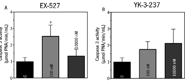
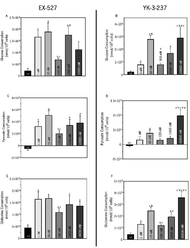
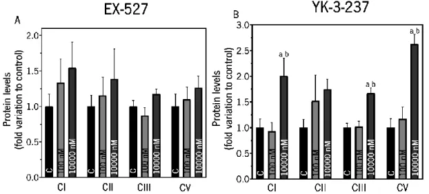
armazenado e reservas lipídicas. Foram também analisados os danos oxidativos (especificamente a peroxidação lipídica) e o perfil apoptótico, determinando a atividade da caspase-3 e a expressão de proteínas anti e pró-apoptóticas. A exposição das mSCs ao ativador ou inibidor da SIRT1 nas doses selecionadas não apresentou qualquer toxicidade para as células. No entanto, observou-se um aumento da atividade de caspase-3 quando as células foram expostas ao EX-527 (100 nM), sugerindo uma ativação da via apoptótica. Por outro lado, o ativador da SIRT1 (10000 nM) modulou a função mitocondrial, favorecendo a fosforilação oxidativa, sendo que as células nessas condições exibiram níveis mais elevados de peroxidação lipídica. Metabolicamente, quando a SIRT1 está ativada (YK-3-237, 10000 nM), há um aumento do consumo de glucose e piruvato, evidenciando um favorecimento do fluxo glicolítico, que se traduziu num aumento da produção de lactato (essencial para a espermatogénese). Adicionalmente, houve uma diminuição da acumulação lipídica, sugerindo a sua metabolização na mitocôndria. Em suma, a modulação da atividade da SIRT1 em mSCs leva a alterações do perfil metabólico dessas células. Em particular, a ativação da SIRT1 com YK-3-237 modula o desempenho metabólico das mSCs, favorecendo o metabolismo mitocondrial, sem afetar a viabilidade e proliferação celular, portanto, pode ser considerado como um ponto de controlo do metabolismo testicular.
vi
SIRT1 AND ENERGY METABOLISM IN RODENT SERTOLI CELLS ABSTRACT
In the last years, sirtuins have been identified as active participants in metabolic coordination of various organs and cells, being involved in the modulation of multiple metabolic pathways. Among all deacetylases, sirtuin 1 (SIRT1) has been the most studied. Still, little is known about its role in regulating testicular metabolism. We hypothesized that SIRT1 might have a regulatory action in Sertoli cells and consequently in spermatogenesis. The aim of this project was to study the role of SIRT1 in mouse Sertoli cells (mSCs). For that purpose, we exposed cells to increasing concentrations of an inhibitor (EX-527) or an activator of SIRT1 (YK-3-237) and we evaluated their cytotoxicity and the metabolic profile of TM4 cell line. The cytotoxic profile was studied by 3-(4,5-dimethylthiazol-2-yl)-2,5-diphenyltetrazolium bromide (MTT), lactate dehydrogenase (LDH) release and sulfhorodamine B (SRB) assays. We evaluated mitochondrial function by determining the mitochondrial complexes expression levels and mitochondrial potential (JC-1 assay). The metabolic profile of mSCs was determined by Nuclear Magnetic Resonance (1H-NMR) technique, and by the quantification of intracellular glycogen content and lipid accumulation.
The oxidative damages in cells was also analyzed (specially lipid peroxidation) and the apoptotic profile was also assessed by determining caspase-3 activity and the expression of anti and pro- apoptotic proteins. The exposure of either SIRT1 activator or inhibitor at the selected doses presented no toxicity to mSCs. However, increased caspase-3 activity was observed when cells were exposed to EX-527 (100 nM), suggesting an activation of apoptotic pathway. On the other hand, SIRT1 activator (10000 nM) modulated mitochondrial function, favoring the functioning of the oxidative phosphorylation, and under these conditions, mSCs exhibited higher levels of lipid peroxidation. Metabolically, when SIRT1 is activated (YK-3-237, 10000 nM) there is an increase in glucose and pyruvate consumption, demonstrating that there is an enhancement in glycolytic flow, which translated into an increase in lactate production (essential for spermatogenesis). Additionally, there was a decrease in lipid accumulation, suggesting its metabolization in mitochondria. In sum, modulation of SIRT1 activity in mSCs leads to changes in metabolic profile. Particularly, the activation of SIRT1 with YK-3-237 modulates the metabolic performance, without affecting cell viability and proliferation and thus, SIRT1 can be considered as control point of testicular metabolism.
vii TABLE OF CONTENTS AGRADECIMENTOS ... iii RESUMO ... v ABSTRACT ... vi LIST OF ABBREVIATIONS ... ix LIST OF FIGURES ... xi
LIST OF TABLES ... xiii
1. INTRODUCTION ... 15
1.1. Male Reproductive Tract – An Overview ... 15
1.2. Sertoli cells (SCs) ... 15
1.3. Sertoli cell metabolism and spermatogenesis ... 16
1.3.1. Energy sources on Sertoli cell metabolism ... 19
1.4. Spermatogenesis ... 20
1.4.1. Hormonal regulation of spermatogenesis ... 21
1.5. Modulators of testicular metabolism ... 23
1.5.1. Sirtuins ... 24
1.5.1.1. Sirtuin 1 (SIRT1) ... 25
1.5.1.2. SIRT1 in the testicular environment... 27
1.6. Activators and inhibitors of Sirtuins ... 29
1.6.1. Sirtuin inhibitors ... 29
1.6.2. Sirtuins activators ... 31
2. AIM OF THE PROJECT ... 35
3. MATERIALS AND METHODS... 37
3.1. Chemicals ... 37
3.2. Cell culture conditions ... 37
3.3. Experimental Groups ... 37
3.4. Sulforhodamine B (SRB) cytotoxicity assay ... 38
3.5. Lactate Dehydrogenase (LDH) release assay ... 38
3.6. MTT viability assay ... 39
3.7. Protein extraction and quantification ... 39
viii
3.9. Slot-Blot – Lipid Peroxidation ... 40
3.10. Mitochondrial Membrane Potential... 41
3.11. Caspase-3 Activity ... 42
3.12. 1H-NMR Spectroscopy ... 42
3.13. Colorimetric Method For Glycogen Quantification ... 43
3.14. Oil red O Staining (ORO) ... 43
3.15. Statistical analysis ... 44
4. RESULTS ... 46
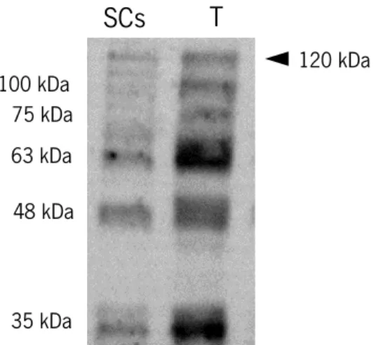
4.1. SIRT1 protein is expressed in TM4 cell line ... 46
4.2. Sirtuin 1 activation and inhibition do not alter cellular proliferation and metabolic viability of TM4 Sertoli cells ... 46
4.3. Exposure to the SIRT1 inhibitor EX-527 promoted a decrease of LDH leakage on TM4 Sertoli cells 48 4.4. SIRT1 inhibition results in the activation of caspase-3 in TM4 Sertoli cells ... 48
4.5. SIRT1 modulates glucose catabolism in mouse TM4 Sertoli cells ... 50
4.6. Exposure to the highest concentrations of SIRT1 inhibitor EX-527 and activator promoted an increased secretion of alanine and lactate ... 53
4.7. Exposure to SIRT1 activator YK-3-237 or Inhibitor EX-527 did not alter glycogen content on TM4 Sertoli cells ... 55
4.8. Exposure of TM4 Sertoli cells to both the SIRT1 inhibitor EX-527 or activator YK-3-237 decreases lipid accumulation in Sertoli cells ... 55
4.9. Exposure to the SIRT1 activator YK-3-237 increases the expression of mitochondrial complexes in mouse Sertoli cells ... 56
4.10. Exposure to the SIRT1 activator YK-3-237 increases mitochondrial membrane potential in mouse TM4 Sertoli cells ... 58
4.11. Inhibition or activation of SIRT1 increases lipid peroxidation in TM4 Sertoli cells ... 58
5. DISCUSSION ... 62
6. CONCLUSION ... 68
ix LIST OF ABBREVIATIONS
ALT – Alanine aminotransferase
AMPK - 5' AMP-activated protein kinase ATP- adenosine triphosphate BSA – Bovine serum albumin
BTB - Blood-testis barrier
DMEM - Ham’s F12- Dulbecos’s modified Eagle Medium Ham’s Nutrient Mixture F12 DMSO – Dimethyl sulfoxide
FBS - Fetal Bovine Serum
FOXO - Forkhead transcription factor FSH - Follicle- stimulating hormone GLUTs- Glucose transporters
GnRH - Gonadotropin releasing hormone HPT - Hypothalamic-pituitary- testis JC-1 - 5,5′,6,6′-tetrachloro-1,1′,3,3′-tetraethylbenzimidazolylcarbocyanine iodide LCs - Leydig cells LDH - Lactate dehydrogenase LH - Luteinizing hormone MCT4 – Monocarboxylate transporter 4 MCT2 - Monocarboxylate transporter 2 mSCs - mouse Sertoli cells
MTT - 3-(4,5-dimethylthiazol-2-yl)-2,5-diphenyltetrazolium bromide NAD+ - Nicotinamide adenine dinucleotide
NAM - Nicotinamide
x ORO - Oil Red O staining
OXPHOS - Oxidative phosphorylation PBS - Phosphate Buffered Saline PDH – Pyruvate dehydrogenase
PGC-1α - Peroxisome proliferator- activated receptor gama coactivator 1 α PPAR - Peroxisome proliferator-activated receptor
RIPA – Radioimmunoprecipitation assay ROS – Reactive Oxygen Species
RSV – Resveratrol SCs - Sertoli cells SIRT1 - Sirtuin 1 SIRT2 – Sirtuin 1 SIRT3 – Sirtuin 3 SIRT4 – Sirtuin 4 SIRT5 – Sirtuin 5 SIRT6 – Sirtuin 6 SIRT7 – Sirtuin 7
STAT3 - Signal transducer and activator of transcription 3 SRB - Sulforhodamine B
xi LIST OF FIGURES
Figure 1. Representative diagram of the metabolic cooperation mechanisms established between Sertoli
cells and developing germ cells. ... 18
Figure 2. Schematic representation of the principal events during spermatogenesis process in seminiferous tubule.. ... 21
Figure 3. Hormonal regulation of the male reproductive tract.. ... 22
Figure 4. Sirtuin 1 (SIRT1) regulation signaling.. ... 27
Figure 5. Identification of SIRT1 and its isoforms (35-120 kDa) .. ... 46
Figure 6. Evaluation of cell proliferation (Panels A and B) and metabolic activity (Panels C and D) on TM4 Sertoli cells after exposure to SIRT1 inhibitor (EX-527) (Panels A and C) or activator (YK-3-237) (Panels B and D).. ... 47
Figure 7. Evaluation of LDH release (Panels A and B) on TM4 Sertoli cells after exposure of SIRT1 inhibitor (EX-527) (Panel A) or activator (YK-3-237) (Panel B).. ... 48
Figure 8. Evaluation of the caspase 3 activity (Panels A and B) on TM4 Sertoli cells after exposure of SIRT1 inhibitor (EX-527) (Panel A) or activator (YK-3-237) (Panel B).. ... 49
Figure 9. Evaluation of the ratio of BAX/BCL-2 (Panels A and B) on TM4 Sertoli cells after exposure to SIRT1 inhibitor (EX-527) (Panel A) or activator (YK-3-237) (Panel B).. ... 50
Figure 10. Evaluation of glucose (Panels A and B), pyruvate (Panels C and D) and glutamine (Panels E and F) consumption on TM4 Sertoli cells after exposure to sirtuin inhibitor (EX-527) (Panels A, C and E) and activator (YK-3-237)(Panels B, D and F).. ... 52
Figure 11. Evaluation of alanine (Panels A and B), lactate (Panels C and D) and acetate (Panels E and F) on TM4 Sertoli cells after exposure of SIRT1 inhibitor (EX-527) (Panels A, C and E) or activator (YK-3-237) (Panels B, D and F).. ... 54
Figure 12. Evaluation of the intracellular glycogen content (Panels A and B) on TM4 Sertoli cells after exposure of SIRT1 inhibitor EX-527 (Panel A) and SIRT1 activator YK-3-237 (Panel B). ... 55
Figure 13. Evaluation of lipid accumulation (Panels A and B) on TM4 Sertoli cells after exposure of SIRT1 inhibitor EX-527 (Panel A) and SIRT1 activator YK-3-237 (Panel B). ... 56
Figure 14. Evaluation of SIRT1 inhibitor EX-527 (Panel A) and SIRT1 activator YK-3-237 (Panel B) in protein expression levels of mitochondria complexes of TM4 Sertoli cells. . ... 57
Figure 15. Evaluation of JC-1 ratio (Panels A and B) on TM4 Sertoli cells after exposure of SIRT1 inhibitor EX-527 (Panel A) and SIRT1 activator YK-3-237 (Panel B) in mitochondria. ... 58
xii
Figure 16. Evaluation of lipid peroxidation (Panels A and B) on TM4 Sertoli cells after exposure of SIRT1 inhibitor EX-527 (Panel A) and SIRT1 activator YK-3-237 (Panel B).. ... 59
xiii LIST OF TABLES
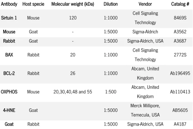
Table 1. Principal sirtuin inhibitors and activators. ... 33 Table 2. List of Primary and Secondary antibodies used in Western Blot and Slot-Blot technique. ... 41 Table 3. Effect of SIRT1 activator YK-3-237 in protein expression levels of mitochondrial complexes on TM4 mouse Sertoli cells from the control group and treated with 10000 nM. ... 57
14
Chapter I
15 1. INTRODUCTION
1.1. Male Reproductive Tract – An Overview
The male reproductive tract provide the appropriate conditions for the development of germ cells, which can generate an entirely new organism, through the transmission of genetic information to the next generation, required for the survival of the species (1).
In order to maintain a normal male reproductive health, the coordination between the different type of cells, accessory glands and tissues is essential. In general, the mammalian testes are paired organs, covered by a layer, tunica vaginalis and an inner layer, tunica albuginea. Testis has lobules that include the seminiferous tubules, recognized as the functional units of the testis, which are immersed in loose connective tissue and some interstitial cells, namely Leydig cells (LCs) (2). The seminiferous tubules are compartmentalized by junctions between adjacent Sertoli cells (SCs), creating the blood-testis barrier (BTB), that physically divides the seminiferous epithelium into basal and adluminal compartments. The BTB is responsible for restricting or allowing the passage of substances that are present in the interstitial fluids into the tubular lumen (3). BTB represent an anatomical, immunological and physiological barrier, essential for the well-functioning of male reproductive tract. Blood and lymphatic vessels that are present in the interstitial space participate in the homeodynamics of hormones and nutrients in the testes (4), with the interstitial LCs being responsible for the synthesis of main male steroid hormone, testosterone (5). So,the testes are responsible for the synthesis of male sex hormones and the production of male
gametes, spermatozoa. (6, 7). An impaired coordination between particularly SCs and LCs, interferes with the mature of spermatozoa (8, 9), and hence, has repercussions on male fertility.
1.2. Sertoli cells (SCs)
Sertoli cells were first described by Enrico Sertoli in the 19 th century (10) and the evidence of their
fundamental involvement in the establishment of male fertility has been confirmed by several studies. Indeed, one SC can support up to 30 at 50 germ cells at different stages of development (11), although there is a variation on this number between species. Hence, the number of SCs is associated to the rate of spermatogenesis and therefore to the daily production of sperm per testis, that posteriorly will result on male fertility capacity (12-14). SCs are also responsible for the phagocytosis of residual bodies and degenerating germ cells (2, 15, 16). Moreover, SCs provide nutritional support that male germ cells need, through the production and release of specific metabolites to environment surrounding developing germ
16
cells. SCs must also produce different growth factors, signaling molecules, cytokines, bioactive peptides and several glycoproteins and peptides that form the molecular basis for Sertoli-germ cell interactions (17, 18).
Specialized junctions, like occludins, claudins and desmossomes, between adjacent SCs, are highly organized, located near the basement membrane, creating the BTB. Collagen, laminin and extracellular matrix components, also contributes to these specialized junctions. All those interactions contribute to the structural and physiological function of BTB (19).
BTB regulates the diffusion of water, electrolytes, nutrients and biomolecules (it specially prevents the movement of large molecules) from systemic circulation in to lumen of seminiferous tubules (20). It also limits the movement of immune cells and regulates the level of cytokines in the seminiferous epithelium (21, 22).
1.3. Sertoli cell metabolism and spermatogenesis
As already mentioned above, SCs are vital for the occurrence of a normal spermatogenesis. They are known as nurse cells, once they provide unique characteristics like the structural and nutritional support for the development of germ cells (23, 24). In fact, there is a metabolic dependence relationship between SCs and developing germ cells. SCs can secrete peptides, nutrients and metabolic intermediates (18, 25). A correlation between the levels of energetic substrates that SCs produce with the germ cells developed can be made, so they are essential for the development of spermatogenesis and hence for male fertility. (26).
Over the past years, multiple works pointed out that cultured SCs use preferably glycolysis and convert the majority of the resulting pyruvate to lactate, that is then exported from the cell. Indeed, Grootegoed et al., demonstrated that only 25% of the pyruvate produced from glucose go to Krebs cycle (27). Like cancer cells, it is known that SCs have a Warburg-like metabolism and prefer the fermentative rather than the oxidative metabolism of glucose (26). They exhibit a high glycolytic flux, however, is not accompanied with enhanced cellular proliferation, representing different aspect comparing to cancer cells.
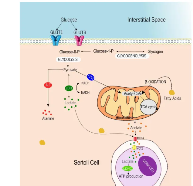
As a starting point, specific carriers are needed to facilitate the diffusion of glucose across the membrane, since glucose is a hydrophilic and polar molecule and hence, it crosses the lipid bilayer very slowly by simple diffusion (Figure 1). There are specific membrane glucose transporters, named as sodium dependent glucose transporters (or solute carrier family 5 transporters - SLC5) and glucose transporters (GLUTs) (or solute carrier family 2 transporters – SLC2) (28, 29). Regarding GLUTs family, they are
17
divided in three sub-families: class I, which include GLUT1-4; class II (GLUT 5, GLUT7 and GLUT9 and GLUT11); and class III (GLUT6, GLUT8, GLUT10 and GLUT12). GLUTs are widely expressed in different tissues and cells. In testis, GLUT1 (30), GLUT2 (31), GLUT3 (32), GLUT5 (33) and GLUT8 have already been identified (34). Specially in SCs, the presence of GLUT1 to GLUT4 has also been confirmed (31, 35, 36). Curiously, Riera et al., reported that when glucose is removed from the culture medium, the expression of GLUT1 increase and GLUT3 expression decreases, demonstrating that glucose acts as signal to adequate an appropriate glycolytic flux in response to the glucose levels in the extracellular space (37).
Once inside the cell, glucose is metabolized by multiple enzymes, resulting in the production of pyruvate. The pyruvate can follow different ways: it can enter the Krebs cycle, it can be converted to alanine by the action of alanine aminotransferase, or it can be converted to lactate by the action of lactate dehydrogenase (LDH), with the simultaneous oxidation/reduction of NADH to NAD+ (Figure 1). In fact, multiple authors,
described that the majority of pyruvate produced by SCs is converted to lactate by LDH (26, 38, 39). Later, this lactate is exported via monocarboxylate transporters (MCTs). This family has 14 members and are expressed in different tissues and cells. Regarding male reproductive tract, MCT2 in found in elongated spermatids (40), MCT1 and MCT4 are present in SCs (41). In fact, MCT1 has a higher affinity for lactate than MCT4. MCT1 is most associated to the import of lactate from the extracellular space (42), unlike MCT4, which is primarily a lactate exporter, since it has lower affinity for lactate (41, 43). Thus, MCT4 seems to play an important role in SCs (39, 42), as it is expressed in cells with high glycolytic profile. In fact, the production of lactate is essential for the normal development of germ cells.
In adverse conditions, when glucose is decreased, SCs can adapt their metabolic behavior to guarantee an appropriate production and export of lactate into the adluminal compartment, they modulate GLUTs expression, trough different signaling pathways (37). Deprivation of insulin has also repercussions on lactate production (42), since it induces an adaptation in the expression of GLUTs. All of these evidences support the fact that SCs are essentials to ensure the metabolic needs of germ cells, in order to support properly the spermatogenesis.
Indeed, lactate has been reported to principal energetic substrate of germ cells and is essential to the maintenance of spermatogenesis in vivo. To support that, Trejo et al, demonstrated that pharmacological deprivation of lactate decreases the viability of male germ cells (44). Then, Courtens and Ploen, described that testicular perfusion of lactate was capable to suppress the loss of spermatocytes and spermatids in adult cryptorchid rat testicle (45). Furthermore, lactate was characterized to exert an anti-apoptotic effect on germ cells (46). All these data demonstrate that lactate is essential to spermatogenesis.
18
Figure 1. Representative diagram of the metabolic cooperation mechanisms established between Sertoli cells and developing germ cells. Glucose from interstitial space is taken preferentially through glucose transporters, GLUT1 and GLUT3, and is converted into glucose-6 phosphate (Glucose–6-P). Although, when glucose is not available glycogen can be used as an energy fuel, where it is converted into glucose-1-phosphate (Glucose-1-P) and then in glucose-6-P. Glucose is converted to pyruvate through multiples reactions that are compromised in glycolysis. The resulting pyruvate can follow three possible pathways. It can be converted in alanine by the action of alanine aminotransferase (ALT), it can be converted in lactate by the action of lactate dehydrogenase (LDH) or it can be converted into Acetyl-CoA through the action of pyruvate dehydrogenase (PDH). Acetyl-CoA can be converted into acetate or it can be used in the Krebs cycle in mitochondria. Acetate and lactate are exported to the intracellular fluid by monocarboxylate transporter 4 (MCT4) and then taken up by developing germ cells trough monocarboxylate transporter 2 (MCT2). The lactate produced by Sertoli cells, can be used for germ cells to ATP production.
19
In fact, it is characterized as the preferred substrate for developing germ cells, even if these cells possess all the machinery to metabolize glucose (26).
LDH enzyme is responsible for the interconversion of pyruvate into lactate, with the concomitant oxidation/reduction if NADH to NAD+, a step essential for the continued production of ATP by glycolysis
(47). LDH has different isoforms (LDHA, LDHB and LDHC). LDHC is exclusively expressed in tumors (48) and in the testis (49), being particularly abundant in spermatids and spermatozoa (50, 51). A disruption in Ldhc gene leads to male infertility and is associated with a decrease in sperm motility and a decline in the levels of ATP in germ cells (52).
In addition to the high amount of lactate being produced by SCs, these cells also produce high amounts of acetate (53). This metabolite seems to have a role in sustaining of the lipid metabolism (particularly phospholipid metabolism) in germ cells (54, 55). In fact, the synthesis of lipids in germ cells is maintained due to the high rates of acetate produced by SCs (53).
1.3.1. Energy sources on Sertoli cell metabolism
There are several substrates that can be used for energy production by SCs, in order to produce ATP. Glucose is the frequently used by different cell types, and SCs are not an exception. Still, some authors described that these cells use other alternative energetic fuels. For instance, the metabolism of lipids has been pointed to be crucial for a proper spermatogenesis, as an inactivation of some genes related to lipid metabolism, disrupted the normal course of spermatogenesis (56).
Glutamine and glycine are other substrates that yield much of the energy required by SCs (27), demonstrating that amino acids can have a role in metabolism in SCs.
Another energetic substrate available in SCs is glycogen. At the end of the 20th century, it was reported
the presence of glycogen and glycogen phosphorylase in SCs (57, 58). Later, Vilarroel- Espíndola et al., demonstrated that glycogen has a role during testicular development and it also acts as a modulator of germ cell survival (59).
These multiple, energetic substrates that are available to SCs, demonstrate that they are flexible in metabolites used to produce ATP and thus, to be able to sustain spermatogenesis. Still, this is a subject that deserves more attention in the future.
20 1.4. Spermatogenesis
Spermatogenesis begins at puberty and continues throughout the entire humans’ life. It takes place inside the seminiferous tubules, through a close association between germ cells and SCs (4, 60). All the stages of spermatogenesis are regulated by the niche of the testis, which is mainly established by SCs (23, 24). SCs are vital to spermatogenesis, since they are responsible for the correct nutritional and physical support for developing of germ cells (9).
The spermatogenic event is divided in different biological phases, comprising, among others, a mitotic and meiotic event. Basically, it starts with the differentiation of diploid cells (spermatogonia), through mitosis, into spermatocytes, which suffer a reduction of the chromosome number, trough meiosis, later originating the spermatozoa (haploid cells) (61, 62). Spermatogenesis only occurs due to the controlled environment that maintain the correct proliferation and differentiation of germ cells. To do so, a tight hormonal control must be maintained, with the participation of the hypothalamus and pituitary being imperative. This happens through the interconnected role of various hormonal and paracrine/autocrine regulation factors (63).
Spermatogenesis can be divided into four different phases such as mitosis, meiosis, spermiogenesis and spermiation (11). Initially, it is essential that germ cells pass through the BTB, moving to the basal into
the adluminal compartment. Here, the germ cells continue to develop in a defined and immunoprivileged microenvironment (64) to continue this complex process. Briefly, the first event that occur is mitosis. During this process, the undifferentiated type A spermatogonia divides in different ways: in one way, it originates new type A spermatogonia, the cells that keep their function as stem cells; and in other way, after some divisions, some of those cells are differentiated into B spermatogonia (Figure 2). After mitosis, the final product is a preleptotene spermatocyte, that enter in the meiotic phase. This phase is separated in two different periods, Meiosis I and Meiosis II. At the end of this first meiotic division, each daughter cells contains only one partner of the homologous chromosome pairs, being called secondary spermatocyte (n), however, the DNA is still doubled. In Meiosis II, a phase with a very short duration, each secondary spermatocyte produces two spermatids (n), with a haploid number of single chromosomes (65, 66). Finally, for the transformation into highly structured spermatozoa, the spermatids undergo a process called spermiogenesis, in which multiple events that induce morphological transformations (for instance, the establishment of the flagellum, the formation of the acrosome and the elongation of the nucleus) (67). At this point, spermatozoids are released into the lumen of the seminiferous tubule (68) (Figure 2).
21 1.4.1. Hormonal regulation of spermatogenesis
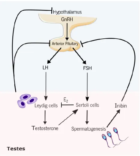
Spermatogenesis is tightly regulated by the hypothalamus-pituitary-testis (HPT) axis. The neurons of the hypothalamus produce the gonadotropin releasing hormone (GnRH), that stimulate gonadotroph cells to secret the follicle-stimulating hormone (FSH) and the luteinizing hormone (LH). These two pituitary hormones are responsible for the connection between brain and testis (69). Under physiological conditions, LH acts on LCs through LH receptors in the surface of these somatic cells and promotes the biosynthesis of testosterone. Testosterone then diffuses into the seminiferous tubules and systemic Figure 2. Schematic representation of the principal events during spermatogenesis process in seminiferous tubule. Successive mitosis occurs at a first stage in which each spermatogonia A, is divided and developed into two other cells, called spermatogonia B, which enter meiotic prophase and differentiate in primary spermatocytes. Primary spermatocytes undergo a first meiotic event occur whereas secondary spermatocytes are formed (haploid cells). A second meiotic event happens, and spermatids are formed. Spermatids go through a maturation process where it is formed fully developed spermatozoa. Then, spermatozoa are released in the lumen of seminiferous tubule.
22
circulation. Indeed, LH levels are positively correlated with testosterone plasma levels (60, 70). Additionally, FSH binds to its receptor in SCs (60) and its main goal is to sustain the male reproductive potential (60, 71).
The HPT axis is tightly regulated through feedback mechanisms to maintain the proper hormonal homeodynamics. In fact, the production of testosterone and 17β- estradiol (E2) by LCs and the production
of inhibin by SCs lead to a negative feedback loop that reduces the secretion of LH and FSH (72) (Figure 3).
Figure 3. Hormonal regulation of the male reproductive tract. The gonadotropin releasing hormone (GnRH) is synthesized by the hypothalamus, which will stimulate the anterior pituitary to produce the luteinizing hormone (LH) and follicle-stimulating hormone (FSH). LH and FSH bind membrane receptor on Leydig and Sertoli cells, respectively, leading to the stimulation of testosterone production and spermatogenesis. High testosterone levels inhibit the release LH and FSH by the anterior pituitary. Inibin production by Sertoli cells regulates FSH and LH production by anterior pituitary in a negative feedback on pituitary. 17β- estradiol (E2) acts on Leydig cells to inhibit the production of testosterone.
23
Besides FSH and LH, there are other hormones who deserves special attention, for instance, steroid hormones, thyroid hormones and insulin. Concerning the sex steroid hormones, androgens and estrogens, they are needed for a properly initiation and maintenance of spermatogenesis (73) and are important for development of the reproductive tract (74, 75). Androgens, particularly testosterone, is essential for normal spermatogenesis, in different stages, from initiation, maintenance to reinitiating of the whole process (76). They are necessary to maintain the integrity of the BTB and the assembly of junctional complexes (77, 78).
As well as androgens, estrogens are critical in the development and maintenance of the male reproductive function (4, 79, 80). Its synthesis is assured essentially by the LCs in adult men (81). Moreover, estrogens are able to regulate the HPT axis and thus, indirectly regulate LH and testosterone equilibrium. In fact, estrogens can disrupt the developmental of fetal LCs, leading to deficient proliferation and differentiation of gonocytes. They are also able to inhibit testosterone production by adult LCs (4, 82).
Insulin receptors have also been identified in SCs (83), and it has been described that insulin stimulates the proliferation of LCs, spermatogenesis and spermatic maturation (84). In fact, under insulin deprivation, SCs present an altered expression of metabolism-associated genes involved in the consumption of glucose (more particularly in GLUT1 and GLUT3 receptors), production and export of lactate (42).
Finally, thyroid hormones have an important role in testicular physiology and their deregulation leads to erectile dysfunction issues that could result in a decreased libido or even impotence (85). Thyroid glands produces tri-iodothyronine (T3), that controls the maturation and growth of the testis, by stimulating immature SCs to differentiate (86). These hormones play a critical role in the onset of LCs differentiation and stimulation of steroidogenesis in postnatal rodent testes (87).
1.5. Modulators of testicular metabolism
The modulation of SCs metabolism must ensure a cooperation between testicular cells and the correct function of several metabolic pathways. To achieve that, there is a complex of signals that count with the participation of multiple players, such as hormones, proteins, metabolic products, growth factors, cytokines and so on, that can trigger different signaling cascades. Despite that, other external factors, such as environmental factors, the lifestyle habits, and pathological conditions, that are linked to a disruption of signals and that can lead to infertility.
24
Over the last years, sirtuins came out as key modulators and targets in physiological and pathological events, such as obesity and cancer (88, 89). Nevertheless, only recently sirtuins were suggested as key metabolic sensors for testicular homeodynamics. Still, the information available is scarce.
1.5.1. Sirtuins
Acetylation and deacetylation are processes required for the post-translational regulation of protein activity. Acetylation is catalyzed by acetyltransferases that transfer an acetyl residue from acetyl-CoA to specific lysine residues in other proteins responsible for multiple cellular processes (e.g. histones). Opposite to acetylation, deacetylation corresponds to the removal of acetyl group from the lysine of acetylated proteins. This processes only happens due to enzymes known as lysine deacetylases (KDACs), particularly sirtuins (90).
Sirtuins belong to a highly conserved family of deacetylases, specifically class III (in a total of four classes), that depend on nicotinamide adenine dinucleotide (NAD+). This cofactor is essential for electron transfers
in an intermediate metabolism that is posteriorly converted into NADH, the reduced form (91). Thus, sirtuins are sensitive to fluctuations in the levels of NAD+, which will interfere with their activity, and with
substrate preference (92). In fact, the ratio NAD+/NADH is the essential piece in multiple metabolic processes such as glycolysis, Krebs cycle and electron transport in mitochondria (93).
Sirtuins respond to different signals like inflammatory signals or hypoxic/oxidative stress (88), metabolic challenges, such as type 2 diabetes and obesity, associated with insulin secretion (94-96). They are also associated with aging and longevity (97, 98), linked to processes like apoptosis and cell survival (99, 100), fatty acid oxidation (101), DNA repair, development and neuroprotection (95) and mitochondrial biogenesis (102).
This family of proteins is homologous to yeast transcriptional repressor, sir2 (90), which is highly conserved from bacteria to humans (103). Indeed, the first evidence on the existence of sirtuins arose when it was demonstrated that the human ortholog of yeast Sir2 can transfer 32P from NAD+ to bovine
serum albumin, indicating the role of Sir2 in mono ADP rybolysation of proteins (104). In the same year, Kaeberlein et al., demonstrated that an overexpression of Sir2 was associated to an increased lifespan (105). Posteriorly, the knowledge of this proteins was amplified and nowadays sirtuins have been found in different subcellular locations and have been classified in a family of seven members (SIRT1 to SIRT7), wherein SIRT1 is the closest phylogenetically to yeast Sir2 (106). Briefly, SIRT1 is predominantly nuclear (107), whereas SIRT2 is located mainly in cytoplasm (108), although it can translocate into the nucleus
25
as well (109, 110). SIRT3 to SIRT5 are mitochondrial proteins. Nevertheless, SIRT3, during cellular stress, can also translocate from the nucleus to the mitochondria. Finally, SIRT6 and 7 are nuclear sirtuins (111, 112). In terms of activity, SIRT1 to SIRT3 seems to have strong deacetylase activity, while SIRT4 to SIRT7 have weak or no detectable deacetylase activity (113-115). In addition to deacetylation, there are other enzymatic activities for sirtuins. For instance, SIRT4 is mostly responsible for the transfer of ADP-ribose to a specific protein - glutamate dehydrogenase (GDH), rather than releasing the deacylated products (deacetylation) (116, 117) and SIRT5 may act as a demalonylase, dessuccinylase and deglutarylase, in which the acid acyl moieties linked to the lysine residues are removed (114, 118).
The testicular metabolism is not an exception with regard to the maintenance of redox balance in order to guarantee the functioning of all processes of the male reproductive tract, particularly in what concerns to the NAD+ ratio. In testes, NAD+ appears to be a vital cofactor and can rewire testicular metabolism
events between cytoplasm and mitochondrial compartments (119).
Despite not knowing exactly its role in testicular metabolism, sirtuins are highly expressed in testicular tissue (120), and it seems that the high energy demand of testes is in part related with the restoring of NADH levels.
In 2003, the interest on the role sirtuins in male fertility was first aroused and their potential impact on the regulation of male fertility was addressed. The strategy adopted was to inhibit the activity of sirtuins in mice, creating several null mice transgenic models (121). Since then, many explanatory attempts were done to highlight effectively the role of sirtuins in male fertility.
1.5.1.1. Sirtuin 1 (SIRT1)
Sirtuin 1 is the mammalian homolog of the yeast silent information regulator 2 and is the most studied sirtuin (122). As mentioned above, SIRT1 is a NAD+ dependent histone deacetylase that has roles in
multiple biological processes, resulting in modification of the acetylation status of histones and other proteins (123). Physiologically, SIRT1 seems to have a role in the regulation of energy metabolism, autophagy, apoptosis, inflammation and senescence (124, 125). Chromatin remodeling and DNA repair, with the involvement in cellular processes ranging from stress responses to the activation of survival or death pathway, are also linked to sirtuins (126). For instance, SIRT1 deacetylates specific residues in histones, such as lysine 16 of histone 4 (H4K16), lysine 9 of histone 3 (H3K9), lysine 56 of histone 3 (H3K56) (127). The non-histone protein substrates are diverse, such as: the tumor suppressor p53 that has a role in regulating apoptosis (107, 128) and the nuclear factor kB (NF-kB) that controls inflammation
26
through deacetylation of p65 subunit, thereby inhibiting NF-kb signaling. The relationship between NF-kb signaling and SIRT1 has repercussions in many inflammatory diseases. When SIRT1 is inhibited, the acetylation levels of the p65 subunit increase, as well as the levels of nitric oxide, which are associated with the inflammation process (129). Moreover, SIRT1 is important as tumor suppressor, with the capacity to deacetylate and inhibit β-catenin transcriptional activity (130). Although, SIRT1 activity in cancer is still on debate, as high levels of SIRT1 could be linked to a resistance to chemotherapy (131, 132). Furthermore, Yeung et al., described the SIRT1 paradox, that consists of the ability of mediating pro-apoptotic or antiapoptotic effects (133).
Regarding the relationship that sirtuins have with cell metabolism, it is already known that SIRT1 has a role in linking diet and cell metabolism. For instance, 5' AMP-activated protein kinase (AMPK), which is a critical regulator of mitochondrial biogenesis in energy deprivation conditions (134), was descried as an enhancer of SIRT1 activity by increasing cellular NAD+ levels. Higher levels of NAD+ leads to the
deacetylation and modulation of the activity of downstream SIRT1 targets, that include peroxisome proliferator-activated receptor gamma coactivator (PGC-1-α) and the forkhead transcription factor (FOXO1) (135). PGC-1-α is a member of a family of transcriptional regulators that handle the expression of genes related to energy homeostasis, fatty acid oxidation, glucose metabolism and mitochondrial biogenesis (136, 137). In fact, SIRT1 can control mitochondrial biogenesis though regulation of PGC-1-α pathway (138). In the liver, SIRT1 is capable to maintain lipid homeostasis through PPARα mediated β-oxidation of fatty acids, evidencing that it has a role in glucose and lipid metabolism in fasting (139), and suggesting that gluconeogenesis in the liver is regulated by SIRT1 (140). Hence, SIRT1 controls the gluconeogenic/glycolytic pathways trough co-activator PGC-1- α.
Another key feature of SIRT1 is its negative regulation on the expression and phosphorylation of signal transducer and activator of transcription 3 (STAT3). Bernier et al., identified STAT3, which is a contributor to cellular respiration, as an additional SIRT1 target (141).
Nowadays, due to the western lifestyle, diabetes type 2 becomes to be like a pandemic disease, and SIRT1 seems to be involved in this pathology. It is mentioned that SIRT1 positively regulates insulin secretion in pancreatic β-cells by repressing uncoupling protein 2 (UCP2), that bounds directly to the UCP2 promoter. Its function is to uncouple oxygen consumption during respiration from the production of ATP (142).
27 1.5.1.2. SIRT1 in the testicular environment
Due to their role as metabolic sensors, sirtuins, and in particularly SIRT1, might have a relevance in the maintenance of the spermatogenesis (89). Several studies were done, and when the activity of SIRT1 is inhibited, there are immediate percussions on male lifespan. Moreover, SIRT1 knockout (KO) presented a reduction on testes size and sperm quality (121, 143). HPT axis was also affected, with decreased levels of the LH and FSH. So, SIRT1 appear to regulate spermatogenesis at postnatal stages by controlling HPT axis signaling (143). Cakir et al., (144) showed that SIRT1 affects spermatogenesis primarily by Figure 4.Sirtuin 1 (SIRT1) regulation signaling. Increased expression of SIRT1 results in deacetylation of non-histone and histone substrates, that affect multiple cellular functions. The deacetylase reaction catalyzed by sirtuins is based on the removal of the acetyl group of lysine residue in a reaction that consumes nicotinamide adenine dinucleotide (NAD+), releasing nicotinamide (NAM). Enhanced levels of
5' AMP-activated protein kinase (AMPK) lead in enhanced SIRT1 activity. SIRT1 activation can mediate PPARα, that has important roles in maintaining lipid homeostasis; forkhead transcription factor (FOXOs) which is important in the regulation of oxidative stress; peroxisome proliferator-activated receptor gamma coactivator 1 -α (PGC1-α) that regulates multiple signaling pathways related with fatty acid oxidation, glucose metabolism mitochondrial biogenesis. Furthermore, SIRT1 regulates insulin secretion by repressing uncoupling protein 2 (UCP2) gene. SIRT1 is also able to regulate apoptosis by deacetylation of p53 and controls inflammation through regulation of the nuclear factor kB (NF-kB) signaling. It also negatively regulates the expression and phosphorylation of signal transducer and activator of transcription 3 (STAT3).
28
disrupting normal hypothalamus-pituitary complex, since this enzyme is highly expressed at the hypothalamus and its inactivation reduces hypothalamic GnRH expression. To support this information, another study was done, showing that SIRT1 KO mice present a reduced number of mature LCs and, thus, an impairment of steroidogenisis was detected due to the decrease in protein levels of steroidogenic acute regulatory (StAR) protein, the enzyme responsible for a rate-limiting step of steroidogenesis. Anatomically, an absence of tubular lumen formation was observed in the testis in SIRT1 KO mice, which results from a loss of differentiation in SCs, illustrating that loss of SIRT1 could result in arrested spermatogenesis. Concomitantly, Coussens et al., demonstrated that SIRT1 KO mice display decreased sperm counts, with a high aberrant morphology and increased DNA damage (145). In addition, epididymal sperm from SIRT1 KO animals were immature and with reduced motility (146).
Failure of SIRT1 can also be correlated to the unbalance of the oxidative stress, which is one of the main causes of male infertility (147). Reduced levels of SIRT1 are associated to oxidative stress and it is believed that SIRT1 mediates cell apoptosis. Indeed, SIRT1 KO mice exhibit defects in chromatin condensation as well as defects in histone to protamine transition leading to sperm DNA prone to apoptotic/oxidative damage (148). Wu and Bratton showed that reactive oxygen species (ROS) has an impact in the mitochondrial pathway of apoptosis (149). Later on, Rato et al (2016) described that the loss of testicular SIRT1 and SIRT3 results in a lower antioxidant defenses and leads to a decreased mitochondrial function in the testes (119).
More recently, Liu et al. described that SIRT1 KO mice had severe alterations in germ cell development, with early mitotic and meiotic phases not being affected, but with the subsequent spermiogenesis event being compromised. Germ cell exhibited a defective acrosome biogenesis. Additionally, increased acetylation levels of LC3 (a central protein in autophagy) were detected. This affected the recruitment of acrosome biogenesis-related proteins to the acrosomal vesicles, leading to a decrease of autophagy. These data suggest a novel function for SIRT1, by regulating the acrosome biogenesis through SIRT1- mediated LC3 nucleocytoplasmic transportation (150). All these reports revealed that sirtuins have a role in male fertility, particularly in glycolysis, oxidation of fatty acids and oxidative stress, which are crucial pathways for spermatogenesis. They showed also that sirtuins deficiency leads to the formation of deficient spermatozoa. For these reasons, it is off great relevance to fully clarify the role of this protein, which can potentially be of assistance in the development of better in pharmacological treatments for male fertility and extend the reproductive lifespan of males.
29 1.6. Activators and inhibitors of Sirtuins
Sirtuins, and particularly SIRT1, are linked to the modulation of different signaling cascades. Sirtuins act as metabolic sensors and respond to the fluctuation of NAD+/ NADH, which is the fuel for the function of
multiple metabolic pathways, intrinsically related to several diseases and conditions, such as infertility. Furthermore, the role of some sirtuins, such as SIRT1 and SIRT3, in regulating mitochondrial and cellular energetics makes them a potential target for molecular therapeutics. The modulation of their functional status may have immediate impact on several aspects of cell physiology. So, either natural or synthetic, the inhibitors or activators of sirtuins activity may prove to be an important tool for improving the health status of individuals.
1.6.1. Sirtuin inhibitors
Over the last few years, several sirtuin inhibitors have been developed, and some have target specifically SIRT1, SIRT2, SIRT3 or SIRT5, with some compounds inhibiting more than one sirtuin although with different affinities. Sirtuin inhibitors can be potentially useful as therapeutic strategies. For example, SIRT1 is usually up-regulated in cancer cell lines (151, 152) and thus, its inhibition might repress cancer cell proliferation.
Sirtinol is an inhibitor of SIRT1 and SIRT2, with the capacity of reducing inflammation in capillary endothelial cells of the skin, modulating the expression of dermal cells, and preventing skin disorders (153). This compound is also known to have anticancer potentials, reducing the proliferation of breast cancer (MCF7 cell line) and lung cancer lines (H1299 cell lines) (154).
Another example is cambinol, which is used to inhibit SIRT1 or SIRT2 and SIRT5 (although with less inhibitory effect). It was discovered in 2001 and it is characterized by a weak sirtuin inhibitory activity (155). To overcome that, it has been administrated in a combination of other sirtuin inhibitors, cambinol and EX-527. The results were quite positive. Indeed, Livore et al., demonstrated that SIRT1 and SIRT2 are overexpressed in hepatocellular carcinoma, and that their expression is correlated with tumoral progression and multidrug resistance. When cambinol and EX-527 were administrated together, tumor cell viability and cell migration were reduced, and apoptosis was increased (156).
There are some reports of new compounds that inhibit other sirtuins, such as Nicotinamide and GW5074 that can inhibit SIRT5’s desuccinylation activity (157). Furthermore, there are some compounds with the ability to inhibit different sirtuins but with comparable potencies. Dish et al., found that ELT-11c exerts an
30
inhibitory effect over SIRT1, 2 and 3 (158). More recently, a resveratrol-related compound SDX-437 was described, having a stronger effectiveness against SIRT3 than against SIRT1 (159).
There are some more selective inhibitors, i.e. that present a considerable isoform-specificity. For instance, AGK2 and MIND4 are SIRT2 inhibitors that have protective effects in neurological disorders, being used as pharmacological agents for Parkinson’s and Huntington’s disease models, respectively (160, 161). Another aspect to consider is the solubility of the compound. Lain et al., described Tenovin-6, which is water-soluble and has more affinity for SIRT2, then for SIRT1, and less for SIRT3. It was reported as a pharmacological agent for inducing apoptosis in gastric cancer cells (162) and slow down the progression in models of chronic myeloid leukemia (163).
The sirtuin inhibitor EX-527, also known as Selisistat is the only selective inhibitor to SIRT1 used in the clinic environment, that easily penetrates into cells. It was tested both in healthy humans volunteers and in Huntington’s disease (HD) patients and no toxicity was detected (164). This later study demonstrated that EX-527 is well tolerated with no adverse effects in circulating levels (165). In sum, SIRT1 inhibition in HD with EX-527 seemed to alleviate the symptoms of this disease (166).
Moreover, a protective effect of EX-527 on cerebral ischemia-reperfusion was observed (167). EX-527 was also used with necrostatin-1, an inhibitor of necroptosis, to investigate what were the percussions at neuroprotective levels. Necroptosis is a type of programmed cell death, that is involved in ischemia-reperfusion-induced brain injury. It was previously known that SIRT1 has a crucial role on neural loss and this study demonstrated it can relieve ischemia, having a neuroprotective potential and thus preventing brain stroke. Concerning neuronal/cognitive disorders, it was further described that EX-527 has a positive effect on depression or anxiety-like behaviors (168).
Additionally to this, it was demonstrated that EX-527 has also benefic effects on acute lung injury (169), and block the amplification of human papillomavirus (170). In cancer, the combination of EX-527 and AGK2, inhibiting SIRT1 and SIRT2 respectively, was able to reduce cell migration by suppressing the HSF1 protein. This protein affects multiple molecular pathways involved in the regulation of cellular migration and cell protection (171-173). It is believed that activation of HSPF1/HSP27 (Heat shock protein 27) is dependent of SIRT1 and 2 pathway and, when these are inhibited, HSF1 ubiquitination and degradation in vitro is induced, demonstrating that these SIRTs are important for heat shock response signaling activation (174).
In the fertility field, more specifically in the female reproductive system, the activity of sirtuins was described in early stages of oocyte maturation, in events such as histone deacetylation (175). Indeed, when EX-527 is administrated in vitro to mouse oocytes, it causes an increase in ROS production and
31
abnormal metaphase II plates, demonstrating that SIRT1 has an important role in oocyte maturation (176).
As shown in Table 1, more attention has been given to the pharmacological applications of sirtuins inhibitors, and a new era of therapeutics possibilities has arisen. Still, further studies must be made to provide new selective molecules in this pharmacological field.
1.6.2. Sirtuins activators
Caloric restriction was described to improve the health and extend the lifespan of mammals. This is associated with increased levels of SIRT1 (177), and thus the discovery and the development of new activating drugs has attracted great interest in order to achieve better outcomes. So, over the last few years, attempts were made to increase the pharmacokinetic and pharmacodynamic efficiency of several compounds known to be sirtuin activators.
Resveratrol (RSV) is a natural polyphenol that can be found in red wine and grape skins (178). It was the first sirtuin activator to be described and it was said to increase the lifespan in a range of models, from yeast to worms (179, 180). RSV seems to prevent also neurodegenerative diseases that are correlated with aging (181). It can mimic caloric restriction, preventing the deleterious effects of high-fat diets (182). In mice under a high-fed diet, Person et al., found out that RSV administration improved aortic elasticity, motor coordination and preserved bone mineral density. It also decreased inflammation and apoptosis in vascular endothelium (183).
The fact that RSV has a role in aging and energy spending, aroused interest in the discovery of new molecules, with higher efficacy that this natural polyphenol. One example is SRT1720, which has higher specificity for SIRT1 than SIRT2 and SIRT3 and can counteract multiple metabolic disorders. Indeed, it has been reported that the use of this compound causes a reduction of liver triglyceride content and expression of lipogenic genes in obese an insulin resistant mice, resulting also in an extension of lifespan (184-186). Moreover, it exhibits anti-cancer properties, by inducing apoptosis and a decreased of cell growth in myeloma cells (187). There are also available some nutraceutical formulations with RSV. For instance, ResVida formulation (administration of 150 mg/day) exhibited positive effects in obese men, decreasing circulating levels of glucose and triglycerides, and increasing lipid breakdown (188).
SRT2104 is one of the most studied sirtuin activators. Different studies were made, and some of them demonstrated that it has an effect on lipid metabolization (189), although with no effects in terms of insulin control or improved glucose handling, indicating that SRT2014 might has a modest activity on
32
SIRT function. It showed some promising results, although it is necessary to improve pharmacokinetics and the bioavailability of this compound upon oral administration (190).
SRT501 has a stronger impact than RSV on SIRT1, enhancing several metabolic signaling pathways, blunting pro-inflammatory pathways, and enhancing mitochondrial biogenesis. In addition, it can lower blood glucose levels and improve insulin sensitivity in patients with type 2 diabetes (191, 192).
Another polyphenol with SIRT activating activity is quercetin, (which can be found in Black mulberry, Morus nigra). This compound has hypoglycemic, hypotensive, anti-tumoral and anti-inflammatory effects (193, 194). Still further research is necessary in order to develop new derivatives of quercetin because this compound has low bioavailability. Up until now, two quercetin derivates, diquercetin and 2-chloro-1,4-naphtoquinone-quercetin, were studied. These compounds exhibited an action on SIRT6, which is involved in metabolism and has a role in DNA damage signaling and repair (195, 196). The available data demonstrated that quercetin based derivates are an alternative approach in the regulation of some of these mechanisms. Interestingly quercetin has been reported with a synergy with resveratrol, causing an upregulation of SIRT1 and SIRT2 activities (197).
Curcumin is a natural SIRT activator that belongs to ginger family. It displays lots of benefits on cancer (198, 199), diabetes (200), nonalcoholic fatty liver diseases (201), respiratory diseases (202), anxiety and depression (203). In a very general way, when curcumin is present, the reduction of ATP promoted by curcumin leads to AMPK activation, which in turns increases the NAD+ levels and consequently leads
to the activation of SIRTs, which mediate protective effects against different disorders (204).
The influence of SIRTs activators is still an issue that deserves special attention, particularly when reduced SIRTs levels are associated with some disorders. An example is YK-3-237, that initially was identified as a compound with antiproliferative effects in different cancer cell lines. However, its mechanism of action was unknown (205). Yi et al., were able to describe its SIRT promoting activities, after inducing deacetylation of a triple negative breast cancer (TNBC) cell line carrying different p53 status, a mutant form (mtp53). This resulted in a suppression of cell proliferation and arresting cell growth (206). Moreover, as SIRT1 activation has been shown to have a role in renal fibrosis by protecting the kidney from acute injury, it was described that exposure to YK-3-237 resulted in an enhancement of α-SMA and fibronectin expression (fibroblast activation markers), which in turns aggravates renal fibrosis in a dose-dependent manner (10 µM). This data demonstrates that SIRT1 has an important role in mediating activation of renal interstitial fibroblast (207).
33 Table 1. Principal sirtuin inhibitors and activators.
Compound Sirtuin Target Effects Reference Inhibitors
Sirtinol Sirtuin 1 Sirtuin 2
Skin disorders
Slows down proliferation I breast and lung cancer line
(153, 154) Cambinol
Sirtuin 1 Sirtuin 2
Sirtuin 5* Weak effects (155, 156) ELT-11c
Sirtuin 1 Sirtuin 2
Sirtuin 3 ND (158) MIND4 Sirtuin 2 Huntington’s disease (160, 161) AGK2 Sirtuin 1 Sirtuin 2 Parkinson’s disease and Huntington’s disease
Suppresses cell migration in cancer
(160, 161, 172, 173) Tenovin-6
Sirtuin 1* Sirtuin 2 Sirtuin 3**
Induces apoptosis in gastric cancer cells
Slows down proliferation in chronic myeloid leukemia cells
(162, 163) EX-527 Sirtuin 1 Sirtuin 2 *
Sirtuin 3 *
Neuroprotective effects Anxiety and depression Suppress cell migration
(166, 168, 173, 174) Activators
Resveratrol Sirtuin 1 Neurodegenerative disorders Positive effects in obesity (182, 183) SRT1720 Sirtuin 1 Sirtuin 2*
Sirtuin 3*
Effects on metabolic disorders Induces apoptosis
Decreases cell growth
(184-187) SRT2104 Sirtuin 1 Psoriasis Improves lipid parameters (189, 190) SRT501 Sirtuin 1
Improves metabolic pathways Enhances mitochondrial biogenesis Improve insulin sensitivity in type 2 diabetes (191, 192) Quercetin Sirtuin 1 Sirtuin 2 Sirtuin 6
DNA damage and repair (195-197) Curcumin Sirtuin 1
Cancer Diabetes
Nonalcoholic fatty liver diseases Respiratory diseases
Anxiety and depression
(198-202) YK-3-237 Sirtuin 1 TNBC Renal fibrosis (206, 207)
34
Chapter II
35 2. AIM OF THE PROJECT
Over the past years, sirtuins have been recognized as sensors in multiple cellular events, being able to integrate a network of signaling pathways. SIRT1, in particular, when activated/ or inhibited translates into different behaviors that are relevant in the normal functioning of living cells, particularly to their metabolic phenotype. There are several studies made with SIRTs in cancer-related areas, however less attention has been given to their role in non-cancerous cells, particularly in cells that present a “Warburg-like metabolism”, “Warburg-like SCs.
YK-3-327 and EX-527 are sirtuin modulators (activator and inhibitor, respectively) of SIRT1. Up to date there is no evidence of the impact of these compounds in testicular metabolism. This work aimed to unravel the actions of these two compounds on testicular cells metabolism, particularly in mouse SCs (mSCs), since spermatogenesis is extremely dependent on the metabolism of these cells, as described previously. Considering that SIRT1 may modulate glycolytic metabolism, we hypothesized that its activation or inhibition may have percussions on testicular metabolism and can control male fertility. Specifically, we aimed to:
1. Identify SIRT1 expression in the TM4 Sertoli cell line;
2. Evaluate the cytotoxic profile of the exposure to a SITR1 activator (YK-3-237) and inhibitor (EX-527) in the TM4 Sertoli cell line;
3. Evaluate the glycolytic profile of TM4 Sertoli cells after the exposure of EX-527 and YK-3-237; 4. Evaluate the oxidative status of TM4 Sertoli cell after the exposure of EX-527 and YK-3-237; 5. Determine the glycogenic and lipid reserves on TM4 Sertoli cells after the exposure of EX-527
36
Chapter III
37 3. MATERIALS AND METHODS
3.1. Chemicals
Fetal Bovine Serum (FBS) was obtained from Millipore (Darmstadt, Germany). Dulbecco’s Modified Eagle Medium, Ham’s Nutrient Mixture F12 (DMEM: Ham’s F12), Ethylene Diamine Bovine Serum Albumin (BSA), trypsin–EDTA, Oil Red O solution, EX-527 and YK-3-237 were all purchased from Sigma–Aldrich (St. Louis, MO, USA). Sulforhodamine B (SRB) was purchased from Biotium (Hayward, CA, USA). 3-(4,5-dimethylthiazol-2-yl)-2,5-diphenyltetrazolium bromide (MTT) was purchased from Amresco (Solon, OH, USA). LDH CitoxTM Assay Kit was obtained from BioLegend® (San Diego, CA, USA).
5,5′,6,6′-tetrachloro-1,1′,3,3′-tetraethylbenzimidazolylcarbocyanine iodide (JC-1) dye was purchased from Life Tecnhologies (Gaitherburg, MD, USA). RIPAS Lysis Extraction Reagent and BCA Protein Assay Kit were obtained from Thermo Scientific (Whalthan, MA, USA). WesternBrightTM ECL substrate was purchased from Advansta (Menlo Park, CA, USA). Dried milk was obtained from Regilat (Saint-Martin-Belle-Roche, France). EFCTM
Western Blotting Reagent was obtained from GE HealthCare Life Sciences (USA). 3.2. Cell culture conditions
Cell line of TM4 mouse Sertoli cells (mSCs) were purchased from ATCC (Manasas,VA, USA). In brief, mSCs were cultured in 75-cm2 flasks (VWR collection, Amadora, Portugal) at 37ºC, 5% CO2 and maintained
in Sertoli culture medium (DMEM:Ham’s F12 1:1, supplemented with 10% heat inactivated FBS, 50 µm/mL gentamicin, 1% Pen-Strep, 1% amphotericin B, 15 mM HEPES, 14 mM NaHCO3 and 18 mM
Glucose). TM4 is a Sertoli cell line derived from the prepurbertal BALB/c nu/+ mouse exhibiting the capacity to respond to FSH with an increase in cAMP production and does not respond to LH. It is recognized as non-tumorigenic cell line and is negative for ectromelia virus (208).
3.3. Experimental Groups
Sertoli cell were allowed to grow until they reached 90-95% confluence, and then washed thoroughly with phosphate buffered saline (PBS) solution. The medium was replaced by the culture medium supplemented with increasing concentrations of the sirtuin inhibitor (EX-527) or activator (YK-3-237). To evaluate the effect of EX-527 and YK-3-237 we defined five concentrations (1 nM, 10 nM, 100 nM, 1000
38
nM and 10000 nM) and as a control condition we used culture medium with same volume of DMSO (the vehicle compound where EX-527 and YK-3-237 were dissolved). These concentrations were chosen based on the IC50 value of EX-527 (nearly 100 nM). The same range of concentrations were used with YK-3-237
and were chosen based on the maximum inhibitory concentration used in a renal fibrosis case study (10000 nM) (207). Then, the total number of cells was determined using a Neubauer chamber and the cells were collected for protein extraction and enzymatic assays. The extracellular medium was collected for 1H-NMR analysis.
3.4. Sulforhodamine B (SRB) cytotoxicity assay
The cytotoxicity of EX-527 and YK-3-237 was evaluated by the colorimetric SRB assay (209). Briefly, SCs were seeded in a 24-well culture plate, let to grow until reaching 60-70% confluence and then treated with the different concentrations of either the sirtuin activator or inhibitor during 24 hours. After treatment, cells were washed with PBS and fixed in 1% acetic acid in methanol for 1 hour at -20ºC. Next, cells were stained with 0,05% (w/v) SRB dissolved in 1% of acetic acid for 1 hour at 37ºC. Afterwards, unbound SRB was removed through washing with 1% acetic acid solution. SRB bound to cell proteins was extracted afterwards, with 10 mM Tris solution (pH 10) in a shaker for 10 minutes at room temperature. Then, 100 µL of this solution were transferred to a 96-well culture plate. The optical densities of the resulting media were determined at 490 nm. The percentage of cell proliferation in each treated group was calculated with a normalization to control group.
3.5. Lactate Dehydrogenase (LDH) release assay
LDH release was determined by measuring the extracellular activity using a commercial assay kit following the manufacturers’ instructions (LDH CitoxTM Assay Kit, BioLegend®, San Diego, CA, USA). To sum up,
after 24 hours of the treatment, 100 µL of extracellular media were collected into a 96 well plate, to which were added 100 µL of working solution. LDH assay substrate was added to all samples in a dark environment and this mixture was incubated at 37ºC for 15 minutes. After that time, a 50 µL stop solution was added to stop the enzymatic activity and absorbance was read at 490 nm using Bio-Rad model 680 microplate reader. LDH release of each treated group was calculated with a normalization to control group