Contribution of CD30/CD153 but not of CD27/CD70,
CD134/OX40L, or CD137/4-1BBL to the optimal induction
of protective immunity to Mycobacterium avium
Manuela Flo´rido,* Margarida Borges,* Hideo Yagita,
†and Rui Appelberg*
,‡,1*Laboratory of Microbiology and Immunology of Infection, Institute for Molecular and Cell Biology, and
‡ICBAS,
Instituto de Cieˆncias Biome´dicas de Abel Salazar, University of Porto, Portugal; and
†Department of Immunology,
Juntendo University School of Medicine, Tokyo, Japan
Abstract:
A panel of monoclonal antibodies
spe-cific for CD27 ligand (CD70), CD30 ligand
(CD153), CD134 ligand (OX40L), and CD137
li-gand (4-1BBL) were screened in vivo for their
ability to affect the control of Mycobacterium
avium infection in C57Bl/6 mice. Only the
block-ing of CD153 led to increased mycobacterial
bur-dens. We then used CD30-deficient mice and found
an increase in the proliferation of two strains of M.
avium in these mice as compared with control
an-imals. The increased mycobacterial growth was
as-sociated with decreased T cell expansion and
re-duced interferon-
␥ (IFN-␥) responses as a result of
reduced polarization of the antigen-specific,
IFN-␥-producing T cells. At late times but not early in
infection, the lymphoid cuff surrounding
granulo-mas was depleted in the CD30-deficient animals.
This report expands our knowledge about tumor
necrosis factor superfamily members involved in
the immune responses to mycobacterial infection
by identifying CD30 –CD153 interactions as
re-quired for optimal immune control of M. avium
infection. J. Leukoc. Biol. 76: 1039 –1046; 2004.
Key Words:
mycobacteria
䡠macrophages
䡠cell-mediated
immu-nity
INTRODUCTION
Tumor necrosis factor (TNF) and its receptors are structurally
related to an increasing number of molecules belonging to two
superfamilies: the TNF superfamily (TNFSF) and the TNF
receptor superfamily (TNFRSF). These receptor-ligand pairs of
molecules play diverse roles in inflammation, in the immune
response, in organogenesis of lymphoid and bone tissues and
other body structures, and in apoptosis [1, 2]. Although most
information about the role of these molecules has been
ob-tained studying the cytokines TNF and lymphotoxin and their
receptors, newer data about other members of these families
have highlighted their participation in the regulation of the
immune response. Thus, the engagement of certain members of
the TNFRSF appears to be important for costimulation of T cell
immunity [2]. Among these, recent data have emerged about
the role played by the TNFRSF members CD27 (TNFRSF7),
CD30 (TNFRSF8), CD134 (TNFRSF4 or OX40), and CD137
(TNFRSF9 or 4-1BB) and their ligands CD70 (TNFSF7),
CD153 (TNFSF8), OX40L (TNFSF4), and 4-1BBL (TNFSF9),
respectively.
CD27/CD70 interactions participate in the early phases of T
cell responses [2]. Engaging CD27 on T cells activates these
cells, leading to cytokine secretion and proliferation, and
po-tentiates the activity of other accessory molecules [3]. Mice
deficient in CD27 have impaired responses to influenza virus
and reduced T cell memory generation [4]. Conversely, chronic
in vivo triggering of CD27 by expression of a CD70 transgene
led to massive activation and proliferation of T cells with
conversion of naı¨ve cells into cells with an activated/memory
phenotype [5, 6]. In vivo experimental models of experimental
autoimmune encephalomyelitis (EAE), an inflammatory
condi-tion dependent on a type 1 T cell response, have highlighted a
role for CD27/CD70 in the induction of the T cells responsible
for such neurological pathology [7].
The function of CD30 is controversial, as it has been said to
be a marker for T helper cell type 2 (Th2) cells [8] and required
for T cell-negative selection [9], and both claims have been
questioned subsequently [10, 11]. CD30 expression on T cells
is induced by interleukin (IL)-4 and CD28 stimulation [12, 13],
and its engagement may lead to enhanced proliferation [12] or
on the contrary, prime the lymphocyte for apoptotic death by
promoting TNFR-associated factor (TRAF)2 degradation [14,
15]. A virally encoded homologue of CD30 was shown to block
the development of inflammation and interferon-
␥ (IFN-␥)
production during priming with mycobacterial antigens [16],
but nothing is known about the role of CD30 in mycobacterial
infections.
Early T cell responses appear not to require the participation
of OX40/OX40L or 4-1BB/4-1BBL, but subsequent
accumu-lation of effector and memory cells does involve the activity of
these molecules [2]. The role of OX40/OX40L in the
develop-ment of Th2 responses is well docudevelop-mented, namely in an
1Correspondence: Laboratory of Microbiology and Immunology of Infection,
Institute for Molecular and Cell Biology, Rua do Campo Alegre 823, 4150-180 Porto, Portugal. E-mail: [email protected]
Received November 19, 2003; revised May 14, 2004; accepted July 14, 2004; doi: 10.1189/jlb.1103572.
experimental model of asthma [17] and in an infection model
with Leishmania major [18]. In contrast, OX40 deficiency did
not affect the protective Th1 responses in resistant mice
in-fected with L. major [19]. Also, blocking the activity of OX40L
did not affect the development of pathogenic, IFN-
␥-producing
T cells during EAE but still protected against the disease by
reducing the migration of those cells [20]. Similar protection
from EAE was observed after administration of soluble OX40
[21]. In contrast, IFN-
␥ responses to lymphocytic
choriomen-ingitis virus (LCMV) were reduced in OX40-deficient animals
as compared with control mice [22], and OX40 ligand-deficient
animals had impaired IFN-
␥ responses during hypersensitivity
reactions and in mixed leukocyte reactions initiated by
allo-geneic dendritic cells [23]. Triggering of 4-1BB (CD137) on T
cells appears to stimulate IFN-
␥ responses [24–26]. In one
study, 4-1BBL-deficient mice were particularly deficient in the
CD8 T cell response to infection by LCMV [27], whereas in
another study, 4-1BBL-deficient mice mounted normal,
cyto-lytic responses to LCMV but reduced responses to influenza
virus [28].
Here, we studied the effects of neutralizing single pairs of
the TNFRSF/TNFSF members discussed above in the
devel-opment of protective immunity to Mycobacterium avium. Of the
four pairs of interacting molecules, we identified CD30/CD153
interactions as necessary for the optimal induction of immunity
to M. avium and confirmed these observations using a mouse
model where the CD30-encoding gene was disrupted.
MATERIALS AND METHODS
Mice
Female C57Bl/6 mice were purchased from Harlan Iberica (Barcelona, Splain). C57B1/6-CD30–/–(B6.CD30⫺/⫺) mice were kindly supplied by Dr. Tak Mak
(Amgen, Toronto, Canada) and bred in our facilities.
Bacteria
M. avium strain 25291, exhibiting a smooth transparent (SmT) morphotype, was obtained from American Type Culture Collection (Manassas, VA). M. avium strains 2447 and 1983, with SmT morphotype, were isolated from an AIDS patient and a human immunodeficiency virus-negative patient, respec-tively [29]. Mycobacteria were grown in Middlebrook 7H9 medium (Difco, Detroit, MI) containing 0.04% Tween 80 (Sigma Chemical Co., St. Louis, MO) at 37°C until the mid-log phase of growth. Bacteria were harvested by cen-trifugation and resuspended in a small volume of saline containing 0.04% Tween 80. The bacterial suspension was sonicated briefly with a Branson sonifier (Danbury, CT) to disrupt bacterial clumps, diluted, and stored in aliquots at –70°C until used. One aliquot was thawed at 37°C and used to determine the concentration of mycobacteria in the inocula after plating serial dilutions of the suspension in a Middlebrook 7H10 agar medium (Difco) supplemented with oleic acid-albumin-dextrose-catalase. Before inoculation, bacterial aliquots were thawed at 37°C and diluted in saline to the desired concentration.
In vivo infection
Mice were infected intravenously (i.v.) with 106colony-forming units (CFU) of
M. avium through a lateral tail vein. Infected mice were killed at different time-points of infection, and the livers, spleens, and lungs were aseptically collected and homogenized in a 0.04% Tween 80 solution in distilled water. The number of CFU of M. avium in the organs of the infected mice was determined by serial dilution and plating of the tissue homogenates on 7H10 agar medium. The number of bacterial colonies was counted after culture for
2 weeks at 37°C. Statistical comparisons of the mycobacterial loads between deficient and control mice were performed using the Student's t-test.
In vivo antibody treatments
C57Bl/6 mice were treated with monoclonal antibodies (mAb) specific for CD70 [FR70, rat immunoglobulin G (IgG)2b; ref. 30], OX40L (RM134L, rat IgG2b) [31], CD153 (RM153, rat IgG2b) [32], or 4-1BBL (TKS1, rat IgG2a) [33]. All these mAb were purified from ascites by protein G affinity chroma-tography. Nonimmune IgG was purified from sera of normal rats by protein G chromatography. mAb or nonimmune IgG were given intraperitoneally, starting on the day of the infection and treating twice a week for up to 3 months, using 250g antibody per dose.
In vitro stimulation of splenic cells
Single-cell suspensions from spleens of each of the infected mice were pre-pared by teasing portions of the spleen with forceps in Dulbecco’s modified Eagle tissue-culture medium (DMEM; Life Technologies, Paisley, UK), sup-plemented with 10% fetal calf serum (FCS; Life Technologies). Erythrocytes were lysed by incubation of the cell suspensions with hemolytic buffer (155 mM NH4Cl, 10 mM KHCO3, pH 7.2) for 5 min at room temperature. The cell
suspensions were then thoroughly washed with Hanks’ balanced salt solution (Life Technologies) and resuspended in DMEM with 10% FCS. Cells were cultivated at a density of 2⫻ 105cells/well in a U-bottom, 96-well microtiter
plate. Cells were incubated in triplicate in DMEM with 10% FCS with no further stimulus or in the presence of mycobacterial envelope proteins (4 g/ml). Supernatants from the cultures were collected after 96 h of culture, and the IFN-␥ produced was quantified by a two-site sandwich enzyme-linked immunosorbent assay (ELISA) method using anti-IFN-␥-specific, affinity-pu-rified mAb (R4-6A2 as capture and biotinylated AN-18 as detecting antibody), and a standard curve was generated with known amounts of recombinant murine IFN-␥ (Genzyme, Cambridge, CA). The sensitivity of the assay was 30 pg/ml. To determine the frequency of IFN-␥-producing cells, CD4⫹T cells were purified as described below and studied in an ELISpot assay. Microtiter plates were coated with 0.25g/well monoclonal anti-mouse IFN-␥ (cell line R4-6A2), and after overnight incubation at 4°C, plates were emptied and blocked for 2 h with phosphate-buffered saline (PBS) containing 3% bovine serum albumin (BSA) and 0.05% Tween 20 and washed four times with PBS/Tween 20. Cells were cultured directly in the microtiter plates in dupli-cates in the presence of 4g/mL mycobacterial envelope proteins for 24 h at 37°C in 7% CO2atmosphere. For each cell sample, six serial, twofold dilutions
were done from a starting concentration of 4 ⫻ 105CD4⫹ cells plus 2⫻
106-irradiated antigen-presenting cells (APC)/well. Cells were removed by
washing the plates, and cytokine secretion was detected using 0.25g/well biotin-labeled rat anti-mouse mAb (cell line AN18) and 0.1g/well phos-phatase-conjugated streptavidin. The enzyme reaction was developed with a solution of 0.9 mg 5-bromo-4-chloro-3-indolylphosphate per ml substrate buffer (0.74 mM MgCl2, 0.1% Triton-X405, 9.6% 2-amino-2-methyl-1-propa-nol, pH 10.25) containing 0.6% agarose. Blue spots were counted microscop-ically, and the relationship between the number of spots developed per well and the number of input cells was determined. Mycobacterial envelope pro-teins were prepared from M. avium as described previously [34].
Purification and culture of CD4
⫹T cells
Cells were suspended in PBS containing 0.5% BSA and 2 mM EDTA, pH 7.2, at a concentration of 108cells/ml and incubated with micro-beads coated with
anti-CD4 mAb (10l/107cells; clone L3T4; Miltenyi Biotec, Germany) for 20
min at 4°C. Cells were washed, resuspended in PBS/BSA/EDTA at a concen-tration of 2⫻ 108cells/mL, and filtered through a 70-m nylon cell strainer
(BD Falcon, BD Biosciences, San Jose, CA) to remove cell debris and cell clumps. CD4⫹T cells were positively selected by using MidiMACS separation columns (Miltenyi Biotec), as described in the instructions from the manufac-turer. The selected cells were resuspended in complete DMEM medium and cultured in triplicates in a 96-well plate at a density of 1⫻ 106cells/ml in the
presence of irradiated (5000 Rad), nucleated spleen cells as APC at a density of 5⫻ 106cells/mL and stimulated with mycobacterial envelope proteins (4
g/mL) or concanavalin A (Con A; 4 g/mL) for 72 h at 37°C in a 7% CO2
Lymphocyte proliferation assay
Cell proliferation was measured by [3H]-labeled deoxy-thymidine ([3H]-TdR)
incorporation. Briefly, proliferative responses were assessed after 48 h of culture in a humidified atmosphere of 7% CO2. Cultures were pulsed with 0.5
Ci [3H]-TdR (Amersham, Little Chalfont, UK) for 24 h before harvesting, and
incorporation of [3H]-TdR was measured using liquid scintillation. Results are
expressed as mean counts per minute (cpm) of triplicate cultures.
Reverse transcriptase-polymerase chain reaction
(RT-PCR)
In vivo production of IL-12p40 was studied by RT-PCR analysis. Total RNA was extracted from a small portion of liver tissue of individual mice after lysis in guanidinium isothiocyanate buffer. Reverse transcription was performed using an Invitrogen kit. The message for hypoxanthine phosphorybosyltrans-ferase (HPRT) was amplified using specific primers. All samples were stan-dardized for approximately the same HPRT expression level, and this level was verified to be below saturation point by comparison with a curve generated by serial dilutions of one of the cDNAs. The same amounts of cDNA were then used to PCR-amplify the IL-12p40 message, using primers with the following sequences: sense: CGT GCT CAT GGC TGG TGC AAA; antisense: CTT CAT CTG CAA GTT CTT GGG. The PCR products of both messages were run in an agarose gel containing ethidium bromide. The gel was photographed, and the image was analyzed to evaluate band intensity using ImageQuant software.
Flow cytometry
For the immunofluorescence staining, 106cells were incubated in a 96-well
microtiter plate with fluorescein isothiocyanate (FITC)-conjugated anti-CD4 antibody (dilution 1:100) and phycoerythrin (PE)-conjugated CD8 anti-body (dilution 1:100) or FITC-conjugated anti-CD19 antianti-body (dilution 1:100) and PE-conjugated antimembrane-activated complex 1 antibody (dilution 1:100) in PBS containing 3% fetal calf serum (FCS). All antibodies were from BD PharMingen (San Diego, CA). The cells were washed twice with PBS containing 3% FCS, and propidium iodide (Sigma Chemical Co.) was added to the cells at a final concentration of 1g/mL to allow the exclusion of dead cells. The analysis of the cell populations was based on the acquisition of 10,000 events in a Becton Dickinson FACSort equipped with CELL-Quest software.
Histology
Portions of the organs of the infected mice were fixed in buffered formaldehyde and embedded in paraffin. Sections were stained with hematoxylin and eosin or stained for acid-fast bacteria with carbol-fuchsin and counterstained with methylene blue.
RESULTS
We initially screened our panel of mAb in two independent
experiments using mice infected with strain 2447 of M. avium.
Neither anti-CD70 (FR70) nor anti-OX40 ligand (RM134L)
mAb affected the course of the infection in terms of
mycobac-terial growth (Fig. 1, upper row). In contrast, administration of
anti-CD153 mAb (RM153), which blocks its interaction with
CD30, resulted in the exacerbation of the infection as detected
at the 12th week post-infection (Fig. 1, upper row). In the
second experiment, we studied the effect of the neutralization
of interactions between CD137 and its ligand (4-1BBL)
follow-ing administration of the TKS-1 mAb and found no effect on
mycobacterial proliferation (Fig. 1, lower row). When T cells
from spleens of these groups of animals were stimulated in vitro
with M. avium antigen, no effects on the amounts of IFN-
␥
secreted were observed for any group as compared with
con-trols (data not shown).
Given the protracted nature of the infection and the need for
rather long periods of antibody administration, which may
result in ineffectiveness of the treatment along its course, we
then used CD30-deficient mice to validate our results obtained
from the antibody treatments. B6.CD30
⫺/⫺mice were infected
with the strain of M. avium used in the first set of experiments
as well as with two additional ones, a less virulent strain (M.
avium 1983) and a highly virulent strain, which is not
con-trolled by the immune response of the infected mice (M. avium
ATCC 25291) [35, 36]. As shown in Figure 2, B6.CD30
⫺/⫺mice were more susceptible to strains 2447 and 1983 than
control B6 animals. Not surprisingly, both mouse strains
al-lowed similar growth of the highly virulent 25291 strain, which
resists the immune response effector mechanisms [35].
Histologically, there were no differences in the overall
ar-chitecture of the granulomas formed in the wild-type as
com-pared with the CD30-deficient mice until the 3rd month of
infection (data not shown). However, at later time-points of
infection (238 days) with strain 2447, a marked depletion of
the lymphoid cuff surrounding the granulomas was evident
(Fig. 3). The area depleted of lymphocytes appeared as an
empty area, where fibrotic material could be visualized, and
corresponded to where the lymphocytes were previously found
in the granulomas of the mutant strain. When flow cytometric
analysis of the spleen cell populations was performed, some
differences were detected early in infection (Table 1). CD4
⫹T cells expanded less in CD30-deficient than in control mice
infected with strain 2447 (2.7-fold as compared with 3.4-fold at
day 30 of infection) and did not expand in CD30-deficient mice
in response to infection with strain 1983, whereas a delayed
expansion was observed in the controls. Both mouse strains
Fig. 1. Proliferation of M. avium 2447 in C57Bl/6 mice treated with nonim-mune rat IgG (f) or specific antibodies directed against CD153 (e, upper row), OX40L (CD134L;䡬, upper row), CD70 (‚, upper row), or the ligand for 4-1BB (CD137; e, lower row). Data points represent the means of the log10 CFU in the organs of five mice per group, and the bars represent theSD. Statistically significant differences between the treated and control groups are labeled with * (P⬍0.05) or ** (P⬍0.01).
exhibited profound T cell loss during infection with the highly
virulent 25291 strain as previously shown [35, 36]. A similar
deficiency was evident for B cells during infection with strain
1983 and at 90 days, with strain 2447. Some reduction in the
accumulation of macrophages was also evident in
CD30-defi-cient mice during the infection with strain 1983. At 238 days
of infection with strain 2447, the numbers of lymphoid cells
were further reduced in the infected CD30-deficient mice as
compared with control-infected mice (Table 2): CD4
⫹and
CD8
⫹T cells from infected B6.CD30
⫺/⫺mice were 31% of
those in infected C57Bl/6 mice and 55% and 35%,
respec-tively, of uninfected B6.CD30
⫺/⫺mice. CD19
⫹B cells from
infected B6.CD30
⫺/⫺mice were 8% of those in infected
C57Bl/6 mice and 18% of those in age-matched, noninfected
B6.CD30
⫺/⫺animals. No reduction in lymphoid cell numbers
was induced by infection in C57Bl/6 mice. At this late
time-point of infection, the increased mycobacterial loads observed
in CD30-deficient mice as compared with B6 animals were still
apparent (Table 2). In vitro proliferation assays did not provide
any evidence for a defect in lymphocyte proliferation in the
mutant mice. Spleen cells from infected mice showed reduced
proliferation in response to Con A as compared with cells from
noninfected controls, a defect previously reported to be
asso-ciated with the extensive macrophage activation induced by
infection [37]. The reduction was bigger in C57Bl/6 mice (from
53,160
⫾13,007 cpm in controls to 6648⫾2009 cpm in
in-fected mice, corresponding to an 87.5% reduction) than in
B6.CD30
⫺/⫺mice (from 59,212
⫾9971 cpm in controls to
24,240
⫾99,776 cpm in infected mice, corresponding to a
58.4% reduction). Proliferation of spleen cells from infected
animals in response to specific antigen was closer to
back-ground levels and showed no significant differences between
the two mouse strains (2038
⫾449 cpm in controls and
3353
⫾1093 cpm in CD30
⫺/⫺mice).
To further understand the basis of the increased
suscepti-bility of CD30-deficient mice to M. avium, we studied the
production of IFN-
␥ during infection, as this is a pivotal
cytokine in the protective immunity against this
mycobacte-rium [35, 38]. No differences in the amounts of IFN-
␥ found in
the sera of infected animals were found (data not shown).
However, when spleen cells were stimulated in vitro, IFN-
␥
secretion was consistently lower in CD30-deficient mice as
compared with wild-type animals during infection with strain
2447 at all time-points tested and at day 60 of infection with
the other two strains (Table 3). In contrast with what has been
described in M. avium infections of other immune-deficient
mice, such as those lacking expression of CD40 or of the
Toll-like receptor 2 [39, 40], no decrease in IL-12p40
expres-sion was observed during infection of CD30-deficient as
com-pared with control animals (Fig. 4).
To get an insight into the nature of the deficient cytokine
response, CD4
⫹T cells from day 30 M. avium 2447-infected
mice were purified by magnetic cell sorting, and the number of
antigen-specific IFN-
␥-producing cells was determined upon
culture of serial 1:2 dilutions of these cells plated with fixed
numbers of irradiated spleen cells as APC using an ELIspot
assay (Fig. 5). As shown, the decrease in the bulk IFN-
␥
responses in whole spleen cell cultures described before and
confirmed in this experiment (not shown) was not a result of a
decrease in the frequency of responding cells (Fig. 5A) but of
a decreased ability of those cells to secrete IFN-
␥ on a per-cell
basis (Fig. 5B).
DISCUSSION
Many elements of the TNFSF and TNFRSF play a role in
modulating the immune response [1, 2]. Here, we performed a
preliminary screening of the possible role played by four
different ligand-receptor pairs using a panel of mAb previously
Fig. 3. Histological analysis of infected livers at 8 months of infection of C57Bl/6 (A) or B6.CD30⫺/⫺(B) mice with M. avium 2447. Representative areas were chosen from the two strains studied. Four animals of each strain were used in this study.
Fig. 2. Proliferation of M. avium strains 2447 (top row), 1983 (middle row), or 25291 (bottom row) in C57Bl/6 (closed symbols) or B6.CD30⫺/⫺ (open symbols) mice. Statistically significant differences between the two groups of mice (n⫽5) are labeled with * (P⬍0.05) or ** (P⬍0.01). The results with strain 2447 were confirmed in an independent experiment.
shown to be effective in vivo in altering the immune response
in different models of infectious or inflammatory diseases [7,
17, 18, 20]. Of the four antibodies, the one that specifically
targets CD153, the ligand for CD30, was found to exacerbate
the infection by one strain of M. avium. We then used mice
bearing a genetic deficiency in CD30 to further study the role
of these molecules in immunity to M. avium. We found that
CD30 was required for the optimal generation of protective
immunity to two different strains of M. avium, which are
normally controlled by the host. The increased susceptibility
was associated with a smaller expansion of the T cells during
infection, namely of the CD4
⫹subset, which includes the
protective T cells. The increased susceptibility was also
asso-ciated with decreased priming of T cells for the secretion of
IFN-
␥, and there was evidence for a late defect in granuloma
maintenance. These immune deficiencies associated with the
lack of CD30 were of small magnitude and were associated
TABLE 1. Spleen Cell Populations from C57B1/6 or B6.CD30–/–Mice (n⫽5 Per Group)during Infection with M. avium Strains 2447, 1983, and 25291
Infection
CD4⫹cells CD8⫹cells CD19⫹cells CD11b⫹cells
C57B1/6 CD30–/– C57B1/6 CD30–/– C57B1/6 CD30–/– C57B1/6 CD30–/– None* 12.7⫾ 3.5 9.4⫾ 3.4 9.4⫾ 2.2 6.9⫾ 2.2 43.6⫾ 10.9 38.3⫾ 13.3 4.9⫾ 1.3 4.1⫾ 2.1 30 days: 2447 43.0⫾ 5.7 25.7⫾ 6.3 19.6⫾ 2.3 17.2⫾ 4.2 93.7⫾ 19.5 88.0⫾ 20.9 34.4⫾ 5.0 27.8⫾ 7.5 1983 15.8⫾ 0.9 7.6⫾ 1.6 11.6⫾ 1.0 5.6⫾ 0.7 52.3⫾ 5.2 25.4⫾ 5.1 7.2⫾ 0.8 3.7⫾ 0.7 25291 13.1⫾ 2.0 11.4⫾ 2.9 7.7⫾ 0.8 7.4⫾ 1.4 56.6⫾ 6.4 59.4⫾ 14.0 16.1⫾ 3.5 19.1⫾ 4.6 60 days: 2447 19.7⫾ 5.5 14.5⫾ 2.7 11.6⫾ 2.4 7.9⫾ 1.7 66.3⫾ 13.9 61.3⫾ 5.5 9.7⫾ 2.9 6.6⫾ 2.9 1983 20.7⫾ 2.9 9.4⫾ 2.4 14.8⫾ 2.1 7.1⫾ 2.0 70.8⫾ 9.7 37.3⫾ 9.7 7.3⫾ 2.5 3.4⫾ 0.5 25291 5.9⫾ 1.4 4.5⫾ 1.3 3.2⫾ 0.7 2.2⫾ 0.7 23.4⫾ 4.1 24.0⫾ 3.4 31.6⫾ 10.1 12.6⫾ 4.6 90 days: 2447 19.7⫾ 1.7 8.6⫾ 1.8 10.9⫾ 2.0 4.7⫾ 1.1 72.0⫾ 12.8 35.7⫾ 10.7 7.1⫾ 1.7 6.8⫾ 4.6 1983 14.9⫾ 3.8 6.9⫾ 2.2 10.6⫾ 2.2 5.6⫾ 1.1 52.2⫾ 6.9 32.6⫾ 7.5 5.1⫾ 0.6 3.4⫾ 0.7 25291 3.0⫾ 0.6 2.6⫾ 1.2 2.1⫾ 0.2 1.2⫾ 0.5 8.0⫾ 2.8 9.6⫾ 4.6 24.9⫾ 5.5 22.1⫾ 8.2 At the indicated time-points of infection by any of the three strains of M. avium, spleen cells were analyzed by flow cytometry. Data are shown as mean⫾SD, and statistically significant differences between the two mouse strains are indicated as single-underlined (P⬍0.05) or double-underlined (P⬍0.01) values, after performing Student’s t-test. * The means andSDwere calculated from of all groups of uninfected mice used in parallel with the infected animals at all time-points of the three independent infections (n⫽ 28 for C57B1/6; n ⫽ 30 for CD30–/–).
TABLE 2. Consequences of Long-Term Infection with M. avium 2447 C57B1/6 B6.CD30–/– Mycobacterial loads* in Spleen 6.44⫾ 0.15 6.86⫾ 0.15 Liver 5.95⫾ 0.42 6.70⫾ 0.23 Lung 5.63⫾ 0.18 6.11⫾ 0.45
Splenic cell counts**
CD4⫹cells 12.3⫾ 3.1 3.8⫾ 1.3 CD8⫹cells 7.2⫾ 1.5 2.2⫾ 1.1 CD11b⫹cells 6.9⫾ 1.4 6.0⫾ 2.9 CD19⫹cells 72.1⫾ 15.5 5.8⫾ 3.7 C57B1/6 and B6.CD30–/–mice were infected i.v. with 106CFU of M. avium
and studied 8 months post-inoculation by performing viable counts, analyzing spleen cell populations, and performing histological analysis of granuloma structure (see Fig. 3). Data are shown as mean⫾SD, and statistically signif-icant differences between the two mouse strains are indicated as single-underlined (P⬍0.05) or double-underlined (P⬍0.01) values, after performing Student’s t-test. * Mycobacterial loads are shown as the mean log10 CFU/ organ⫾ 1SD. ** The values shown represent the arithmetic means of the numbers of the different cell populations in millions per spleen.
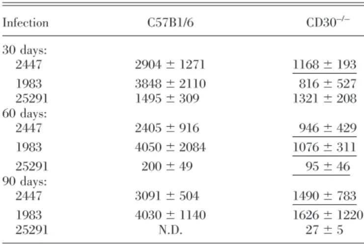
TABLE 3. Priming of T cells from C57B1/6 or B6.CD30–/–Mice for Secretion of IFN-␥ during Infection with M. avium
Strains 2447, 1983, and 25291 Infection C57B1/6 CD30–/– 30 days: 2447 2904⫾ 1271 1168⫾ 193 1983 3848⫾ 2110 816⫾ 527 25291 1495⫾ 309 1321⫾ 208 60 days: 2447 2405⫾ 916 946⫾ 429 1983 4050⫾ 2084 1076⫾ 311 25291 200⫾ 49 95⫾ 46 90 days: 2447 3091⫾ 504 1490⫾ 783 1983 4030⫾ 1140 1626⫾ 1220 25291 N.D. 27⫾ 5
At the indicated time-points of infection by any of the three strains of M. avium, spleen cells were stimulated in vitro with homologous mycobacterial envelope proteins, and the amounts of IFN-␥ secreted into the supernatants were measured by ELISA and shown here as mean⫾SDin pg/ml culture medium. Cells from noninfected mice or unstimulated cells from infected animals produced negligible amounts of cytokine. N.D. ⫽ Not detectable. Statistically significant differences between the two mouse strains are indicated as single-underlined (P⬍0.05) values after performing Student’s t-test.
with significant, albeit limited, exacerbation of the infection.
Thus, although impacting on the immune response and
resis-tance to M. avium, CD30 plays a marginal role as compared
with other elements of the immune system. However, this is the
first time such an involvement in immunity to infection is
shown. It will be interesting to analyze other infection models
and assess whether more important contributions may be found
with other pathogens. The deficiency in CD30 expression did
not affect the course of the infection by a strain of high
virulence, which escapes protective immunity. The infection
with this strain induces a strong IFN-
␥ response and marked
macrophage activation, but the mycobacterial strain escapes
the effector mechanisms of the immune response through
as-yet unidentified mechanisms. It is thus not surprising that a
reduction in the development of the immune response had no
effect on the growth of a strain able to resist such response.
Several TNF and TNFR family members have been shown to
be involved in the in vivo induction of immune responses,
namely during viral infections and experimentally induced
autoimmune disorders [4, 7, 20, 21, 27, 28]. In models of
infectious diseases, CD134 was incriminated in the induction
of disease-promoting Th2 responses during experimental,
cu-taneous leishmaniasis [18], and a soluble viral homologue of
CD30 produced by ectromelia virus was shown to interfere with
the generation of Th1 responses [16]. Our data extend these
observations about the role of TNFRSF members in the
induc-tion of the immune response to a mycobacterial species.
Sev-eral members of the TNFSF and TNFRSF have been shown to
be involved in protective immunity to mycobacteria with
spe-cial relevance to TNF itself and lymphotoxin [41– 47]. We also
showed that CD40 is required for optimal induction of
protec-tive immunity to M. avium [36]. It is thus apparent that
different molecules of these families cooperate to orchestrate
an efficient, protective immune response. Future work should
make use of combinations of more than one deficiency to assess
how redundant these mechanisms are.
The mechanism whereby CD30 promotes protective
immu-nity to M. avium is still undefined. It has been shown that IL-4
may increase CD30 expression in T cells [13] and that during
CD30 signaling, the adaptor molecule TRAF2 is rapidly
de-pleted, leading to increased sensitivity to death signals [14].
Consistent with these findings, Seah and Rook [15] described
an in vitro system where peripheral blood leukocytes from
tuberculosis patients had increased IL-4 secretion in response
to the mycobacterial antigens and that CD30
⫹cells showed
increased susceptibility to TNF-induced apoptosis correlating
with decreased amounts of intracellular TRAF2 levels. A
sim-plistic analysis of these data would thus suggest that
CD30-deficient T cells would survive longer. This is in contrast with
our findings where the CD30-deficient animals had lower
num-bers of T cells responding to infection. Also, the amounts of
IL-4 induced in our system are always low to undetectable.
Thus, we favor the opposite hypothesis that CD30 triggering
activates survival pathways rather than promoting T cell death.
Preliminary studies using flow cytometric analysis following
staining with annexin V failed to show an increased rate of
apoptosis of lymphocytes from CD30-deficient mice during
infection (unpublished data). However, given the long
time-frame of the response to infection and the rapid removal of
apoptotic cells, the assay may be inadequate to address the
mechanism of T cell loss. Although a decrease in lymphocyte
proliferation or diminished cell recruitment could explain the
reduced lymphocyte counts detected in CD30-deficient mice,
several data from this report favor a role of CD30 in lymphocyte
survival. First, granulomas in the CD30
⫺/⫺mice appear to
form normally early in infection but appear to lose the
lym-phoid cells surrounding the central core of macrophages later
on, consistent with a decreased survival of those cells. It should
be stressed however that this occurred over a protracted period
of time of up to 8 months, suggesting that the defect is only
minor as compared with other examples of reduced T cell
survival. Second, we found that lymphoproliferation was not
reduced in uninfected or infected B6.CD30
⫺/⫺mice as
com-pared with control animals. Yet, further studies are still
re-Fig. 4. RT-PCR products from samples of RNA taken from the livers of uninfected mice or from mice infected for 15 days with M. avium 2447. Primers specific for HPRT or IL-12p40 were used in nonsat-urating conditions. The time-point chosen was
previ-ously shown to be one when other immune-deficient animals showed defects in expression of the gene.
Fig. 5. IFN-␥ produced by purified CD4⫹ T cells from C57Bl/6 or B6.CD30⫺/⫺mice infected for 30 days with M. avium 2447. (A) Frequency of IFN-␥-producing CD4⫹T cells stimulated with M. avium antigen as deter-mined by ELISpot assay. (B) The amount of IFN-␥ produced by fixed numbers of CD4⫹T cells was corrected for the frequency of responsive cells, and the values obtained show the amount of IFN-␥ produced per 1000 cells. Both cultures were performed from the same purified CD4⫹T cells, and the data represent the means and SDfrom values obtained from individual animals. Irradiated spleen cells from uninfected C57Bl/6 mice were used as APC.
quired to definitely exclude the possible defects in lymphocyte
migration and proliferation in the mutant mice.
In addition to possible effects on T cell survival, CD30
signaling may as well promote differentiation of T cells to a
protective phenotype, namely by increasing the ability of these
cells to produce IFN-
␥. We presented evidence that the lack of
CD30 impacted negatively on the ability of CD4
⫹T cells to
polarize adequately into type 1, IFN-
␥-producing subsets. The
amount of cytokine produced on a per-cell basis was
signifi-cantly reduced in CD30-deficient mice as compared with the
control animals. This defect was not explained by reduced
IL-12p40 expression, but expression of other cytokines not
tested here and required for such polarization may be affected.
The lower numbers of CD4
⫹T cells plus the reduced ability of
each cell to secrete IFN-
␥ are expected to lead to overall
reduced concentrations of this cytokine at the infected sites.
Yet, sufficient quantities of this cytokine were still produced to
lead to normal granuloma structuring and to control of
infec-tion, albeit at slightly higher loads than in control animals.
Although CD8
⫹T cells may produce IFN-
␥, and their numbers
appeared to be reduced in infected CD30-deficient mice, we
have shown previously that this T cell population does not
contribute to resistance to infection by M. avium [38] and
therefore did not characterize here their function in this model.
Our data are consistent with the recent work of Saraiva et al.
[16], who have shown that a poxvirus homologue of CD30 was
able to block CD30 –CD153 interactions and to inhibit the
generation of IFN-
␥ responses. In vitro generation of
alloreac-tive T cells and in vivo sensitization with mycobacterial
anti-gens were studied by these authors, and the presence of the
soluble, viral CD30 molecule was shown to reduce priming of
IFN-
␥-producing T cells and in the latter case, to reduce
granuloma formation [16]. In contrast, no effect on Th2
re-sponses was found.
In summary, we report about the role of CD30 –CD153
interactions in the costimulation of T cell-mediated immunity
to mycobacterial infection through a pathway that involves
IFN-
␥.
ACKNOWLEDGMENTS
This work was supported by Grants POCTI/32629/99 and
PSIDA/MGI/49647/2003 from the Portuguese Science and
Technology Foundation (FCT; Portugal) and SDH.IC.I.01.15
from the Calouste Gulbenkian Foundation (Portugal). M. F.
received a fellowship from the FCT. The authors are indebted
to Isabel Dantas, Alexandra Reˆma, Fa´tima Faria, and Ce´lia
Lopes for their technical help.
REFERENCES
1. Locksley, R. M., Killeen, N., Lenardo, M. J. (2001) The TNF and TNF receptor superfamilies: integrating mammalian biology. Cell 104, 487– 501.
2. Croft, M. (2003) Co-stimulatory members of the TNFR family: key to effective T-cell immunity? Nat. Rev. Immunol. 3, 609 – 620.
3. Hintzen, R. Q., Lens, S. M. A., Lammers, K., Kuiper, H., Beckmann, M. P., van Lier, R. A. W. (1995) Engagement of CD27 with its ligand
CD70 provides a second signal for T cell activation. J. Immunol. 154, 2612–2623.
4. Hendriks, J., Gravestein, L. A., Tesselaar, K., van Lier, R. A. W., Schumacher, T. N. M., Borst, J. (2000) CD27 is required for generation and long-term maintenance of T cell immunity. Nat. Immunol. 1, 433– 440.
5. Arens, R., Tesselaar, K., Baars, P. A., Schijndel, G. M. W., Hendriks, J., Pals, S. T., Krimpenfort, P., Borst, J., van Oers, M. H. J., van Lier, R. A. W. (2001) Constitutive CD27/CD70 interaction induces expansion of effector-type T cells and results in IFN␥-mediated B cell depletion. Immunity 15, 801– 812.
6. Tesselaar, K., Arens, R., van Schijndel, G. M. W., Baars, P. A., van der Valk, M. A., Borst, J., van Oers, M. H. J., van Lier, R. A. W. (2003) Lethal T cell immunodeficiency induced by chronic costimulation via CD27-CD70 interactions. Nat. Immunol. 4, 49 –54.
7. Nakajima, A., Oshima, H., Nohara, C., Morimoto, S., Yoshino, S., Kobata, T., Yagita, H., Okumura, K. (2000) Involvement of CD70-CD27 interac-tions in the induction of experimental autoimmune encephalomyelitis. J. Neuroimmunol. 109, 188 –196.
8. del Prete, G., de Carli, M., Almerigogna, F., Daniel, C. K., d’Elios, M. M., Zancuoghi, G., Vinante, F., Pizzolo, G., Romagnani, S. (1995) Preferential expression of CD30 by human CD4⫹T cells producing Th2-type cyto-kines. FASEB J. 9, 81– 86.
9. Amakawa, R., Hakem, A., Kundig, T. M., Matsuyama, T., Simard, J. J. L., Timms, E., Wakeham, A., Mittruecker, H-W., Griesser, H., Takimoto, H., Schmits, R., Shahinian, A., Ohashi, P. S., Penninger, J. M., Mak, T. W. (1996) Impaired negative selection of T cells in Hodgkin’s disease antigen CD30-deficient mice. Cell 84, 551–562.
10. Bengtsson, A., Joahansson, C., Linder, M. T., Hallde´n, G., van der Ploeg, I., Scheynus, A. (1995) Not only Th2 cells but also Th1 cells express CD30 after activation. J. Leukoc. Biol. 58, 683– 689.
11. de Young, A. L., Duramad, O., Winoto, A. (2000) The TNF receptor family member CD30 is not essential for negative selection. J. Immunol. 165, 6170 – 6173.
12. Gilfillan, M. C., Noel, P. J., Podack, E. R., Reiner, S. L., Thompson, C. B. (1998) Expression of the costimulatory receptor CD30 is regulated by both CD28 and cytokines. J. Immunol. 160, 2180 –2187.
13. Nakamura, T., Lee, R. K., Nam, S. Y., Al-Ramadi, B. K., Koni, P. A., Bottomly, K., Podack, E. R., Flavell, R. A. (1997) Reciprocal regulation of CD30 expression on CD4⫹T cells by IL-4 and IFN-␥. J. Immunol. 158, 2090 –2098.
14. Duckett, C. S., Thompson, C. B. (1997) CD30-dependent degradation of TRAF2: implications for negative regulation of TRAF signaling and the control of cell survival. Genes Dev. 11, 2810 –2821.
15. Seah, G. T., Rook, G. A. W. (2001) IL-4 influences apoptosis of Myco-bacterium-reactive lymphocytes in the presence of TNF-␣. J. Immunol. 167,1230 –1237.
16. Saraiva, M., Smith, P., Fallon, P. G., Alcami, A. (2002) Inhibition of type 1 cytokine-mediated inflammation by a soluble CD30 homologue encoded by ectromelia (mousepox) virus. J. Exp. Med. 196, 829 – 839. 17. Salek-Ardakani, S., Song, J., Halteman, B. S., Jember, A. G-H., Akiba, H.,
Yagita, H., Croft, M. (2003) OX40 (CD134) controls memory T helper 2 cells that drive lung inflammation. J. Exp. Med. 198, 315–324. 18. Akiba, H., Miyahira, Y., Atsuta, M., Takeda, K., Nohara, C., Futagawa, T.,
Matsuda, H., Aoki, T., Yagita, H., Okumura, K. (2000) Critical contribu-tion of OX40 ligand to T helper cell type 2 differentiacontribu-tion in experimental leishmaniasis. J. Exp. Med. 191, 375–380.
19. Pippig, S. D., Pena-Rossi, C., Long, J., Godfrey, W. R., Fowell, D. J., Reiner, S. L., Birkeland, M. L., Locksley, R. M., Barclay, A. N., Killeen, N. (1999) Robust B cell immunity but impaired T cell proliferation in the absence of CD134 (OX40). J. Immunol. 163, 6520 – 6529.
20. Nohara, C., Akiba, H., Nakajima, A., Inoue, A., Koh, C-S., Ohshima, H., Yagita, H., Mizuno, Y., Okumura, K. (2001) Amelioration of experimental autoimmune encephalomyelitis with OX40 ligand monoclonal anti-body: critical role for OX40 ligand in migration, but not development, of pathogenic T cells. J. Immunol. 166, 2108 –2115.
21. Weinberg, A. D., Wegmann, K. W., Funatake, C., Whitham, R. H. (1999) Blocking OX-40/OX40 ligand interaction in vitro and in vivo leads to decreased T cell function and amelioration of experimental allergic en-cephalomyelitis. J. Immunol. 162, 1818 –1826.
22. Kopf, M., Ruedl, C., Schmitz, N., Gallimore, A., Lefrang, K., Ecabert, B., Odermatt, B., Bachmann, M. F. (1999) OX40-deficient mice are defective in Th cell proliferation but are competent in generating B cell and CTL responses after virus infection. Immunity 11, 699 –708.
23. Chen, A. I., McAdam, A. J., Buhlmann, J. E., Scott, S., Lupher, M. L., Greenfield, E. A., Baum, P. R., Fanslow, W. C., Calderhead, D. M., Freeman, G. J., Sharpe, A. H. (1999) OX40-ligand has a critical costimu-latory role in dendritic cell:T cell interactions. Immunity 11, 689 – 698.
24. Kim, Y-J., Kim, S. H., Mantel, P., Kwon, B. S. (1998) Human 4-1BB regulates CD28 co-stimulation to promote Th1 cell responses. Eur. J. Im-munol. 28, 881– 890.
25. Cannons, J. L., Lau, P., Ghumman, B., de Benedette, M. A., Yagita, H., Okumura, K., Watts, T. H. (2001) 4-1BB ligand induces cell division, sustains survival, and enhances effector function of CD4 and CD8 T cells with similar efficacy. J. Immunol. 167, 1313–1324.
26. Shuford, W. W., Klussman, K., Tritchler, D. D., Loo, D. T., Chalupny, J., Siadak, A. W., Brown, T. J., Emswiler, J., Raecho, H., Larsen, C. P., Pearson, T. C., Ledbetter, J. A., Aruffo, A., Mittler, R. S. (1997) 4-1BB costimulatory signals preferentially induce CD8⫹ T cell proliferation and lead to the amplification in vivo of cytotoxic T cell responses. J. Exp. Med. 186,47-55.
27. Tan, J. T., Whitmire, J. K., Ahmed, R., Pearson, T. C., Larsen, C. P. (1999) 4-1BB ligand, a member of the TNF family, is important for the generation of antiviral CD8 T cell responses. J. Immunol. 163, 4859 – 4868. 28. DeBenedette, M. A., Wen, T., Bachmann, M. F., Ohashi, P. S., Barber,
B. H., Stocking, K. L., Peschon, J. J., Watts, T. H. (1999) Analysis of 4-1BB ligand (4-1BBL)-deficient mice and of mice lacking both 4-1BBL and CD28 reveals a role for 4-1BBL in skin allograft rejection and in the cytotoxic T cell response to influenza virus. J. Immunol. 163, 4833– 4841.
29. Pedrosa, J., Flo´rido, M., Kunze, Z. M., Castro, A. G., Portaels, F., Mc-Fadden, J. J., Silva, M. T., Appelberg, R. (1994) Characterization of the virulence of Mycobacterium avium complex isolates in mice. Clin. Exp. Immunol. 98, 210 –216.
30. Oshima, H., Nakano, H., Nohara, C., Kobata, T., Nakajima, A., Jenkins, N. A., Gilbert, D. J., Copeland, N. G., Muto, T., Yagita, H., Okumura, K. (1998) Characterization of murine CD70 by molecular cloning and mAb. Int. Immunol. 10, 517–526.
31. Akiba, H., Oshima, H., Takeda, K., Atsuta, M., Nakano, H., Nakajima, A., Nohara, C., Yagita, H., Okumura, K. (1999) CD28-independent costimu-lation of T cells by OX40 ligand and CD70 on activated B cells. J. Im-munol. 162, 7058 –7066.
32. Shimozato, O., Takeda, K., Yagita, H., Okumura, K. (1999) Expression of CD30 ligand (CD153) on murine activated T cells. Biochem. Biophys. Res. Commun. 256, 519 –526.
33. Futagawa, T., Akiba, H., Kodama, T., Takeda, K., Hosoda, Y., Yagita, H., Okumura, K. (2002) Expression and function of 4-1BB and 4-1BB ligand on murine dendritic cells. Int. Immunol. 14, 275–286.
34. Pais, T. F., Cunha, J. F., Appelberg, R. (2000) Antigen specificity of the T cell response to Mycobacterium avium infection in mice. Infect. Immun. 68,4805– 4810.
35. Flo´rido, M., Gonc¸alves, A. S., Silva, R. A., Ehlers, S., Cooper, A. M., Appelberg, R. (1999) Resistance of virulent Mycobacterium avium to
interferon-␥-mediated antimicrobial activity suggests additional signals for induction of mycobacteriostasis. Infect. Immun. 67, 3610 –3618. 36. Flo´rido, M., Cooper, A. M., Appelberg, R. (2002) Immunological basis of
the development of necrotic lesions following Mycobacterium avium in-fection. Immunology 106, 590 – 601.
37. Appelberg, R., Soares, R., Ferreira, P., Silva, M. T. (1989) Induction of nonspecific immunosuppression in mice by mycobacterial infections and its relation to macrophage activation. Scand. J. Immunol. 30, 165–174. 38. Appelberg, R., Castro, A. G., Pedrosa, J., Silva, R. A., Orme, I. M., Mino´prio, P. (1994) The role of␥ interferon and tumor necrosis factor-␣ during the T cell independent and dependent phases of Mycobacterium avium infection. Infect. Immun. 62, 3962–3971.
39. Flo´rido, M., Gonc¸alves, A. S., Gomes, S., Appelberg, R. (2004) CD40 is required for optimal induction of protective immunity to Mycobacterium avium. Immunology 111, 323–327.
40. Gomes, M. S., Flo´rido, M., Cordeiro, J. V., Teixeira, C. M., Takeuchi, O., Akira, S., Appelberg, R. (2004) Limited role of the Toll-like receptor (TLR)-2 in resistance to Mycobacterium avium. Immunology 111, 179 – 185.
41. Flynn, J. L., Goldstein, M. M., Chan, J., Triebold, K. J., Pfeffer, K., Lowenstein, C. J., Schreiber, R., Mak, T. W., Bloom, B. R. (1995) Tumor necrosis factor-␣ is required in the protective immune response against Mycobacterium tuberculosis in mice. Immunity 2, 561–572.
42. Kindler, V., Sappino, A. P., Grau, G. E., Piguet, P. F., Vassalli, P. (1989) The inducing role of tumour necrosis factor in the development of bacte-ricidal granulomas during BCG infection. Cell 56, 731–740.
43. Roach, D. R., Briscoe, H., Saunders, B., France, M. P., Riminton, S., Britton, W. J. (2001) Secreted lymphotoxin-␣ is essential for the control of an intracellular bacterial infection. J. Exp. Med. 193, 239 –246. 44. Bean, A. G. D., Roach, D. R., Briscoe, H., France, M. P., Korner, H.,
Sedgwick, J. D., Britton, W. J. (1999) Structural deficiencies in granuloma formation in TNF gene-targeted mice underlie the heightened suscepti-bility to aerosol Mycobacterium tuberculosis infection, which is not com-pensated for by lymphotoxin. J. Immunol. 162, 3504 –3511.
45. Bopst, M., Garcia, I., Guler, R., Olleros, M. L., Rulicke, T., Muller, M., Wyss, S., Frei, K., le Hir, M., Eugster, H-P. (2001) Differential effects of TNF and LT␣ in the host defense against M. bovis BCG. Eur. J. Immunol. 31,1935–1943.
46. Jacobs, M., Brown, N., Allie, N., Ryffel, B. (2000) Fatal Mycobacterium bovis BCG infection in TNF-LT-␣-deficient mice. Clin. Immunol. 94, 192–199.
47. Appelberg, R., Sarmento, A., Castro, A. G. (1995) Tumor necrosis factor ␣ (TNF␣) in the host resistance to mycobacteria of distinct virulence. Clin. Exp. Immunol. 101, 308 –313.