UNIVERSIDADE DE LISBOA FACULDADE DE CIÊNCIAS DEPARTAMENTO DE FÍSICA
TELOMERE BIOLOGY IN METAZOA
NUNO M. V. GOMES
DOUTORAMENTO EM ENGENHARIA BIOMÉDICA E BIOFÍSICA 2011
ii
UNIVERSIDADE DE LISBOA FACULDADE DE CIÊNCIAS DEPARTAMENTO DE FÍSICA
TELOMERE BIOLOGY IN METAZOA
NUNO M. V. GOMES
Thesis supervised by: Prof. Doutor Jerry W. Shay
The University of Texas Southwestern Medical Center at Dallas Prof. Doutor Eduardo Ducla-Soares
Institute of Biophysics and Biomedical Engineering University of Lisbon
DOUTORAMENTO EM ENGENHARIA BIOMÉDICA E BIOFÍSICA 2011
iv DEDICATION
Dedicated to my wonderful family for brightening every day of my life.
v Copyright
by
Nuno M. V. Gomes, 2011 All Rights Reserved
ACKNOWLEDGMENTS
I am grateful to my mentors Jerry Shay and Woodring Wright, from the Department of Cell Biology of the University of Texas Southwestern Medical Center at Dallas, for the opportunity to train in their laboratory and their daily teachings and support. Their training provided solid foundations for the development of my scientific skills and critical thinking, turning my mental clock from deductive thinking into inductive reasoning.
I would like to thank the Faculty of the Institute of Biophysics and Biomedical Engineering, in particular Professor Ducla-Soares for its advice and supervision. I would also like to thank my fellow colleagues of the IBEB with whom I had the privilege to study. It was an honor to study side by side with these bright and creative group of physicists, engineers and chemists.
I would also like to thank the Shay/Wright lab members, past and present, for their teachings and kind help. In particular would like to acknowledge Michael Wang, Maeve Hsieh, William Walker and Donna Meng, for their great help advancing this project. I thank also Nicholas Forsyth, for his insightful discussions and Ying Zou for the excellent imaging training. I‟m thankful to the amazing Kevin Kennon, for making sure all administrative issues were properly and timely done, for his friendship and good spirit.
I‟m grateful to Oliver A. Ryder, Marlys L. Houck, Suellen J. Charterand other from the Conservation and Research for Endangered Species, Genetics Division of the San Diego Zoo, for their contribution with the animal cells and useful information provided.
I thank Steven N. Austad and his team from the Barshop Center for Longevity and Aging Studies in San Antonio, Texas for providing animal cells and for their insighfull discussions and ideas.
I also thank John Wise and the National Marine Sanctuary Foundation for providing the Bowhead Whale lung cells.
I thank Chris Vendetti from the University of Reading and Mark Pagel from the University of Reading and Santa Fé Institute in New Mexico for their amazing help with the filogenetic statistical analyses.
To the faculty and students “happydauers” of the Aging course of the Woods Hole Marine Biological Laboratory for opening my mind to the exciting research in the aging field and for their past and present aging research discussions and updates.
This work was supported by the European Union Programs POCI 2010 & FSE and by national funds from the Portuguese Ministry for Science, Technology and Superior Education ((N.M.V.G) and by the Keck Foundation and the National Institute on Aging (W.E.W. & J.W.S.).
I am deeply grateful to my family for always being there for me, brightening my life and supporting my crazy scientific endeavors.
TELOMERE BIOLOGY IN METAZOA
Nuno M. V. Gomes, D.V.M., M.Sc. University of Lisbon, 2011
Supervising Professors: Jerry W. Shay, Ph.D. and Eduardo Ducla-Soares, Ph.D.
ABSTRACT
Telomerase, the enzyme that maintains telomeres, is absent from most adult human somatic cells, producing a progressive telomere shortening that limits the proliferative potential of primary human cell cultures (Shay and Wright 2007). This programmed telomere shortening, replicative aging, functions as a tumor suppressor program that generates a barrier for the outgrowth of tumors. Remarkably, this telomere tumor suppressor program is not conserved in laboratory rats and mice, which have long telomeres and constitutive telomerase (Sherr and DePinho 2000; Wright and Shay 2000). The present study examines over 60 mammalian species to determine the phylogenetic distribution of the telomere tumor suppressor pathway. Phylogeny based statistical analysis demonstrates that telomere length inversely correlates with lifespan but not body size, while telomerase expression inversely correlates with body size but not lifespan. The ancestral mammalian phenotype was determined to have short telomeres and
repressed telomerase. At least 5-7 independent times in different orders smaller, shorter lived species changed to having long telomeres and expressing telomerase, suggesting tradeoffs between the advantages and drawbacks of using replicative aging as a tumor suppression mechanism. We show that one advantage is consistent with reducing the energetic/cellular costs of specific oxidative protection mechanism needed to maintain short telomeres. We propose that the telomere tumor suppressor pathway represents an initial adaptation to the increased mutational load of homeothermy by ancestral mammals, has adaptive advantage in large and long-lived animals, but has been abandoned by many species. These observations resolve a longstanding confusion about the use of telomeres in humans and mice, support a role for telomere length in limiting lifespan, provide a critical framework for interpreting studies of the role of oxidative protection in the biology of aging, and identify which mammals can be used as appropriate model organisms for the study of the role of telomeres in human cancer and aging.
RESUMO
As células somáticas humanas normais exibem uma capacidade proliferativa limitada, um fenómeno conhecido como “limite de Hayflick”. As células fetais dividem-se mais vezes em cultura do que as de uma criança, que por sua vez dividem-se dividem mais do que as de um adulto. Os telómeros são os relógios moleculares que permitem às células contarem o seu número de divisões. Os telómeros são as sequências repetitivas de ADN encontradas nos extremos dos cromossomas lineares. Cada um dos 92 telómeros humanos contém milhares de repetições da sequência de seis nucleótidos TTAGGG e as proteínas associadas aos telómeros. O comprimento dos telómeros diminui quer em função da idade dos tecidos do dador, quer com o número de divisões celulares em cultura.
A telomerase é uma ribonucleoproteina celular transcriptase reversa que utiliza o seu componente catalítico (hTERT) para sintetizar ADN telomérico (TTAGGG)n directamente nas extremidades dos cromossomas. Em humanos, esta enzima é expressa em tecidos embrionários e em células germinais específicas, mas não é detectada na maioria das células somáticas normais, o que conduz a um encurtamento progressivo dos telómeros que limita o potencial proliferativo das células primárias humanas.
Este encurtamento programado dos telómeros - senescência replicativa – funciona como um programa supressor tumoral que gera uma barreira contra o sobrecrescimento tumoral (85% dos tumores humanos possuem actividade da enzima telomerase e são capazes de manter os seus telómeros). Notavelmente, este programa supressor tumoral telomérico não se encontra preservado nas ratazanas e nos ratos de laboratório, que têm
telómeros longos e telomerase constitutiva. O estudo presente examina mais de 60 espécies de mamíferos de modo a determinar a distribuição filogenética deste mecanismo supressor tumoral.
A expressão de telomerase em culturas de fibroblastos em divisão provenientes de dadores adultos foi usada para determinar a força da repressão da telomerase em células somáticas mesenquimatosas. As células cultivadas em condições não ideais (ex: falta de um micronutriente, oxigénio a 20%), exibem frequentemente uma paragem de divisão chamada estase (“STASIS”- stress or aberrante signaling induced senescence), que é independente do encurtamento telomérico. A presença de estase cedo (dentro de 15 duplicações) também forneceu um fenótipo adicional.
Neste estudo examinei a expressão da telomerase, o comprimento dos telómeros, o peso corporal e a longevidade. Usando o resultado da análise de modelos de regressão que levam em linha de conta a ascendência comum, prevista pela filogenia dentro de uma matriz filogenética dos quadrados mínimos (PGLS) verifica-se que a expressão da telomerase se correlaciona de modo significativo com o inverso da massa corporal (p=0.0082), mas não apresenta efeitos independentes com a longevidade (p=0.34). A mesma análise demonstrou que o comprimento dos telómeros apresenta uma significativa correlação negativa com a longevidade (p=0.0032) acima do previsto pela massa corporal por si só, mas não se observou uma associação independente entre o comprimento dos telómeros e a massa corporal (p=0.71).
Um controlo rigoroso da senescência replicativa como mecanismo de supressão tumoral requer telómeros curtos juntamente com repressão da telomerase. Contudo, estes
resultados sugerem que um decréscimo dos níveis de expressão da telomerase pode, por si só, conferir vantagens à medida que o número de células do corpo aumenta (com o tamanho). Isto pode dever-se à capacidade da telomerase para reparar telómeros que sofreram eventos de deleção “catastróficos” (por exemplo, a expressão da telomerase pode permitir que uma célula pré-maligna em que se encontrem ausentes pontos de controlo celular sobreviva a deleções resultantes de paragens dos garfos de replicação ao nível dos telómeros). Por outro lado, a expressão da telomerase pode ter efeitos adicionais independentes da manutenção dos telómeros. Existe uma correlação bem estabelecida entre a massa corporal e a longevidade. Quando o número de células atinge um certo patamar, a associação independente entre telómeros curtos com um aumento da longevidade sugere que o estabelecimento completo da senescência replicativa é necessário de modo a suprimir a formação tumoral durante períodos de tempo mais prolongados. Observações anteriores levaram a concluir que a expressão da telomerase in vivo decresce com o aumento da massa corporal em roedores, mas não foi observada uma relação com a longevidade quer para a actividade da telomerase, quer para o comprimento telomérico. Os resultados deste projecto demonstram que, numa análise global da classe dos mamíferos, o comprimento telomérico coevolui com a longevidade.
O comprimento telomérico ancestral na base dos mamíferos placentários foi reconstruído usando modelos de estimação de probabilidade máxima que geram o valor mais provável na raiz de uma determinada árvore filogenética sob o modelo evolutivo de movimento Browniano juntamente com o parâmetro “lambda”, que mede a força do sinal filogenético. O estado ancestral determinado foi de 18.6 kb com um lambda igual a 1,
indicando um signal filogenético muito forte. O estado ancestral de repressão da telomerase foi estimado usando uma matriz de transição probabilística (Markov). A probabilidade de o mamífero placentário ancestral reprimir a telomerase foi calculada como sendo alta (1), em comparação com a probabilidade de a telomerase ter sido expressa (0). Estes resultados permaneceram qualitativamente iguais mesmo se cada ordem for analisada separadamente. A frequência de transição de expressão para repressão foi estimada como sendo perto de zero, o que significa que apenas ocorreram transições de repressão para expressão. Apesar de haver apenas um número limitado de estudos, um extenso grupo de espécies aquáticas poiquilotérmicas (de equinodermes a peixes cartilagíneos ou ósseos) possuem telómeros curtos e expressam a telomerase em muitos dos seus tecidos. No entanto, conseguimos determinar que é provável que o fenótipo ancestral dos mamíferos placentários ancestrais consistisse em ter telómeros curtos e reprimir a enzima telomerase. Isto sugere que uma das primeiras adaptações à homeotermia, (com o aumento da carga mutacional que a acompanha), foi a repressão da telomerase nas células somáticas adultas que possuíam já os telómeros curtos, levando assim ao início do encurtamento telomérico como um mecanismo de protecção tumoral.
O mamífero ancestral foi provavelmente mais semelhante aos mamíferos não placentários, mas não foi possível esclarecer por completo o seu fenótipo telomérico. A sequência TTAGGG não possui sítios de restrição. O comprimento telomérico é normalmente determinado pela digestão do ADN genómico com uma mistura de quatro enzimas de restricção reconhecedoras de quatro bases de modo a remover ADN de sequência diversa do lado telomérico interno (centromérico), sendo o tamanho dos
telómeros medido em géis de agarose. Os mamíferos não placentários possuem telómeros em que longas extensões de repetições teloméricas foram interrompidas por ADN contendo sítios de restrição. O comprimento dos telómeros de wombats e coalas parece ser menor que 2 kb quando digerido com a nossa mistura de rotina de seis enzimas, mas exibem padrões completamente diferentes quando digeridos com enzimas individuais. O comprimento dos telómeros variou de longo a muito curto dependendo de qual enzima de restrição reconhecedora de quatro bases fosse utilizada para digerir o ADN, não tendo assim sido possível determinar o comprimento telomérico. A natureza das sequências intrateloméricas encontradas nos mamíferos não placentários e se estas reflectem acontecimentos passados de recombinação/inserção ou um processo corrente envolvido na manutenção dos telómeros ainda não foi determinado. Contudo, a ausência de expressão da telomerase por células de coala, a sua paragem de crescimento em cultura após apenas 38 divisões, mesmo após o bloqueio da função de outras barreiras do ciclo celular, e a sua subsequente imortalização após a introdução de hTERT, sugere que pelo menos um marsupial utiliza a senescência replicativa e que apenas a porção mais terminal da sequência telomérica não interrompida se encontra a funcionar neste processo.
Acredita-se que as espécies ancestrais de mamíferos eram pequenas. Contudo a maioria das pequenas espécies actuais (com menos de 1 kg) possuem telómeros longos e expressam a telomerase. A análise filogenética sugere que o fenótipo ancestral dos mamíferos consistia em ter telómeros curtos e reprimir a telomerase como uma adaptação inicial à homeotermia, e que a aquisição de telómeros longos conjuntamente com a ausência de repressão da telomerase representam alterações secundárias que
proporcionaram vantagens adaptativas a espécies que, ou permaneceram pequenas ou que evoluíram de precursores maiores (tal como se pensa ter ocorrido em morcegos). Estas espécies mais pequenas adquiriram telómeros mais longos e expressão de telomerase pelo menos 5-7 vezes e de modo independente durante a evolução. Uma vantagem pode reflectir um compromisso entre os benefícios da supressão tumoral e os custos de limitar a regeneração. Doenças humanas envolvendo mutações na telomerase causam esgotamento prematuro das células estaminais e uma variedade de doenças associadas com a idade tais como insuficiência esporádica da medula óssea, disqueratose congénita e fibrose pulmonar idiopática, levantando a hipótese de que o encurtamento telomérico pode contribuir para alguns aspectos do envelhecimento humano. Uma vantagem da não utilização da senescência replicativa seria um aumento da capacidade regenerativa. Um compromisso adicional pode ser um investimento mais elevado de recursos destinados à protecção contra o dano oxidativo em espécies com telómeros curtos. Os radicais livres danificam preferencialmente a sequência GGG, que nos mamíferos se encontra presente a cada seis pb nas repetições teloméricas TTAGGG. Além disso, uma propriedade fundamental dos telómeros (a supressão local de indicadores de lesões do ADN de modo a que os extremos de cromossomas lineares não sejam reconhecidos como quebras de cadeia dupla) leva a que lesões teloméricas oxidativas sejam reparadas de um modo muito mais lento do que no restante genoma. Este facto, aumenta a probabilidade de que quebras de cadeias simples sejam convertidas em quebras de cadeia dupla e a consequente perda de sequências teloméricas. Telómeros muito longos (demasiado longos para contar as divisões celulares de modo eficiente), permitem contudo que
grandes segmentos teloméricos sejam perdidos sem que seja comprometida a divisão celular. De modo semelhante, a expressão da telomerase poderia permitir a reparação e o alongamento de telómeros com encurtamentos abruptos. A manutenção de telómeros suficientemente curtos para limitar a capacidade proliferativa das células e funcionar como um mecanismo supressor tumoral pode levar a uma necessidade acrescida de investir recursos em mecanismos de protecção oxidativa. Para examinar esta hipótese de trabalho, determinámos a sensibilidade de 15 espécies a dois tipos de stresse oxidativo, tert-Butilhidroperóxido e arseniato de sódio. A relação entre a sensibilidade e o comprimento telomérico como variável independente da massa corporal/longevidade foi significativa usando análise de contraste independente da filogenia para ambos os agentes (tert-Butilhidroperóxido p=0.032; e arseniato de sódio p=0.017). Esta relação também se manteve considerando quer a longevidade, quer a massa como variáveis independentes. A resposta ao arseniato de sódio é mais dramática que ao tert-Butilhidroperóxido. As espécies podem ser agrupadas em dois grupos cuja sensibilidade difere aproximadamente 6 vezes sem que as espécies desta análise limitada exibam valores intermédios. A resistência ao arseniato de sódio foi independente da capacidade das células crescerem bem sob as condições normais de cultura, dado que duas espécies com telómeros curtos que exibiram estase (a baleia cinzenta e o tapir) ainda apresentaram o fenótipo de resistência. Apesar do mecanismo para o diferente comportamento do tert-Butilhidroperóxido e do arseniato de sódio ser desconhecido, pode reflectir compartimentalização celular ou diferentes propensões a lesar lípidos, proteínas ou ADN.
Há muito que se colocou a hipótese de as lesões oxidativas serem um dos principais determinantes da longevidade. Os resultados presentes mostrando uma relação de dependência entre o comprimento dos telómeros e o arseniato de sódio / tert-Butilhidroperóxido demonstram que irá ser importante considerar o comprimento telomérico como uma variável independente ao comparar o nível de resistência a diferentes stresses oxidativos como uma função da longevidade.
Propõe-se assim que o mecanismo supressor tumoral telomérico representa uma adaptação inicial dos mamíferos ancestrais ao aumento de mutações associado à homeotermia tem vantagens adaptativas em animais grandes / de maior longevidade, mas foi abandonado por muitas espécies. Estas observações resolvem uma longa confusão acerca do uso dos telómeros em humanos e ratos, definem uma importante característica distintiva das ordens filogenéticas em mamíferos, fornecem um cenário crítico para a interpretação de estudos do papel da protecção oxidativa na biologia do envelhecimento, e identificam que mamíferos podem ser usados como organismo modelo apropriado para o estudo do papel dos telómeros no cancro e envelhecimento humanos.
TABLE OF CONTENTS Title …... i Title Page ... ii Dedication ... iv Copyright ... v Acknowledgements ... 6 Abstract ... 8
Resumo (Abstract in Portuguese) ... 10
Table of Contents ... 18
List of Figures ... 21
List of Tables ... 22
List of Abbreviations ... 23
CHAPTER ONE: General Introduction and Literature Review ... 27
1. Summary ... 27 2. Introduction ... 27 3. Evolution of telomeres ... 36 3.1 Unicellular organisms ... 36 3.2 Plants ... 39 3.3 Metazoa ... 42 3.3.1 Invertebrates ... 42 3.3.1.1 Lower Metazoan ... 42 3.3.1.2 Bilateralia invertebrates ... 45
3.3.1.3 Ecdysozoa (Platyhelmintes and Acanthocephala) ... 46
3.3.1.4 Lophotrochoa (Nematodes and Arthropods) ... 46
3.3.1.5 Deuterostomia ... 50 3.3.2 Vertebrates ... 54 3.3.2.1 Fish ... 54 3.3.2.2 Amphibians ... 59 3.3.2.3 Reptiles ... 60 3.3.2.4 Birds ... 63 3.3.2.5 Mammals... 70 4. Animal Cloning ... 81 5. Conclusion ... 82
CHAPTER TWO: The Comparative Biology of Mammalian Telomeres ... 84
1. Introduction ... 84
2. Materials and Methods ... 85
2.1. Cell culture ... 85
2.2. Viral transfections ... 86
2.3. Telomere length analysis ... 86
2.4. Non-denaturing in-gel hybridization ... 87
2.5. Telomerase activity measurements ... 88
2.6. Metaphase spread preparation ... 89
2.7. FISH analysis ... 89
2.8. Cytotoxicity assay……… ... 90
2.9. DNA damage/repair assay……… ... 91
2.10. Criteria for groupings ... 92
2.11. Statistical analysis ... 92
3. Results and Discussion ... 95
3.1 Evolutionary distribution of telomere length, telomerase activity & stasis ... 95
3.3 Ancestral phenotypes ... 102
3.4 Non-placental mammal telomere grouping ... 103
3.5 Evolutionary trade-offs ... 105
3.6 Telomere strategy & cellular oxidative stress ... 106
3.7 Telomere lenght & oxidative DNA damage ... 110
4. Conclusion ... 115
5. Future Directions ... 116
BIBLIOGRAPHY ... 118
LIST OF FIGURES
Figure 1.1. A metaphase spread of a normal human BJ fibroblast. ... 28
Figure 1.2. The telomeric T-loop and associated protein complex ... 32
Figure 1.3. Replicative aging and cancer. ... 34
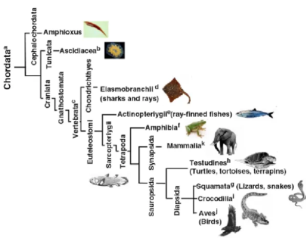
Figure 1.4. Phylogenetic tree of metazoa ... 45
Figure 1.5. Phylogenetic tree of the phylum chordate ... 56
Figure 1.6. Telomeres in vertebrates. ... 69
Figure 2.1. Evolutionary distribution of telomere length, telomerase activity and stasis. ... 96
Figure 2.2. Evolutionary Relationship of telomeres and telomerase to mass and lifespan. . 99
Figure 2.3. Telomere/ Telomere length, telomerase, lifespan and body massdistributions..101
Figure 2.4. Non-placental mammals have discontinuous telomeres. ... 104
Figure 2.5. Resistance to tert-Butyl hydroperoxide and sodium arsenite. ... 108
Figure 2.6. Oxidative DNA damage versus telomere length , longevity. ... 111
Supplementary Figures. ... 142
Supplementary Figure 1 Data on growth, telomere length and telomerase expression for 44 species versus maximum lifespan specie identification ... 143
LIST OF TABLES
Table 1.1. Telomere function. ... 36 Table 1.2. Telomere sequences and replicative aging during evolution.. ... 53 Supplementary Tables.. ... 137 Supplementary Table S1.Species, Mass and Lifespan . ... 138 Supplementary Table S2. PLGS analysis of individual non-placental mammalianorders.139 Supplementary Table S3. Species analyzed for resistance to oxidative stressors. ... 140 Supplementary Table S4. Species analyzed for resistance to different oxygen
LIST OF ABBREVIATIONS
a-MEM Minimum Essential Medium (MEM) Alpha Medium ALT Alternative Lengthening of Telomeres
At-TERT Arabidopsis thaliana Telomerase Reverse Transcriptase (Protein Component) BJ Human Foreskin Fibroblast Cell line
bp Base Pair(s)
BSA Bovine Serum Albumin CDK4 Cyclin-dependent Kinase 4
chTERT Chicken Telomerase Reverse Transcriptase DAPI 4‟, 6-Diamidino-2-Phenylindole
DMEM Dulbecco's Modified Eagle's Medium DNA Deoxyribonucleic Acid
ds Double-stranded FBS Fetal Bovine Serum
FGM Clonetics Fibroblast Growth Medium FIGE Field Inversion Gel Electrophoresis FISH Fluorescence in situ Hybridization FITC Fluorescein Isothiocyanate
FLARE Fragment Length Analysis using Repair Enzymes fmol Femtomole
Fpg E. coli Formamidopyrimidine-DNA Glycosylase (Fpg)
fTERT Fugo Telomerase Reverse Transcriptase (Protein Component) Hpv Human Papilloma Virus
hTERT Human Telomerase Reverse Transcriptase (Protein Component) hTR/ hTERC Human Telomerase RNA (template RNA Component)
mTR Mouse Telomerase RNA (template RNA Component) ITAS Internal Telomerase Assay (TRAP) Standard
kb Kilobase Pair(s)
LD90 Lethal dose that Kills 90% of the cells M1 Mortality Stage 1
M2 Mortality Stage 2 Mb Megabase Pair(s)
MEFS Mouse Embryo Fibroblasts
mRNA Messenger RNA
mTERT Mouse Telomerase Reverse Transcriptase (Protein Component) PBS Phosphate Buffered Saline
PCR Polymerase Chain Reaction PD Population Doublings
RLgT Retrovirus expressing SV40 LgT RNA Ribonucleic Acid
ROS Reactive Oxygen Species Sc Saccharomyces cerevisiae
SCGE Single Cell Gel Electrophoresis SE (SEM) Standard Error
Sp Schizosaccharomyces pombe
STASIS STress or Aberrant SignalingInduced Senescence SV40 Simian Virus 40
TAS Non-coding Sub-telomere region of Plasmodium falciparum tBH tert-Butyl hydroperoxide
TPE Telomere Position Effects
TRAP Telomeric Repeat Amplification Protocol TRF Telomere Restriction Fragment
CHAPTER ONE
General Introduction and Literature Review
1. Summary
Telomere-based replicative senescence is thought to function as a potent mechanism of tumor protection in humans. Whether this mechanism is conserved in other species is still unclear. In this general introduction I present an inter-species critical overview of some of the available literature on the fundamental biology of telomeres and telomerase during development, regeneration, cancer and aging of living organisms during their evolutionary journey through time.
2. Introduction
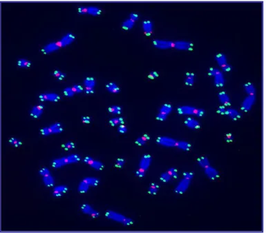
Telomeres are the repetitive DNA sequences found at the ends of linear chromosomes (Muller 1938; McClintock 1941). Each of the 92 human telomere ends is formed by thousand of repeats of the six nucleotide sequence TTAGGG bound by telomere-associated proteins such as the shelterin complex (Blackburn and Gall 1978; Moyzis, Buckingham et al. 1988; deLange 2005) (Fig 1.1).
During DNA replication the leading strand of linear chromosomes is synthesized as a continuous molecule that can potentially replicate all the way to the end of a linear template. The lagging strand is made as a discontinuous set of short Okazaki fragments,
each requiring a new RNA primer to be laid down on the template that are then ligated to make a continuous strand. As there is no DNA beyond the end for a priming event to fill the gap between the last Okazaki fragment and the terminus, the lagging strand cannot replicate all the way to the end of a linear chromosome. This leaves a 3‟ overhang that cannot be filled, and this has been called the “end replication problem” (Watson 1972; Olovnikov 1973). The leading strands are also processed to leave a 3‟overhang (Wright and Shay 2000). Since one strand cannot replicate its end, telomere shortening will occur, and once inherited by the daughter cells, the process repeats itself in subsequent divisions (Olovnikov 1973). Humantelomeres sizes range from ~15 kb at birth to sometimes less than 5kb in chronic disease states (Shay and Wright 2004).
Fig. 1.1- A metaphase spread of a normal human male BJ fibroblast cell. Chromosomes
and shown in Green. Centromeres are probed with a centromeric probe and shown in Red. (Courtesy of Ying Zou)
Normal human somatic cells display a limited capacity to proliferate, a phenomenon known as the “Hayflick limit” (Hayflick and Moorhead 1961). Fetal cells divide more times in culture than those from a child, which in turn, divide more than those from an adult. Telomeres provide the molecular clock that determines this replicative lifespan (Harley, Futcher et al. 1990). Human telomere length decreases both as a function of donor age in tissues and number of cell divisions in culture (Harley, Futcher et al. 1990; Hastie ND, Dempster M et al. 1990; Allsopp, Vaziri et al. 1992; Chang and Harley 1995). Replicative aging can be divided into 2 stages: Mortality stage 1 (M1 or Senescence) and Mortality stage 2 (M2 or Crisis). M1 occurs when most chromosomes still have several thousand base pairs of telomeric sequences left at their ends (Shay and Wright 2001). This stage is thought to be induced by DNA damage signals produced by one or a few particularly short telomere ends. DNA damage signaling from short telomeres,loss of the 3' G-rich telomere single-strandoverhangs, and telomere position effects have all been suggested as potential inducers of M1. In the absence of cell-cycle checkpoint pathways (e.g. p53 and or p16/Rb), cells bypass M1 senescence and telomerescontinue to shorten eventually resulting in M2/crisis (Shay and Wright 2001). M2 represents the result of multiple critically short telomeres when cells are no longer able to protect the ends of chromosomes so that end-to-end fusions occur,
leading to genomic instability and growth arrest or cell death. Rarely cells escape from M2 and become immortal almost universally due to the upregulation or reactivation of the enzyme telomerase, which is able to repair and maintain the telomeres. Senescent cells (due to telomere shortening as well as other inducers of irreversible growth arrest) can be stained by senescence associated β-galactosidase, and exhibit alterations in protein expression, such as increased secreted growth factors, cytokines, extracellular matrix, and degradative enzymes (Krtolica, Parrinello et al. 2001).
Telomerase is a ribonucleoprotein cellular reverse transcriptase that uses its catalytic component (hTERT) to synthesize telomeric DNA (TTAGGG)n directly onto
chromosome ends (Feng J, Funk WD et al. 1995; Nakamura TM, Morin GB et al. 1997). The internal RNA component (hTR or hTERC) contains the template complementary to the telomeric single-strand overhang (Greider and Blackburn 1985; Morin 1989). After adding six bases, the enzyme pauses while it translocates the template RNA for the synthesis of the next 3‟ DNA repeat. This leads to additional rounds of replication of the 3‟end of the G-rich strand (i.e. telomerase is a processive enzyme), thus compensating for telomeric losses due to the end replication problem and perhaps other end processing events (Shay and Wright 2001). In humans, this enzyme is expressed in embryonic tissues and specific germline cells. Telomerase is detected in fetal and adult testis but is neither found in most normal somatic cells, nor in non-dividing oocytes and mature spermatozoa(Shay and Wright 2004; Liu, Bailey et al. 2007). The exceptions are specific proliferative cells of renewal tissues (e.g. hematopoietic stem cells, activated
lymphocytes, basal cells of the epidermis, proliferative endometrium, and intestinal crypt cells) (Shay and Wright 2004). Many of these stem or stem-like cells in adult humans can activate telomerase activity when stimulated to divide. Low levels of telomerase activity may be sufficient to slow but not to prevent telomere shortening. Human intestine or skin telomeres shorten as a function of age although low levels of telomerase can be found in crypt cells and basal keratinocytes. In normal somatic cells and even in stem-like cells expressing telomerase, progressive telomere shortening occurs, eventually leading to senescence (Shay and Wright 2004). Introduction of the telomerase catalytic protein component (hTERT) into normal telomerase negative cells results is restoration of telomerase activity and telomere maintenance or elongation and immortalization (Bodnar, Ouellete et al. 1998). In some cell types in which the culture conditions are inadequate, it has been demonstrated that growth inhibitory genes can beactivated due to a variety of environmental stresses in a process variously termed, premature senescence, culture shock, stress-induced senescence or STASIS (STress or Aberrant Signaling Induced Senescence) (Shay and Wright 2004). In cell culture if the conditions are inadequate, hTERT alone will not immortalize cells.
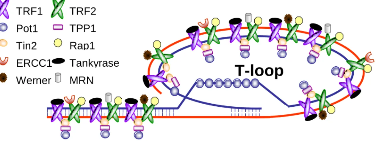
There are specific proteins (shelterin) associated with human telomeres. TTAGGG is recognized directly at least by the three shelterin subunits, TRF1, TRF2, and POT1. These are interconnected by at least three additional shelterin proteins, TIN2, TPP1, and Rap1, forming a structure that enables cells to distinguish telomeres from sites of DNA damage. Without TRF2, telomeres are no longer hidden from the DNA damage
surveillance and chromosome ends are inappropriately processed by the DNA repair machinery (deLange 2005). Shelterin is implicated in the formation of T-loops, first identified in human and mouse cells (Griffith, Comeau et al. 1999). The telomeric overhang has been proposed to invade the double-stranded telomeric DNA forming a lariat structure, base pairing with the C-strand and displacing the G-strand (Fig. 1.2). T-loops are a conserved aspect of telomere structure and have been speculated to protect telomeres and regulate telomerase (deLange 2005).
Fig. 1.2- The telomeric T-loop and associated protein complex. (Courtesy of Agnel Sfeir)
Telomere-based replicative senescence is thought to have evolved as a tumor protection mechanism in long-lived organisms such as humans, preventing the early development of cancer (Wright and Shay 2000). Normal human fibroblasts essentially
T-loop
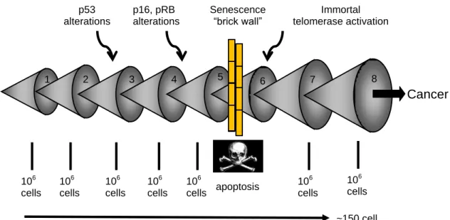
Tankyrase ERCC1 Rap1 Tin2 TPP1 Pot1 TRF2 TRF1 Werner MRN complexnever immortalize in culture in part because at least three independent tumor prevention pathways (p53, p16INK4a/pRB, telomere shortening) have to be altered to allow immortal cell growth (Wright and Shay 2000). Cancer cells must acquire many mutations before they became malignant (Shay and Roninson 2004). Replicative aging blocks this progression by halting cell division before many mutations are able to accumulate within a single cell (Fig. 1.3). The cell containing an initial mutation must expand to a population size of perhaps one million cells beforethere is a reasonable probability for a second mutation to occur, so each mutation would require at least 20 divisions (220=106). Since most mutations are recessive, an additional clonal expansion is required to eliminate the remaining wild-type allele (usually through loss of heterozygosity). Limiting thenumber of available cell divisions to less than 100 would thusprevent pre-malignant cells from dividing after accumulatingonly a few mutations, and thus block their progression (Shay and Wright 2004). This hypothesis is supported by the finding that ~85% of human tumors have upregulated or reactivated telomerase activity and are able to maintain their telomeres Immortalization may occur by gene(s) mutation in the telomerase repression pathway (Tanaka H, Horikawa I et al. 2005).
Fig. 1.3- Replicative aging and cancer. Multiple mutations are required before a cell can
become malignant. This occurs as a series of clonal expansions. This uses a sufficient number of cell doublings so that senescence imposed by telomere shortening forms a barrier to the progression of tumor cells.
Another way telomeres can be maintained is through telomerase independent mechanisms known as alternative lengthening of telomeres (ALT) (Bryan, Englezou et al. 1997). This ALT pathway is only detected in a few rarer cancers (e.g. sarcomas), but is low in the more frequent epithelial neoplasias (carcinomas). This may reflect tighter telomerase regulation in mesenchymal versus epithelial tissues (Henson JD and RR. 2010). The ALT pathway is characterized by an array of phenotypes such as a very heterogeneous distribution of telomere sizes and length fluctuations, ALT-associated PML bodies (APBs), higher levels of telomere sister chromatic exchanges (T-SCE), and
106 cells 106 cells 106 cells p53 alterations Cancer ~150 cell doublings 1 2 3 4 5 6 7 106 cells 106 cells 106 cells p16, pRB alterations Senescence “brick wall” Immortal telomerase activation 8 106 cells apoptosis
raised levels of C-circles (Henson JD and RR. 2010). Recent studies in mice have suggested that telomerase-independent telomere elongation plays a role in normal development (Liu, Bailey et al. 2007). Mice oocyte telomere elongation following fertilization seems to be achieved through a recombination based mechanism characterized by extensive T-SCE. At the blastocyst stage, telomerase appears to take control of telomere maintenance (Liu, Bailey et al. 2007). Undifferentiated mouse ES cells expressing a gene cluster (Zscan4) undergo rapid telomere extension and long-term genomic stability, probably by telomere recombination or T-SCE. Unlike other cells that display T-SCE, such ALT tumor cells and survivors of telomerase knockout Terc2/2 ES cells, telomerase activity is detected in Zscan4 ES cells (Zalzman, Falco et al. 2010).
Telomeres are essential to prevent chromosome ends from being recognized as double-strand breaks. In addition, telomeres regulate cellular proliferation, survival, chromosome positioning, prevent DNA recombination, and participate in proper mitotic and meiotic divisions (Table 1.1) (Teixeira and Gilson 2005). As telomeres shorten during cellular aging there may be de-repression of genes near telomeres eventually leading to reactivation of other previously silenced genes.This process could occur on all or only in a subset of chromosome ends and is known as telomere position effects (TPE) (Baur, Zou et al. 2001). Telomere dysfunction has been implicated in a variety of human age related diseases (e.g. Werner syndrome) (Crabbe, Jauch et al. 2007). Mutations in telomerase genes have also been linked to some pathologies such as idiopathic pulmonary
fibrosis, aplastic anemia and dyskeratosis congenita (Armanios, Chen et al. 2007; Blasco 2007).
3. Evolution of telomeres 3.1. Unicellular organisms
Telomerase-based end maintenance is likely to be a very ancient mechanism since it is found in widely divergent species that represent many of the major eukaryote lineages (ciliates, animals, fungi, green plants). The loss of telomerase is a catastrophic event unless there is immediate (within a few generations) replacement by an alternative system.
In 1978, Elizabeth Blackburn found that the telomeres of the ciliated protozoan Tetrahymena thermophila, consisted of a simple sequence of the hexameric repeat of nucleotides TTGGGG (Blackburn and Gall 1978). Telomerase is necessary for the replication of chromosome ends in this protozoan, and telomeric elongation activity
Table 1.1 Telomere function
(Rocco, Costagliola et al. 2001; Zou, Yi et al. 2002; Teixeira and Gilson 2005)
Prevent chromosome ends from being recognized as double-stand breaks.
Regulate cellular proliferation (Replicative Aging/tumor prevention)
Regulate cellular survival
Chromosome positioning
Prevent DNA recombination
Role in mitotic division
Role in meiotic division
Telomere Position Effect (TPE)
occurs massively during the macronuclear development when telomeres are formed and replicated (Greider and Blackburn 1985). Elongation by recombination is also seen as a backup mechanism in yeast (Lundblad and Blackburn 1993; DeLange 2004). In the protozoan Oxytricha fallax, the telomeric sequence is similar to that of Tetrahymena but the terminal sequence is very short (36 bp) (Pluta, Kaine et al. 1982). Gene conversion based on strand invasion and copy-choice replication has also been observed in Tetrahymena (Walter, Bozorgnia et al. 2001).
Easy laboratory cultivation conditions and powerful genetics have resulted in Saccharomyces cerevisiae, Kluveromyces lactis and Schizosaccharomyces pombe being used as crucial model organisms for telomere biology research. Saccharomyces cerevisiae (Sc) and Schizosaccharomyces pombe (Sp) are almost as different from each other as either is from vertebrates: their ancestors separated about 420-330 million years ago. The telomeric proteins of S. pombe are more similar to the mammalian ones (Teixeira and Gilson 2005). In the yeast Saccharomyces, (TG1-3) or TG2-3(TG1-6)
telomere repeats are observed (Teixeira and Gilson 2005). In other fungi (TTAGGG)n is
observed in Cladosporium but more complex repeats such as (ACACCAAGAAGTTAGACATCCGT)n are found in Candida albicans (Table 1.2) (Shampay, Szostak et al. 1984; Coleman, McHale et al. 1993; McEachern and Hicks 1993; Sinclair, Richmond et al. 2007). Today‟s yeast telomerase enzymatic activity appears to be adapted for both TTAGGG and TG-degenerated sequences (Forstemann, Zaug et al. 2003). Telomeres of Candida parapsilosis are composed of long tandem
repeats and also t-circle intermediates (Tomaska, McEachern et al. 2004; Nosek, Rycovska et al. 2005). The widespread occurrence of t-circles across eukaryote lineages suggests that t-circles (which permit telomere elongation by rolling-circles replication) may not only represent a backup if telomerase dysfunction occurs, but also may be the ancestral system for telomere maintenance (Fajkus, Sykorova et al. 2005). Telomeres also play an important role in the nuclear architecture in some organisms. In yeast, telomeres are anchored to nuclear membranes through a protein complex (Galy, Olivo-Marin et al. 2000).
In the causative agent of malaria, the intracellular protozoa Plasmodium falciparum, telomeres are followed by a non-coding sub-telomere region (TAS), and telomerase not only maintains telomeres, but also participates in the repair of broken chromosome ends. One of P. falciparum’s telomere associated proteins, a homologue of the yeast Sir2, is required for the establishment of a heterochromatic structure at the telomeres, leading to silencing of sub-telomeric genes. PfSir2 associates with promoter regions of silenced genes involved in antigenic variation (Figueiredo and Scherf 2005). In kinetoplastid pathogens such as Trypanosoma brucei, Trypanosoma cruzi and Leishmania major subtelomeres are closely related to antigenic variation, a process which allows the clonal switch of surface antigens, enabling escape from acquired immune responses (Horn and Barry 2005). T-loops have been found in Oxytricha fallax and Trypanosoma brucei. Although trypanosome telomeres have the same size as human
telomeres, their t-loops are very small (less than 1 kb in length) (Munoz-Jordan, Cross et al. 2001; DeLange 2004).
Other ways exist to overcome terminal telomere loss and are exhibited by viruses, prokaryotes and some eukaryotes. Poxvirus has a covalently-closed hairpin at each end of its dsDNA genome. Controlled nicking of the hairpin provides the 3′OH group that is necessary for DNA replication. The linear DNA of the spirochete Borrelia burgdorferi displays a similar strategy. A complication of this replication strategy is the generation of circular dimers requiring a specialized conversion into monomers (DeLange 2004). Retroviruses reverse transcriptase executes a complex terminal jump in order to maintain their chromosome ends and in adenoviruses the solution to the end-replication problem is provided by a terminal protein primer, which is covalently attached to the 5′ ends of its genome (de Jong, van der Vliet et al. 2003; DeLange 2004)
3.2. Plants
In most plants the telomeric sequence (TTTAGGG)n is observed (Table 2) (Cox,
Bennett et al. 1993; Fuchs, Brandes et al. 1995). Both needle and root samples of long-lived trees such as the coastal redwood (Sequoia sempervirens) and the bristlecone pine (Pinus aristata) (2000 to 5000 year lifespan) were found to have higher average telomere lengths of the longest, mean, and shortest telomeres compared with aged matched medium-lived and short lived trees such as the longleaf pine (Pinus palustris) (100-200 years lifespan) (Flanary and Kletetschka 2005). In needle, root, and core samples,
long-lived trees also display higher telomerase activity compared with both short and medium-lived trees. A direct correlation has been found between telomere length and telomerase activity and the expected lifespan of these trees. In the longest lived tree, the Great Basin bristlecone pine (P. longaeva) there was no evidence of overall telomere shortening or decrease in telomerase activity with age (up to 3500 years). One living bristlecone tree “Methuselah” had estimated germination at 2838 BC (Schulman 1958; Flanary and Kletetschka 2005; Flanary and Kletetschka 2006).
In almost all angiosperms, telomeric DNA is composed of many repeats of the heptanucleotide TTTAGGG(McKnight, Riha et al. 2002). However, Alliacaeae, a group of monocots that includes the onions and Aloe seems to be an exception, and several alternative telomeric DNA structures have been proposed (Pich and Schubert 1998). Thus in Asparagales (includes Allium and Aloe) there have been at least two switch-points in the evolution of telomeres. The first occurred with the replacement of the Arabidopsis-type telomere for a “TTAGGG vertebrate-like” sequence. A low fidelity of telomerase (with implications for telomere-binding proteins) may have favored a second switch point in the ancestor to Allium, leading to a still unclear mechanism (Fajkus, Sykorova et al. 2005). It has been proposed that elongation of minisatellite repeats using recombination/replication processes initially compensated for the loss of telomerase function. In more established ALT groups, subtelomeric satellite repeats may replace the telomeric minisatellite repeat while keeping the recombination/replication mechanisms
for telomere elongation in place. Retrotransposition-based mechanisms may also subsequently become established (Fajkus, Sykorova et al. 2005).
Telomeric length is variable among species, from very short telomeres in the plant model Arabidopsis (Arabidopsis thaliana) (2–4 kb) to the extremely long telomeres of tobacco (Nicotiana tabacum) (up to 150 kb) (Richards and Ausubel 1888; Fajkus, Kovarik et al. 1995). Telomere length also varies within the same species (McKnight, Riha et al. 2002). Despite having much shorter telomeres than mice, telomerase null Arabidopsis generated through a T-DNA disruption of the single At-TERT gene can survive up to ten generations (Fitzgerald, Riha et al. 1999; Riha, McKnight et al. 2001; McKnight, Riha et al. 2002). The last five generations of telomerase deficient mutant plants display increased cytogenetic damage and in late-generation chromosome fusions occur in over 40% of the cells, with some cells surviving with only half of their chromosomes. Amazingly, some plants manage to flower and set seeds until the ninth generation (McKnight, Riha et al. 2002). Differences in the consequences of the massive genome damage probably reflect the greater developmental and genomic plasticity of plants. It is known, for example, that chromosomal rearrangements and ploidy changes are better tolerated in plants (Walbot 1996; Fitzgerald, Riha et al. 1999). Telomere dysfunction in plants, leading to end-to-end chromosome fusions, can have a profound effect on chromosome evolution and even speciation (Fajkus, Sykorova et al. 2005). T loops have been found in plants. Extremely large t-loops, up to 50 kb in size, are seen in peas (Pisum sativum) (Cesare, Quinney et al. 2003; deLange 2005).
In plants, telomerase is expressed abundantly in reproductive organs and dividing tissues such as the dedifferentiated callus cells but it is expressed at low or undetectable levels in most post-mitotic vegetative organs (McKnight, Riha et al. 2002). Most cell division takes place in the apical meristem, a group of stem cells that gives rise to all tissues including germ-line cells. It is believe these cells and can undergo approximately 1000 divisions from seed to seed and differentiate into an array of cell types that make a shoot, root, and flower (Fajkus, Kovarik et al. 1995; Oguchi, Liu et al. 1999). Therefore we can conclude that it is unlikely that plants use telomere shortening as a tumor protection mechanism (Oguchi, Liu et al. 1999; Forsyth, Wright et al. 2002).
3.3. Metazoa 3.3.1 Invertebrates 3.3.1.1 Lower Metazoan
As an evolutionary bridge between fungi and higher animals, there are the Lower Metazoan includes the phyla Porifera (sponges), Placozoa (Trichoplax adhaerens), Cnidaria (corals and jellyfish) and Ctenophora (comb jellies) and are considered an evolutionary bridge between fungi and higher animals (Sinclair, Richmond et al. 2007) (Fig. 1.4). All these phyla display the “vertebrate” telomeric motif, also found in the unicellular metazoan sister group Choanozoa (Traut, Szczepanowski et al. 2007).
The lowest metazoan phylum is Porifera (Fig. 1.4.a) in which many species are reported to present negligible senescence (Finch 1990). Sponge species usually show
continuous growth, long lifespans, and a highly flexible cell lineage determination (Koziol, Borojevic et al. 1998). Species from this phyla are known for their extensive regenerative capacity and use of both sexual and vegetative forms of reproduction (Finch 1990). In vivo and in vitro studies in marine demosponges Suberites domuncula and Geodia cydonium exhibit telomerase activity in their somatic and immortal germ tissues. After dissociation into single cell suspensions, isolated cells retain their proliferative capacity but lose telomerase activity, possibly due to lack of contact/adhesion factors. However, telomerase activity is recovered after aggregation of the cells to form primmorphs (Koziol, Borojevic et al. 1998).
These simple multicellular animals provide excellent models for the study of the separation of soma and cell lineages. In the sponges studied, the number of germ-cells is much reduced or null, so the levels of telomerase observed should come from elevated levels of telomerase in the somatic cells that display unlimited replication potency. Alternatively, there might be a high number of somatic stem cells capable of unlimited replication that would undergo subsequent differentiation. Although Archaeocytes in sponges are pluripotent (stem-cell like), with the potential for differentiation into all major cell types, morphological data seem to support the hypothesis that the proliferation of all major somatic cells types is the major contributor for tissue growth. Furthermore, the plasticity of sex determination and the ability of fully differentiated cells to produce gametes also favor the first hypothesis (Koziol, Borojevic et al. 1998; Muller and Muller 2003).
In Calcarea (Leucosolenia sp and Sycon sp.) (Fig. 1.4.a) telomere sizes seem to range from below 1 kb to over 20 kb. One study in Calcarea that also examined the demosponge Suberites failed to detect telomerase activity in either species (Traut, Szczepanowski et al. 2007). This is unexpected and conflicts with the Suberites study cited above, so it is premature to conclude that Calcarea do not express telomerase.
Among Cnidarians (Fig. 1.4.d), the Anthozoans (Corals) are the most basal organism reported to exhibit the (TTAGGG)n telomeric sequence. This repeat is found in
DNA from several Scleractian order corals: Acropora surculosa, Leptoria phrygia, Favia pallida and Goniastrea retiformis. Average telomere length of Acropora surculosa is 3.5 kb (Sinclair, Richmond et al. 2007). Reef corals display vegetative growth of hundreds of years, their rate of mortality decreases as coral body mass increases and several species tend to behave as plants, increasing fecundity as the colonies grow larger (Finch 1990). In spite of these properties, which are characteristic of negligible senescence, reef corals show signs of aging, with declining growth, calcification and reproduction before colony death in Stylophora pistillata (Rinkevich and Loya 1986; Finch 1990).
Cnidaria Scyphozoa species (Fig. 1.4.d) such as compass jellyfish (Chrysaora hysoscella) and blue jellyfish (Cyanea lamarckii) and Ctenophora (Pleurobrachia pileus) (Fig. 1.4.c) reported telomere sizes range from less than 1 kb to over 20 kb. In the Cnidaria Hydra vulgaris (Fig. 1.4.d) sizes seem to be around 20 kb. Telomerase activity has been found in gonad extracts of Cnidaria moon jelly (Aurelia aurita) and the ctenophore (Pleurobrachia pileus). However, similar studies in Cnidarians such as hydra
or in Placozoan (Trichoplax) (Fig. 1.4.b) did not detect telomerase activity (Traut, Szczepanowski et al. 2007).
Fig. 1.4- Phylogenetic tree of metazoa (animalia). The tree and chart shows the
relationships of the different species whose telomere biology is discussed, keyed to superscript letters.
3.3.1.2 Bilateria invertebrates
Among Bilateria (Fig. 1.4), the phyla Onychophora, Platyhelminthes, most Annelida and Mollusca, Echinodermata and the subphylum Urochordata (Fig. 1.4.e),
seem to share the “vertebrate” telomere motif (TTAGGG)n (Jha, Dominguez et al. 1995;
Joffe, Solovei et al. 1998; Vitturi, Colomba et al. 2000; Wang and Guo 2001; Castro and Holand 2002; Plohl, Prats et al. 2002; Laird and Weissman 2004; Vitkova, Kral et al. 2005).
3.3.1.3 Ecdysozoa (Platyhelmintes and Acanthocephala)
In the Trematode Schistosoma mansoni (Fig. 1.4.h) chromosomes are also protected from degradation by telomeres (Hirai and LoVerde 1996). A telomeric study of parasitic worms including the Platyhelminthes flatworm groups Monogenea and Cestoda, and thorny-headed worms (Syndermata: Acanthocephala) revealed conservation of the (TTAGGG)n sequence, in Monogenea (Paradiplozoon homoion) and Cestoda (Caryophyllaeus laticeps, Caryophyllaeides fennica, and Nippotaenia mogurndae. However neither this motif or the nematode motif were present in the parasitic Acanthocephala (Pomphorhynchus laevis and Pomphorhynchus tereticollis) (Fig. 1.4.g) suggesting the existence of an as yet unknown telomeric repeat sequence or an alternative mechanism of telomere maintenance (Bombarová, Vítková et al. 2009).
3.3.1.4 Lophotrochoa (Nematodes and Arthropods)
The so called nematode motif (TTAGGC)n, is found in the Secernentea
roundworms Ascaris lumbricoides, Ascaris sum and Parascaris univalens (Fig. 1.4.j) (Niedermaier and Moritz 2000). In Ascaris, chromatin fragmentation involves a complex molecular mechanism that includes site-specific chromosome breaks, telomeric synthesis,
and degradation of DNA (Muller, Wicky et al. 1991; Traut, Szczepanowski et al. 2007). In Parascaris univalens the haploid germline genome is contained in a single large chromosome and the somatic genome is surrounded by heterochromatin (HET) blocks constituted by segments of the repeats TTGCA and TTTGTGCGTG. However, in both species, the ends of the germline chromosomes are said to be capped by the same (TTAGGC)n tracts, which are added to all the new somatic ends after removal of the old
ones during the complex chromatin diminution process (Niedermaier and Moritz 2000). Chromosome capping in the free-living nematode Caenorhabditis elegans, is achieved by the 4-9 kb telomeric repeats (TTAGGC)n (Wicky, Villeneuve et al. 1996).
All the major arthropod Subphyla (Chelicerata--except spiders, Myriapoda, Crustacea and most Hexapoda) (Fig. 1.4.i) have the (TTAGG)n telomere motif (Traut,
Szczepanowski et al. 2007). Unlike mammals that stop growing after adulthood, some invertebrates, such as the Decapoda crustacean lobster (Homarus americanus) grow continuously throughout life, although growth rates seem to decrease with age. Lobsters show asymptotic growth and can occasionally weigh over forty pounds, and seem to present negligible or very slow gradual senescence. Lobsters have very long lifespans of 50 to 100 year and neither sex exhibits a post-reproductive phase nor molting cessation. They are also able to regenerate their limbs even at advanced ages (Finch 1990; Klapper, Kuhne et al. 1998). Telomere analysis reveals the sequence (TTAGG)n and telomerase
expression has been found in fully differentiated tissues of all organs, with high levels detected in the hepatopancreas and heart and moderate levels in skin and muscle tissues (Klapper, Kuhne et al. 1998). Tumors are rare in adult lobsters and do not seem to
correlate with size or lifespan (Finch 1990). Another Decapoda crustacean, the green sea crab (Carcinus maenas) also has the pentameric (TTAGG)n telomere sequence and high
telomerase activity in its tissues (Elmore, Norris et al. 2008).
The low number of tumor reports in decapod crustaceans may represent a truly low incidence of neoplasia compared to other well studied animal groups rather than insufficient information. This is a large animal group of more than 10,000 species, many commercially important and well investigated, such as lobsters, crabs, shrimp and crayfish. Despite many of these species having long lifespans, some reaching almost 100 years, neoplasias are said to be extremely rare (Vogt 2008). Furthermore, many of these species are benthic, and have an elevated exposure to carcinogens but the frequencies of tumors are remarkably different from mollusks, bottom feeding fish and other fish and even insects (Vogt 2008).
The reason for the low cancer incidences observed in this Phyla are unknown, but many mechanisms may play a role in this event. Decapod crustaceans exhibit some remarkable carcinogen detoxification pathways such as rapid elimination of PAH-related DNA adducts from the tissues. Their immune system includes only innate responses and is reported to be able to either phagocytose or melanize and encapsulate all kinds of foreign material. Arthropods use this rigid melanin barrier to isolate and, together with quinolone cellular toxicity, eliminate cancer cells and damaged tissue areas (Vogt 2008). Stem cell maintenance until the end of life, for example by telomere protection due to high telomerase activity in tissues throughout life, has also been suggested as contributors
for the virtual absence of age-related cancer in the Decapoda (Vogt 2008). These species may provide excellent models for tumor protection mechanism studies.
With the exception of the heterogeneous Coleoptera, most insect orders can be divided into those that use the telomeric repeat (TTAGG)n (e.g. Lepidoptera) or the ones
that do not (e.g. Diptera) (Okazaki, Tsuchida et al. 1993; Meyne and Imai 1995; Sahara, Marec et al. 1999; Frydrychova, Grossmann et al. 2004; Sinclair, Richmond et al. 2007).
Telomerase activity has recently been detected in crickets, cockroaches, and species of Lepidoptera (Sasaki and Fujiwara 2000). The telomerase reverse transcriptase (TERT) subunit has been identified and characterized in the domestic silkworm (Bombyx mori) and the flour beetle (Tribolium castaneum) (Osanai, Kojima et al. 2006). In the group of insects with the largest number of species, the beetle (order Coleoptera), the telomerase-dependent (TTAGG)n motif has been repeatedly lost (5 to 6 times) in different phylogenetic branches and was likely replaced with the alternative mechanisms of telomere elongation (Frydrychova and Marec 2002). The order Diptera seems to be an exception from the general pattern of having short G-rich repeats at their telomeres, and instead often has arrays of complex long satellite repeats at the ends of their chromosomes (e.g. Chironomus & Anopheles gambiae) (Rosen and Edstrom 2000; Walter, Bozorgnia et al. 2001; Traut, Szczepanowski et al. 2007). Elongation of telomeres in the mosquito (Anopheles) is done through gene conversion between complex terminal satellite repeats that are present at natural telomeres (Walter, Bozorgnia et al. 2001). One hypothesis is that Diptera may have lost the telomerase gene and was forced
to use alternative mechanisms of telomere elongation (Biessmann and Mason 1997; Walter, Bozorgnia et al. 2001). The fruit fly (Drosophila melanogaster) uses telomerase independent mechanisms such as chromosome end capping with non-LTR retrotransposons. Chromosome end-elongation is predominantly achieved by terminal insertion of two classes of telomere-specific LINE-like retrotransposable elements, HeT-A and THeT-ART (Mason and Biessmann 1995). However, Drosophila telomeres can also be extended by gene conversion (Mikhailovsky, Belenkaya et al. 1999) and perhaps by recombination between telomeric HeT-A elements (Kahn, Savitsky et al. 2000). The telomeric structure of Damselflies (Zygoptera) and spiders (Araneae) is still unclear (Frydrychova, Grossmann et al. 2004; Vitkova, Kral et al. 2005). Sea spiders (Pycnogonida) also have the (TTAGG)n telomeric motif (Traut, Szczepanowski et al. 2007).
3.3.1.5 Deuterostomia
In Deuterostomia, which includes the phyla Chordata (Fig. 1.4.e and 1.5.a) and Echinodermata (e.g. sea urchins) (Fig. 1.4.f), many examples of long-lived species have been found. Longevities of a decade or more are found in many sea urchins, and in fact, mortality rates decrease with size in adults (Finch 1990). The Red Sea urchin (Strongylocentrotus franciscanus) (Fig. 1.4.f) grows indeterminately during a lifespan that can go beyond 100 years without evidence of age-related disease or decline in reproductive potential, while other species such as the green sea urchin (Lytechinus variegatus) are fast growing and short lived, with a maximum lifespan of 3 to 4 years.
Telomere studies in the Red Sea urchin reveals telomerase activity in mature eggs, and also during early stages of development of L. variegatus and in tissues during adulthood in both species (Aristotle‟s lantern muscle, ampullae, esophagus, intestine, tube feet, male and female gonads). The (TTAGGG)n telomeric sequence has been found in the
moderately long-lived species S. purpuratus. Long telomere lengths (>20 kb) were found both in germ and somatic tissues of L. variegatus. The adult tissues of S. franciscanus have short telomere lengths (≈ 5 kb), similar to the California purple sea urchin (S. purpuratus) (6 kb), and no telomere shortening occurs throughout life of these species (Lejnine, Makarov et al. 1995; Francis, Gregg et al. 2006). It is also known that sea urchin embryo telomeres need to be maintained. The use of cationic porphyrins as telomere interfering agent decreases the rate of cell proliferation and leads to increased chromosome destabilization (Izbicka, Nishioka et al. 1999). These results seem to indicate that neither short nor long-lived sea urchins use replicative aging as a tumor protective mechanism (Francis, Gregg et al. 2006). Furthermore, the number of reported cases of neoplasia in sea urchins, a very intensively studied model organism, is very low (www.pathology-registry.org). This suggests that these species have evolved other mechanisms of tumor prevention/suppression, such as efficient cellular or molecular protection against damage or free radicals and/or a good capacity of replenishment to damaged cells (Francis, Gregg et al. 2006). These species may be excellent candidates for future senescence and tumor protection mechanism studies (Francis, Gregg et al. 2006).
The golden star tunicate (Botryllus schlosseri), the model Urochordate (Fig. 1.4.e and Fig. 1.5.b), is a colonial organism that propagates both asexually and sexually during the 2 to 5 years of colony life. Asexual budding occurs continually from the progenitor body wall and when the colony reaches a critical size sexual reproduction initiates with the production of gonads. It has been proposed that pools of stem cells assure renovation throughout the lifespan. Heterogeneous telomeres of 6-15 kb protect the chromosome ends and high levels of telomerase have been reported in germ and embryonic tissues (Laird and Weissman 2004). Telomerase activity peaks in tissues containing bud rudiments, then decreases in buds that are going through organogenesis and drops to even lower levels in functional zooids, in individual organs and blood (Laird and Weissman 2004). It has been hypothesized that telomerase activity needs to be retained in progenitor and stem cells, is downregulated during differentiation, and is not necessary to maintain the relatively short-lived somatic tissues of Botryllus (Laird and Weissman 2004).
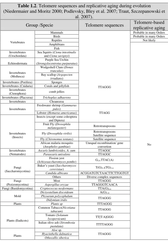
Information about telomere sequences and telomerase TERT and TR/TERC sequences and structure in invertebrates and vertebrates is now readily available online (Table 1.2) (Podlevsky, Bley et al. 2007).