FACULDADE DE MEDICINA
T Follicular Regulatory Cells
in Human Adaptive Immunity and Autoimmunity
Válter Bruno Ribeiro Fonseca
Orientadores: Prof. Doutor Luís Ricardo Simões da Silva Graça Prof. Doutor João Eurico Cortez Cabral da Fonseca Prof. Doutor Rui Manuel Martins Victorino
Tese especialmente elaborada para obtenção do grau de Doutor em Medicina, Imunologia.
Júri:
Presidente: Doutor José Luís Bliebernicht Ducla Soares, Professor Catedrático em regime de tenure e Vice-Presidente do Conselho Científico da Faculdade de Medicina da Universidade de Lisboa
Vogais:
Doutor Hideki Ueno, Professor of Microbiology da Ichan School of Medicine, Nova Iorque, Estados Unidos da
América;
Doutor Peter The Sage, Postdoctoral Fellow da Harvard University, Massachusetts, Estados Unidos da América;
Doutor Luís Ricardo Simões da Silva Graça, Professor Associado com Agregação da Faculdade de Medicina da Universidade de Lisboa (orientador);
Doutora Ana Cristina Gomes Espada de Sousa, Professora Associada Convidada com Agregação da Faculdade de Medicina da Universidade de Lisboa;
Doutor Paulo Manuel Leal Filipe, Professor Auxiliar Convidado da Faculdade de Medicina da Universidade de Lisboa.
Fundação para a Ciência e Tecnologia: SFRH/SINTD/96663/2013 2018
A impressão desta tese foi aprovada pelo Conselho Científico da Faculdade
de Medicina de Lisboa em reunião de 20 de dezembro de 2017.
As opiniões expressas nesta publicação são da exclusiva responsabilidade
do seu autor.
Para os quatro pilares da minha vida, Catarina, a minha esposa, Carolina, a minha filha, Aldina, a minha mãe, Luísa, a minha avó.
Comme pour moi je me persuade que si on m’eût enseigné dès ma jeunesse toutes les vérités dont j’ai cherché depuis les démonstrations, et que je n’eusse eu aucune peine à les apprendre, je n’en aurais peut-être jamais su aucunes autres, et du moins que jamais je n’aurais acquis l’habitude et la facilité que je pense avoir d’en trouver toujours de nouvelles à mesure que je m’applique à le chercher.
i
Table of Contents ... i
Figures Index ... v
Table Index ... vii
Acknowledgments ... ix List of Abbreviations ... xi SUMMARY ... 1 SUMÁRIO ... 3 SUMÁRIO EXTENSO ... 5 CHAPTER 1 ... 9 GENERAL INTRODUCTION ... 9
1. The Immune System... 11
2. T Cell Mediated Humoral Immunity ... 13
2.1. Thymic T Cell Development ... 13
2.2. CD4+ T Cell Diversity ... 15
2.3. T Follicular Helper Cells ... 16
2.3.1. Circulating Tfh Cells and Human-Specific Tfh Cell Features ... 19
3. Regulation of Humoral Responses ... 22
3.1. Germinal Centre Reaction and Cell Dynamics ... 23
3.2. Regulatory T Cells ... 24
3.3. T Follicular Regulatory Cells ... 31
3.3.1. Biology of Tfr Cells ... 32
3.3.2. Mechanisms of Tfr Cell Function ... 44
3.3.3. Tfr Cells in Humans ... 52
3.3.4. Tfr Cells in Different Human Diseases ... 54
4. T – B Cross-Talk in Autoimmunity ... 59
4.1. Dysregulation of Germinal Centre Responses... 60
4.2. Ectopic Lymphoid Structures ... 62
4.2.1. Development of Ectopic Lymphoid Structures ... 63
4.2.2. Autoantibody Formation in Ectopic Lymphoid Structures ... 64
ii
CHAPTER 3 ... 73
MATERIALS AND METHODS ... 73
1. Patient Recruitment and Human Samples ... 75
1.1. Healthy Donor’s Blood Samples ... 75
1.2. B-cell Deficient Patients ... 75
1.3. Children Blood and Tissue Samples ... 75
1.4. Sjögren Syndrome Patients ... 76
1.5. Ethical Issues ... 77
2. Cell Isolation and Flow Cytometry ... 77
3. Functional Assays ... 78
3.1. Suppression Assays and in vitro Cultures ... 78
3.2. ELISA ... 79
3.3. Migration Assays ... 80
4. Real-time RT-PCR ... 80
5. Microscopy ... 81
5.1. Human Tonsils ... 81
5.2. Minor Salivary Gland Biopsies ... 81
6. Statistical analysis ... 82
CHAPTER 4 ... 83
RESULTS ... 83
Human Blood CXCR5+Foxp3+ Treg Cells Are Immature Tfr Cells Not Fully Licensed with Humoral Suppressive Function ... 85
Introduction ... 85
Results ... 86
Discussion ... 104
Blood Tfr/Tfh Ratio Marks Ectopic Lymphoid Structure Formation in Primary Sjögren Syndrome ... 107
Introduction ... 107
Results ... 108
iii
Concluding Remarks ... 138
REFERENCES ... 139
ANNEXES ... 179
v
Figure 1: Tfr cell differentiation signals and pathway. ... 35
Figure 2: Signalling and transcriptomics of Tfr cells. ... 40
Figure 3: Mechanisms of Tfr cell-mediated regulation of humoral responses. ... 44
Figure 4: Flowchart of Sjögren syndrome patient recruitment and selection. ... 77
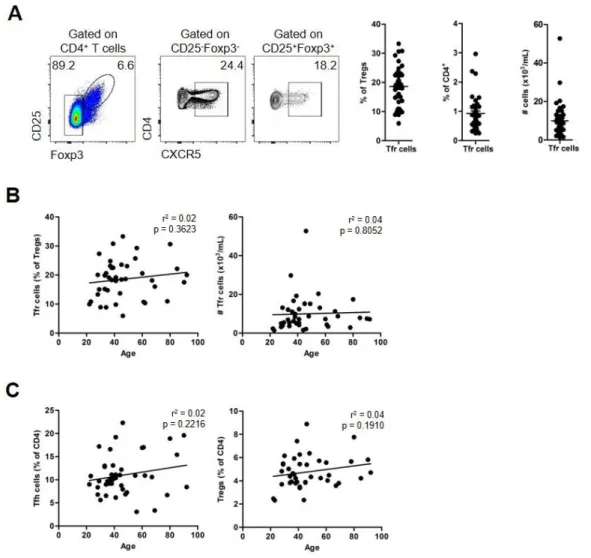
Figure 5: Identification of blood Tfr cells. ... 87
Figure 6: Blood Tfr cells show expression of follicular and regulatory markers. ... 88
Figure 7: Blood Tfr cells are distinct from their tissue counterparts. ... 89
Figure 8: CXCR5+Foxp3+ T cells are localized within GCs in human secondary lymphoid organs. ... 90
Figure 9: Sorting strategy for human blood Tconv, Tfh, CXCR5- Treg, Tfr cells, and naïve B cells. ... 91
Figure 10: Blood Tfr cells are a distinct subset of suppressive Treg cells. ... 92
Figure 11: Stability of blood Tfr cells. ... 93
Figure 12: CXCR5 upregulation by blood Treg cells is not induced by TCR or CD28 signalling. ... 94
Figure 13: Blood Tfr cells suppress Tfh cell proliferation. ... 94
Figure 14: Blood Tfr cells do not show specialized humoral regulatory capacity. ... 96
Figure 15: Blood Tfr cells are immature (resting-like) cells. ... 97
Figure 16: Blood Tfr cells are generated in the periphery after mature adaptive immune responses. ... 99
Figure 17: Tissue Tfr cells are effector cells. ... 100
Figure 18: Sorting strategy for human tonsil Tconv, Tfh, CXCR5- Treg and Tfr cells. .. 101
Figure 19: Blood Tfr cells emerge prior to B-cell interactions. ... 102
Figure 20: Blood Tfr cells are lymphoid tissue derived Tfr precursors. ... 103
Figure 21: Primary Sjögren’s syndrome patients have an increased blood Tfr/Tfh ratio. 110 Figure 22: Circulating Tfh cell subsets are not altered in primary Sjögren syndrome. .... 112
Figure 23: Blood PD-1+ICOS+ activated Tfh and Tfr/Tfh ratio marks distinct features of primary Sjögren syndrome. ... 113
vi patients. ... 115
Figure 26: Blood Tfr/Tfh ratio identifies pathological lymphocytic infiltration in Sjögren’s
syndrome target organ. ... 117
Figure 27: Blood Tfr/Tfh ratio is a marker of primary Sjögren’s syndrome and focal
sialadenitis. ... 118
vii
Table 1: Human blood Tfh cell subsets ... 22
Table 2: Mechanisms that control germinal centre B cells responses ... 24
Table 3: Phenotypic markers of Treg, Tfr and Tfh cells in mice ... 31
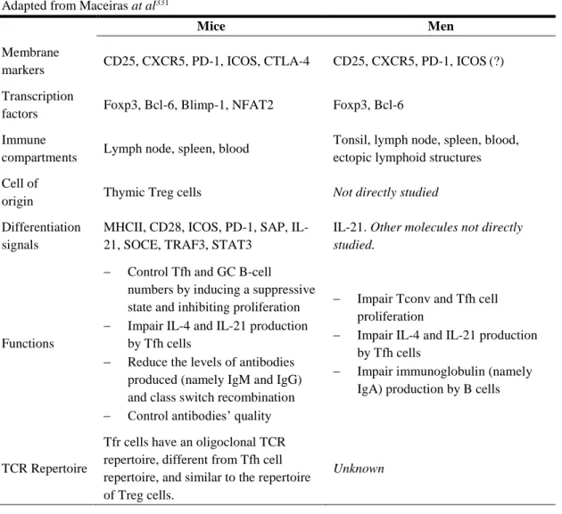
Table 4: Biology of Tfr cells in Mice and Men ... 53
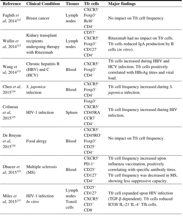
Table 5: Foxp3+ Tfr cells in Human Diseases ... 54
Table 6: Summary of demographic characteristics of primary Sjögren syndrome (SS), non-Sjögren sicca syndrome (non-SSS) patients, and healthy donors ... 108
Table 7: Demographic and clinical characteristics of Sjögren syndrome (SS) and non-Sjögren sicca syndrome (non-SSS) patients ... 109
ix
I decided to acknowledge in Portuguese, my native language, as the cultural and emotional dimensions of language and speech may be lost in translation.
Ao Prof. Doutor Luís Graça, meu orientador, por ter tornado possível este meu percurso na ciência; por me ter mostrado um mundo para além daquele que eu conhecia, e no qual me sinto realizado; por ter criado o pensamento crítico que julgo, só agora, ter começado a ter; pelo seu espírito empreendedor e otimista, absolutamente exemplar; por ter conseguido colocar-me num voo transcontinental.
À Catarina, minha esposa, pela incansável e insubstituível presença a meu lado, sobretudo nos momentos mais difíceis; por tornar, inquestionavelmente, este percurso possível; por esperar por mim; por acreditar em mim; por me manter ligado a este mundo real; por ter garantido a originalidade das ilustrações desta tese. A ti, Carolina, por teres tornado este percurso ainda mais desafiante.
À minha mãe, por garantir quem sou, o maior valor que conheço. Aos meus avós, à minha avó Lisa (ou Luísa, agora que sou adulto), pelo seu espírito único; grande acelerador da minha personalidade; por sempre me ter incentivado a ser mais; espero, agora, ter concretizado também um dos seus sonhos. À Rosa Pinto, pela sua incansável ajuda. À minha família, por continuar a acreditar em mim, mesmo nas minhas constantes ausências e exigências.
À Ana Água-Doce, pela sua imperturbável boa disposição e paciência; por ter sido uma excelente surpresa; por me ter guiado irrepreensivelmente neste mundo novo; por acreditar em mim; pelas suas sábias e concretizáveis previsões. À Saumya Kumar, pela concretização do tão ansiado brainstorming; pelas grandes discussões; por me fazer falar inglês; pelo seu espírito aberto; pela sua riqueza cultural. À Marta Monteiro, por ter acreditado em mim antes de eu próprio o fazer; pelos seus ensinamentos, que ainda uso me guiam. À Raquel Maceiras, pelo seu espírito indomável; por ter feito com que me superasse perante a sua critica feroz. A todos os que encontrei durante o meu percurso no Instituto de Medicina Molecular e que moldaram o que hoje sou: à Prof. Doutora Ana Espada de Sousa, ao Prof. Doutor João Eurico
x Rita Pires, à Sara Matos, à Enfermeira Lurdes.
Ao Prof. Doutor Rui Victorino, pelo seu sábio conselho; por ter destinado este meu percurso, que agora recomeça. Ao Prof. Doutor Carlos Ferreira, também pelo seu sábio conselho; pela sua presença distanciada que ainda sinto; pela saudade que deixa do seu espirito democraticamente revolucionador; por acreditar na expressão romântica da Medicina. À Dra. Marisa Teixeira Silva, por me ter deixado crescer; pela sua permanente tentativa de não me deixar fugir da Medicina Interna minha vida original; pela sua amizade. A todos os que me viram divergir do percurso hospitalar e não deixaram de me ver apenas como aquilo que sou: à Dra. Ana Moleiro, ao Dr. Sérgio Paulo, à Cátia Albino.
A todos os que acreditam em mim.
xi AID/Aicda Activation-induced cytidine deaminase
AECG American European consensus group
AIRE Autoimmune regulator
AKT Akt serine/threonine kinase
Alum Aluminium
anti-dsDNA Antibody anti-double strand deoxyribonucleic acid anti-SSA/Ro52 Antibody anti-Sjögren syndrome-related antigen A/Ro52 anti-SSA/Ro60 Antibody anti-Sjögren syndrome-related antigen A/Ro60 anti-SSB/La Antibody anti-Sjögren syndrome-related antigen B/La AP Red Red alkaline phosphatase substrate
APC Antigen presenting cell
AS Ankylosing spondylitis
Ascl2 Achaete-scute homologue-2
BATF Basic leucine zipper transcription factor BCA-1 B-cell-attracting chemokine 1
Bcl Anti-apoptotic protein B-cell lymphoma
BCR B-cell receptor
BLIMP-1/Prdm-1 PR domain zinc finger protein 1 BTK Bruton’s tyrosine kinase
BTLA B and T-lymphocyte attenuator
BXD2/Tnfs11 Tumour necrosis factor (ligand) superfamily, member 11
C1q Complement factor 1q
cAMP Cyclic adenosine monophosphate
CARMA1 Caspase recruitment domain family member 11
CCL CC chemokine ligand
CCR CC chemokine receptor
CD Cluster of differentiation CFA Complete Freund’s adjuvant
Ciita Class II major histocompatibility complex transactivator c-Maf bZIP transcription factor
xii CTLA-4/CD152 Cytotoxic T-lymphocyte-associated antigen 4
CTV Cell trace violet
CVID Common variable immunodeficiency
CXCL CXC chemokine ligand
CXCR CXC receptor
DAB 3,3’-diaminobenzidine
DC Dendritic cell
DMARDs Disease modifying anti-rheumatic drugs
DN Double negative
DNA Deoxyribonucleic acid
DNAse I Deoxyribonuclease I DOCK8 Dedicator of cytokinesis 8
DP Double positive
DTR Diphtheria toxin receptor EBF Early B-cell factor
EBV Epstein-Barr virus
EDTA Ethylenediamine tetra-acetic acid ELISA Enzyme-Linked Immunosorbent Assay
ELS Ectopic lymphoid structure
ERT Oestrogen receptor induced by tamoxifen
ESSDAI European league rheumatism Sjögren’s syndrome disease activity score
ESSPRI European league rheumatism Sjögren’s syndrome patient reported index
FcγRIIB Fc receptors IIB
FDC Follicular dendritic cell
Flox Floxed
Foxo Forkhead box, subgroup O
Foxp Forkhead box P
FSA Focal sialadenitis
GATA Glutamyl-tRNA amidotransferase, subunit A
xiii
HBV Hepatitis B virus
HCV Hepatitis C virus
HD Healthy donor
HEL Hen egg lysozyme
Helios IKAROS family zinc finger 2 HIER Heat-induced epitopal retrieval
HIV Human immunodeficiency virus
HLA Human leukocyte antigen
HSC Hematopoietic stem cells
ICAM1 Intercellular adhesion molecule 1 ICOS Inducible T-cell co-stimulator
ICOSL ICOS ligand
Id Inhibitor of DNA binging (HLH protein)
IFA Incomplete Freund’s adjuvant
IFN Interferon
Ig Immunoglobulin
IKKβ Inhibitor of nuclear factor kappa B kinase subunit beta
IL Interleukin
ILC Innate lymphocyte cell
IPEX Immune dysregulation, polyendocrinopathy, enteropathy, X-linked syndrome
IRES Internal ribosome entry sites IRF Interferon-regulatory factor
JAK Janus kinase
KLF Kruppel-like factor
KLH Keyhole limper hemocyanin
LAG3 Lymphocyte activating 3
LCK Src family tyrosine kinase proto-oncogene LCMV Lymphocytic choriomeningitis virus LEF-1 Lymphoid enhancer binding factor 1
LPS Lipopolysaccharide
xiv MALT Mucosa-associated lymphoid tissue
MFGE8 Milk fat globule-EGF factor 8 protein
MFI Mean fluorescence intensity
MG Myasthenia gravis
MHC Major histocompatibility complex
MOG Myelin oligodendrocyte
MS Multiple Sclerosis
MSG Minor salivary gland
mTEC Medullar thymic epithelial cells
mTOR Mechanistic target of rapamycin kinase MYC bHLH transcription factor proto-oncogene Myd88 Myeloid differentiation primary response 88
NEMO Inhibitor of nuclear factor kappa B kinase subunit gamma NF-kB Nuclear factor kappa B
non-SSS Non-Sjögren’s sicca syndrome Notch Neurogenic locus notch homolog NP 4-hydroxy-3-nitrophenylacetyl hapten
OPN Osteopontin
ORAI1 Calcium release-activated calcium modulator 1 OT-II Anti-OVA TCR transgenic mice
OVA Ovalbumin
OX40/CD134 Tumour necrosis factor receptor superfamily member 4
OX40L OX40 ligand
PBMC Peripheral blood mononuclear cell PCR Polymerase chain reaction
PD-1/Pdcd1 Programmed cell death receptor 1
PD-L PD-1 ligand
PI3K Phosphatidylinositol-4,5-biphosphate 3-kinase
PKC Protein kinase C
POLH DNA polymerase eta
Pou2af1 POUC class 2 associating factor 1 PSGL-1/CD162 Selectin P ligand 1
xv
Rag Recombination-activating gene
RANKL Receptor activator of nuclear factor kappa-B ligand
RNA Ribonucleic acid
RNASeq Ribonucleic acid sequencing
RORγT RAR-related orphan receptor gamma RPMI Roswell Park Memorial Institute medium
Rptor Regulatory associated protein of mTOR complex 1
RT-PCR Real time PCR
RUNX3 Runt related transcription factor 3
SAP SH2 domain containing 1A
SEA Staphylococcus aureus enterotoxin A
SEB Staphylococcus aureus enterotoxin B
SEE Staphylococcus aureus enterotoxin E
SGEC Salivary gland epithelial cells
SLAM Signalling lymphocytic activation molecule
SLE Systemic lupus erythematosus
SMARTA TCR transgenic cells specific for LCMV SOCE Store-operated calcium entry
SP Single positive
SRBC Sheep red blood cells
SS Sjögren’s syndrome
STAT Signal transducer and activator of transcription STIM Stromal interaction molecule
SYBR SYBR green I, cyanine dye
T-bet T-box 21
TC21 RAS related 2
TCF-1 HNF homeobox A
TCR T-cell receptor
Tfh T follicular helper cell Tfr T follicular regulatory cell TGF-β Transforming growth factor beta
xvi TNF Tumour necrosis factor
TNFR TNF receptor
TOX Thymocyte selection associated high mobility group box
Tr1 T regulatory 1 cell
TRA T-cell receptor alpha
TREC T-cell receptor excision circles Treg T regulatory cell
TSLP Thymic stromal lymphopoietin TSST-1 Toxic shock syndrome toxin 1 tTreg Thymic-derived Treg cell
WT Wild type
Xbp1 X-box binding protein 1 pseudogene 1 YFP Yellow fluorescent protein
1
SUMMARY
Germinal centres (GC) are formed during adaptive immune responses to defend our body against invading pathogens. During GC reactions antigen-specific high-affinity antibodies are produced through an intricated T – B cell crosstalk. In the last decades, the identification of a T cell subset specialized in controlling GC reactions, the T follicular helper (Tfh) cells, was a major scientific breakthrough. Within GCs, Tfh cells support B cell affinity maturation and class switch recombination. In addition, T follicular regulatory (Tfr) cells were recently described as GC regulators. Tfr cells are derived from thymic Foxp3+ Treg cells and undergo
a still poorly defined, Bcl-6-dependent, multistep differentiation pathway within secondary lymphoid tissues. Throughout this process, Tfr cells enforce tolerance and limit autoantibody-mediated autoimmune diseases by regulating Tfh – GC B cell interactions. Although the biology of human blood and tissue Tfh cells has been established, the biology and ontogeny of human blood Tfr cells, defined as CXCR5+Foxp3+ T cells, still remains elusive.
This work focused on Tfr cells in human adaptive immunity and autoimmunity. We have shown that human blood CXCR5+Foxp3+ Treg cells constitute a circulating counterpart of GC bona fide tissue Tfr cells. Indeed, those blood Tfr cells increase after the induction of GC responses by vaccination, and they are absent from human umbilical cord blood, where non-maternal foreign antigens are not present. However, blood Tfr cells are immature cells which fail to fully regulate humoral responses, suggesting these cells are not fully competent Tfr cells. To address the biological significance of blood Tfr cells in autoimmunity, we studied patients with a systemic autoimmune disease (Sjögren’s syndrome) characterized by abnormal generation of autoantibodies within lymphoid structures ectopically formed in exocrine glands. Unexpectedly, Sjögren’s syndrome patients had a striking increase in blood Tfr cells, as well as an increase in blood Tfr/Tfh ratio. In addition, we established the relationship between blood Tfr and Tfh cells and abnormal immune responses in the target organ of an autoimmune disease. Patients with ectopic lymphoid structures in their salivary glands had the highest blood Tfr/Tfh ratio, suggesting this ratio may be a novel biomarker of ectopic lymphoid activity in Sjögren syndrome.
2 discovery of Tfr cells as GC “fine tune” regulators, targeting Tfr cell responses may constitute a novel and highly selective approach for the treatment of autoantibody-mediated autoimmune diseases.
Keywords
Germinal centres; T follicular helper cells; T follicular regulatory cells; Autoimmunity; Sjögren syndrome.
3
SUMÁRIO
Durante as respostas imunológicas adaptativas formam-se centros germinativos, onde, através de interações celulares complexas, entre linfócitos T e linfócitos B, são produzidos anticorpos. Os anticorpos produzidos nos centros germinativos apresentam elevada afinidade e especificidade contra os antigénios que iniciaram a resposta imunológica, constituindo, por isso, um elemento essencial na defesa do organismo face a agentes invasores. Nas últimas décadas foi identificado o tipo de linfócitos T que, por interagir com linfócitos B de forma especializada, é responsável pela formação dos centros germinativos: as células T foliculares de ajuda (Tfh). No centro germinativo, as células Tfh são responsáveis pela maturação de afinidade e mudança de classe das imunoglobulinas dos linfócitos B. Recentemente foi descrito um outro tipo de linfócitos T especializado na regulação das interações entre as células Tfh e os linfócitos B que ocorrem nos centros germinativos, evitando a desregulação destas respostas e prevenindo o desenvolvimento de doenças autoimunes. Estes linfócitos designam-se células T foliculares reguladoras (Tfr). As células Tfr diferenciam-se nos órgãos linfoides secundários a partir de células T reguladoras formadas no timo e que expressam Foxp3. Esta via de diferenciação é ainda pouco conhecida, mas envolve vários processos que dependem da aquisição de expressão de Bcl-6 por parte das células T reguladoras. Contudo, o significado biológico e a função destas células (definidas como células T CXCR5+Foxp3+) no sangue dos seres humanos não é conhecido, contrariamente ao que acontece com as células Tfh, cuja biologia já foi estabelecida em humanos.
Este trabalho foi desenvolvido com o objetivo de estudar as células Tfr nas respostas imunológicas adaptativas (fisiológicas) e nas doenças autoimunes, em seres humanos. O trabalho experimental desenvolvido permitiu estabelecer, pela primeira vez, que as células T CXCR5+Foxp3+ existentes no sangue humano constituem um compartimento circulante das células Tfr formadas nos órgãos linfoides secundários. De facto, verificou-se um aumento destas células sanguíneas estas células após a vacinação e a sua ausência no sangue do cordão umbilical, onde não existem antigénios exógenos, para além dos maternos. Contudo, as células Tfr do sangue são imaturas e não são capazes de regular eficazmente a produção de anticorpos, pelo que as células T CXCR5+Foxp3+ do sangue humano não apresentam a mesma diferenciação funcional que as células Tfr dos órgãos linfoides
4 secundários. Para estudar a importância destas células nas doenças autoimunes estudaram-se doentes com síndrome de Sjögren. Esta doença autoimune sistémica é caracterizada pela formação ectópica de estruturas linfoides nas glândulas exócrinas. A produção de autoanticorpos ocorre maioritariamente nestas estruturas onde existem interações T – B patológicas. Contrariamente ao esperado, as células Tfr, descritas como apresentando funções reguladores, encontram-se aumentadas no sangue dos doentes com síndrome de Sjögren, tal como o rácio Tfr/Tfh. Este trabalho permitiu ainda estabelecer a relação entre as células Tfh e Tfr e os fenómenos imunológicos que ocorrem nos órgão-alvo desta doença autoimune. De facto, os doentes com formação ectópica de estruturas linfoides nas glândulas salivares são os que apresentam os valores mais elevados do rácio Tfr/Tfh, pelo que este rácio pode constituir um novo marcador para a identificação de doentes com formação ectópica de estruturas linfoides.
As doenças autoimunes cuja patogénese está relacionada com a perda de tolerância nos centros germinativos continuam a ser um desafio clinico e terapêutico. O desenvolvimento de fármacos capazes de modular a resposta células Tfr pode constituir uma nova abordagem terapêutica para estas doenças autoimunes.
Palavras-chave
Centros germinais; Células T foliculares de ajuda; Células T foliculares reguladoras; Autoimunidade; Síndrome de Sjögren.
5
SUMÁRIO EXTENSO
Apesar dos avanços científicos das últimas décadas, a complexidade dos mecanismos imunológicos que estão implicados na patogénese de várias doenças continua a ser um dos mais relevantes desafios da medicina moderna.
O sistema imunitário apresenta uma ambiguidade intrínseca: ao mesmo tempo que garante a proteção do organismo humano contra agentes invasores, através de mecanismos celulares e humorais potentes, o sistema imunitário integra mecanismos homeostáticos de tolerância para o self. A indução e manutenção de tolerância é, por isso, uma importante função do sistema imunitário, particularmente relevante na prevenção de respostas imunológicas inapropriadas contra o próprio organismo. As respostas humorais são um dos mais especializados e desenvolvidos mecanismos efetores do sistema imunitário. Estas respostas garantem a produção de anticorpos (específicos para o antigénio que iniciou a resposta) nos centros germinativos, através de interações celulares complexas entre linfócitos B e linfócitos T. As células T foliculares de ajuda (Tfh) constituem um tipo de linfócitos especializados na ativação e diferenciação de linfócitos B nos centros germinativos, sendo caracterizadas pela expressão do gene Bcl-6, do recetor CXCR5 (que permite a sua migração para a zona folicular dos órgãos linfoides) e moléculas diretamente envolvidas na estimulação de linfócitos B, tais como ICOS, CD40L e PD-1. A ausência das células Tfh é suficiente para impedir o estabelecimento de centros germinativos, constituindo uma causa de imunodeficiências primárias. Apesar da sua indiscutível importância na proteção contra agentes invasores, a desregulação das células Tfh pode culminar em respostas humorais excessivas capazes de promover o desenvolvimento de doenças autoimunes mediadas por autoanticorpos.
A manutenção da tolerância imunitária (e a consequente prevenção da autoimunidade) são uma das reconhecidas e amplamente estudadas funções das células T reguladoras (Treg), habitualmente definidas pela expressão do gene Foxp3. Recentemente foi identificado um subtipo de células Treg especializado na regulação das interações que ocorrem entre linfócitos B e células Tfh nos centros germinativos. Estas células T foliculares reguladoras (Tfr) expressam Foxp3 e diferenciam-se a partir de células Treg nos órgãos linfoides secundários, por mecanismos ainda pouco conhecidos. Durante a sua diferenciação as
6 células Tfr, tal como as células Tfh, adquirem a expressão de Bcl-6, CXCR5, PD-1 e ICOS, mas também a de CTLA-4 (um dos seus principais mecanismos efetores). Apesar do seu potencial papel na prevenção de doenças autoimunes mediadas por autoanticorpos, existem vários aspetos da biologia das células Tfr (definidas como células T CXCR5+Foxp3+) que não são conhecidos, sobretudo em seres humanos. O estudo desta população celular em seres humanos é de grande relevância antes de uma eventual translação terapêutica deste conhecimento. Contudo, o estudo das células Tfr em humanos tem sido limitado pelo difícil acesso a órgãos linfoides secundários (onde estas células se formam e exercem a sua função).
Este trabalho foi desenvolvido com o objetivo de estudar, em seres humanos, as células Tfr em respostas humorais (adaptativas) fisiológicas e em doenças autoimunes sistémicas.
Após a identificação das células Tfh nos órgãos linfoides secundários foi descrito um subtipo de linfócitos T circulantes com características fenotípicas e funcionais de células Tfh. Apesar de também terem sido identificados linfócitos T CXCR5+Foxp3+ no sangue humano (antes da primeira descrição das células Tfr) o seu significado biológico e a sua ontogenia não foram esclarecidos. Através da caracterização fenotípica de células T CXCR5+Foxp3+ em vários tecidos humanos e de ensaios funcionais demonstrou-se neste trabalho, pela primeira vez, que as células T CXCR5+Foxp3+ do sangue humano constituem um compartimento circulante das células Tfr formadas nos órgãos linfoides secundários. Esta conclusão fundamenta-se nas seguintes observações: a) a percentagem desta população celular no sangue aumenta após a vacinação contra o vírus da gripe; b) no sangue do cordão umbilical (onde não existem antigénios exógenos para além dos maternos) não existem células Tfr, embora se observem células Treg ativadas; c) estas células não existem no timo humano, pelo que a sua diferenciação ocorre nos órgãos linfoides periféricos; d) a estimulação in vitro com anti-CD3/CD28 não é suficiente para a expressão de CXCR5 pelas células Treg, demonstrando que a expressão de CXCR5 não é um mero marcador de ativação de células Treg (pelo contrário, a sua expressão é estável e funcionalmente relevante: as células Tfr são o único tipo de células Treg sanguíneas capazes de migrar a favor de um gradiente de CXCL13, responsável in vivo pela migração celular em direção às zonas foliculares); e, e) as células Tfr do sangue suprimem in vitro a proliferação de células Tfh. Contudo, contrariamente ao descrito para as células Tfr efetoras (presentes nos tecidos de ratinhos), o compartimento circulante não é capaz de suprimir completamente a produção de anticorpos (apesar de inibir a ativação de linfócitos B). De facto, as células Tfr do sangue humano são
7 imaturas (ou seja, não terminalmente diferenciadas), emergindo – dos tecidos linfoides – antes da interação com as células B, que é necessária para completar a sua diferenciação. Esta observação efetuada em doentes com uma imunodeficiência primária caracterizada pela ausência de células B demonstra que as células Tfr do sangue humanas não refletem a capacidade reguladora de respostas humorais do sistema imunitário.
Os mecanismos fisiopatológicos das doenças autoimunes sistémicas caracterizadas pela presença de autoanticorpos envolvem a perda de tolerância imunitária e podem, por isso, refletir respostas desreguladas de células Tfh e/ou Tfr. A síndrome de Sjögren é uma doença autoimune caracterizada por um processo inflamatório crónico das glândulas exócrinas (responsável pela xerostomia e xeroftalmia típicas desta doença) que pode atingir o sistema nervoso central, o rim e as vias respiratórias, acarretando ainda um risco significativo de evolução para linfoma. No contexto do processo inflamatório crónico desenvolvem-se, nas glândulas salivares, estruturas linfoides ectópicas, onde interações patológicas entre linfócitos T e linfócitos B culminam na produção de autoanticorpos. Para o diagnóstico da SS está recomendada a realização de uma biopsia das glândulas salivares, para além da pesquisa de autoanticorpos no sangue e de uma observação oftalmológica. Para estudar a relevância biológica e clínica das células Tfr do sangue nas doenças autoimunes foram colhidas amostras de sangue e de glândulas salivares de doentes com suspeita clínica de síndrome de Sjögren. Contrariamente ao esperado, as células Tfr, bem como o rácio Tfr/Tfh, encontram-se aumentadas no sangue dos doentes com síndrome de Sjögren (quando comparados com um grupo controlo recrutado simultaneamente). O aumento do rácio Tfr/Tfh correlaciona-se significativamente com a infiltração das glândulas salivares por linfócitos T e linfócitos B, permitindo discriminar os doentes com síndrome de Sjögren que apresentam a lesão histológica típica desta doença (designada sialadenite focal) dos doentes que não apresentam este achado histológico. Desta forma foi possível estabelecer, pela primeira vez, uma relação entre a população de células Tfr do sangue e a ocorrência de fenómenos imunológicos patológicos nos órgão-alvo de doenças autoimunes. Como se identificaram in situ células CXCR5+Foxp3+ nas lesões de sialadenite focal foi possível concluir que não existe exclusão anatómica destas células das estruturas linfoides ectópicas. estes locais. Por isso, integrando o conhecimento obtido quando se investigaram as células Tfr em respostas humorais fisiológicas, é possível que a perpetuação dos centros germinativos (um fenómeno reconhecido na autoimunidade) com respostas humorais persistentes e crónicas esteja na base do aumento destas células no sangue. Do ponto de vista
8 clínico, os resultados deste trabalho apontam para a utilização do rácio Tfr/Tfh do sangue humanos para identificar os doentes com síndrome de Sjögren que apresentam sialadenite focal – um fator prognóstico desta doença – sem recorrer à realização da biopsia.
As doenças autoimunes cuja patogénese está relacionada com a perda de tolerância nos centros germinativos continuam a ser um desafio clinico e terapêutico. O desenvolvimento de fármacos capazes de modular a resposta células Tfr pode constituir uma nova abordagem terapêutica para estas doenças autoimunes. O presente trabalho definiu o ontogenia e a biologia das células Tfr do sangue humano, permitindo, no futuro, que possam ser tentadas estratégias terapêuticas para modular estas respostas. Por outro lado, a utilização de um parâmetro não invasivo, que integra a informação sobre a frequência destas células no sangue humano – o rácio Tfr/Tfh – ao estratificar os doentes com SS que apresentam interações celular patológicas entre linfócitos T e linfócitos B nos órgãos-alvo da doença, pode ser útil na seleção dos melhores candidatos para as novas terapêuticas que se tem desenvolvido para modular estas interações celulares.
9
C
HAPTER
1
11
1.
The Immune System
Some of the greatest discoveries in life sciences and medicine arose from ethically questionable experiments. Edward Jenner launched Modern Immunology in such a way. He injected the material from a cowpox pustule into the arm of an 8-year-old boy. When this boy was later intentionally inoculated with smallpox, the disease did not develop. Janner’s landmark treatise on vaccination was published in 1798 and led to the widespread acceptance of this method for inducing immunity to infectious diseases.
The immune system is historically regarded as the cells and molecules responding in a coordinated way to defend our body against invading threats. Cellular and biochemical defence mechanisms poised to respond rapidly and universally to invaders constitute the first line of defence, called Innate Immunity. Evolving during exposure to pathogens, the
Adaptive Immunity, is characterized by the exquisitely ability to distinguish different
substances (specificity), and to respond more vigorously to repeated exposures to the same pathogen (memory). Albeit immune responses were classically divided into two strands, the fundamental unity of immune responses is now widely recognized.
The cornerstones of the adaptive immune system are two broad sets of antigen-responsive cells: B and T lymphocytes. B lymphocytes are responsible for antibody production, using antibody in a membrane protein form as their antigen-binding receptors. Antibodies are capable of neutralizing pathogens (Humoral Immunity). By contrast, T cell receptors recognize a complex consisting of an antigen-derived peptide bound into a specialized groove in class I or class II major histocompatibility complex (MHC) molecules. As such, T cell recognition of antigen occurs on the surface of cells expressing these peptide/MHC complexes (Cell-mediated Immunity).
To fulfil the complexity of immune responses lymphoid tissues have evolutionally differentiated into highly organized structures, broadly classified as into primary and secondary lymphoid organs. Primary lymphoid organs comprise bone marrow and the thymus and are primarily responsible for the development of B and T lymphocytes.
Secondary lymphoid organs (lymph nodes, spleen, and mucosal-associated lymphoid
12 challenge.
Lymphocytes are derived from pluripotent hematopoietic stem cells (HSC) present in the foetal liver and bone marrow. Common lymphoid progenitor derived from HSC give rise to lymphocytes committed to T cell lineage, under Notch-1 and GATA3 transcription factors, or to lymphocytes committed to B cell lineage, under EBF, E2A and Pax-5 transcription factors1,2. While B cell-committed lymphocyte precursors mature in the bone marrow (B-1 cells derived from foetal liver HSC and B-2 cells from bone marrow HSC), T cell-committed lymphocyte precursors circulate to the thymus, where they complete their maturation (γδ T cells from foetal liver HSC and αβ T cells from bone marrow HSC). Development of mature B and T cells is a complex process that contains numerous intrinsic steps at which the developing cells are tested and continue to mature only if a preceding step has been successfully completed. The rearrangement of antigen receptor genes (BCR in B cells and TCR in T cells), by V(D)J recombination3, is the key event for maturation, as the correct
rearrangement is essential for survival signals delivered to developing lymphocytes.
To mount high-affinity immune responses, B cells interact with T cells in the outer T cell zones of secondary lymphoid organs and differentiate along either the follicular or
extrafollicular pathway. In the follicular pathway, activated B cells form germinal centres
(GC) and exit these specialized structures as long-lived antibody-producing plasma cells or
memory B cells that can respond and re-diversify to secondary challenges. In the
extrafollicular pathway, B cells migrate to splenic bridging channels or junction zones and the borders between T cell zones and the red pulp, forming clusters of short-lived plasmablasts. The mechanisms responsible for this fate decision remain poorly defined, although various studies suggest that BCR affinity for the foreign antigen, the amount of antigen-receptor engagement, and costimulatory signals received from T cells might all be involved4–8.
GCs are transient and specialized structures that form within secondary lymphoid tissues following follicular B cell differentiation. The GC was first described by Walther Flemming in 1884 as one of the major sources of lymphocytes throughout the body9. Although, his
assumption proved to be wrong, GCs were found to be specialized structures where B cells expressing high-affinity antibodies develop and differentiate into antibody-secreting plasma cells and memory B cells under the critical help of T cells (T cell-dependent antibody
13 responses)4,10,11. Indeed, within GCs, B cells proliferate at a rate that is unparalleled in mammalian tissues and their immunoglobulin variable region genes are diversified by
somatic hypermutation. This process results in the generation of mutant clones that have a
broad range of affinity for the immunizing antigen. Throughout these processes of clonal proliferation, somatic hypermutation and selection, the affinity of B cell clones increases, in a phenomenon known as affinity maturation6,12,13. Although, GCs may not be an absolute requirement for affinity maturation, the selective advantage conferred by these specialized structures might be as an adaptation that supports a complex mechanism of cellular amplification and selection in response to antigens, while simultaneously limits the consequent risk of autoreactivity14.
In 1968 a trio of publications by Miller and Mitchell described that GC B cell selection and antibody responses required matching thymus-derived T cell responses. Using cell transfer experiments, they showed that the co-transfer of both T and B cells to irradiated mice was absolutely necessary for robust antibody responses after immunization of mice with sheep red blood cells (SRBC)10.
While the specialized formation of GC and T – B cell crosstalk are critical to provide protection against a broad range of invading pathogens, the stochastic nature of somatic hypermutation makes the generation of self-reactive B cell clones an almost certain by-product of routine GC responses to foreign antigens15. Failure of the immune system to enforce tolerance readily leads to the development of autoimmune disease and allergies. Therefore, the chief challenge of the immune system is to provide robust protection against pathogens while ensuring tolerance to self-antigens and innocuous non-self-antigens.
2.
T Cell Mediated Humoral Immunity
2.1. Thymic T Cell Development
Development of T cells consists of several processes that require the dynamic relocation of developing lymphocytes into, within and out of multiple thymic environments, as well as an intricate crosstalk between T-cell committed lymphocytes, or thymocytes, and thymic
14 stromal cells16,17. Thymus seeding by lymphoid progenitor cells starts by the eight week of human gestation, initially by CCL21 and CCL25-mediated chemotactic attraction18,19, and then through PSGL-1 and P-selectin20. Lymphoid progenitor cells begin their maturation as CD4-CD8- double negative (DN) thymocytes. The cells that succeed in generating in-frame TCRβ rearrangement will assemble the pre-TCR complex, formed by TCRβ, pre-TCRα, CD3 and ζ proteins. The pre-TCR complex mediates the selection of developing DN thymocytes that productively rearranged β chain of the TCR. Along with the Delta-Notch interaction and signals delivered by IL-721,22, the successful expression of the pre-TCR, initiates recombination at the α chain of the TCR, and the signals for further transition to the CD4+CD8+ double positive (DP) stage.
DP thymocytes that are newly generated in the thymic cortex contain the unselected repertoire of T cells. DP thymocytes interact through their TCR with self-peptide-MHC complexes expressed by stromal cells (cTEC and dendritic cells). Following TCR recognition of peptide-MHC ligands at low avidity interactions, DP thymocytes are induced to receive signals for survival and further differentiation into single positive (SP) thymocytes. This process, called positive selection, enriches for “useful” T cells17. By contrast, high avidity interactions elicit signals that lead to deletion of thymocytes, by apoptosis, in a process called negative selection17. This process contributes to elimination of self-reactive T cells, thereby decreasing autoimmunity potential. Positively selected DP thymocytes are induced to differentiate into SP CD4+CD8- (if TCR recognizes MHC class II molecules) or CD4-CD8+ (if TCR recognizes MHC class I molecules) thymocytes and relocate to the thymic medulla, throught CCR7-mediated chemotaxis towards medullary CCL19 and CCL21 gradients. The environmental cues, cellular signals and transcription factors involved in CD4/CD8-lineage choice are still being elucidated. The “kinetic signalling model” proposes that TCR-signalled DP thymocytes first terminate CD8 gene transcription and assess the effect of absent CD8 gene transcription on TCR signalling. If TCR-mediated positive selection signals persist in the absence of CD8 gene transcription (MHC class II signalling), thymocytes differentiate into CD4+ T cells. If TCR-mediated positive selection signalling ceases in the absence of CD8 transcription (MHC class I signalling), thymocytes differentiate into CD8+ T cells. Thus, the CD4/CD8-lineage choice
is not a stochastic phenomenon, but a tightly regulated process by multiple clues, including IL-7 signalling and the orchestrated transcriptional activities of Th-POK, RUNX3, TOX and GATA323.
15 The final stage of thymocyte maturation is accompanied by further deletion of self-reactive thymocytes that have escaped negative selection in the cortex. Such additional deletion in the medulla seems to be particularly important in establishing central tolerance to tissue-specific antigens. This process is mediated by autoimmune regulator (AIRE)-dependent expression of tissue specific antigens by mTEC24–26. Central tolerance is also ensured by production of Foxp3-expressing regulatory T (Treg) cells in thymic medulla27,28.
Mature naïve CD4+ T cells and CD8+ T cells egress from the thymus and circulate towards secondary lymphoid organs. After activation, effector CD4+ T cells (Th cells) are mainly responsible for recruiting effector innate cells and for providing help to B cells leading to autoantibody production29. On the other hand, CD8+ T cells (CTL cells) are mainly
responsible for killing host-infected cells, through cytotoxic mechanisms30.
2.2. CD4+ T Cell Diversity
CD4+ T cells constitute a highly heterogenous population of thymic derived lymphocytes. Besides cytokine-producing effector T-helper (Th) cells derived from naïve CD4+ T cells, CD4+ T cells also comprise thymic derived (or naturally occurring) Foxp3-expressing Treg cells.
The process of forming effector CD4+ T cells begins when the TCR on naïve CD4+ T cells recognises peptide-MHCII complexes on dendritic cells (DCs) in secondary lymphoid organs. Signals through TCR and antigen-presenting cell-derived costimulatory molecules (namely, CD80/CD86) trigger the naïve cells to divide and became effector cells, as postulated by Burnet’s clonal selection theory31,32. Depending on the nature of cytokines produced by the innate immune system, inexperienced CD4+ T cells undergo a differentiation process toward distinct Th lineages able to tailor their responses to the character of the threat encountered29. The initial insights for such diversity came from studies examining antigen-specific T cell clones from immunized mice33,34. It was postulated that
Th1 cells orchestrated a phagocytic and intracellular defence, and Th2 cells orchestrated a nonphagocytic and extracellular defence. This binary division of labour was proved to be wrong after the discovery of Th17 cells35,36.
16 Briefly, INF-γ and IL-12 secretion by DCs promote Th1 program in naïve T cells by inducing the expression of T-bet through STAT4 signaling29,37–40. On the other hand, IL-4 secretion by DCs induces GATA3 expression through STAT6 signalling on naïve T cells29,40–44. These Th2 cells will became specialized producers of IL-4, IL-5 and IL-13. IL-17-producing Th17 fate is instructed by upregulation of RORγT, by TGF-β, IL-6, IL-23 and IL-21 signalling (through STAT3)29,40,45–48. Although this paradigm was a useful construct for understanding cell-mediated immunity, flexibility and plasticity of CD4+ T cells is now widely accepted40.
More recently, a CD4+ T cell subset specialized in B cell-helping functions was identified. These cells upregulate Bcl-6 (through STAT3 signalling) becoming IL-21-producing Tfh cells.
2.3. T Follicular Helper Cells
A CD4+ T cell subset specialized in B cell help was first proposed at the turn of the millenium49–51. Schaerli, Breitfeld and Kim described a subset of CXCR5-expressing T cells in human tonsils co-localized with mantle and light-zone B cells. Using in vitro assays, they showed that CXCR5+CD4+ T cells selectively migrated toward a CXCL13/BCA-1 gradient, secreted IL-4 and IL-10 (but not other Th1/Th2 cytokines) and specifically promoted IgA and IgG production by B cells. These T helper cells were highly prone to apoptosis as almost all cells expressed Fas/CD9549. The impact of CXCR5+CD4+ T cells in IgM production was more controversial in these pioneering studies, as only two studies demonstrated a significant increase in IgM secretion by B cells co-cultured with CXCR5+CD4+ T cells compared to CXCR5-CD4+ T cells49,50. Additionally, a subset of CXCR5-exppressing CD4+ T cells was identified in peripheral blood with the capacity to proliferate in response to allogenic macrophages, but these blood cells did not prove to be specialized in B cell help as compared to blood CXCR5-CD4+ T cells. Conversely, in light of more recent and robust evidence, blood CXCR5+CD4+ T cells were also found to be capable of providing help to B cells (see below)52–56.
Subsequent studies showed that GC T cells in human tonsils highly expressed Bcl-6, providing the first clue of a lineage defining transcription factor57. In fact, the existence of
Tfh cells was only widely accepted after BCL-6 was identified as a lineage-defining transcription factor of Tfh cells58–60. A range of experiments – including the use of Bcl6
-/-17 CD4+ T cells, constitutive expression of BCL-6 in antigen-specific CD4+ T cells, and manipulation of the expression of Blimp-1 (a potent antagonist of Bcl-6) – showed that the expression of Bcl-6 by CD4+ T cells is necessary and sufficient for Tfh cell differentiation and that Tfh cells are the unique providers of T cell help to B cell for the generation of most high affinity class-switched antibodies. Moreover, Bcl-6 expression was found to be induced by IL-6 and IL-21 and to drive Tfh cell differentiation, as it promotes upregulation of CXCR5, ICOS and PD-159–61. Notably, Bcl-6 is not only required for the multifaceted Tfh cell biology, but also to repress alternative Th1, Th2 and Th17 cell fates60,62.
Following the identification of Bcl-6, the study of Tfh cells has markedly increased. Stages of Tfh cell differentiation, inductive signals, migration patterns, Tfh cell memory and plasticity, and Tfh cell function have been extensively studied63.
Antigen presentation by MHCII-expressing cells is absolutely required for priming naïve CD4+ T cells and initiating Th subset differentiation. Likewise, Tfh cells require antigen presentation by DCs for their differentiation64. DCs induce Tfh cell differentiation through antigen presentation, co-stimulation through CD80/86:CD28, ICOSL:ICOS, and OX40L:OX40 signalling, and by generating a favourable cytokine milieu. Compared to Th1, Th2, and Th17 cells, the differentiation of Tfh cells requires persistent TCR stimulation64,65. Moreover, TCR dwell time on peptide:MHCII complex may also instruct the fate of naïve T cells, as naïve T cells with longer TCR-peptide:MHCII dwell time preferentially differentiate into Tfh cells66,67. CD28 and ICOS signalling are both critical checkpoints during Tfh cell differentiation68–72. The unique dependence of Tfh cells on ICOS co-stimulation is thought to be mediated through PI3K signalling. Activation of PI3K-Akt pathway by ICOS engagement phosphorylates Foxo1 and leads to its nuclear exportation and degradation. Foxo1 inactivation removes its repression of Bcl-6 expression, and its activation of KLF2 expression, thus promoting Tfh cell formation70,73,74. Additionally, these co-stimulatory molecules are also important for Tfh cell maintenance69,74. Besides CD28 and ICOS, OX40 was also shown to amplify Tfh cell development and GC responses, by cooperating with ICOS75,76. A suitable cytokine microenvironment is also essential for Tfh
cell differentiation. IL-6 secreted by DCs potentiates Tfh cell lineage commitment and differentiation through STAT1 and STAT3 pathways, although other factors, such as high IL-21 and low IL-2, are also required77–80. While, IL-21 have a redundant action with IL-6,
18 Among all cytokines which orchestrate GC responses, IL-2 has been regarded as one key regulator of T cell progenies upon DC priming. Indeed, low IL-2 concentration is required to maintain Bcl-6-dependent transcriptional program in developing Tfh cell and to allow GC formation, as IL-2 suppress Bcl-6 and induce Blimp-1 (via STAT5 activation)83–86. Thus, Tfh cell development may not rely so much on a particular set of instructions from DCs. Instead, their development may depend more on a chance escape from a IL-2 enriched environment that favours terminal Th effector cell differentiation combined with a chance encounter with B-cell follicle environment83. Interestingly, some activated DCs were found to be able to quench T-cell-derived IL-2 by producing membrane bound and soluble CD25 (the high affinity Il-2 receptor), thus favouring Tfh cell differentiation87.
Although, DC role in Tfh cell differentiation is unquestionable, DC priming alone is not sufficient to induce fully competent Tfh cells. Indeed, developing Tfh cells require sequential antigen presentation and cell-to-cell interactions with cognate B cells at the T-B border64,88. Following the initial DC priming, developing Tfh cells migrate to the T-B border and form cognate T-B cell conjugates. These conjugates are stabilized by homotypic interactions between the SLAM family members CD84 and Ly108, which thereafter signal via SAP89,90. In the absence of SAP, or antigen-presenting cognate B cells, GC Tfh cells do not form, demonstrating the absolute requirement for cognate T-B interactions in full development of Tfh cells63,89,90. At this stage of the immune response, activated B cells act as the dominant antigen presenting cell (APC) further supporting Tfh cell development through CD80/CD86, ICOSL, and CD40 co-stimulatory signals61,64,69,91,92. Recently, Watanabe and colleagues found that while CD80/CD86 was required on DCs, CD40 was required on B cells for the generation of antigen-specific Tfh cells92. Additionally, bystander B cells may act independently of cognate interactions, by providing ICOSL to developing Tfh cells, thereby promoting their migration toward the B cell follicle72.
All these biological processes shape the transcriptional profile of a naïve T cell toward a distinctive Tfh cell gene profile. In several murine studies it was established that Bcl-6, c-Maf, BATF, IRF4, STAT1, STAT3, STAT4, Notch1, Notch2, Ascl2, TCF-1, and LEF-1 positively influence Tfh cell program; while Foxo1, Foxp1, Blimp1, KLF2, negatively influence Tfh cell program58–61,63,73,74,79,86,93–103. This molecular machinery is responsible for
the acquisition of specific migration capacity toward B cell follicles on secondary lymphoid tissues and of specialized effector functions by developing Tfh cells. Ascl2 and Bcl-6 (via
19 Blimp-1 repression) induce CXCR5 upregulation which, along with downregulation of CCR7 directly by Bcl-6, promotes Tfh cell migration toward CXCL13-abundant follicles62,98,104,105. As described above, Tfh cells get access to GC light zone after previous interactions with cognate B cells at the T-B border. Within GCs the provision of help from Tfh cells to GC B cells comes in the form of CD40 ligation and cytokine signals, namely IL-2163. Interaction with CD40L is required for maintenance of established GC responses, continued survival of GC B cells, and formation of high-affinity bone marrow plasma cells106–110. Tfh cell-derived IL-21, by maintaining Bcl-6 expression, in GC B cells, controls the maintenance and optimal affinity maturation of the GC response111,112. Besides IL-21, Tfh cells can shape their cytokine secretion accordingly with the stimulus that induced the GC reaction. Tfh cells were found to secrete IL-4 in type 2 induced responses and IFN-γ in type 1 induced responses, thereby modulating class switch of B cells toward IgG1 and IgG2a, respectively113–115. It is still debatable whether Tfh cells modulates class switch recombination taking advantage of cytokines produced by Th cells outside the GC or two distinct IFN-γ-producing and IL-4-producing Tfh cells directly modulate class switch recombination in type 1 and type 2 immune responses, respectively. Our group is currently approaching this important question using RNA-Seq data generated from sorted TCR-specific Tfh cells differentiated under type 1 and type 2 conditions.
2.3.1. Circulating Tfh Cells and Human-Specific Tfh Cell Features
Tfh cell discovery remarkably uncovered the biology of CXCR5+CD4+ T cells in human blood, known since 1994116. Several independent observations have recently established that Tfh-committed cells can egress from lymph nodes comprising a memory compartment of Tfh lineage cells52–56. The concept that blood CXCR5+CD4+ T cells are generated from cells committed to Tfh fate is based on two main lines of evidence.
First, blood CXCR5+CD4+ T cells were shown to selectively promote in vitro activation and class switch recombination of B cells53–56. Chevalier and colleagues found that CXCR5+ T cells sorted from human blood were more efficient at providing B cell help (compared to CXCR5- T cells), as a higher proportion of activated B cells, as well as higher supernatant
IgA and IgG concentrations, were found in co-cultures upon superantigen stimulation53.
CXCR5+ T cells provided help to B cells in a ICOS and IL-10-dependent manner, as the use
20 B cells53. In those experimental conditions, CXCR5+ T cells secreted high amounts of IL-17, IL-10, and IL-2153,82,111,112. Moreover, CXCR5+ T cells were found to upregulate Bcl-6 and c-Maf upon in a greater extended than in CXCR5- T cells upon in vitro stimulation53. In another report, Morita and colleagues clearly showed human blood CXCR5+ T cells were necessary and sufficient to induce activation-induced cytidine deaminase (AID) upregulation and Ig secretion by B cells, which survive and upregulate activation markers (such as CD38) upon in vitro stimulation with superantigens and CXCR5+ T cells, but not with CXCR5- T cells54. They further validated that human blood CXCR5+ T cells produce IL-21 and IL-10 upon superantigen stimulation54. Furthermore, IgA and IgM titres in co-culture supernatants markedly decreased when IL-21-blocking antibodies were used54. The use of superantigens (such as SEA, SEB, SEE and TSST-1) greatly improved the experimental design to study T-B interactions, as superantigens promote cognate interactions more similar to physiological cell interaction in vivo52. The second evidence relies on studies with blood samples from primary immunodeficient patients with severely impaired GC formation due to deficiency of IL-10R, CD40L, NEMO, BTK, ICOS, IL-12R, STAT3 loss of function; presenting also a profound and selective reduction of blood CXCR5+CD4+ T cells71,117–119.
Although, this compelling evidence suggests human blood CXCR5+ T cells are counterparts of bona fide GC Tfh cells, there are many differences between these two cell populations. Whereas, the vast majority of blood Tfh cells express CD45RO, CD62L and CCR7, tissue Tfh cells downregulate CCR7 to gain access to GC, as described above49–51,105. In great contrast to GC Tfh cells, most blood Tfh cells are in a quiescent state and lack the expression of Bcl-6, PD-1 and ICOS, suggesting that Bcl-6-dependent transcriptional program is dispensable for the maintenance of blood memory Tfh cells52,54,56,120. Hence, human blood Tfh cells express elevated levels of c-Maf, suggesting that while Bcl-6 is required for Tfh cell differentiation, c-Maf is required for the maintenance of Tfh cell phenotype and/or function53,55,121.
Human Blood Tfh Cell Diversity
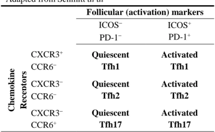
Extensive analysis of human blood Tfh cells have further revealed phenotypic and functionally distinct subsets (Table 1).
Ki-21 67- quiescent cells which lack expression of ICOS and PD-1, while expressing CCR752,55,56. He and colleagues reported that blood Tfh cells are heterogeneous and are composed of CCR7+PD-1- and CCR7-PD-1+ subsets. These two subsets showed distinct kinetics following seasonal influenza vaccination, as only CCR7-PD-1+ Tfh cells increased upon vaccination56. By comparing these results with mice transferred with congenic marked Ovalbumin (OVA)-specific OT-II cells and then immunized with OVA in alum, He and colleagues proposed that CCR7-PD-1+ Tfh cells reflect tissue Tfh cell responses56. Additionally, they demonstrated that CXCR5+CCR7- Tfh cells sorted from human blood were more efficient in supporting plasma cell differentiation and IgG production, compared to CXCR5+CCR7+ Tfh cells, upon in vitro TCR stimulation56. Using a different experimental design, Morita and colleagues demonstrated that human blood CXCR5+ Tfh cells were
composed of three subsets according to expression of CXCR3 and CCR6 chemokine receptors54. The T-bet+CXCR3+CCR6- Tfh1, GATA-3+CXCR3-CCR6- Tfh2, and RORγt+CXCR3-CCR6+ Tfh17-like cells were proved functionally distinct54. Whereas Tfh1,
Tfh2 and Tfh17-like cells were able to secrete the defining cytokines of their Th counterparts, namely IFN-γ (Tfh1), IL-4, IL-5 and IL-13 (Tfh2), and IL-17 and IL-22 (Tfh17), only Tfh2 and Tfh17-like cells produced IL-21, induced activation of naïve B cells, and supported class switch recombination upon in vitro stimulation with superantigens54. This was further confirmed in GC Tfh cells from human tonsils in which T-bet was found to diminish the Tfh cell program required to provide help to B cells122. Taking advantage of these findings, Locci and colleagues used gene expression microarrays to demonstrate that PD-1+CXCR3- Tfh cells were the blood Tfh cell subset which most closely resemble GC Tfh cells (sorted from human tonsils)55. Using in vitro co-cultures under the presence of superantigens, PD-1+CXCR3- Tfh cells displayed the strongest capacity to induce IgG and IgM secretion by memory B cells55.
The clinical relevance of blood Tfh cell subsets is supported by vaccination studies. Following seasonal influenza vaccination, the emergence of blood ICOS+CXCR3+ Tfh cells correlated with the development of protective antibodies120,123. Thus, Tfh cell subsets could serve as potential biomarkers for monitoring antibody responses in vaccination and infectious diseases. However, it is somehow unexpected that Bentebibel and colleagues correlated blood CXCR3+ Tfh cells with humoral responses. Indeed, CXCR3-expressing Tfh
cells were previously shown to be non-efficient helpers54,55,122. The help activity of blood
22
vitro stimulation with superantigens120. Therefore, it is likely that ICOS+CXCR3+ Tfh cells specifically evoke memory humoral responses which bypass de novo GC reactions.
Table 1: Human blood Tfh cell subsets
Adapted from Schmitt at al52
Follicular (activation) markers
ICOS PD-1 ICOS+ PD-1+ Chemo kin e Rec ept o rs CXCR3+ CCR6 Quiescent Tfh1 Activated Tfh1 CXCR3 CCR6 Quiescent Tfh2 Activated Tfh2 CXCR3 CCR6+ Quiescent Tfh17 Activated Tfh17
Distinctive Features of Human Tfh Cells
Although, several studies have established that Tfh cells from humans and mice are similar at the transcription level, a parallel body of work implies a largely different group of cytokines in human Tfh cell differentiation62,117,121,124–128. Schmitt and colleagues systematically analysed the upregulation of Tfh-signature molecules by sorted naïve CD4+ T cells from human blood cultured in the presence of several combinations of cytokines plus anti-CD3/CD28 stimulation126. TGF-β plus IL-12 or TGF-β plus IL-23 induced upregulation of CXCR5, ICOS, Bcl-6, and downregulation of Blimp-1. These cytokine combinations also increased the proportion of IL-21-expressingT cells in the end of culture and efficiently enabled those T cells to provide help to memory B cells, measured by IgG concentration in culture supernatants126. These findings suggest that TGF-β co-opts redundant signalling via STAT3/STAT4 (induced by IL-12 and IL-23) to promote Tfh cell differentiation in humans. While, preformed in artificial systems, the reduced Tfh cell and GC responses observed in patients lacking functional 12Rβ1, the receptor for 12 and 23, corroborates the IL-12 requirement for human Tfh cell differentiation in physiological conditions117.
3.
Regulation of Humoral Responses
To ensure clonal selection of high affinity B cell clones, GC reaction is tightly regulated by complex molecular signals at multiple stages.
23
3.1. Germinal Centre Reaction and Cell Dynamics
Following immunization, IgD+ naïve B cells are activated and move to the outer follicular zone of secondary lymphoid organs. Here, they present antigenic peptides on MHCII to specialized subsets of separately-activated CD4+ T helper cells5,129. The antigen-activated B cells harbouring the highest BCR affinity for the immunizing antigen eventually gain access to the follicle centre and populate the specialized follicular dendritic cell (FDC) network5,6,130. After these initial steps of GC reaction, two main compartments become evident: the dark and light zones, as initially proposed by Röhlich in 1930. The dark zone is formed by densely packed B cells (classically called centroblasts) undergoing rapid proliferation and somatic hypermutation. This Darwinian process results in the generation of B cell clones (classically termed centrocytes) expressing surface antibodies with a wide range of affinities for the immunizing antigen. These cells then travel to the light zone where they compete for binding to immune complexes presented by highly ordered surface units of FDC (termed iccosomes), and for survival and differentiation signals provided by T helper cells4,63,131–135. During the course of cognate T-B cell interaction in the light zone, a subset of B cells additionally undergo activation-induced cytidine deaminase (AID)-mediated class
switch recombination136,137. A great work by Victora and colleagues confirmed that T cell help, and not direct competition for antigen is the limiting factor for B cell selection within GCs135,138. Therefore, it seems that within the context of the GC , the role of the BCR is primarily to capture and internalize antigen rather than to induce BCR signalling6,135. Ultimately, high-affinity GC B cell clones differentiate into plasmablasts and memory B cells after repeated rounds of expansion, diversification and selection. This cyclic re-entry sustains the population of cells that proliferate within the dark zone and maintains the GC reaction over time5,6,136. Although, the precise mechanisms driving the fate decision of GC B cells are still being elucidated, Tfh cell help was found to favour plasma cell differentiation (and not memory B cell differentiation) in a CD40L-dependent manner108–110,139,140.
The dark and light zones of the GC are organized by expression of CXCR4 and CXCR5, respectively. These chemokine receptors on B cells are required to guide B cell migration toward CXCL12-enriched dark zone (populated by CXCR4hi B cells) and CXCL13-enriched
light zone (populated by CXCR4lo B cells)131,135. These phenotypic states are the result of a
timed B cell-intrinsic program: Bcl-6 is required for the establishment of the specialized dark zone-associated gene expression programme that allows somatic hypermutation, MYC and