Insira o título da
dissertação, projeto
ou relatório de
estágio, letra Arial
Bold, tamanho
ajustado a caixa
de texto 12x12
cm, justificado
à esquerda
Ana Isabel Lavoura Puga
Dissertação de Mestrado apresentada à
Faculdade de Ciências da Universidade do Porto, Instituto de
Ciências Biomédicas Abel Salazar
Bioquímica
2013
C ha racter iz atio n of an ex pe rim en tal m od el of ne on atal m en ing iti s ind uce d by G rou p B S tr ep toco ccus An s a Is ab el La v ou ra P ug aMSc
FCUP ICBAS 2013 2.º CICLOCharacterization
of an
experimental
model of neonatal
meningitis
induced by Group
B Streptococcus
Insira o título da
dissertação, projeto
ou relatório de
estágio, letra Arial
Bold, tamanho
ajustado a caixa
de texto 12x12
cm, justificado
à esquerda
Ana Isabel Lavoura Puga
Mestrado em Bioquímica
Laboratório de Imunologia Mário Arala Chaves 2013
Orientador
Professor Paula Ferreira, PhD, Instituto de Ciências Biomédicas Abel
Salazar
Coorientador
Elva B. Andrade, PhD, Instituto de Ciências Biomédicas Abel Salazar
Characterization
of an
experimental
model of
neonatal
meningitis
induced by
Group B
Streptococus
Todas as correções determinadas pelo júri, e só essas, foram efetuadas. O Presidente do Júri,
This work was supported by funds from European Regional Development Fund (FEDER) through the Operational Competitiveness Program (COMPETE) underProject FCOMP – 01-0124-FEDER-015841 and by National funds from the Foundationfor Science and Technology (FCT), under Project PTDC/SAU – MIC/ 111387/2009.
ii FCUP
“If you're going to try, go all the way. Otherwise, don't even start. This could mean losing girlfriends, wives, relatives and maybe even your mind. It could mean not eating for three or four days. It could mean freezing on a park bench. It could mean jail. It could mean derision. It could mean mockery - isolation. Isolation is the gift. All the others are a test of your endurance, of how much you really want to do it. And, you'll do it, despite rejection and the worst odds. And it will be better than anything else you can imagine. If you're going to try, go all the way. There is no other feeling like that. You will be alone with the gods, and the nights will flame with fire. You will ride life straight to perfect laughter. It's the only good fight there is.”
iv FCUP
FCUP Characterization of an experimental model of neonatal meningitis induced by Group B Streptococcus
i
Acknowledgments
Em primeiro lugar, quero agradecer à minha orientadora, a Professora Paula. Palavras não chegam para lhe agradecer a oportunidade que me deu. Muito obrigada por me ter aberto a porta com um sorriso e por me ter aceitado na sua equipa. Quero agradecer-lhe pela preocupação, pela exigência e por todo o apoio ao longo destes anos. Sob a sua supervisão aprendi a ser uma cientista (on going work) melhor e uma pessoa melhor. Obrigada por ter acreditado em mim, e por me fazer acreditar que este é o meu lugar, que a Imunologia é o que
eu quero fazer, aprender e explorar. Professora, obrigada por ser assim, positiva e bem-disposta. Mesmo quando o mundo parece mais cinzento, bastam 5 minutos consigo, que
as nuvens vão todas embora. Não sei como o faz, mas muito obrigada por isso e por tudo, tudo!
Como é óbvio, em segundo lugar, quero agradecer a minha co-orientadora, a Elva.
Sweet Master, obrigada por todo o tempo e paciência que investiste em mim. Espero que no
final desta jornada estejas orgulhosa da tua pequena padawan. Obrigada por teres partilhado o teu trabalho e a tua paixão pela Ciência comigo, obrigada por me teres deixado ser parte de um trabalho tão bonito e tão interessante como o teu. Sinto-me uma sortuda por ter sido duplamente tua estagiária. É um prazer trabalhar com alguém tão metódico e tão elegante naquilo que faz. Claro, obrigada por teres sido além de mentora, uma amiga com quem tive ataques de riso e momentos parvos e divertidos. Guardo-os todos com muito carinho. Um grande, gigante, enorme e super obrigado!
À minha Joaninha. Jo, não há palavras. A sério. Pessoas como tu já não são feitas. És
one of a kind. Obrigada por me aturares todos os dias da semana, incluindo alguns sábados
esporádicos e quiçá uns feriados. Obrigada por não seres só uma colega de laboratório e de trabalho, mas uma amiga, daquelas com A grande. Obrigada por me apoiares e por me dares confiança, por acreditares em mim e por dizeres me ‘Tu és capaz!’. O teu altruísmo e a tua bondade são qualidades quase extintas, e eu sou uma sortuda por me ter cruzado contigo. Obrigada por me alimentares, não só com comida Master Chef, mas por me alimentares o sonho de ser melhor. Não podia no seguimento deixar de agradecer ao Tiago. Rapaz, tu és um castiço. É impossível estar cabisbaixa ao teu lado, impossível. Obrigada por todos nerdanços, e pelas boleias. Citando o eloquente Miguel Gonçalves ‘Puque tu és muito potente!’
Ao Melo, também carinhosamente Pedro ‘Melito’. Fico muito contente que a vida tenha dado uma daquelas voltas e que nos tenha colocado aos dois no Arala-Chaves. És uma pessoa cinco estrelas, em todos os sentidos. Obrigada pela paciência, pelas gargalhadas, pelas conversas científicas, pelas conversas não cientificas, pelas parvoíces, por tudo. Mesmo! Espero, e tenho quase a certeza que o teu futuro vai ser brilhante, porque tu és sem dúvida das
ii FCUP
Characterization of an experimental model of neonatal meningitis induced by Group B Streptococcus
pessoas mais competentes, prestáveis, humildes e amigas que alguma vez conheci. Foi um prazer e um privilégio ter partilhado as câmaras de fluxo e a mesa do almoço contigo!
Ao Pedro ‘A Wikipédia’ Madureira. És um poço de saber! Obrigada por todas as explicações (todas mesmo!) e pelo tempo que perdeste comigo a explicar-me citometria de fluxo e coisas afins. Mas obrigada também pelas piadas ‘a la Pedro Madureira’ e por me perceberes nas alturas aleatórias em que falo de blasers da Balmain (sério, este teu conhecimento mudou a minha vida!) e sandes do Guedes!
A todas as pessoas do laboratório, a Encarnação pela boa disposição e ajuda, sem si o laboratório não era o mesmo. A Adília por pôr sempre tudo a funcionar e pelas histórias hilariantes que contou, muito obrigada! Ao Pedro Ferreirinha, por ser das pessoas mais prestáveis que já conheci, obrigada pelas ajudas e pelos discussões saudáveis! Claro, às três meninas, a Alex, a Bego e a Ângela, por serem super agradáveis e deixarem usar sempre ‘o cantinho da câmara’.
A todos os meus amigos, artistas e cientistas, pelas conversas, pelos insights e diferentes perspectivas. ‘Nenhum homem é uma ilha’, obrigado a todos pelo apoio e por me ouvirem falar em dialecto tese nos últimos tempos. Vocês são todos muito lindos.
Mas claro, um especial obrigado em néon cor-de-rosa para a minha querida Matilde. Obrigada por me apoiares em todos os momentos decisivos da minha vida. Obrigada por me transmitires calma. Miga, nem que te pague 500 jantares, nunca vão ser suficientes para te agradecer por tudo o que fizeste por mim, durante todos estes anos, mas principalmente nesta recta final. Obrigada por tudo, e quando digo tudo incluo paciência, amizade, cumplicidade e por teres feito comida durante um mês seguido, sem nunca pedir nada em troca. És uma pessoa muito linda. Claro está que para o ano quero um parágrafo só para mim. Migas são como migas, migas forever. (Sim escrevi isto na minha tese! Ahahahaha!) Sério, um grande, grande obrigada!
E por fim, quero agradecer às pessoas que mais amo neste mundo, os meus pais e o meu irmão. Mano, muito do que sou hoje devo-to a ti, por teres sido um porreiro comigo, um verdadeiro cool older brother. És das pessoas que mais admiro no mundo, e obrigada por me dares na cabeça quando preciso, mas também por me apoiares incondicionalmente e me defenderes com unhas e dentes. Mãe e Pai, esta tese é o fruto de todo o apoio incondicional que me deram ao longo de 23 anos. Obrigada por me terem educado da forma que educaram, por se terem sentado a fazer os trabalhos de casa comigo, por me estimularem desde miúda a querer ser melhor, a querer fazer melhor, a ser curiosa. Obrigada por TODOS os sacrifícios e pela dedicação. Vocês são sem dúvida os melhores pais do mundo. Obrigada pelas injecções de energia e de tranquilidade. E claro, à avó Alzira, melhor ‘tartaruguinha’.
Espero que estejam tão orgulhosos e felizes quanto eu, porque esta tese é dedicada a vocês, família. Muito obrigada!
FCUP Characterization of an experimental model of neonatal meningitis induced by Group B Streptococcus
iii
Resumo
A meningite neonatal induzida por bactérias é uma importante causa de mortalidade e morbilidade em todo o mundo. Streptococcus agalactiae, mais comummente designado por Estreptococos do Grupo B (do Inglês GBS) é o responsável pela maioria dos casos de meningite neonatal. A colonização materna dos tratos gastrointestinal e genitourinário são a principal fonte de infeção dos recém-nascidos. Apesar das melhorias nos cuidados neonatais e da implementação de medidas antimicrobianas, a taxa de mortalidade associada a este microrganismo é de 10%. Além disso, até cerca de 50% dos sobreviventes da meningite causada por GBS desenvolvem graves sequelas neurológicas. Ainda existe uma grande lacuna no conhecimento da patogénese desta doença, apesar da existência de vários modelos de experimentação animal de meningite neonatal. Uma possível explicação é que nenhum destes modelos utiliza a via de infeção que ocorre nos humanos. No nosso laboratório estamos a desenvolver um modelo que mimetiza a via de infeção humana. Neste sentido, fêmeas BALB/c grávidas foram infetadas intra-vaginalmente com 105células de GBS BM110, estirpe responsável pela maioria dos casos de meningite humana. Após o parto, a colonização do trato vaginal das fêmeas e a carga bacteriana presente nos descendentes foram monitorizadas em diferentes tempos. Os resultados obtidos mostraram que as fêmeas estavam altamente colonizadas até ao quarto dia após o parto e que, a bactéria foi transmitida às ninhadas. De fato, GBS foi encontrado nos pulmões, no sangue, no fígado e no cérebro dos ratinhos recém-nascidos, a diferentes tempos após o parto. Análise histopatológica dos cérebros de ratinhos infetados mostrou que estes apresentavam as caraterísticas típicas de meningite: espessamento das meninges, hemorragias cerebrais e influxo de células inflamatórias. Aproximadamente 40% da descendência morreu após o parto, um valor semelhante ao descrito em humanos antes da implementação da profilaxia antibiótica intrapartum. De modo a confirmar se os animais sobreviventes à semelhança do descrito em humanos apresentavam sequelas neurológicas, foram avaliados, na idade adulta, o seu desempenho cognitivo e motor bem como os padrões de neurotransmissores em diferentes secções de cérebro. A avaliação cognitiva foi determinada usando o Radial
Maze Test, e a atividade locomotora e comportamento exploratório foram avaliados
usando o Open Field Test. Após os testes, os animais foram sacrificados e diferentes secções dos seus cérebros foram utilizadas para medir as concentrações de neurotransmissores. Ratinhos sobreviventes à infeção por GBS apresentaram um maior número de erros cometidos quando comparados com os controlos. Estes erros
iv FCUP
Characterization of an experimental model of neonatal meningitis induced by Group B Streptococcus
estão associados com os níveis de glutamato no hipocampo que se encontravam diminuídos no grupo de ratinhos que sobreviveram à infeção. Os resultados obtidos no
Open Field Test indicaram que estes ratinhos são menos ativos, uma vez que
apresentaram atividade locomotora e comportamento exploratório diminuídos quando comparados com o grupo controlo. Estes resultados estavam associados com os níveis diminuídos de glutamato no tálamo e com os níveis diminuídos de dopamina e seus metabolitos no hipocampo e no estriado dos ratinhos sobreviventes, quando comparados com os controlos. Foram assim observadas alterações neurológicas nos ratinhos que sobreviveram à infeção neonatal por GBS.
Em conjunto, estes resultados indicam que o nosso modelo mimetiza o que acontece nos humanos, sendo por isso um bom modelo para ser usado na caracterização da patogénese e patofisiologia da meningite induzida por GBS.
Palavras-Chave: Meningite neonatal; Streptococcus do Grupo B; modelo
FCUP Characterization of an experimental model of neonatal meningitis induced by Group B Streptococcus
v
Abstract
Bacterial meningitis is a substantial cause of morbidity and mortality in neonates. Group B Streptococcus (GBS), a common designation for Streptococcusagalactiae, is the main agent of neonatal meningitis. Maternal colonization with GBS in
the genitourinary and/or gastrointestinal tracts is the source of the neonatal infection. Despite early antimicrobial treatment and improvement in neonatal intensive care, up to 10% of neonatal invasive GBS infections are lethal and up to 50% of surviving infants with meningitis present neurological sequelae. Significant gaps in knowledge of the pathogenesis of this disease still remain despite several animal models have been used. All these models use artificial routes of infection. Our research team is developing a murine model of neonatal GBS-induced diseases, which addresses the natural course in human pathogenesis of ascending infection from the lower genital tract to neonates. Here, we characterize the suitability of this animal model to study the neonatal meningitis induced by GBS. For that purpose, pregnant BALB/c female mice were infected intra-vaginally with 105cells of GBS BM110, a strain responsible for the
majority of human cases of meningitis. Pregnant female were allowed to deliver and their vaginal tract colonization and the bacterial transfer into the newborns, were monitored at different time points after birth. The obtained results showed that mothers remained highly colonized until the fourth day after delivery and the bacterium was transmitted to their progeny. Indeed, GBS was found in the lungs, blood, liver and brain, of pups at different time points after birth. Histopathological analysis of the brains of infected pups showed the classical features of meningitis such as: meningeal thickening, cerebral bleeding and massive influx of inflammatory cells. Approximately 40% of the progeny died, a value close to that reported for the initial case-fatality observed in humans, before the introduction of intrapartum antibiotic therapy. To confirm that the surviving animals present neurological sequelae, we determined their cognitive and motor performance in adult life, and identified whether an altered neurotransmission pattern was involved. The cognitive evaluation was performed in a complex learning task using the Radial Maze test and the anxiety levels, locomotor activity and exploratory behavior were measured through Open Field Test. After the tests, the animals were sacrificed and the different brain regions were used for determination of neurotransmitter levels. Mice that survived to neonatal GBS infection presented increased working and reference memories errors when compared with the non-infected controls, which were associated with the decrease in the glutamate levels in the hippocampus. Results from Open Field Test, showed that mice survivors to GBS infection, were less active since they present decreased locomotor activity and
vi FCUP
Characterization of an experimental model of neonatal meningitis induced by Group B Streptococcus
exploratory behavior than the non-infected controls. These results are associated with the lower levels of glutamate in the thalamus and also with the diminished levels of dopamine and its metabolites in the hippocampus and in the striatum of GBS-survivors comparatively with non-infected controls.
Altogether these results indicate that our mouse model closely mimics the characteristics observed in humans and, therefore, could be used to characterize the pathogenesis and pathophysiology of meningitis induced by GBS.
Keywords: Neonatal meningitis; Group B Streptococcus; experimental model;
FCUP Characterization of an experimental model of neonatal meningitis induced by Group B Streptococcus
vii
Table of Contents
Acknowledgments ... i
Resumo ... iii
Abstract ... v
Tables and Figures ... ix
Abbreviations ... x
Introduction ... 1
1. Neonatal Bacterial Meningitis ... 1
1.1 Overview ... 1
1.2 Epidemiology ... 2
1.3 Streptococcus agalactiae ... 3
2. GBS Disease ... 5
2.1 Risk Factors ... 5
2.2 Early-Onset and Late-Onset Disease ... 5
2.3 Prophylaxis for neonatal GBS Infections ... 7
3 Meningitis ... 9
3.1 Pathophysiology of Meningitis ... 9
3.2 Homeostatic Barriers of the Brain ... 9
3.3 Pathogen Translocation across the Blood Brain Barrier ... 12
3.4 GBS Infection and Virulence ... 13
3.5 GBS translocation across the BBB ... 14
3.6 Experimental Models of Neonatal Meningitis ... 14
Objectives ... 17
Materials and Methods ... 21
Bacterial strains and growth conditions ... 23
Animals and Ethics statement ... 23
viii FCUP
Characterization of an experimental model of neonatal meningitis induced by Group B Streptococcus
Neonatal mouse model of GBS-induced meningitis ... 23
DNA extraction... 24
PCR assays ... 24
Histopathology ... 25
Behavioral assessments ... 25
Open field (OF) ... 27
Neurotransmitter Determination ... 27
Amino acids ... 27
Monoamines and their metabolites ... 28
Total protein determination ... 28
Statistical analysis ... 28
Results ... 31
1 Vaginal and gastrointestinal tracts of the female mice remains colonized with GBS after delivery ... 33
2 GBS is vertically transmitted to the progeny... 34
3 Brain histopathological changes in GBS infected neonates ... 35
4 Mice survive to neonatal GBS infection present behavior alterations in adult life ……….36
5 GBS-induced meningitis leads to an altered neurotransmitter pattern ... 39
Discussion ... 43
Concluding Remarks... 51
FCUP Characterization of an experimental model of neonatal meningitis induced by Group B Streptococcus
ix
Tables and Figures
Table 1 | Pathogens commonly causing central nervous system infections in humans..3
Table 2 | GBS-specific conventional PCR assay characteristics…... 25
Figure 1 | Schematic view of the stages of neonatal GBS infection ... 7
Figure 2 | Incidence of invasive early and late-onset group B streptococcal disease ... 8
Figure 3 | Homestoatic Barriers of the Brain... 10
Figure 4 | The blood–brain barrier. ... 11
Figure 5 | Mechanisms involved in microbial traversal of the blood–brain barrier. ... 13
Figure 6 | Colonization of the murine vaginal mucosa with GBS. ... 33
Figure 7 | Murine model of neonatal GBS-induced meningitis. ... 34
Figure 8 | Murine model of neonatal GBS-induced meningitis. ... 35
Figure 9 | Murine model of neonatal GBS-induced meningitis. ... 36
Figure 10 | Effects of neonatal GBS-induced meningitis on learning and memory performances in adulthood... 37
Figure 11 - Effects of neonatal GBS-induced meningitis in the locomotor and exploratory behaviors in adulthood, distance travelled in OFT. ... 38
Figure 12 | Effects of neonatal GBS-induced meningitis in the locomotor and exploratory behaviors in adulthood. ... 38
Figure 13 | Effects of neonatal GBS-induced meningitis in glutamatergic function. ... 39
Figure 14 | Effects of neonatal GBS-induced meningitis in dopaminergic function in the hippocampus. ... 40
Figure 16 | Effects of neonatal GBS-induced meningitis in dopaminergic function in the striatum. ... 41
x FCUP
Characterization of an experimental model of neonatal meningitis induced by Group B Streptococcus
Abbreviations
5-HIAA - 5-hydroxyindolacetic acid
5-HT - Serotonin
ABC - Active Bacterial Core
BBB - Blood brain barrier
BCSFB - Blood Cerebrospinal Fluid Barrier
BECS - Brain Extracelular Fluid
BMEC - Brain MicrovascularEndothelial Cells
BSA - Bovine Serum Albumin
CAMP - Christie Atkins Munch-Petersen
CDC - Center for Disease Control
CFU - Colony-forming units
CNS - Central Nervous System
CP - Choroid Plexus
CSF - Cerebrospinal Fluid
E - Epinephrine
EOD - Early onset disease
DA - Dopamine
DOPAC - 3,4-diydroxiphenylacetic acid
FbsA - Fibrinogen-binding protein A
G - Gestation
GABA - Gamma-Aminobutyric Acid
GAPDH - Glyceraldehyde 3-phosphate dehydrogenase
GBS - Group B Streptococcus
HBMEC - Human Brain Microvascular Endothelial Cells
HPLC - High Performance Liquid Chromatography
HPLC/ED - High Performance Liquid Chromatography with
Electrochemical Detection
HVA - Homovanillic Acid
HvgA - Hypervirulent GBS adhesion
IAP - Intrapartum antibiotic prophylaxis
IgG - Immunoglobulin G
IL-10 - Interleukin 10
FCUP Characterization of an experimental model of neonatal meningitis induced by Group B Streptococcus
xi
LOD - Late onset disease
Lmb - Laminin-binding protein
MLST - Multi-locus sequence typing
MOI - Multiplicity of infection
NE - Norepinephrine
OF - Open Field
OFT - Open Field Test
PND - Postnatal Day
ST - Sequence type
ST-17 - Sequence type 17
TH - Todd-Hewitt
2 FCUP
FCUP
Characterization of an experimental model of neonatal meningitis induced by Group B Streptococcus 1
1. Neonatal Bacterial Meningitis
1.1 Overview
Neonatal bacterial meningitis is a serious life-threatening disease and a major cause of disability worldwide, despite advances in health care, in the development of more effective antibiotics, and in greater tools for rapid pathogen identification (Gaschignard et al., 2011).
At birth, all organ systems of the neonate switch from a highly controlled intra-uterine environment to the drastically different surroundings of the outside world.
This uttermost transition is then followed by a gradual, age-dependent maturation. Actually, prenatal immune system maturation cannot be fully conducted until neonates lose their status of privileged allograft, surrounded by a great number of maternal antigens, the fetus remains in his ‘sterile world’, unresponsive to any challenge. Yet, the immune system is prepared to cope with the dramatic changes that occur during birth, as newborns respond to microbes from the circling new non-sterile environment.
Nevertheless, neonates are at greater risk of developing infections, sepsis and meningitis when compared to other age groups as their immune system is not fully mature. Deficiencies in humoral and cellular immunity are setbacks that make them more susceptible to infections. The defects in adaptive immunity are well described [reviewed in (Adkins et al., 2004)], as they do not have immunological memory, are unable to produce high levels of IgG against encapsulated bacteria, produce less efficient inflammatory cytokines and also they are committed to produce the immunosuppressive/immunoregulatory cytokine IL-10 (Madureira et al., 2007, Madureira et al., 2011). Thus, neonates must rely on their innate immune system for protection against pathogens (Krishnan et al., 2003, Firth et al., 2005). Nevertheless, newborns have a diminished bone marrow pool, and therefore their ability to accelerate neutrophil production in response to infection is limited (Koenig and Yoder, 2004). Also, neutrophil recruitment in neonates is reduced (Koenig and Yoder, 2004). Moreover, the phagocyte function is diminished in neonates, especially in premature babies. (Falconer et al., 1995).
2 FCUP
Characterization of an experimental model of neonatal meningitis induced by Group B Streptococcus
1.2 Epidemiology
The incidence value for neonatal bacterial meningitis may be underestimated as it is very hard to determine accurately due to a range wide of factors, which include: the difficulty in diagnosing neonatal meningitis, variations between hospital-based and community studies, regional differences and unregistered deaths in areas where the access to proper health care is poor or almost inexistent (Osrin et al., 2004). Moreover, although most data evidence derives from developed countries, the major burden of neonatal sepsis and meningitis occurs in the developing world (Furyk et al., 2011), where health care and data acquisition are unevaluated. Thus, it is obvious that the existing data does not reflect the true incidence of bacterial meningitis.
In industrialized developed countries, bacterial meningitis incidence is approximately 0.3 per 1,000 live births (Brouwer et al., 2010). The World Health Organization (WHO) estimates that there are 5 million neonatal deaths per year and that approximately 98%, an overwhelming majority, occurs in developing countries (Furyk et al., 2011). Neonatal meningitis contributes significantly to this shocking statistics, as it is recognized as one of the top ten causes of infection-related death worldwide (Fauci, 2001), with 126,000 cases annually and more than 50,000 deaths (Stoll, 1997, Weber et al., 2003).
Mortality from neonatal meningitis in developing countries is estimated to be 40-58%, against 10% in developed countries (Furyk et al., 2011). A study conducted throughout Asia reported an estimated incidence of neonatal meningitis from 0.48 per 1000 live births in Hong Kong, whereas in Kuwait, an incidence rate of 2.4 per 1,000 births (Tiskumara et al., 2009). In another study focused in neonatal infections in Africa and in South Asia, an incidence of neonatal meningitis ranging from 0.8 to 6.1 per 1,000 live births was reported (Thaver and Zaidi, 2009).
Group B Streptococcus is, beyond the neonatal period, the most common cause of bacterial meningitis, accounting for approximately 80% of cases among those less than 2 month of age (Schrag, 2011). Escherichia coli is the second pathogen (Klinger et al., 2000) and Listeria monocytogenes, the third, and it possesses the unique characteristic of transplancental transmission (Heath et al., 2003). In Portugal, GBS is the most common isolate in early onset neonatal sepsis. A survey from 2008 in Portuguese infants younger than 90 days, estimated an overall incidence of invasive GBS disease of 0.54 per 1,000 live births, with a mortality rate of 6.6% (Neto, 2008). Moreover, this incidence also varies from one geographic area to another, as approximately 35% of pregnant women from the north are colonized with GBS, contrasting with the 13% found in the south (Neto, 2008).
FCUP
Characterization of an experimental model of neonatal meningitis induced by Group B Streptococcus 3
Table 1 summarizes the most common pathogens responsible for central nervous system (CNS) infections and meningitis.
Table 1 – Pathogens commonly causing central nervous system infections in humans
Pathogen Reference
Streptococcus agalactiae (Group B Streptococcus) (Brochet et al., 2008, Heath et al., 2009, Edmond et al., 2012)
Escherichia coli (Klinger et al., 2000, Gaschignard et al., 2012)
Listeria monocytogenes (Heath et al., 2003, Fayol et al., 2009)
Streptococcus pneumoniae (O'Brien et al., 2009)
Neisseria meningitidis (Kurlenda et al., 2010)
Haemophilus influenzaetype b (Watt et al., 2009)
Mycobacterium tuberculosis (Farinha et al., 2000)
Borrelia burgorferi (Avery et al., 2005)
Candida albicans (Aleixo et al., 2000)
Trypanossoma spp. (Finsterer and Auer, 2013)
Although the mortality rate in developed countries declined almost from 50% in the 1970’s to approximately 10% in the late 90’s (Berardi et al., 2010), the morbidity rates are still high, as meningitis remains a major source of disability and long term sequelae (Puopolo et al., 2005). Neonatal meningitis survivors are at risk for developing moderate to severe disability, which include: problems in language, motor function, hearing, vision and cognition and also 5 to 20% have future epilepsy. Besides, survivors may also present more subtle symptoms: visual deficits, middle-ear disease and behavioral problems (Bedford et al., 2001). In a 2001 study, regarding a sample superior to 1500 neonates from the England and Wales with surviving until the age of 5, several neuro-motor disabilities were reported as cerebral palsy (8.1%), learning disability (7.5%), seizures (7.3%) and hearing problems (25.8%) (Bedford et al., 2001).
1.3 Streptococcus agalactiae
Streptococcus agalactiae is a Gram positive encapsulated bacterium which
appears as diplococci or as a chain (Doran and Nizet, 2004, Nandyal, 2008). It is a catalase negative, beta-hemolytic microorganism and it is a facultative anaerobic,
4 FCUP
Characterization of an experimental model of neonatal meningitis induced by Group B Streptococcus
growing preferentially in oxygen absence. During the 30’s, Rebecca Lancefield observed that hemolytic Streptococcus from human and animal origins could be divided and distinguished serologically according to their polysaccharide capsule composition (Lancefield, 1933, 1934a). Moreover, five Streptococcus groups were then discovered: A, B, C, D and E (Lancefield, 1934b). In line with this type of classification, the most common designation for S. agalactiae, Group B Streptococcus (GBS), was implemented. Lancefield was also able to sub-divide GBS into different serotypes using the antigen immunogenicity of the polysaccharide capsule as criteria (Lancefield, 1934b). Currently there are ten known GBS serotypes: Ia, Ib, II, III, IV, V, VI, VII, VIII (Doran and Nizet, 2004, Shet and Ferrieri, 2004, Slotved et al., 2007).
Still during the 30’s, Lancefield and Hare isolated GBS for the first time from the vaginal tract of a woman after having birth [reviewed by (Mulder and Zanen, 1984)] and three years later, Fry observed cultured GBS collected from women during delivery. Despite these facts, little knowledge of GBS role in neonatal infections was obtained during the next thirty years. It was only in 1964 that Eichkoff highlighted the importance of this pathogen in the previously stated subject. In the late 70’s, GBS outstripped
E. coli as the most important cause of newborn septicemia in the United States of
America. Nowadays, GBS continues to be one of the major pathogens responsible for neonatal bacterial meningitis and septicemia (Stoll et al., 2011).
The Lancefield division does not demonstrate the true biodiversity regarding the existing GBS strains, as strains that belonging to different serotypes can be more related at a genetic level than strains from the same serotype (Davies et al., 2004, Tettelin et al., 2005).
A more profound knowledge about serotype relations was needed and multilocus sequence typing (MLST) was applied. This is a very important molecular biology technique and it is used to type multiple loci. This procedure characterizes microbial isolates using the DNA sequences of internal fragments of housekeeping genes (Davies et al., 2004). Approximately 450-500 bp internal fragments of each gene are used, as these can be accurately sequenced on both strands using an automated DNA sequencer. For each housekeeping gene, the different sequences present within a bacterial species are assigned as distinct alleles and, for each isolate, the alleles at each of the loci define the allelic profile or sequence type (ST). After the characterization of the GBS MLST profile, it was observed that isolates with the same sequence can actually belong to different capsular serotypes. From all known serotypes, serotype III seems to be the most virulent and it is very closely associated with most of the cases of neonatal meningitis (Nandyal, 2008).
FCUP
Characterization of an experimental model of neonatal meningitis induced by Group B Streptococcus 5
2. GBS Disease
2.1 Risk Factors
GBS maternal colonization is the primary risk factor for the development of bacterial infections (Verani et al., 2010). The gastrointestinal tract is a natural reservoir for GBS and is likely the source of vaginal colonization (Dillon et al., 1982, Hoogkamp-Korstanje et al., 1982). In addition, a study suggested that the vagina becomes colonized with GBS as a result of its transfer from the rectum into the vagina, stating that rectal GBS colonization is a major predictor and risk factor for vaginal colonization (Meyn et al., 2009). Up to 30% of both sex individuals are commonly colonized with this bacteria in the gastrointestinal and genital tracts but remain asymptomatic. Importantly, 30% of the asymptomatic individuals are pregnant women colonized in the vaginal tract (Verani et al., 2010). Moreover, the carriage rate in the vaginal and rectal microbiota ultimately ranges between 10 to 37% and it’s the same in both developing and developed countries (Bergeron et al., 2000, Verani and Schrag, 2010).
GBS colonization during pregnancy can be transient, intermittent, or persistent (Lewin and Amstey, 1981, Hoogkamp-Korstanje et al., 1982, Hansen et al., 2004). Some women with GBS colonization during a pregnancy will be colonized during subsequent pregnancies (Cheng et al., 2008, Turrentine and Ramirez, 2008). Nevertheless, previous delivery of an infant with invasive GBS disease is a risk factor for early-onset disease in subsequent deliveries (Carstensen et al., 1988, Faxelius et al., 1988, Schrag et al., 2002b). In addition to maternal colonization with GBS, other parameters increase the risk for neonatal infections such as: gestational age inferior to 37 completed weeks, abnormally longer duration of membrane rupture, intra-amniotic infection, young maternal age, black race, and low maternal levels of GBS-specific anti-capsular antibody (Baker et al., 1981, Boyer et al., 1983, Schuchat et al., 1994, Schuchat et al., 2000, Zaleznik et al., 2000, Oddie and Embleton, 2002, Adair et al., 2003).
2.2 Early-Onset and Late-Onset Disease
Epidemiologically, neonatal GBS disease is divided in two distinct forms, early-onset disease (EOD), occurring between 0-6 days after birth, and late early-onset disease (Barichello et al.), starting after the first week of life until day 89 (Phares et al., 2008).
6 FCUP
Characterization of an experimental model of neonatal meningitis induced by Group B Streptococcus
Infants with GBS EOD generally present respiratory distress, apnea, or other signs of sepsis within the first 24–48 hours of life (Franciosi et al., 1973, Baker, 1978). The most common clinical syndromes of early-onset disease are sepsis and pneumonia but even though less frequently, early-onset infections can lead to meningitis. The case-fatality ratio of EOD has declined from as high as 50% in the 1970s (Baker and Barrett, 1974). In contrast, the progression of the infection to a state of meningitis, which is the most common manifestation of LOD, did not decline (Nandyal, 2008). Early-onset infections develop mainly in utero because GBS is able to ascend to the amniotic cavity, contaminating the amniotic fluid (Desa and Trevenen, 1984). Also, neonatal infection can be acquired during labor through exposure to GBS present in the vagina of a colonized woman (Katz and Bowes, 1988).
In relation to GBS LOD cases, the route of infection is not well known. It can be acquired from birth, as approximately 50% of the infants with LOD are colonized at delivery time with the same GBS serotype as the mother (Dillon et al., 1987), or it can be acquired from environmental sources (Berardi et al., 2013b). Horizontal transmission during the perinatal period may occur from mother to infant or from hospital or community sources (Melin, 2011). Another reported source of infection is breastfeeding (Godambe et al., 2005, Gagneur et al., 2009), although this has been under discussion in the scientific community as opinions differ (Berardi et al., 2013b). Nevertheless, it is thought that the bacterium is acquired by swallowing of infected maternal vaginal secretions during birth, leading to a persistent infection that culminates in its dissemination and in the later development of meningitis (Nandyal, 2008).
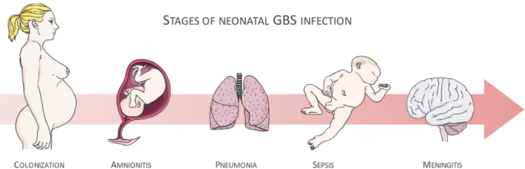
As illustrated in Figure 1, the first focus of infection is the lungs, as both vaginal fluid and amniotic fluid can be inspired. GBS caused pneumonia is characterized by severe pulmonary lesions, with bacterial infiltrates, hemorrhage and protein exudates into the alveolar space (Doran and Nizet, 2004). Thereby, lungs are an important gateway for bacterial infections, giving access to the bloodstream. The main consequences are bacterial dissemination to other anatomical places, such as the liver; bacteremia and septicemia and the consequent penetration into the brain (Doran and Nizet, 2004, Maisey et al., 2008).
FCUP
Characterization of an experimental model of neonatal meningitis induced by Group B Streptococcus 7
Figure 1 | Schematic view of the stages of neonatal GBS infection
Approximately 75% of the EOD cases are associated with type Ia, III and V serotypes (Nandyal, 2008). Serotype III GBS is the main responsible for the majority of infections in neonates worldwide (Edmond et al., 2012).
In LOD, it has been shown that most cases are associated with a singular capsulated serotype III GBS clone, designated as ST-17 (Jones et al., 2003, Lin et al., 2006, Tazi et al., 2010). The ST-17 clones are referred has being from the hyper-virulent type (Davies et al., 2004). Around 25-50% of GBS meningitis surviving infants experience permanent neurologic sequelae, including mental retardation, cerebral palsy, seizure activity, deafness and/or blindness (Edwards et al., 1985, Gibbs et al., 2004). Yet, these findings are most likely to underestimate the real incidence of GBS-induced meningitis, as signs of meningitis are often subtle in the neonate and therefore difficult to diagnose (Garges et al., 2006).
2.3 Prophylaxis for neonatal GBS Infections
In 1996, the Centers for Disease Control and Prevention (CDC), in association with both the American Congress of Obstetricians and Gynecologist and the American Academy of Pediatrics, presented guidelines for intrapartum antibiotic prophylaxis (IAP). According to these new measures, all pregnant women between the 35th and
37th week must be subjected to GBS screening and, whether positive, they are
administered with IAP. In addition, the guidelines were meant for all the women that presented, at time of birth, risk factors of disease transmission (Verani et al., 2010).
In 2002, these guidelines were reviewed (Schrag et al., 2002a) and, in November of 2010, a new CDC document was emitted (Verani et al., 2010). Some key-points in the combat against GBS disease were reinforced, such as: upgrading and improving the laboratorial methods for rapid and more accurate GBS identification, change in the penicillin dose used in prophylaxis, alternate antibiotic options for penicillin allergic women, actualized IAP and screening algorithms for preterm delivery
STAGES OF NEONATALGBS INFECTION
AMNIONITIS
8 FCUP
Characterization of an experimental model of neonatal meningitis induced by Group B Streptococcus
women, and lastly algorithm improvement for newborn babies with risk of contracting EOD (Verani et al., 2010, Schrag and Verani, 2013).
Such measures were accepted worldwide in developed countries and, although they aided the reduction of EOD rates, they did not contributed significantly for the lowering of the LOD rates (Verani et al., 2010, Berardi et al., 2013a, Schrag and Verani, 2013). Actually, in the period of widespread IAP use, the incidence of invasive early-onset GBS disease in Active Bacterial Core surveillance (ABCs) decreased by more than 80% from 1.8 cases/1000 live births in the early 1990s to 0.26 cases/1000 live births in 2010 (Figure2) (Schrag and Verani, 2013). However, GBS meningitis among patients under 2 months of age did not change in a significant fashion after the introduction of universal GBS screening of pregnant women and improvement in neonatal intensive care (Thigpen et al., 2011). Furthermore, the casualties from both EOD and LOD remain high and the morbidity associated with LOD has not changed substantially over decades (Gibbs et al., 2004) (Figure 2).
Figure 2 | Incidence of invasive early and late-onset group B streptococcal disease, Active Bacterial Core surveillance, United States, 1990–2010 (Schrag and Verani, 2013).
FCUP
Characterization of an experimental model of neonatal meningitis induced by Group B Streptococcus 9
3 Meningitis
3.1 Pathophysiology of Meningitis
Bacterial meningitis is an inflammation of the meninges that affects the pia, the arachnoid and the subarachnoid space (Hoffman and Weber, 2009). It is a complex mechanism that occurs in a series of steps which include: the adherence of the pathogen to the host mucosal surfaces and subsequent colonization; the bacterial invasion to adjacent intravascular spaces; and, of course, the survival and multiplication inside the host, resulting in bacteremia. Once microorganisms reach the bloodstream, they can cross into the subarachnoid space and they can enter in places where the Blood Brain Barriers (BBB) is vulnerable or inexistent, such as the choroid plexus (CP) (Daum et al., 1978). In addition, bacteria leaving cerebral capillaries enter the cerebrospinal fluid (CSF) and initiate an inflammatory cascade in the subarachnoid space (Spellerberg, 2000).
Although bacteremia is very important for meningitis development, it’s not sufficient by itself to microorganism penetration into the Central Nervous System (CNS). To cause meningitis, the pathogen must be able to leave the bloodstream and cross the barrier that separates it from the CNS.
3.2 Homeostatic Barriers of the Brain
The CNS is protected at three key sites: the endothelium of brain parenchymal blood capillaries, the choroid plexus epithelium and the arachnoid epithelium of the meninges (Abbott, 2005). These barriers play critical roles in controlling the movement of a series of metabolites, but also drugs, between the blood and the brain (Saunders et al., 2013).
Figure 3 displays the locations of the different homeostatic barriers in the brain, with (A) representing the Cerebrospinal Fluid Barrier (BCSFB); (B) the Blood-Brain Barrier (BBB); (C) the fetal CSF-Blood-Brain Barrier and (D) the Outer-CSF Barrier.
10 FCUP
Characterization of an experimental model of neonatal meningitis induced by Group B Streptococcus
Figure 3 | Homestoatic Barriers of the Brain. (A) The blood-CSF barrier between the choroid plexus blood vessels and the CSF. (B) The Blood-Brain Barrier between the lumen of the cerebral endothelium and the brain parenchyma. (C) The inner CSF-brain barrier, present in only during development, between the CSF and neuroependyma. (D) The outer CSF-brain barrier between the subarachnoid space and overlying structures. [Adapted from (Saunders et al., 2013)].
The BBB is a functional and structural barrier which separates the brain extracellular fluid (BECF) from the blood in the CNS. As shown in Figure 4, three cellular elements of the brain microvasculature compose the BBB: brain microvascular endothelial cells, astrocyte end-feet, and pericytes (Kim, 2003, Ballabh et al., 2004).
The BBB establishes both a physical as well as a metabolic barrier, isolating the CNS from systemic circulation and thus creating a unique and stable environment for an optimal neuronal activity (Correale and Villa, 2009). Moreover, this structure maintains the neuronal microenvironment homeostasis in a biochemical fashion, as endothelial cells regulate the molecular traffic in the brain, protecting it from microorganisms and circulating toxins in the bloodstream (Kim, 2008).
Endothelial cells present unique characteristics that make them so efficient in their functions of protection. Tight junctions, found between the adjacent cerebral endothelial cells, form a diffusion barrier, which selectively excludes most blood-borne substances from entering the brain (Ballabh et al., 2004). Furthermore, their cytoplasm lacks fenestrations present in most of the peripheral tissues (Correale and Villa, 2009). These special features make the BBB virtually impermeable to ions and molecules. In addition, the almost inexistent pinocitosis also contributes to the tight control of the molecular trafficking (Correale and Villa, 2009).
While endothelial cells are the main keepers of the brain homeostasis, astrocytes and pericytes help maintaining the barrier properties, but their functions are not fully understood. Yet, evidence from cell culture studies indicate that astrocytes upregulate many BBB features: they tightly sustain the vessel wall and appear to be critical for the induction and maintenance of the tight junction barrier (Hayashi et al.,
FCUP
Characterization of an experimental model of neonatal meningitis induced by Group B Streptococcus 11
1997, Sobue et al., 1999). As for pericytes, they are not believed to function as a barrier in the mammalian brain (Ballabh et al., 2004).
Figure 4 | The blood–brain barrier. The blood–brain barrier is formed by brain microvascular endothelial cells, astrocytes and pericytes. It maintains the neural microenvironment by regulating the passage of molecules into and out of the brain, and protects the brain from any microorganisms and toxins that are circulating in the blood.
The BCSFB displays fundamentally different properties when compared with the BBB, either structurally or functionally (Johanson et al., 2011). The CP operates jointly with blood brain barrier, as the choroid epithelial interface on the BCSFB has distinctive structural features that facilitate regulatory functions for ultimately promoting neuronal environment maintenance. The endothelium in the choroid plexus is fenestrated and forms a non-restrictive barrier. In addition, choroid plexus epithelial cells are joined by functional tight junctions towards the apical surface that stop the movement of hydrophilic molecules (Saunders et al., 2013). Two prominent areas where the blood is near the CSF are the arachnoid membrane blanket over the subarachnoid space and the CP invagination of the ventricles. The lack of true lymphatic capillaries in the inner surface of the brain makes the CSF the main cleanser of harmful proteins and anionic catabolites in young adulthood (Johanson et al., 2011). Interestingly, inner CSF-brain barrier (Figure 4) is present only during early development, between the CSF and the neuroependyma (Dziegielewska et al., 2000). In adulthood due to a loss of strap junctions and due to the switch from neuropendyma
Neuron Astrocyte Microglia Pericyte Blood Vessel CSF Endothelial Cell
12 FCUP
Characterization of an experimental model of neonatal meningitis induced by Group B Streptococcus
to ependyma, this structure is not present. So, there’s no restriction of molecular trafficking in the adult brain across this interface.
Apart from the described barriers, another barrier plays an important role in the regulation of neuronal homeostasis, the outer CSF-brain barrier (Figure 4). This barrier is located between the CSF-filled subarachnoid space and overlying structures (Saunders et al., 2013). Endothelium is fenestrated and therefore its functions as barrier are not the most efficient. On the other hand, outer cells of the arachnoid membrane are connected by tight junctions.
3.3 Pathogen Translocation across the Blood Brain Barrier
Theoretically, all pathogenic microorganisms are able to access the CNS but only a small fraction of these pathogens cause infections in the CNS (Kim, 2008). The BBB relies on a very complex equilibrium that maintains the neuronal environment homeostasis. The microorganism crossing and invasion reflect a series of tight interactions between the host and pathogen. Simultaneously, bacteremia and consequent bacterial translocation to the BBB (Spach and Jackson, 1999) are also a route of CNS infection. Even though high levels of bacteria are important for meningitis development, it is not enough on its own for microorganism entrance in the CNS (Kim, 2003).
Microorganisms can bypass the BBB by three different methods [reviewed by (Kim, 2006) and (Drevets et al., 2004)]: transcellularly, paracellularly and/or by means of infected phagocytes, so called Trojan horse mechanism (Figure 5). Transcellular traversal, refers to bacterial entrance thought the cells without tight junction rupture of the BBB; it has been demonstrated for several bacterial pathogens, such as E. coli (Kim, 2001, 2002, Kim et al., 2003, Kim, 2003), GBS (Nizet et al., 1997), Streptococcus
pneumoniae (Ring et al., 1998), L. monocytogenes (Greiffenberg et al., 1998),
Neisseria meningitidis (Unkmeir et al., 2002), and for some fungal pathogens such as
Candida albicans (Jong et al., 2001). As for the paracelular mechanism, it has been
defined as microorganism passage between the endothelial cells of the BBB, with or without tight junction destruction. Paracellular penetration of the BBB has been suggested for the protozoans Trypanosoma spp. (Grab et al., 2005). Lastly, in the Trojan horse mechanism, infected phagocytes carry the pathogen through the BBB and into the CNS; this mechanism has been suggested for L. monocytogenes (Join-Lambert et al., 2005) and Mycobacterium tuberculosis (Drevets et al., 2004). Recently our group showed that recruitment of the host plasminogen to the GBS surface
FCUP
Characterization of an experimental model of neonatal meningitis induced by Group B Streptococcus 13
generates a proteolytic bacterium that after conversion to plasmin, can traverse the BBB (Magalhaes et al., 2013).
Figure 5 | Mechanisms involved in microbial traversal of the blood–brain barrier. Pathogens can cross the blood– brain barrier transcellularly (Transcelullar traversal), paracellularly (Paracelullar traversal) and/or in infected phagocytes (the Trojan-horse mechanism).
3.4 GBS Infection and Virulence
The CNS tropism that some GBS strains present and its ability to invade the CNS reflects a series of complex interactions between the host cells and many surface-associated and secreted bacterial components. Briefly, some GBS molecules, like fibrinogen-binding protein A (FbsA) (Tenenbaum et al., 2005), pili A (PilA), (PilB) (Maisey et al., 2007), laminin-binding protein (Lmb) (Tenenbaum et al., 2007), β-hemolysin/cytolysin toxin (β-h/c) (Doran et al., 2003), serine-richrepeat-1 (van Sorge et al., 2009), and glycosyltransferase involved in the production of a cell-membrane glycolipid anchor for lipoteichoic acid (Doran et al., 2005), mediate interaction of the pathogen with brain microvascular endothelial cells (BMEC) and penetration through the BBB. Many of these GBS ligands are known to bind to extracellular matrix molecules such as fibronectin, fibrinogen and laminin, which successively bind host-cell-surface proteins such as integrins. It has been recently identified a hypervirulent GBS adhesin (HvgA), which significantly impacts on intestinal colonization by translocation across the intestinal barrier and mediates adhesion to BMEC (Tazi et al., 2010). Trojan-horse mechanism BLOOD BRAIN Transcellular traversal Endothelial Cell Microorganism Paracellular traversal Macrophage Microorganism
14 FCUP
Characterization of an experimental model of neonatal meningitis induced by Group B Streptococcus
3.5 GBS translocation across the BBB
Once in the bloodstream, GBS has the ability to cross the BBB and cause meningitis. The mechanisms from which GBS establishes CNS infection remain unknown. Some studies state that GBS is able to penetrate the BBB through a transcelullar mechanism, also described to E. coli (Kim et al., 2003), since GBS was observed intracellularly within vaculae connected to human BMEC membranes (Nizet et al., 1997). Electron microscopy also shows that GBS migrates through the cell, from the apical surface to the basal surface with no sign of free bacteria in the cytoplasm. GBS requires the presence of live cells to cross the BBB, as protein and nucleic acid synthesis is necessary and also modifications of the cytoskeleton, microtubules and microfilaments of the host cell are needed (Huang et al., 2000). However, conducted studies in other culture-types, such as respiratory and gastrointestinal epithelium, indicate that the bacteria could enter through a paracelullar mechanism, once GBS is found between adjacent cells (Pezzicoli et al., 2008). We recently showed that hijacking of the host plasminogen system to generate a proteolytic bacterium constitutes another mechanism used by GBS to penetrate into the brain (Magalhaes et al., 2013)
3.6 Experimental Models of Neonatal Meningitis
Concerning bacterial meningitis, namely the induced by GBS, there’s a lack of clinical data, as sampling procedure is often invasive and complex. The limitation of a suitable animal model, essential for a better understanding of the pathogenesis and pathophysiology of neonatal meningitis induced by this microorganism could explain the worldwide high mortality and morbidity rates caused by GBS infection (Gaschignard et al., 2011). Most of the data is derived from severe cases of neonatal meningitis, normally associated with death or severe health complications of the studied individuals (Brochet et al., 2008). In addition, the current knowledge about the pathogenic mechanisms that contribute to CNS complications and neuronal damage are largely derived from experimental models of meningitis, either in vitro or in vivo. However, none of the experimental animal models developed to date mimic the route of infection used by GBS in humans. Although these studies are valuable, as they helped to understand some points of this microorganism pathogenesis, they have serious limitations and drawbacks, as they bypass the normal bacteremia-meningitis sequence of GBS infection (Ferrieri et al., 1980, Mancuso et al., 1994, Leib et al., 1996, Reiss et al., 2011, Patterson et al., 2012, Barichello et al., 2013).
FCUP
Characterization of an experimental model of neonatal meningitis induced by Group B Streptococcus 15
Therefore, the aim of my project was the characterization of our animal model of neonatal GBS infection that uses an approach that addresses human pathogenesis of ascending infection of neonates acquired from the lower genital tract of their mothers.
16 FCUP
FCUP
Characterization of an experimental model of neonatal meningitis induced by Group B Streptococcus 17
18 FCUP
FCUP
Characterization of an experimental model of neonatal meningitis induced by Group B Streptococcus 19
Objectives
The main aim was to characterize the suitability of our animal model to the study of neonatal meningitis induced by GBS. Thus, in this project the specific aims were:• To assess vaginal and gastrointestinal tracts maternal colonization and GBS infection on their offspring;
• To evaluate the meningitis characteristics signs in the brains of the pups born from colonized mothers;
• To evaluate the cognitive and motor performance of the animals that survive to GBS infection, in adulthood.
20 FCUP
FCUP
Characterization of an experimental model of neonatal meningitis induced by Group B Streptococcus 21
22 FCUP
FCUP
Characterization of an experimental model of neonatal meningitis induced by Group B Streptococcus 23
Bacterial strains and growth conditions
GBS strain BM110, capsular serotype III, and MLST sequence type ST-17 is a well-characterized isolates from human with invasive infections. GBS BM110 was cultured at 37°C in Todd-Hewitt (TH) broth or agar (Difco Laboratories) containing 5 µg/mL of colistin sulphate and 0.5 µg/mL of oxalinic acid (Streptococcus Selective Supplement, Oxoid).
Animals and Ethics statement
Six- to eight-week-old male and female BALB/c mice were purchased from The Jackson Laboratory. All animals were kept at the ICBAS animal facilities during the time of the experiments. All procedures were performed according to the European Convention for the Protection of Vertebrate Animals used for Experimental and Other Scientific Purposes (ETS 123) and 86/609/EEC Directive and Portuguese rules (DL 129/92). All efforts were made to minimize animal suffering and to reduce the number of animals used.
Gestation time and pregnancy monitoring
Detection of the vaginal plug and measurement of body weight were jointly used to determine the time of gestation. Two to three females were put together with one male and examined for the presence of vaginal plug every morning. The finding day of the vaginal plug was considered as gestation (G) day one (G1) and the pregnancy progression was monitored every other day by weighting the females.
Neonatal mouse model of GBS-induced meningitis
Pregnant BALB/c mice were intra-vaginally (i.vag.) infected at G17 and G18 with 106 GBS BM110 cells, in a 30 µL volume. This infection period was determined to
be the optimal, as early infections did not allow pregnancy to reach term (data not shown). Pregnant females were allowed to deliver and newborns were kept with their mothers during the entire time of the experiment. Survival curves were determined in a 30-day period. Pups were weaned after postnatal day (PND) 30. To assess bacterial colonization, the liver, lungs and brain were aseptically removed at the indicated PND and homogenized in PBS. Serial dilutions were prepared in sterile saline, plated on TH agar and incubated overnight at 37°C.Blood was collected in heparinized containers
24 FCUP
Characterization of an experimental model of neonatal meningitis induced by Group B Streptococcus
and centrifuged to collect the sera. When possible, 10 µL of blood were saved for CFU counts. The sera were stored at -80˚C until analysis. At the indicated times, stool samples were collected and serial dilutions were plated on selective medium (Granada, Biomérieux) for CFU counts.
DNA extraction
Stool Samples
Stool samples were placed in 200 µL pre-heated PBS and subsequently homogenized. Samples were centrifuged, 2 min, at 8000 rpm (Biofuge Fresco, Heraeus Instruments) and the supernatant was rejected. The pellet was resuspended in 150µL in TE (Tris 10 mM, EDTA 1 mM) solution and centrifuged, 10 min, at 8000 rpm. The supernatant was discarded and the pellet resuspended in 150 µL of TE-Sucrose (Tris 10 mM, EDTA 1 mM, 7% TE-Sucrose) solution. 60 µL of Lysozime (Sigma) (10 ng/mL) were added and incubated for 1h, in water bath, at 56ºC, with periodical shaking. 36 µL of EDTA (Merck) 0.25 M+ 24 µL SDS 10% (Sigma) solution was added and the solution was resuspended several times. 40 µL of Proteinase K (Affimetrix) (5 mg/mL) were added, gently mixed and incubated during 30 min at 37ºC (until solution appeared clear). 27µL of NaCl (Merck) 5 M was added and gently mixed. 37.5 µL of CTAB (previously heated) was added and incubated for 20 min at 65ºC. Genomic DNA was obtained through Phenol-Chlorophorm (Sigma) extraction. Samples were centrifuged and the aqueous phase was transferred to a new tube and precipitated in isopropanol (Merck) and shaken until a DNA precipitated was formed. Thereafter, 0.5 mL of ethanol (Sigma) was added, samples were centrifuged, the supernatant was discarded and DNA was rehydrated in 50 µL of DNA-Hydration Solution (Qiagen).
PCR assays
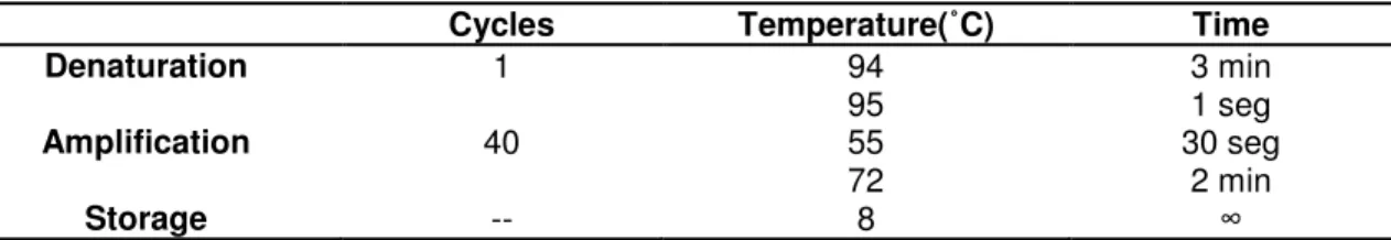
The cfb gene encoding the Christie-Atkins-Munch-Petersen (CAMP) factor is virtually present in all GBS isolates, and therefore is suitable for PCR detections (Wilkinson, 1977). The GBS cfb gene (CAMP factor) was amplified from the extracted genomic DNA. Specific primers were used in the PCR assay: Sag59 (T T T C A C C A G C T G T A T T A G A A G T A) e Sag190 (G T T C C C T G A A C A T T A T C T TT G A T) (Ke et al., 2000).
Escherichia coli K1 was used as a negative control. In addition, purified GBS genomic
FCUP
Characterization of an experimental model of neonatal meningitis induced by Group B Streptococcus 25
(Gibco) were used in the PCR mix. MWG Biotech thermocycler was used according with Table 2. 1.5% ultra-pure agarose (Life Sciences) gel electrophoresis was performed to analyze amplification products.
Table 2 |GBS-specific conventional PCR assay characteristics.
Cycles Temperature(˚C) Time Denaturation 1 94 3 min Amplification 40 95 1 seg 55 30 seg 72 2 min Storage -- 8 ∞
Histopathology
Brain sections were fixed in 10% buffered formalin, routinely processed, and embedded in paraffin. 4-5 µm-thick sections were cut and stained with hematoxylin and eosin (H&E).
Behavioral assessments
The mice that survived to GBS-induced meningitis were evaluated to determine their cognitive and motor performance. The behavioral tests were performed in a sound attenuated, temperature (21 ± 1ºC) and humidity (70%) controlled room with a 12-h light–dark cycle (lights at 7 a.m.), using male animals at PND60 (begin of adulthood), from different litters. The animals were kept at approximately 90% (26.64 ± 1.28 g) of their free feeding body weight and began training after reaching this weight. During the whole test time mice had restricted access to food (they were only fed following testing) and their weight and general health were carefully monitored every day to prevent more than a 10% body weight loss.
Radial maze test
The cognitive status of the animals as regards spatial learning and memory was assessed with the 8-arm radial maze. Working and reference memory were assessed simultaneously through a fixed position reward task, in which half of the arms were baited and their positions were fixed throughout the training trails. The radial maze consists in a central area (22 cm in diameter) giving access to eight equally-sized arms in transparent acrylic (length, 25 cm; width, 6.5 cm). Identical food wells (2.5 cm deep
26 FCUP
Characterization of an experimental model of neonatal meningitis induced by Group B Streptococcus
and 3 cm in diameter) were placed at the distal end of each arm. Commercialized sugar pellets (Bioserv F0042 - DPP'S 45MG SUGAR 50TH) located at the end of each baited arm were used as rewards. Two days prior to the beginning of the test, the rewards were placed once a day in the animal cages, to allow them to explore and eat the pellets. There were extra-maze clues including free-standing laboratory equipment, and geometric pictures in walls to help mice navigation. A digital stopwatch was used to record the amount of time taken to a mouse to complete a trial.
Habituation/training - On the first habituation day, each animal was placed alone in the center of the starting platform and allowed to freely explore the maze. On the second habituation day, the mice were allowed to explore the apparatus with randomly placed food pellets throughout the maze, for a 5 min period. On the third habituation day, the food pellets were placed at the distal end of each arm and the animals were allowed to explore for 5 min. The habituation period ended after the mouse ate at least four rewards or when 5 min had elapsed.
Test – During the test phase, four out of eight arms were baited and randomly assigned for each mouse, but always constant for the same animal throughout the trial period, and the mice were given a maximum of 5 min to complete the maze. Geometrical figures placed on the walls and the researcher were the only visual extra maze cues present. In each training session, the mouse was placed within a transparent cylinder on the platform in the middle of the maze for few seconds. The cylinder was then lifted and the animal was allowed to move freely in the maze. It was considered that an animal had entered an arm when the four paws and the tail were inside it. Trials ended when the animal had either eaten all four rewards or 5 min had passed, whichever came first. Once the animal returned to the central platform and the cylinder was lowered; a minute later the next trial took place. One session of two trials was performed per day over fourteen days. The maze was rotated and wiped with a dextran solution at 2% between animals to eliminate or reduce olfactory cues from different individuals.
Behavioral analysis - There were four measures used in behavioural analysis. The number of errors made: working memory error, defined as re-entering an arm already visited within a trial; and a reference memory error, defined as entering a never baited arm. Trial completion time was also included. It started when the cylinder was lifted, in which the mouse had full access to explore the maze, and stopped when the animal entered the fourth baited arm. Response latency was also measured and defined by the total session duration divided by the number of arms entered (seconds per entry). Seven blocks were calculated by average measure of four trials per day.