INSTITUTO POLITÉCNICO DE LISBOA
ESCOLA SUPERIOR DE TECNOLOGIA DA SAÚDE DE LISBOA
Co-60 source - a study on induction of damages and repair
kinetics in a PC3 cell line
Filipe Fernandes Pires
Supervised by Ana Belchior, PhD
Co-Supervised by Margarida Eiras, PhD
M
ASTER INR
ADIATIONSA
PPLIEDT
OH
EALTHT
ECHNOLOGIES-
R
ADIATIONT
HERAPY-
Lisbon, 2018
INSTITUTO POLITÉCNICO DE LISBOA
ESCOLA SUPERIOR DE TECNOLOGIA DA SAÚDE DE LISBOA
Co-60 source - a study on induction of damages and repair
kinetics in a PC3 cell line
Filipe Fernandes Pires
Supervised by Ana Belchior, PhD
Co-Supervised by Margarida Eiras, PhD
Dissertation submitted in partial fulfillment of the requirements for the Degree of
M
ASTER INR
ADIATIONSA
PPLIEDT
OH
EALTHT
ECHNOLOGIES-
R
ADIATIONT
HERAPY-
Declaration
I, Filipe Pires, declare that this thesis, submitted in the fulfillment of the Master’s degree at Escola Superior de Tecnologia da Saúde de Lisboa, is wholly my own work unless otherwise referenced or acknowledged appropriately.
--------------------------- Filipe Pires August 2018
ACKNOWLEDGMENTS
I’m lucky enough to be indebted to a number of people.First and foremost, I would like to express my deepest gratitude to my supervisor, Professor Ana Belchior, for an amazing opportunity and for a great deal of support and patience throughout the course of the dissertation.
A word of appreciation to Professor Margarida Eiras, as co-supervisor, for the promptness and availability that always characterized her.
I will keep a debt of gratitude to the researchers of GPSR and to the C2TN in a broader sense, for allowing me a peak in the world of investigation and for keeping the doors of their laboratories and offices always open.
During my time at the center I got acquainted with a couple of fellow students, with whom I had the pleasure to share a well-furnished and organized workspace. I must thank Ana, for being a great teacher and a greater friend and for being such a vast source of wisdom and support. A special mention to João, for being a brave companion in the student’s room as well as in the lab. I won’t succeed to say “thank you” to Jorge nearly as eloquently as I would like to or as he would be able to, but still, I’d like to emphasize how grateful I am for having him on the adjacent desk. I’d say I didn’t know how to repay him, but fortunately I do – take him to David’s hole (its tastier than one could assume). My thankfulness to Valerio, for choosing Lisbon as an Erasmus destiny, and for his inconspicuous curiosity toward the meaning of Portuguese vocabulary;; if he experienced half of the joy we felt for sharing this year with him, he had a great time. More than personal friends, all of them contributed in some way, either by discussing ideas, helping in research or programming, or reviewing drafts and they prove to be enormously helpful and unselfish with their time.
My sincere gratitude to Alex, Inês and Rita for the moments, adventures and for the laughter in between. I cherish those and I’m longing for new ones, even if I’m forced to endure some appalling music choices.
To my grandmother, “vó”, my uncles, Paulo and Di, and my cousins, Miguel and Pedro, I would like to express how deeply lucky I feel for having such unconditional sources of strength and support.
To my mother. Some people claim its actually hard to raise your offspring by means of example. I wouldn’t know. It came so natural for her. I’m grateful for that.
CONTENTS
ACKNOWLEDGMENTS ... ICONTENTS ... II
LIST OF TABLES ... IV
LIST OF FIGURES ... V
LIST OF ACRONYMS ... VII
ABSTRACT ... VIII
RESUMO ... IX
1. INTRODUCTION ... 1
1.1. MOTIVATION ... 1
1.2. STUDIES UNDERTAKEN ... 3
1.3. CELL LINE ... 4
1.4. RADIATION SOURCE ... 4 1.5. OBJECTIVES……….. 5 1.6. THESIS OUTLINE ... 5
2. BIOLOGICAL EFFECTS OF RADIATION ... 7
2.1. DIRECT AND INDIRECT EFFECT OF RADIATION ... 7
2.2. DNA AND CHROMATIN ... 8
2.3. CELL CYCLE ... 9
2.4. BIOMARKERS OF RADIATION EXPOSURE ... 9
2.5. BIOMARKERS OF DNA LESIONS ... 11
2.5.1. g-H2AX/ 53BP1 FLUORESCENCE ASSAY ... 11
2.5.2. CLONOGENIC ASSAY ... 13
3. INTERACTION OF RADIATION WITH MATTER ... 15
3.1. INTERACTION OF PHOTONS IN MATTER ... 15
3.2. INTERACTIONS OF A CHARGED PARTICLE IN MATTER ... 17
3.3. PHYSICAL QUANTITIES ... 18
3.3.1. ABSORBED DOSE ... 18
3.3.2. STOPPING POWER ... 18
3.3.3. LET ... 19
4. MATERIALS AND METHODS ... 23
4.1. CELL CULTURE ... 23
4.1.1 CULTIVATION OF CELLS ... 23
4.1.2. CELL VIABILITY / CELL COUNTING ... 24
4.2. IRRADIATION PROCEDURE ... 24
4.2.1. BRIEF DESCRIPTION OF THE IRRADIATION SOURCE ... 24
4.2.2. IRRADIATION SETUP ... 25
4.2.3. DOSIMETRY ... 26
4.3. QUANTIFICATION OF THE NUMBER OF DSB'S LESIONS IN THE DNA ... 26
4.3.1. g-H2AX/ 53BP1 ASSAY ... 26
4.3.2. ACQUISITION AND ANALYSIS ... 27
4.3.2.1. Foci Counting ... 27
4.3.2.2. Co-localization ... 28
4.4. QUANTIFICATION OF THE SURVIVAL CURVE ... 29
4.4.1. COLONY COUNTING ... 30
4.4.2. ANALYSIS ... 31
4.4.2.1 Linear-Quadratic (LQ) model ... 31
4.4.2.2 Statistical analysis ... 31
5. RESULTS AND DISCUSSION ... 33
5.1. QUANTIFICATION OF THE NUMBER OF DSB LESIONS IN THE DNA ... 33
5.2. ANALYSIS OF FOCI CO-LOCALIZATION ... 38
5.3. QUANTIFICATION OF THE SURVIVAL FRACTION ... 40
6. CONCLUSION ... 42
ANNEXES ... 45
PROTOCOL CELL COUNTING ... 45
PROTOCOL g-H2AX/ 53BP1 ASSAY ... 46
PROTOCOL CLONOGENIC ASSAY ... 47
REFERENCES ... 49
LIST OF TABLES
Table 1: Typical LET values of various types of radiation. . ... 19Table 2: Mean of induced damage in a mammalian cell after administration of 1 Gy, by a low-LET photon beam and by a beam of low-energy a particles (high-LET).. ... 20
Table 3: Values of SF, obtained through a clonogenic assay.. ... 40
LIST OF FIGURES
Figure 1: Decay process from Cobalt-60 into Nickel-60.. ... 4
Figure 2: Representation, depicting the DNA double helix, unwound, and the building blocks, the phosphate group (P), the deoxyribose sugar (S), and the bases.. ... 8
Figure 3: Schematic representation of some DNA damage examples: (I) alteration of bases;; (II) SSB;; (III) DSB.. ... 10
Figure 4: Digital image illustrating IRIF originating from g-H2AX and 53BP1 in PC3 cells;; (I) a control image and (II) a 4 Gy image. ... 13
Figure 5: Digital image showing a plate with 4 cultures with colonies derived from a clonogenic survival assay carried out with PC3 cells exposed to a dose of 6 Gy. ... 14
Figure 6: Schematic representation of the main interactions with photons in matter, where (I) corresponds to the photoelectric effect, (II) to the Compton scattering, (III) to the pair production, and (IV) to the Rayleigh scattering.. ... 16
Figure 7: Representation of an electron trajectory in relation to an atom, where a is the radius of the atom and b is the impact parameter.. ... 17
Figure 8: Relation between relative biological effectiveness (RBE) values and linear e nergy transfer (LET) values.. ... 21
Figure 9: Scheme representing the experimental chamber, with the irradiation setup. (I)The culture plate, (II) the rotation mechanism with a base upon which the culture plates were placed, fixed with adhesive tape, and (III) the interior portion of the chamber. ... 25
Figure 10: Digital image showing the Ionization Chamber (IC) Farmer Type Chamber FC65-P (Scanditronix, Wellhofer). ... 26
Figure 11: Digital images of (I) nuclei, (II) g-H2AX, (III) 53BP1, and (IV) the merged image.. ... 28
Figure 12: Digital image, exemplifying an output set of images from ImageJ, after being spplited into separated channels.. ... 29
Figure 13: Shape of survival curve.. ... 31
Figure 14: Induction of g-H2AX foci per nucleus in control cells and in cells irradiated with 2 Gy, after 0.5 (30 minutes), 2, 6, and 24 hours.. ... 34
Figure 15: Induction of 53BP1 foci per nucleus in control cells and in cells irradiated with 2 and 4 Gy, after 0.5 (30 minutes), 2, 6, and 24 hours.. ... 35
Figure 18: Mean value of colocalization as measured by the correlation coefficient from
Figure 19: Survival curve for the PC3 cell line obtained from a clonogenic assay.. ... 41
LIST OF ACRONYMS
g-H2AX Phosphorylated H2AX
53BP1 Tumor Suppressor p53 Binding Protein 1
BSA Bovine Serum Albumin
DAPI 4’,6- diamidino-2-phenylindole
DDR DNA Damage Response
DNA DeoxyriboNucleic Acid
DSB Double Strand Break
FBS Fetal Bovine Serum
FITC Fluorescein Isothiocyanate
IR Ionizing Radiation
IRIF Ionizing Radiation-Induced Foci
H2AX LET
Histone family member X Linear Energy Transfer
LQ Linear Quadratic
PBS Phosphate-Buffered Saline
PCC Pearson’s Correlation Coefficient
PE Plating Efficiency
PSA RBE
Prostate Specific Antigen
Relative Biological Effectiveness
ROS Reactive Oxygen Species
RPMI Roswell Park Memorial Institute medium
SF Survival Fraction
SSB Single Strand Break
WHO World Health Organization
ABSTRACT
This dissertation was designed to address the study of induction and repair kinetics of Co-60-induced deoxyribonucleic acid (DNA) damages in human prostate tumor cells (PC3 cell line). The analysis aims to investigate the number of complex damage induced in this cell line, to test its repair ability and its influence on cell survival capacity. It is intended that this study contributes to the characterization of the response to a specific radiation source by these malignant prostate tumor cells.
The present work proposes to evaluate the radio-induced effects in the following 24 hours and to observe the integrity of the survival capacity. In the first part, an immunofluorescence assay is performed. Two biomarkers are used, phosphorylated H2AX (g-H2AX) and tumor suppressor p53 binding protein 1 (53BP1), in order to determine the number of double strand breaks (DSBs) through their correspondence with the foci, identified by antibodies specific to the biomarkers, over several time- points from the first half hour after irradiation up to 24 hours. Subsequently, co- localization between g-H2AX and 53BP1 is tested using the correlation coefficient provided by CellProfiler. In a second part a clonogenic assay is performed, observing the evolution of the survival fraction with increasing dose.
The results showed that cellular repair after induction of damage allows a decrease of the number of DSBs, but up to 24 hours’ post-irradiation there is a level of residual damage present. Between g-H2AX and 53BP1 there appears to be a partial level of co- localization, with a tendency for decreasing throughout the repair process.
In summary, the work described in this dissertation demonstrates the level of induced damage and repair kinetics of the PC3 cell line, suggesting a dose and time repair dependence.
Keywords: PC3 cell line, low-LET radiation, DSB, g-H2AX, co-localization
RESUMO
Esta dissertação foi desenhada para abordar o estudo da indução e cinética de reparação de danos na molécula do ácido desoxirribonucleico (DNA), induzidos por Co-60, em células humanas de tumor da próstata (linha celular PC3). A análise pretende investigar o número de danos complexos induzidos nesta linha celular, testar a sua capacidade de reparação e a sua influência na capacidade de sobrevivência. Pretende-se que este estudo contribua para a caracterização da resposta a uma fonte de radiação específica por estas células malignas de tumor de próstata.
O presente trabalho propõe fazer uma avaliação dos efeitos radio-induzidos nas 24 horas seguintes e observar a integridade da capacidade de sobrevivência. Na primeira parte, é realizado um ensaio de imunofluorescência. Dois biomarcadores são utilizados, a H2AX fosforilada (g-H2AX) e a 53BP1, com o objectivo de determinar o número de duplas quebras de cadeia (DSB) através da sua correspondência com os focos identificados por anticorpos específicos aos biomarcadores, ao longo de diversos pontos no tempo desde a primeira meia hora após irradiação até às 24 horas. Posteriormente, é avaliada a co-localização entre a g-H2AX e a 53BP1, com recurso ao coeficiente de correlação providenciado pelo CellProfiler. Numa segunda parte é realizado um ensaio clonogénico de sobrevivência, observando a evolução da fracção celular sobrevivente com o aumento da dose.
Os resultados mostraram que a reparação celular após a indução de danos permite diminuir o número de DSB, mas até 24 horas pós-irradiação existe um nível de dano residual presente. Entre os anticorpos, g-H2AX e a 53BP1, parece haver um nível parcial de co-localização, com uma tendência para este diminuir ao longo do processo de reparação.
Em suma, o trabalho descrito nesta dissertação demonstra o nível de danos induzido e a cinética de reparação da linha celular PC3, sugerindo uma dependência em relação à dose e tempo de reparação.
1. INTRODUCTION
1.1. MOTIVATION
It was after the discovery of X-rays in 1985, by Wilhelm Conrad Röntgen, that the role of ionizing radiation (IR) began to be appreciated for its potential in diverse applications. The first documented utilization with a clinical application occurs in less than one year after Röntgen manages to see a shadow of his wife’s finger bones in a palette. A medical student, Emil Grubbe, used X-rays to treat a 65-year-old woman with a recurrent breast carcinoma, at a factory in Chicago, USA (1).
The World Health Organization (WHO) cancer report places cancer among the major causes of mortality, with a global estimate of 8 million cancer-related deaths annually, with a growing tendency. Prostate cancer presents the second highest incidence rate (31.1 per 100 000), with a mortality rate of 7.8 per 100 000 (2). At present day, the therapeutic application of IR, radiation therapy, is given a protagonist role. Broadly, the therapeutic approaches associated with cancer treatment often include a combination of therapies, of which the more frequent include surgery, chemotherapy, and radiation therapy. More than half of these patients have clinical indication to undergo radiation therapy, with diverse timings and purposes.
The beginning of the story to study the biological effects of IR is marked around the time of Röntgen’s discovery and it was driven by the necessity of assessing the effects and potential risks for human health of the uses of IR that were being discovered. IR has the ability to produce charged particles, through the ionization of atoms, that deposit energy in the surrounding medium. When this “medium” is a cell, ionization events can induce modifications in the constituent biomacromolecules. Resulting damages include losses in function of proteins, deactivation of enzymes, peroxidation of lipids, and ruptures or modifications on the structure of nucleic acids, among others. From all the alterations, deoxyribonucleic acid (DNA) constitutes the critical target because it contains genes/chromosomes that preserve the genetic information, which makes it pivotal for the maintenance of the cellular survival.
Radio-induced DNA changes can vary in frequency and degree of severity and include single strand breaks (SSB), double strand breaks (DSB) or base alterations, among others. DSBs, despite the fact they are not the most common lesions, stand out as
critical. DSBs constitute, by definition, breaks in both strands at a reduced distance from one another, so it’s not viable to use the complementary strand as a template. The nonrepair or misrepair of a lesion, presupposes a loss of information during the process of cell division which may lead to genomic instability and cell death (3).
Although the use of IR for therapeutic purposes is relatively recent this sort of damage is not exclusively associated with it. As a bi-product of aerobic respiration, reactive oxygen species (ROS) are produced and are able to induce damages (4). It is estimated that each typical mammalian cell can acquire, on a daily basis, between 1000 to 1,000,000 DNA lesions (4). This induction of damages under normal physiological conditions has forced the cells to naturally keep signalization and repair processes, in order to maintain the genome’s integrity (5).
DSB creation triggers a set of events, generically known as the DNA damage response (DDR). The actors involved in DDR can function as predictive biomarkers of cell response. Their study is often divided into either techniques that allow the detection of DNA damages or the ones who allow the observation of underlying repair (6).
One of the first observed modifications is the phosphorylation of serine 139 in the H2 histone family member X (H2AX) variant, inducing its phosphorylation in the vicinity of the lesion;; the molecule receives the name of phosphorylated H2AX (g-H2AX). Afterwards, a number of repair proteins will follow. This histone modification is associated to some interesting characteristics, such as the fact that the process is fast, abundant, and presents a good relation with DSB, which makes it a sensitive marker for damage detection (7).
However, although the g-H2AX measurement may be sensitive, it is not a completely specific marker. It has been suggested that phosphorylation of H2AX can occur by diverse processes, such as DNA replication, apoptosis, or residual damage (7). Furthermore, some authors report findings regarding repair kinetics being cell- dependent processes (8). If the measurement of g-H2AX cannot be attributed exclusively to DSBs and the foci only measure the response and not the lesions themselves, there is a degree of uncertainty associated to it. One way to increase the strength of the results is to combine the measurement of g-H2AX with that of other proteins known to be involved in the signaling/repair process of the lesions. Several other proteins were discovered to correlate at the sites of DSBs.
discovered to co-localize with g-H2AX, allowing the detection of false positives. The presence of both g-H2AX and 53BP1 leads to ionizing radiation induced foci (IRIF), providing a spatial indication that signalizes DSBs. The parameters of induction and disappearance of IRIF kinetics reflect the physical characteristics of the IR agent. This way, it is possible to retrieve information about the appearance, repair or nonrepair of DSBs and signalizing DSBs in time.
1.2. STUDIES UNDERTAKEN
The above-mentioned sub-sections aim at describing the studies that were performed in this thesis.
Quantification of DSBs, in PC3 cells, using the g-H2AX and 53BP1 assays and analysis of DNA repair kinetics
This study aimed at quantifying the induction of DSBs, in PC3 cells, after exposure to Co-60. The main goal consisted of measuring the number of foci, both by the phosphorylation of H2AX and 53BP1. Cells were exposed to 2 and 4 Gy, using a Co-60 source. The induced lesions were analyzed at different time-points. After validating the visualization of induced damage, a study regarding its dependence on two variables was performed: (I) A time dependence study. For a certain dose, is there a significant
difference between a number of time-points? (II) A dose dependence study. Is there a significant difference between control cells and cells that were irradiated with different doses?
Analysis of foci co-localization
Measure the co-localization of the two fluorescence signals, g-H2AX and 53BP1, studying its dependence on repair time and dose.
Survival curve
A cell survival curve was evaluated through the use of a clonogenic assay. The aim was to understand and relate the induction od DSBs with the maintenance of clonogenic ability.
1.3. CELL LINE
An article published in 1979 report the establishment of the PC3 cell line derived from bone metastases of a grade IV prostate cancer patient, a 62-year-old Caucasian male. PC3 is a cell line characteristic of prostate small cell carcinoma. It is characterized by the non-expression of androgen receptors or prostate specific antigen (PSA). PC3 cells do not respond to glucocorticoids neither fibroblast growth factors. The cell line exhibits markedly malignant behavior, with high metastatic potential, which stands in contrast with the indolent behavior typically found in the clinical setting. For this reason, PC3 cells tend to be used as a portrayal of the malignant presentation of prostatic tumors (9).
1.4. RADIATION SOURCE
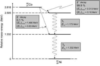
Cobalt-60 is a radioactive isotope of cobalt-59, with an extra neutron and a half-life of 5.26 years. Through a beta- minus process it decays for an excited Ni-60 with an energy of 0.31 MeV and for a stable state emitting two photons, of 1.173 MeV and 1.332 MeV (Figure 1). There are two beta- minus channels;; in 99.88 % of the decays follow from Co-60 to the second excited state de Ni-60 with a maximum electron energy of 0.313 MeV. Only 0.1 % of the decays follow from Co-60 to the first excited state of Ni-60 with a maximum electron energy of 1.486 MeV. Co-60 is produced artificially by neutron activation in nuclear reactors (10).
Figure 1: Decay process from Cobalt-60 into Nickel-60. Extracted from (10).
The linear energy transfer (LET) describes the energy transfer from the incident beam to the surrounding medium (11). Historically, much of the use of IR in cancer therapy has been done with low-LET sources, both in brachytherapy, with sources such as iridium-192, cesium-137, and also in external radiotherapy, with electrons and photons. Cobalt-60 sources have a part in both aforementioned approaches. High- LET IR, beams composed of heavy charged particles, have been playing an increasingly more relevant role in the clinic, due to the growing evidence, that describes a greater efficacy in inducing cell damage (11). Regardless, considering of the past and current role of low-LET IR in areas as diverse as radiotherapy or food irradiation, this type of sources possess a robust body of evidence and, therefore, play a major role on the comparison with other types of IR.
For this work the Precisa 22, an experimental equipment (Graviner, Manufacturing Company, Ltd, U.K.) loaded with Co-60 sources was used. It is located at Campus Tecnológico e Nuclear (CTN), Sacavém, Portugal. It should be noted that all irradiations and associated dosimetric procedures were conducted by PhD. Pedro Santos, from the same institution, who kindly granted his time and expertise.
1.5. OBJECTIVES
The main objective of this project was to contribute to the characterization of the PC3 cell line response to radiation. Secondary golds were defined, in order to accomplish this. A study on the induction of DSBs after irradiation was developed. A number of PC3 cells was irradiated with Co-60 sources. Damage repair was measured, using dose and repair time as variables. The co-localization between g-H2AX foci and 53BP1 foci was evaluated, in order to test their relation and its potential usefulness as biomarkers. Finally, an attempt to relate the induced damages with the maintenance of the cell’s survival was made, by performing clonogenic assays on the same experimental conditions.
1.6. THESIS OUTLINE
The present dissertation is divided into six chapters. The introduction chapter describes: i) the motivation for the thesis, ii) the studies undertaken, iii) the irradiation source, iv) the cell line used and v) a section-by-section summary.
The second and third chapters intend to provide a theoretical background to the results obtained. The second chapter approaches some key concepts related to the damage induced on the cell line, describing the effects of radiation, the DNA and chromatin, biomarkers of radiation exposure and DNA damage and the actual assays. The third chapter relates to the radiation that leaves the source, the way it interacts with the medium and some basic quantities used to characterize the damage from a physical stand point.
The fourth chapter describes the materials, equipment, and methodologies used. From the maintenance of the cell culture, to the irradiation procedure, the assays, as well as the analysis performed to produce the results.
The fifth chapter presents the results obtained with the different assays, as well as a discussion.
The six and final chapter provides a general conclusion to the work performed during this dissertation and a brief discussion regarding possible future work.
2. BIOLOGICAL EFFECTS OF RADIATION
This chapter begins with an introduction to biological effects and an overview of some theoretical concepts associated with DNA damage. This chapter explains the difference between direct and indirect effects to the DNA, what the DNA is and how it interconnects with the proteins histones to form chromatin. A brief description of some biomarkers of radiation exposure and others types of lesions in the DNA is presented, which will be used in this work. A small theoretical introduction is made in regard to the assays that were performed throughout the dissertation.
2.1. DIRECT AND INDIRECT EFFECT OF RADIATION
IR produces damages by direct and indirect effects, depending on whether the initial events occur in a critical target, such as DNA, or in the cellular environment.
A direct event can be induced directly in DNA, with a subsequent breakdown of molecular bonds. In an indirect way, the damage is induced by free radicals that are mainly originated by the radiolysis of the water, due to its relative abundance in mammalian cells (water can account for 70% or more of its total mass) (12). The energy deposition is done by the ejection of orbital electrons that will induce a cascade of events. The target molecule is converted into an ion pair and then into a free radical. The ejected electrons are free to induce further ionizations. The ionization cycle, the production of free radicals and further release of electrons continues until the photons and particles lose their energy.
X-rays and g-rays, typical examples of low- LET IR, tend to be associated to indirect effects, whereas particles such as neutrons, alpha particles and other high-LET particles tend to induce mostly direct effects (13).
2.2. DNA AND CHROMATIN
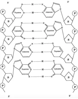
The DNA molecule has two polypeptide chains, consisting of nucleotides (Figure 2). A nucleotide is composed of 3 elements: a sugar, deoxyribose, which is interspersed with a phosphate group forming a backbone, and a base. The base constitutes the only variable element in the composition of a nucleotide and may take one of four forms: adenine, guanine (both purines), cytosine and thymine (two pyrimidines). They follow a rule of complementarity – adenine binds to thymine, whereas citosine binds to guanine;; from one strand, it is possible to construct the complementary one. Thus, the double- stranded structure is created, being composed of two sequences that are coiled around each other, forming a double-helix structure (14).
Figure 2: Representation, depicting the DNA double helix, unwound, and the building blocks, the
phosphate group (P), the deoxyribose sugar (S), and the bases. Extracted from (14).
Chromatin consists of a combination of DNA and proteins. The DNA is compacted because of its length, although it must remain accessible for biological processes, such as mRNA synthesis, replication, repair of lesions in the strands. The main proteins in the chromatin, histones, are small proteins that facilitate DNA binding. There are five main histones types - H1, H2A, H2B, H3 e H4.
The basic unit of chromatin is called the nucleosome. A nucleosome is composed of 147 nucleotides in length, wrapped in a complex of eight histones, two of each of H2A, H2B, H3, and H4. In order to form a nucleosome, the DNA is first coiled in two H3 and
two H4, followed by the addiction of two dimers of H2A and H2B. The complex is sealed by H1 (14).
Chromatin is present with different levels of compaction. Interphase cells tend to present the chromatin in a more diffuse, unwrapped and active form than the more commonly packaged, typical in division and usually silenced. The level of compaction influences factors such as radiosensitivity and the ability to repair. Uncompressed forms tend to be more sensitive, but also more efficiently repaired than more compacted, less sensitive, forms.
2.3. CELL CYCLE
The cell cycle encompasses the various stages in which a cell goes through since birth, until it is ready to divide and give rise to new cells. It is typically divided into interphase and mitosis.
The Gap 1 (G1) and Gap 2 (G2) are associated to the preparation for the events that characterize the cell cycle: the synthesis phase (S) and the cell division. First, the cell begins to grow to a certain volume, producing all of the cellular components it needs for the next phase, with the exception of the nucleus, in order to duplicate the genetic material. In case the environment is poor in nutrients, the cell remains at this stage until the conditions change. There is an evaluation of the physiological conditions and the environment, which determines the continuation of the course. The cell enters the S phase. An extra copy of the genetic material is created. The G2 phase proceeds to mitosis, and the cell will grow, reorganize. During the mitosis, the cell will divide in two daughter cells, each with the respective sets of genetic material and cellular components. At the end of the process, the cell is first subjected to a nucleokinesis process and then to a cytokinesis process, with cleavage of the nucleus and then the cytoplasm (15).
2.4. BIOMARKERS OF RADIATION EXPOSURE
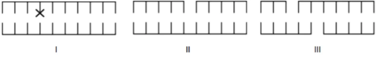
Damage to the genetic material is strongly correlated with IR-induced cell death, but also with tumor mutations and lesions. The IR can induce various types of damages, from changes in bases, strand breaks, SSB or DSB, among others (Figure 3).
Figure 3: Schematic representation of some DNA damage examples: (I) alteration of bases;; (II) SSB;; (III)
DSB. Adapted from (16).
An altered base can occur by disruption or chemical modification of the bases through ROS, a result of indirect effects.
A SSB corresponds to one or more breaks on a single strand of the DNA backbone. There are two general ways of inducing this type of damage: (i) at the phosphodiester bond level between the phosphate group and deoxyribose;; (ii) at the level of the bond between the base and deoxyribose. This type of injury tends to be produced either by ROS or by repairing abasic sites. Taken individually, they are of little biological significance in intact DNA, due to the possibility of repair using the second strand as a template. If the repair is not effective (misrepair), it may result in a mutation. If both strands breaks possess a significant separation, they will be handled separately as individuals SSBs. This type of injury resembles events that occur naturally in the cell. As an example, during the replication phase the double strand must be opened in order to allow access of the replication proteins to the genetic information (12).
DSBs are considered the most relevant type of lesion for the study of radiobiological effects, such as cell death, chromosomal aberrations or carcinogenesis. They consist of two SSBs in opposite strands, at a distance such that base pairing and chromatin structure are insufficient to keep the two joints juxtaposed. DSB can result from two non-time correlated SSBs or two SSBs induced by the same primary event.
A level of complexity can still be added to the repair process if multiple injuries (whether an accumulation of one type of damage or a combination of multiples types) occurs at close distances, in the same strand, or on the opposite one. They form clustered lesions, which, by convention, consists of two or more lesions in a region of 10-20 bp and are usually more difficult to repair because they aren’t dealt with in a separate matter (12). IR induces different proportions of lesions, depending on the type of radiation. Due to the higher density of ionizations and excitations along the path, some types of IR will generate a greater proportion of cluster lesions. The proportion of cluster lesions as well as the degree of complexity of each lesion increases with the increase of LET. For a cluster injury to be completely repaired, all components must be
repaired or removed. In addition, several DSBs in proximity increase the likelihood of an incorrect junction of the DNA ends (11,12).
2.5. BIOMARKERS OF DNA LESIONS
2.5.1.
g-H2AX/
53BP1
FLUORESCENCEA
SSAYDNA damage leads to the modification of the chromatin surrounding the lesions, with the local accumulation of protein complexes, part of a response known as DNA damage response (DRR).
Currently, the quantification of the histone variant X- phosphorylated form H2A, (g- H2AX foci), represents a well-established method to correlate the DSB formation and the repair kinetics process, serving as a biomarker (17).
The H2A family is constituted by a number of variants, including H2AX. This variant is present in a relative abundance of 2-25 % of the H2A variations, depending on tissue and cell line in analysis (18). Instead of being located in a specific region, it is found in a seemingly dispersed form in histones throughout the DNA. The H2AX protein is unique for its C-terminal tail (COOH). Prior to the stop codon, the tail has a highly- conserved sequence, which includes a serine residue at position 139;; this residue is phosphorylated in response to DNA damage.
The g-H2AX foci formation constitute one of the first responses to DSBs, being capable of extending for up 2 megabase chromatin regions around a lesion in mammalian cells (19, 20).
The phosphorylation process can occur within 1 and 10 minutes after irradiation, and the phosphorylated fraction will increase, peaking at 30 minutes. The fraction of phosphorylated H2AX was found to be proportional to the number of DSBs, with around 0.03% of the phosphorylated H2AX per DSB (21).
After repairing the integrity of the chromatin, g-H2AX is reversed. If the g-H2AX signals a destabilization of the chromatin, the signal should be “turned off”, after restoration of the chromatin’s integrity. It has been suggested that this process occurs either by removing the g-H2AX by histone change or by dephosphorylation by a phosphatase (22). In mammalian cells, the phosphatase 2A (PP2A) appears to be involved in the process of dephosphorylation (22).
Several studies highlight a correlation 1:1 between the number of g-H2AX foci and the expected number of DSBs induced (21;; 23).
Considering the rapid induction and amplification of g-H2AX, as well as a good agreement with DSBs, this type of study has been considered the gold standard in the detection of this type of damage. It is known empirically that H2AX plays a role in the cellular response to DSBs, since H2AX-deficient cells and mice exhibit a higher sensitivity to IR and have higher levels of spontaneous genomic instability (24, 25). The literature emphasizes the sensibility of this assay. However, questions have also been raise regarding its specificity, as summarized by Menegakis et al (26).
i. It has been shown that residual g-H2AX foci may persist after the rejoining process of the initial damage (18);;
ii. McManus et al, describes the presence of “small” foci, irrelevant for the DDR and cycle dependent (27);;
iii. A dependence on the level of chromatin’s condensation (18);;
iv. H2AX phosphorylation events may occur without the presence of a DSB lesion (28).
In this context, there is an argument to be made on the validity of using a second biomarker for the fluorescence assay.
TP53 binding protein 1 (53BP1) was first described as a binding partner to the central domain of p53 tumor suppressor protein, it is often mutated in tumors. The 53BP1 gene is located on chromosome15q15-21, encoding a protein consisting of 1972 amino acids, and presents “interaction surfaces” for several proteins involved in DSB repair (29)(30). The first evidence that would play a role in the cellular response to DSBs was the discovery of their migration and accumulation in these breaks following DSB induction treatments. Later, it was observed that, in response to DSB’s, 53BP1 moves to lesion sites, where it plays a role in the acute response and DNA repair.
53BP1 foci begin to form 5 minutes after irradiation, at doses as low as 0.5 Gy. The 53BP1 foci number increases linearly over time, peaking at 15-30 minutes after irradiation, and then decreases, to baseline over the next 16 hours (30).
The relevance of this technique was tested in 53BP1 deficient mices;; which exhibited immune deficiencies, high sensitivity to IR and genomic instability, with a tendency to develop tumors (31).
The co-localization of 53BP1 foci with other foci known to mark sites of DNA DSBs such as g-H2AX foci, supports the hypothesis that the 53BP1 foci can closely relate with the number of DSBs (30).
Figure 4: Digital image illustrating IRIF originating from g-H2AX and 53BP1 in PC3 cells;; (I) a control image and (II) a 4 Gy image (both images obtained throughout the course of this work).
To properly quantify DNA damage using immunofluorescence microscopy, cells were treated with two antibodies for each of the foci used in this work (Figure 4). Both protein-primary antibodies and protein-secondary fluorescence antibodies were used. For identification of g-H2AX foci, a primary antibody (mouse anti g-H2AX) and FITC- conjugated anti-mouse second antibody were used;; whilst for 53BP1 foci, a primary antibody (rabbit anti-53BP1) and Texas Red-conjugated anti-rabbit second antibody were used.
2.5.2.
C
LONOGENIC ASSAYThe clonogenic assay is a cell survival assay and represents the gold standard in vitro method to evaluate the clonogenic potencial of in vivo cells (32). Determination of a cell’s viability is enabled through the ability to form a colony, which is why is frequently categorized as a viability assay. A colony, by definition, consists of a gathering of at least 50 cells. It represents 5-6 potential cell divisions, depending on the growth rate of a specific cell line (33, 34). The assay’s goal is to test the cells for their survival ability –
potential for division, either by inducing sublethal damage or by being in a process of cell differentiation. Survival cells that remain viable after irradiation will form colonies and are subsequently quantified, which allows to withdraw considerations regarding cell survival (Figure 5) (33).
Figure 5: Digital image showing a plate with 4 cultures with colonies derived from a clonogenic survival
assay carried out with PC3 cells exposed to a dose of 6 Gy.
The assay allows for an assessment of differences in survival capacity between cells that serve as controls and cells that are exposed to various cytotoxic agents, such as IR or drugs used in chemotherapy. Likewise, it is often applied to monitor the efficacy of several agents by determining their cytotoxic effects at the level of colony formation in different cell lines (34).
3. INTERACTION OF RADIATION WITH MATTER
In this chapter, a brief overview on photons and charged particles interactions with matter is presented, as well as some relevant physical quantities. A description on the basic functioning of an ionization chamber is included.
3.1. INTERACTION OF PHOTONS IN MATTER
In a simplified way, a medium can be seen as a combination of orbital electrons and atomic nuclei, which is composed of protons and neutrons.
When a particle transverses an absorbing material, it interacts with the material’s constituent atoms. Photons are indirectly ionizing radiation and so the energy transfer occurs in a two-step process. The incoming photon transfers its kinetic energy to secondary directly ionizing particles, such as electrons. The secondary particles will then deposit their energy through Coulomb interactions (this process will be further developed in the next section).
When a photon transverses a medium, either it interacts or does not interact, maintaining its trajectory. When a photon interacts, multiple processes may occur, depending on factors such as photons energy and chemical composition of the material. When transversing a medium, photon beams mainly experience a loss of intensity, as opposed to the loss of energy verified with directly ionizing particle beams. An ionization event occurs when the energy transferred by an incident photon is high enough to remove an electron from its orbital. When the transferred energy is lower than the electron’s binding energy, the electron is transferred to a more energetic orbital and the atom becomes excited. When the transferred energy is higher than the electron’s binding energy, the electron is removed from the atom and the atom becomes ionized (10).
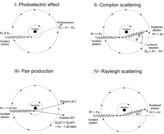
The main physical interactions between photons and matter include the photoelectric effect, Compton scattering, pair production and Rayleigh scattering (Figure 6).
Photoelectric effect. An electron is ejected from one of the orbitals of an atom, induced by the energy absorption of an incident photon. The photon disappears by transferring all its energy, while the orbital electron is ejected with a certain kinetic
energy, leaving the ionized atom. This energy is the result of the difference between the energy of the incident photon and the binding energy of the electron. The electron tends to deposit the energy locally. The vacancy that is open by the ejection of the electron is filled by electrons of outer orbitals, thus inducing one of two competing processes: the emission of characteristic X-rays and of Auger electrons. This process happens when the incident photon’s energy is equal or higher than the electron’s binding energy (10).
Compton scattering. An incident photon collides with an orbital electron, producing a scattered photon with lower energy, as well as a recoil electron that is ejected from the atom. This phenomenon happens when the energy of the incident photon is much higher than the electron’s binding energy (10).
Pair production. A photon with at least 1.022 MeV is converted into an electron and a positron. An electron-positron pair may combine and provoke an annihilation reaction, which, usually results in two photons, each with an energy of 0.51 MeV (10).
Figure 6: Schematic representation of the main interactions with photons in matter, where (I) corresponds
to the photoelectric effect, (II) to the Compton scattering, (III) to the pair production, and (IV) to the Rayleigh scattering. Adapted from (10).
I- Photoelectric effect
IV- Rayleigh scattering III- Pair production
Rayleigh scattering. An incident photon interacts with the absorber atom, being scattered. The scattered photon’s energy is very close to the incident photon’s energy. An orbital electron in these circumstances accelerates, which causes the atom to emit radiation, to return to a stable state. One of the characteristics of this type of interaction is that the atom does not reach an excited or ionized state. This phenomenon happens when incident photon’s energy is approximately equal to the binding energy (10).
3.2. INTERACTIONS OF A CHARGED PARTICLE IN MATTER
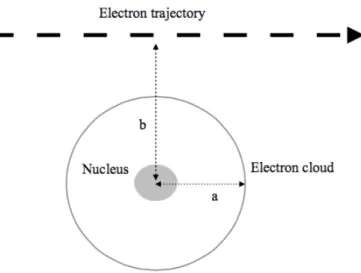
Electrons are directly ionizing radiation, thus interacting with matter in diverse ways through Coulomb interactions, being distinguishable through the impact parameter b. It is defined as the smallest distance between the center of the nucleus and the trajectory of the incident particle as it transverses the atom, being measured in relation to the radius of the atom (Figure 7) (10).
Figure 7: Representation of an electron trajectory in relation to an atom, where a is the radius of the atom
and b is the impact parameter. Adapted from (10).
When an incident electron passes at a considerable distance from an atom, that is, the impact parameter b is much higher than the atomic radius (b >> a) it is called a soft collision. When the particle transverses the atom, the particle’s Coulomb field will interact with the atom’s constituent particles, which causes a small amount of energy to be transferred from the incident electron to an orbital electron, which in turn gets excited. Despite the small energy involved in an individual interaction, it becomes
approximately 50 % of a charged particles’ energy loss.
When an incident electron passes at such a distance from an atom that the impact parameter b is approximately equal to the radius (b » a) it is called a hard collision. The electron tends to interact mostly with orbital electrons, transferring most of its kinetic energy. If the energy transfer is sufficient to exceed the binding energy of the orbital, the electron is released, thus ionizing the atom. When this electron is able to produce a noticeable track, its named delta ray and it has enough energy to undergo its own Coulomb interactions with other atoms. Although they are less likely, the energy transfer involved is much higher and they account for approximately 50 % of the charged particle’s energy loss.
Finally, when the incident electron passes near a nucleus, the impact parameter b is much lower than the atomic radius (b << a) it will experience a Coulomb interaction with the atomic nucleus. An electron may also interact with the orbital electrons of an atom, slowing down due to the repulsive Coulomb interactions and emitting X-rays in the process, known as bremsstrahlung radiation (10).
3.3. PHYSICAL QUANTITIES
3.3.1.
A
BSORBED DOSEWhen a charged particle passes through a medium, it deposits energy along its path. The dose is a measure of the mean energy, E, deposited by IR to matter of mass, m:
𝐷 = ∆$
∆% Eq. 1
Where
DE is the absorbed energy to a mass element Dm and its SI unit is the gray (Gy) (1Gy= 1 J.kg-1) (35).
3.3.2.
S
TOPPINGP
OWERThe loss of energy per path length is defined as stopping power. The total stopping power is the sum of the contributions of two processes: radiative stopping power and collisional stopping power, according to the following expression:
The radiation stopping power or nuclear stopping power includes Coulomb interactions with the nucleus. It is related to the emission of photons of bremsstrahlung when an incident particle changes direction along its path. The contribution of this process is particularly relevant in the case of electrons, because they are light charged particles, in a medium with high atomic number.
The collisional stopping power or electronic stopping power results from Coulomb interactions of charged particles with orbital electrons. The contribution of this process is relevant for both light and heavy charged particles. This type of energy loss is associated with the excitation/ ionization of the atoms in the medium (10).
3.3.3.
LET
A concept related to stopping power is the linear energy transfer (LET). Constitutes a measure of the average energy locally imparted to the medium by a charged particle of specified energy in traversing a distance dl;; it is expressed in keV.µm-1, according to
the following equation (35, 36):
𝑳∆=𝒅𝑬𝒅𝒍∆ Eq. 3
in which,
dE∆, represents the energy transferred to the medium
dl, represents the path length distance
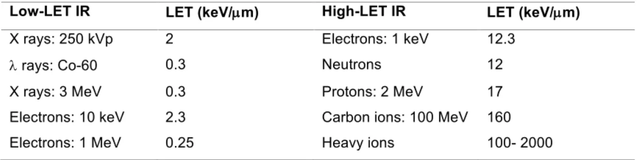
IR is categorized as low-LET or high-LET (Table 1). The low-LET IR usually consists of X-rays. High-LET IR typically includes heavy charged particles and neutrons.
Table 1: Typical LET values of various types of radiation. Usually is considered a value of 10 keV/ µm to separate low from high LET. Extracted from (10).
Low-LET IR LET (keV/µm) High-LET IR LET (keV/µm)
X rays: 250 kVp 2 Electrons: 1 keV 12.3
l rays: Co-60 0.3 Neutrons 12
X rays: 3 MeV 0.3 Protons: 2 MeV 17
Electrons: 10 keV 2.3 Carbon ions: 100 MeV 160 Electrons: 1 MeV 0.25 Heavy ions 100- 2000
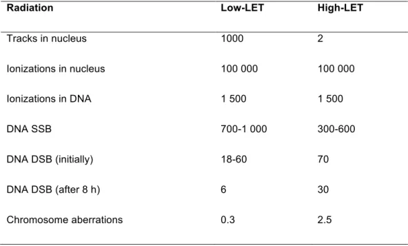
The low-LET and high-LET IR present a different pattern of spatial dose distribution and, consequently, diverse efficiency (Table 2). Low-LET IR deposits the dose relatively homogeneously in the cell’s nucleus, presenting a mean spacing between ionizations events in the order of hundreds of nanometers. The high-LET IR tends to deposit a high dose near the particle track and practically none in the area between the tracks, with a higher ionization density.
Table 2: Mean of induced damage in a mammalian cell after administration of 1 Gy, by a low-LET photon
beam and by a beam of low-energy a particles (high-LET). Adapted from (37). It should be noted that the
induced effects complex with high-LET IR are in greater numbers and a greater part of these damages are not repaired at 8 hours.
Radiation Low-LET High-LET
Tracks in nucleus 1000 2
Ionizations in nucleus 100 000 100 000
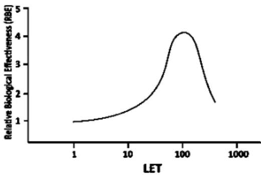
Ionizations in DNA 1 500 1 500
DNA SSB 700-1 000 300-600
DNA DSB (initially) 18-60 70
DNA DSB (after 8 h) 6 30
Chromosome aberrations 0.3 2.5
For low-LET IR the ionization events are too spaced relative to the size of a DNA molecule. The result is that a photon can transverse it without depositing energy. For high-LET IR, energy loss events can occur more frequently, as a result, a significant percentage of energy is deposited along the track.
Due to higher ionization density associated with a higher LET value, it is considered to be biologically more effective to have a higher LET value, until around 100 keV/µm. It corresponds to a spatial density of ionization events coincident with the diameter of the DNA strands (about 2 nm).