Universidade de Aveiro Ano 2016
Departamento de Biologia
Andreia Filipa Afonso
Magalhães
Avaliação da atividade antimicrobiana de isolados
bacterianos de pele de rã de zonas urbanas
Assessment of the antimicrobial activity of bacterial
isolates from frogs’ skins from urban zones
DECLARAÇÃO
Declaro que este relatório é integralmente da minha autoria, estando
devidamente referenciadas as fontes e obras consultadas, bem como
identificadas de modo claro as citações dessas obras. Não contém, por isso,
qualquer tipo de plágio quer de textos publicados, qualquer que seja o meio
dessa publicação, incluindo meios eletrónicos, quer de trabalhos académicos.
Universidade de Aveiro Ano 2016
Departamento de Biologia
Andreia Filipa Afonso
Magalhães
Avaliação da atividade antimicrobiana de isolados
bacterianos de pele de rã de zonas urbanas
Assessment of the antimicrobial activity of bacterial
isolates from frogs’ skins from urban zones
Dissertação apresentada à Universidade de Aveiro para cumprimento dos requisitos necessários à obtenção do grau de Mestre em Biologia Molecular e Celular, realizada sob a orientação científica do Doutor Sérgio Miguel Reis Luís Marques, (Investigador Pós-Doc) do Departamento de Biologia da Universidade de Aveiro e co-orientação do Doutor Fernando José Mendes Gonçalves, Professor Associado c/ Agregação do Departamento de Biologia da Universidade de Aveiro e do Doutor Mário Jorge Verde Pereira, Professor Auxiliar do Departamento de Biologia da Universidade de Aveiro
o júri
presidente Doutora Maria Helena Abreu Silva
Professora Auxiliar do Departamento de Biologia da Universidade de Aveiro
Doutor Nelson José Cabaços Abrantes
Investigador Auxiliar do CESAM – Centro de Estudos do Ambiente e do Mar da Universidade de Aveiro
Doutor Sérgio Miguel Reis Luís Marques
Investigador de Pós-Doutoramento do Departamento de Biologia da Universidade de Aveiro (Orientador)
agradecimentos Em primeiro lugar gostaria de agradecer ao meu orientador, Dr. Sérgio Marques, pelo apoio científico, aos meus co-orientadores Dr. Fernando Gonçalves e Dr. Mário Pereira principalmente pela oportunidade que me concederam para desenvolver este trabalho.
Em segundo lugar, gostava de agradecer a disponibilidade da Raquel e da Ana pela ajuda nas saídas de campo bem como no laboratório ao nível dos ensaios de inibição de crescimento.
Por último, ao meu namorado João pelo apoio e motivação nos momentos mais desesperantes.
palavras-chave Amphibia, Pelophylax perezi, microbiota da pele, atividade antimicrobiana, biomarcadores, stress oxidativo, sistema enzimático antioxidante
resumo As populações de anfíbios têm decaído ao longo dos últimos anos devido a inúmeros fatores, tais comos, a perda de habitat, a contaminação/poluição e um dos mais importantes, as doenças. Estas perdas originam também a perda de diversidade genética das espécies, podendo comprometer a sua aptidão e também capacidade de adaptação. Tendo em conta todos estes fatores, é necessário proceder à preservação das populações de anfíbios,
independentemente do local em que se encontram ser contaminado, pristino, rural ou urbano. Sabendo que os anfíbios de zonas urbanas podem ser uma fonte importante para a diversidade genética da espécie e que estão expostos, tal como as populações de zonas naturais, a agentes patogénicos, sendo que normalmente são populações negligenciadas a nível de proteção, urge a necessidade de as avaliar e proteger, nomeadamente contra agentes patogénicos. De uma forma geral, esta proteção é conferida de uma forma inata por estruturas ao nível da pele, que fazem parte do seu sistema imunitário. Estas são glândulas granulares responsáveis pela produção de compostos peptídicos capazes de inibir o crescimento de agentes patogénicos. Em acréscimo, a microbiota existente na pele estimula e complementa a atividade destas secreções. Com base nestes factos, este trabalho teve como objetivos: i) avaliar de que forma fatores como as estações do ano (Primavera e Outono) e o género, podem influenciar a microbiota cultivável da pele de
Pelophylax perezi de zonas urbanas, ii) avaliar se os isolados bacterianos da pele apresentam atividade antimicrobiana e iii) avaliar o potencial dos isolados bacterianos com atividade antimicrobiana enquanto possíveis agentes
probióticos, na presença de um agente patogénico. Os resultados obtidos mostraram diferenças entre locais ao nível das espécies isoladas, sendo poucas as espécies comuns entre locais. Além disso, foi evidenciado que num total de 120 isolados, 19 possuíam atividade antimicrobiana face a Bacyllus aquimaris e Aeromonas salmonicida. Também se verificaram diferenças na atividade antimicrobiana entre estações do ano, existindo um maior número de espécies com atividade antimicrobiana no Outono. Dos isolados com atividade antimicrobiana, os três com maior atividade, Pseudomonas rhizosphaerae,
Pseudomonas fluorescens e Bacillus mycoides foram selecionados para a segunda fase do trabalho, em que se avaliou o seu potencial enquanto possíveis agentes probióticos. Após exposição, in vivo, de girinos aos probióticos, em simultâneo com A. Salmonicida, verificou-se que estes evitavam mortalidade dos girinos, bem como diminuíam o dano peroxidativo quando comparados com os valores do agente patogénico. Dos três
probióticos B. mycoides mostrou ser aquele com maior capacidade de
estimular as enzimas antioxidantes, sendo o agente probiótico com os valores mais baixos de dano peroxidativo.
keywords Amphibia, Pelophylax perezi, skin microbiota, antimicrobial activity, biomarkers, oxidative stress, antioxidant enzymatic system
abstract Amphibian populations have declined over the past few years due to numerous factors such as habitat loss, contamination / pollution and one of the most important, diseases. These losses also result in the loss of genetic diversity of the species, which may compromise their fitness and ability to adapt. Taking all these factors into account, it is necessary to preserve amphibian populations, regardless of being found in contaminated, pristine, rural or urban sites. Given that urban amphibian populations can be an important source for genetic diversity of the species and that they are exposed, such as populations of natural areas, to pathogens, there is a need for assess and protect them against pathogenic agents. Generally, this protection is conferred in an innate way by skin structures, which are part of your immune system. These are granular glands responsible for the production of peptidic compounds capable of inhibiting the growth of pathogens. In addition, the microbiota in the skin stimulates and complements the activity of these secretions. Based on these facts, this work had as objectives: i) to evaluate how factors such as
seasonality (spring and autumn) and gender can influence the cultivable microbiota of Pelophylax perezi skin in urban areas; ii) assess the ability of the bacterial skin isolates to present antimicrobial activity and iii) evaluate the potential of bacterial isolates with antimicrobial activity as potential probiotic agents. The obtained results showed differences between sites at the level of the isolated species, with few common species between sites. In addition, it was evidenced that in a total of 120 isolates, 19 had antimicrobial activity against Bacyllus aquimaris and Aeromonas salmonicida. There were also differences in antimicrobial activity between seasons, with a higher number of species with antimicrobial activity in the autumn. Of the isolates with
antimicrobial activity, the three with the highest activity, Pseudomonas rhizosphaerae, Pseudomonas fluorescens and Bacillus mycoides were selected for the second phase of the study, in which their potential action as probiotic agents was evaluated. After in vivo exposure of the tadpoles to the probiotics, along with A. salmonicida, these were found to decrease the mortality of tadpoles as well as to decrease the peroxidative damage, when compared to the values obtained from the exposure to the pathogen. From the three probiotics B. mycoides revealed to be the one with the greatest capacity to stimulate the antioxidant enzymes, being the probiotic agent with the lowest values of peroxidative damage.
Table of Contents Chapter I
1. General Introduction... 19
1.1 References ... 22
Chapter II 2. Influence of anthropogenic factors in the cultivable microbiota of amphibians and their antimicrobial potential ... 27
2.1 Introduction ... 28
2.2 Materials and Methods ... 30
2.2.1 Sampling Sites ... 30
2.2.2 Study organism ... 32
2.3 Field collection procedures and microbiota sampling ... 33
2.3.1 Frog sampling ... 33
2.3.2 Microbiota sampling... 33
2.3.3 Water abiotic parameters... 34
2.4 Identification of bacteria from frog skin, isolation and preservation ... 34
2.5 DNA extraction ... 35
2.6 DNA amplification and sequencing ... 35
2.7 Inhibition growth assays ... 36
2.8 Results ... 37
2.9 Discussion ... 46
2.10 References ... 48
Chapter III 3. In vivo assessment in Pelophylax perezi tadpoles of the antimicrobial potential of bacterial isolates against the pathogenic agent Aeromonas salmonicida ... 53
3.1 Introduction ... 54
3.2 Material and Methods ... 57
3.2.1. Test organisms ... 57
3.2.3 Bacterial cell culture ... 57
3.3 Experimental assay ... 58
16 3.5 Statistical analysis ... 60 3.6 Results ... 61 3.7 Discussion ... 65 3.8 References ... 68 Chapter IV 4. Final Remarks ... 75 4.1. References ... 77
17 Chapter I General Introduction
19 1. General Introduction
The Amphibians are a large class of vertebrates and are grouped into three orders:
Gymnophiona, Caudata and Anura. The order Gymnophiona also called Apoda
includes caecilians or limbless amphibians. The order Caudata includes newts and salamanders and the order Anura includes frogs and toads. (Beebe, 1996; Wells, 2010). The term Amphibia means "double life" which refers to life in both aquatic and terrestrial habitats. Generally, during their life cycle they suffer metamorphosis passing from water dependent larval stage to a mainly terrestrial adult stage. Amphibians are cold-blooded which means they are ectothermic organisms, depending on external factors to regulate and maintain their temperature (Demas and Nelson, 2012). This factor along with the distribution of water bodies are crucial to their geographical distribution. Few are the ones that can live in extremely low temperatures. Therefore, the higher diversity of amphibians is located in tropical areas like the American Southwest and West of Africa (IUCN 2016; Vitt and Caldwell, 2014). These characteristics make them one of the most sensitive groups of animals to environmental alterations. In recent times we have witnessed a worldwide decline in their populations and presently they are one of the most threatened groups. According to the IUCN Red List Assessment, the order Caudata has a higher threatened extinction rate (49.8%) corresponding to 275 species in a total of 552. The order Anura from 5532, 1749 are endangered species which corresponds to a rate extinction of 31.6%. The order
Gymnophiona, in contrast, have just 3.4% (6 species in a total of 176 are threatened).
The main factors for this occurrence are habitat loss, contamination / pollution, infectious diseases and invasive species (IUCN 2016).
Even suffering worldwide decline, with diseases being one of the main factors (Stuart et al., 2004), amphibians have structures that are part of their innate immune system and help them in their defense. One of these structures and which is common to all of them is their skin. It is one of their most important organs being used for respiration and osmoregulation and providing chemical protection against the
20
environment. This organ is rich in granular and mucous glands that are widespread on the head, body and limbs (Rollins-Smith, 2001). Their role determines their density and location. Mucous glands are the most abundant and are essentially located under the dorsum. They play an important role in maintaining the moisture of the outer surface of the skin through the production of mucous substances. Granular glands in general are more concentrated in the head and shoulders and are known to secrete antimicrobial peptides (Duellman and Trueb, 1994). These peptides have the ability to inhibit the growth of pathogenic organisms. One example is the growth inhibition of the pathogenic chytrid fungus Batrachochytrium dendrobatidis (Bd). In Rana pretiosa, previously exposed to the Bd fungus, were collected skin secretions containing antimicrobial peptides. They verified that all peptides inhibited the growth of the chytrid fungus completely at concentrations lower than 100 μM (Conlon et al. 2016). This ability to inhibit the growth of pathogens is also observed in gram positive and negative bacteria strains (Walke et al., 2014). These results may provide new templates for the development of new antimicrobial treatments. In addition to the glands and the antimicrobial peptides they produce, the skin also shows the presence of bacterial communities that establish symbiotic relationships (McKenzie et al., 2012). The bacterial communities that are part of the microbiota, can suffer changes in its composition, being environmental conditions one of the factors that might contribute for such alterations. Furthermore, these communities also present the particularity of undergoing through changes during the development of the organisms being these host-specific (Myers et al., 2012). The microbiota also plays an important role in amphibian’s immune defense system. In Lauer et al 2008 bacteria were isolated from
Hemidactylium scutatum skin and tested in two fungal pathogens: Mariannaea elegans
and Rhizomucor variabilis. They verified that the isolates belonging to the Bacillaceae,
Oxalobacteraceae, Pseudomonadaceae, Flavobacteriaceae, and
Sphingobacteriaceae families had antifungal activity against the fungi. This type of
research is important because the discovery and study of these organisms with antimicrobial ability can help on the implementation of new techniques of preservation of species. One of them is bioaugmentation which is a technique that uses
21
microorganism’s inoculation on the skin of amphibians to increase their resistance to pathogens’ exposure. There are bacteria that are usually found in amphibians and which are used as probiotics. One is Janthinobacterium lividum and is known for producing violacein whose compound exhibits antifungal properties inhibiting the growth of pathogens. (Becker et al., 2009). Other is Pseudomonas reactans and it was successfully introduced into the skin of Plethodon cinereus salamander. The animals which were successfully inoculated with the bacteria decreased mortality and reduction of the effects, such as loss of body mass, caused by the infectious disease Chytridiomycosis. (Harris et al., 2009). Taking into consideration this background information, and knowing that amphibians inhabiting urban areas might be an essential source of genetic variation for the respective species and also, the fact that populations in these areas are usually overlooked in terms of need for protection, our study aims at filling these gaps.
Therefore, the main objectives of this work are:
• The identification of cultivable bacterial communities present in the skin of
Pelophylax perezi individuals from areas with different levels of urbanization and
compare them between seasons (Autumn and Spring).
• The assessment of the antimicrobial activity of the bacterial isolates.
• To assess the probiotic potential of the bacterial isolates with higher antimicrobial potential.
22 1.1 References
Beebe, T.J.C., 1996, Ecology and Conservation of Amphibians, First Edition
(Vol. 7). Springer Science & Business Media.
Becker, M.H., Brucker, R.M., Schwantes, C.R., Harris, R.N., Minbiole, K.P.C., 2009. The Bacterially produced metabolite violacein is associated with survival of amphibians infected with a lethal fungus. Applied and Environmental Microbiology,
75(21), 6635–6638.
Conlon, J.M., Reinert, L.K., Mechkarska, M., Prajeep, M., Meetani, M.A., Coquet, L., Jouenne, T., Hayes, M.P., Padgett-Flohr, G., Rollins-Smithwang, L.A., 2016. Evaluation of the skin peptide defenses of the Oregon spotted frog Rana pretiosa against infection by the chytrid fungus Batrachochytrium dendrobatidis. Journal of
Chemical Ecology, 39, 797–805.
Demas G., Nelson R., 2012, Ecoimmunology, Oxford University Press. Retrieved from https://books.google.pt/books?isbn=019987624X
Duellman, W.E., Trueb, 1994, L., Biology of Amphibians, JHU press. Encyclopedia of Life, (n.d.), accessed (06-2016)
http://eol.org/pages/1552/details
Harris, R.N., Lauer, A., Simon, M.A., Banning, J.L., Alford, R.A., 2009. Addition of antifungal skin bacteria to salamanders ameliorates the effects of chytridiomycosis.
Diseases of aquatic organisms, 83, 11–16.
Lauer, A., Simon, M.A, Banning, J.L, Lam, B.A., Harris, R.N., 2008.Diversity of cutaneous bacteria with antifungal activity isolated from female four-toed salamanders.
The ISME Journal, 2, 145–157.
McKenzie, V.J., Bowers, R.M., Fierer, N., Knight, N., Lauber, C., 2012. Co-habiting amphibian species harbor unique skin bacterial communities in wild populations. The ISME Journal, 6, 588-596.
Myers, J.M., Ramsey, J.P., Blackman, A.L., Nichols, A.E., Minbiole, K.P.C., Harris, R.N., 2012. Synergistic inhibition of the lethal fungal pathogen Batrachochytrium
23
dendrobatidis: The combined effect of symbiotic bacterial metabolites and antimicrobial
peptides of the frog Rana muscosa. Journal of Chemical Ecology, 38, 958–965. New World Encyclopedia, (2016), accessed (06-2016)
http://www.newworldencyclopedia.org/entry/Caecilian
Rollins-Smith, L.A., 2001. Neuroendocrine-immune system interactions in amphibians: implications for understanding global amphibian declines. Immunologic
Research, 23(2/3), 273-280.
Stuart, S.N., Chanson, J.S., Cox, N.A., Young, B.E., Rodrigues, A.S.L., Fischman, D.L., Waller, R.W., 2004. Status and trends of amphibian declines and extinctions worldwide. Science, 306, 1783–1786.
The IUCN Red List, Guiding Conservation for 50 years, (2016), accessed (06-2016)
http://www.iucnredlist.org/initiatives/amphibians/analysis/geographic-patterns Vitt, L.J., Caldwell, J.P., 2014. Herpetology an introductory biology of
amphibians and reptiles, Second Edition, Academic Press. Retrieved from
https://books.google.pt/books?isbn=012386920X
Walke, J. B.; Becker, M.H; Loftus, S.C.; House, L.L.; Cormier, G., Jensen, R.V.; Belden, L.K., 2014. Amphibian skin may select for rare environmental microbes. The
ISME Journal, 8, 2207–2217.
Wells, K.D., 2010. The ecology and behavior of amphibians, The University of
25 Chapter II
Influence of anthropogenic factors in the cultivable microbiota of amphibians and their antimicrobial potential
27
2. Influence of anthropogenic factors in the cultivable microbiota of amphibians and their antimicrobial potential
Abstract
Amphibians have an immune system in which their skin plays a vital role involved. This is due both to the presence of granular glands as well as the presence of bacterial communities that further protect them against potential pathogens. In recent years’ amphibians’ pathogens have played an important role, contributing for the mortality of large numbers of animals and even the disappearance of entire populations. The reducing number of animals contribute for a decrease in genetic variation and a consequent reduction of fitness and adaptability. In a world where natural habitat loss is also a constraining factor for amphibians, every amphibian population is essential to help maintain genetic variation and thus essential to protect. In this context amphibians from urban areas have been overlooked, despite their role as links between populations from natural sites, promoting gene flow. Taking into consideration the previously mentioned, in this study we assessed the cultivable microbiota and its antimicrobial activity from Pelophylax perezi from two areas with different levels of urbanization in a pre and a post-hibernation season with the double objective of screening for potential probiotic species and also to verify if seasonality could be an important factor contributing for the vulnerability to pathogens. Our results revealed nineteen isolates with antimicrobial activity and this was mainly observed in females and specially in the Autumn (pre-hibernation). Bacteria with this feature were predominantly belonged to Pseudomonadaceae, Enterobacteriaceae, Bacillaceae,
Comamonadaceae and Paenibacillaceae families. Considering only skin microbiota,
these results may indicate a potential immune vulnerability of these animals during spring (post-hibernation), and surely a great dependence of environmental conditions to modulate skin microbiota. Nonetheless the isolated bacteria with antimicrobial activity may be of potential use as probiotics for this urban areas.
28 2.1 Introduction
Amphibians are found in different habitats having the ability to adapt to different ecosystems. These include both pristine and contaminated sites, as well as rural and urban sites (Marques et al., 2009; Kouamé et al., 2015) However, they have characteristics that could apparently make them vulnerable namely their extremely permeable skin, devoid of any protective structures such as feathers, scales or hair. Nonetheless, to cope with external threats, amphibians depend on it. Skin which plays an important role in respiration, osmoregulation and body temperature control (Kueneman, Parfrey, & Woodhams, 2014). Furthermore, one of its most important functions is as a physical barrier protecting from pathogens and consequent infections. This line of defense may be associated with structures present in the dermis layer, such as granular glands, that are responsible for producing antimicrobial secretions (Zug et al 2001; Stuart et al. 2008). An example of granular compounds secreted by granular glands are antimicrobial peptides which prevent infections by pathogens. Depending on the species, antimicrobial peptides (AMPs) can be produced in larger or smaller quantities and also vary in their diversity. In hybrid P. esculentus was verified an increased production of these peptides as well as a more efficient antimicrobial activity against Bd fungus compared to the parental species P. lessonae and P. ridibundus. The hybrid species by presenting a wide variety of peptides, in part acquired by the parent species, gives to the hybrid an advantage fighting against potential infections (Daum et al., 2012). In addition to the presence of the skin granular glands of amphibians are also symbiotic bacterial communities that may also have a role in immune defense of amphibians (Woodhams et al., 2014). Their stability is linked to the way they respond to an external disturbance and how they return to their normal structure after it (Shade et al., 2012). The existing microbiota in the skin will vary depending on the habitat where the amphibians are located because they dependent on both aquatic and terrestrial environments. Alterations in these environments can change the microbiota. (Kueneman et al., 2014; Lozupone et al., 2007; Costello et al., 2009). In addition to the environmental changes like temperature and factors
29
associated with the host, the host-microbiota ratio factor can also influence the efficiency of protection against pathogens (Grice et al., 2011). When there is an imbalance between this host-microbiota relationships, the individuals became more susceptible to diseases. In these cases, a probiotic therapy treatment through bioaugmentation can mitigate this condition (Bletz et al., 2013). In the case of amphibians, it is based on the implementation of microorganisms in the skin, extrinsic to the host, in order to increase their resistance to pathogens. An essential factor in this treatment is that a probiotic cannot interfere with the existing microbiota of the host, and must symbiotically interact with it to its protective efficacy increase. It has been showed through experiments that the addition of probiotic species can increase immunity (Woodhams et al., 2011; Bletz et al., 2013). In Becker et al. (2009)
Janthinobacterium lividum, which produces an antifungal metabolite violacein, was
added successfully to red-backed salamanders (red back salamander). They found that animals treated with J.lividum increased their concentrations of violacein in their skin, which was strongly associated with survival after experimental exposure to B.
dendrobatidis.
Even though some works have already studied the composition of skin microbiota and its importance in disease resistance, the studies focused on amphibians from urban areas are almost nonexistent despite their potential to work as a bridge for gene flow between populations from rural or natural areas. This gene flow is essential to maintain genetic variation which, as reviewed by Allentoft and O’Brien (2010) is closely and positively correlated with fitness and adaptability of amphibians Furthermore, the influence of sampling seasonality has also been neglected. Bearing this in mind, the aim of this study was to collect and identify cultivable bacteria from Pelophylax perezi inhabiting urban areas and assess their variation between two sampling seasons (pre and post-hibernation), as well as their antimicrobial potential.
30 2.2 Materials and Methods
2.2.1 Sampling Sites
Sampling sites were selected according to the degree of urbanization. The most urbanized site is S. António Park because it is inside the city of Aveiro. The less urbanized location is the river beach Olhos de Fervença.
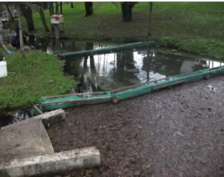
The river beach Olhos de Fervença (Fig. 1) is located in the parish of Cadima, in Coimbra city. The natural springs known as "Olhos de Fervença" are a source of water supply in the area but also in surrounding areas like Mira, Montemor-o-Velho and Coimbra. In 2000, this beach was established through excavation and widening of an existing bedstead of a river and the construction of a small dam. In figure 2 are the three ponds where the samples were collected ( http://solagasta.com/passeio-pedonal-praia-fluvial-dos-olhos-da-fervenca-cantanhede/).
Fig. 1 river beach Olhos de Fervença location (N: 40º20'5, W:8º41'42)
Fig. 2. Ponds in Olhos de Fervença beach lakes
31
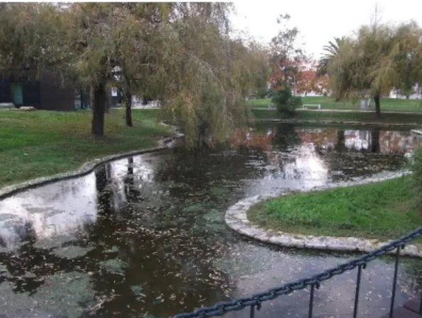
The S. António Park (Fig. 3) is located in the city of Aveiro on the opposite site from Infante D. Pedro Park. It took advantage of Baixa da Ribeira de Santo António stream that allowed the extension of the park. (http://www.rotadabairrada.pt/irt/show/baixa-de-santo-antonio_pt_1629). In figures 4, 5 and 6 are images from the three ponds where the samples were collected.
Fig. 3. S. António Park location (N: 40º38'13, W: 8º39'15)
32
Fig. 6. S. António Park location pond 3
2.2.2 Study organism
Pelophylax perezi Seoane (Fig.7) is commonly known as the Green Frog and
it’s distributed through the Iberian Peninsula. It is the most abundant amphibian in Portuguese territory being present in all bioclimatic regions and associated with various water bodies such as ponds and streams.
Its geographical distribution is limited by the high altitude and it can be found even in humanized areas. In this species, males are smaller than females and have external vocal sacs located below the tympanum on both sides of the mouth. During the
33
breeding period males have black ridges on the inside of the first anterior limbs fingers. (Loureiro, Almeida, Carretero, & Paulo, 2010).
2.3 Field collection procedures and microbiota sampling
2.3.1 Frog sampling
Adult green frogs were collected in two different seasons: autumn (pre-hibernation) and spring (post-(pre-hibernation). The pre-hibernation sampling was made in November 2014 at both sites and the post-hibernation at May and June 2015. A total of 31 individuals were collected: 11 animals in pre-hibernation season (6 females in river beach Olhos de Fervença and 3 males and 2 females in S. António Park) and 20 animals in post-hibernation season (3 females and 7 males in river beach Olhos de Fervença and 5 males and 5 females in S. António Park). Animals were collected with a hand net and handled with the help of a smaller net previously washed with 70% alcohol and rinsed with sterile distilled water. Nitrile gloves were used and changed between individuals to avoid cross contamination
2.3.2 Microbiota sampling
Skin microbiota sampling wasimmediately made at the field after the capture of the adults. They were washed three times with sterile distilled water to remove transient bacteria from the skin (Culp et al., 2007), being collected some of the water from the last wash to confirm the effectiveness of the washes. After washing, sterile swabs were used for microbiota sampling. Each individual was swabbed three times, one per each culture medium used (TSA, TSB 1% and PCA). Each swab consisted in 3 strokes from snout to vent direction, in lateral, dorsal and ventral surfaces of the torso. The swab was slightly turned when changing within body surfaces to avoid saturating the swab with only one surface of the body. As a control for the accidental inoculation of the swab with bacteria from the air on a swab without passing through the skin was left exposed to air for thirty seconds and then swabbed in a plate with TSA medium. After finished
34
microbiota sampling, the length of the animal was determined using a ruler. The sex of the individuals was also determined. All swabs were immediately inoculated in the respective media. The inoculation for the solid media consisted streaking the swabs in the plate, while for the liquid medium TSB 1% the swab was placed into a 1.5 ml Eppendorf tube with 500 microliters of TSB 1% (495 microliters of distilled water: 5 microliters of TSB) and immediately preserved in a refrigerated container (4ºC) for further laboratory processing.
Growth was conducted in an incubating chamber at a temperature of 22 º C. The TSB medium, due to its liquid state at room temperature, was treated differently. Immediately after arriving at the laboratory and in a flow chamber, Eppendorf tubes were agitated in the vortex for several seconds. After this, 50 microliters of the content were pipetted and inoculated, with the help of sterile glass beads, in a Petri dish containing TSA medium. Afterwards the samples were incubated at 22 ° C in a controlled temperature room. The use of the TSB 1% intended to avoid overgrowing of fast growing bacteria, with the aim of attaining slower growing bacteria.
2.3.3 Water abiotic parameters
For every pond where animals were collected the pH (WTW330/SET-2 pH meter), dissolved oxygen (WTW315i/SET Oxi meter) and conductivity (LF 330/SET conductivity meter) were measured.
2.4 Identification of bacteria from frog skin, isolation and preservation
After bacterial growth for two-three days, each plate (TSA inoculated from TSB 1%, TSA and PCA) was verified for subsequent isolation. Colonies were distinguished according to color, size, border and texture. After observation, different colonies where isolated onto new Petri plates containing TSA. After complete isolation the bacteria isolates were preserved at -20ºC in a 20% glycerol solution for subsequent work. These steps were carried out in a laminar flow chamber.
35 2.5 DNA extraction
For the bacterial DNA extraction the following procedure was carried out. The isolates grew overnight in sterilized Eppendorf tubes with TSB medium (1 ml). Afterwards 100 µl were removed from the Eppendorf, placed into a new one and centrifuged at 15000 G for 15 minutes. The supernatant was then discarded and the pellet was resuspended with 40 µl of sterile water. Posteriorly it was centrifuged at 15000 G for 10 minutes and the supernatant discarded again. The pellet was then resuspended with 40 µl of sterile water and incubated at 100º C for 10 minutes and afterwards cooled on ice. New centrifugation was followed at 15000 G for 1 minute. In the end, 5 µl were run on agarose gel (1%) stained with 2 µl of SYBR® Safe DNA gel stain to check if total DNA was present. This was prepared by boiling 0.85 g of agarose in 85 ml of 1xTAE buffer (dilution of a 50x TAE stock solution).
2.6 DNA amplification and sequencing
In order to allow the identification of the bacterial isolates the amplification of the 16S rRNA genes for sequencing was performed. The PCR reactions were performed in 25 µL reactions containing 0.2 µM each primer (27F and 1492R) 1x PCR buffer, 0.2 mM each dNTP, 2 mM MgCl2,1U Taq polymerase and 2 µl of cell lysate as template
DNA. PCR reaction was set as follows: an initial desnaturating step, at 95 ºC, for 3 minutes, followed by 95ºC for 1 minute, 1 minute in the annealing phase, at 54 ºC, and 1 minute in extension phase at 72 ºC. This was repeated for 34x. Next was 1 minute at 72 ºC and then finished at 4 ºC. After the end of the incubation program, PCR products were analyzed by electrophoresis on agarose gel (1%) stained with SYBR® Safe DNA gel stain. The electrophoresis conditions were the follow: 45 minutes run time, 75 V and 400 Ma. Sequencing was performed by Stab Vida (Portugal) and the sequences were compared with databases, using BLASTn from the National Center for Biotechnology Information (NCBI)
36 2.7 Inhibition growth assays
Before carrying out the tests, it was necessary to prepare the culture medium, sterilize tweezers and small discs of paper (Whatman filter G 1) and proceed with bacteria growth (both frogs’ bacterial isolates, as well as the bacteria for the test bacterial lawn). The standardized culture medium Mueller-Hinton was used in these tests. The Muller-Hinton test plates were prepared according to the disk diffusion method (Lalitha, 2008). Briefly, the bacterial cultures were left growing overnight with agitation in 15 ml Falcon tubes containing TSB medium. Then, to standardize the inoculum a BaSO4 turbidity
standard, equivalent to a 0.5 McFarland standard, was used. For an absorbance value of 0.008 to 0.1 determined spectrophotometrically at 625 nm the bacterial suspension was considered to contain approximately 1 to 2x10⁸ CFU/ml. After adjusting the cell
concentration, the inoculation of the plates was made. After inoculating the plate with the bacterial lawn (Bacillus aquimaris or Aeromonas salmonicida) and allowing the plate to dry, disks where placed with a minimum distance of 20 mm between them and then on each disk a different bacterial isolate was inoculated by pipetting 5 µl of the standardized inoculum. On one of the disks only TSB was pippeted as a negative control. The plate was sealed with parafilm and placed to growth for two days at 22 ° C. The plates were then examined to see whether or not an inhibition zone was visible around each disc. The choice of test agents was based, on one hand, by the pathogenic potential (A. salmonicida) and on the other hand by their sensitivity (B. aquimaris) (S. Marques, unpublished data).
37 2.8 Results
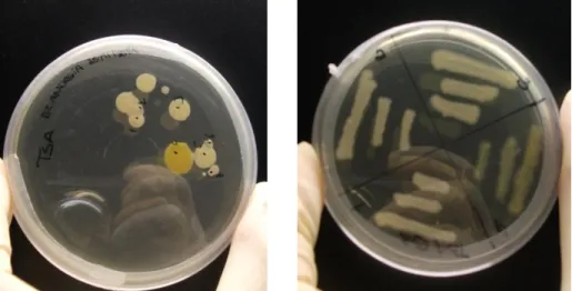
In the pre-hibernation season a total of 137 isolates were collected from the river beach Olhos de Fervença (Fig. 8) and 127 isolated from S. António Park. In the post-hibernation season, in S. António Park were collected 204 samples and 257 samples from river beach Olhos de Fervença.
Fig. 8 Left image- Bacteria from P.perezi skin after growth; Right image- isolation step.
For this work and due to the number of samples in total, all isolates used in this work were among TSA medium because they were the most representative. From the autumn in river beach Olhos de Fervença were selected 38 samples and in Santo António Park 23 samples. From the spring and in river beach Olhos de Fervença were selected 26 samples and in the Santo António Park 33 samples (Table 1). The average lengths of the individuals from river beach Olhos de Fervença in the winter were from 4.8 to 6.5 centimeters. In S. António Park were between 4.2 to 4.8 centimeters. In the Spring in river beach Olhos de Fervença the sizes varied between 5 to 6.3 centimeters. In S. António Park were from 4.3 to 8.2 centimeters.
38
Table 1. Number of isolates per season, local, sex and with antimicrobial activity Local
Number of isolates in females
Number of isolates in males
Autumn Spring Autumn Spring
river beach Olhos de Fervença Total 38 11 - 15 Antimicrobial activity 11 - - 1 S. António Park Total 13 17 10 16 Antimicrobial activity 4 - 2 1
The isolates were from Pseudomonadaceae, Bacillaceae, Oxalobacteraceae,
Moraxellaceae, Paenibacillaceae, Rhizobiaceae, Comamonadaceae,
Enterobacteriaceae, Planococcaceae, Shewanellaceae, Aeromonadaceae,
Microbacteriaceae, Sphingobacteriales, Micrococcaceae and Deinococcaceae. (Table
2.)
Table 2. Identification of bacterial morphotypes obtained from P. perezi skin; RBO- River beach Olhos de Fervença; S- Spring; A-Autumn; M-Male; F-female; IGA- inhibitions growth assays; (+) shows antimicrobial activity in the inhibitions growth assays; (%)- percentage of identity.
Sample Site Season Sex Bacteria
I G A Family (%) 1 RBO S M Pseudomonas fluorescens Pseudomonadaceae 96
4 STP S M Serratia liquefaciens Enterobacteriaceae 97
5 STP S M Bacillus pumilus Bacillaceae 94
7 STP S F Massilia timonae Oxalobacteraceae 96
8 RBO S M Aeromonas
hydrophila Aeromonadaceae 98
9 STP S M Bacillus megaterium Bacillaceae 98
12 RBO A F Pseudomonas
39
13 STP A F Providencia rettgeri Enterobacteriaceae 94
15 STP A F Pseudomonas
fluorescens Pseudomonadaceae 95
16 RBO A F Paenibacillus pabuli Paenibacillaceae 94
17 STP A F Comamonas
testosteroni + Comamonadaceae 93
18 STP A M Pseudomonas
fluorescens Pseudomonadaceae 93
19 RBO A F Bacillus thuringiensis + Bacillaceae 96
20 RBO A F Pseudomonas putida Pseudomonadaceae 92
23 RBO A F Citrobacter freundii + Enterobacteriaceae 96
24 STP A F Obesumbacterium proteus Enterobacteriaceae 94 25 STP A M Rouxiella chamberiensis Enterobacteriaceae 93 26 STP A F Pseudomonas syringae Pseudomonadaceae 87
27 STP A M Pseudomonas putida Pseudomonadaceae 97
28 RBO A F Paenibacillus pabuli Paenibacillaceae 95
29 STP A F Rouxiella chamberiensis Enterobacteriaceae 96 31 RBO A F Rouxiella chamberiensis Enterobacteriaceae 89 32 RBO A F Pseudomonas rhizosphaerae + Pseudomonadaceae 96 33 STP S F Sporosarcina sp. Planococcaceae; 96 34 RBO S F Aeromonas hydrophila Aeromonadaceae 95
35 RBO S F Flavobacterium sp. Flavobacteriaceae 90
36 STP S F Aeromonas
hydrophila Aeromonadaceae 97
37 RBO S F Pseudomonas sp. Pseudomonadaceae 94
38 STP S F Bacillus megaterium Bacillaceae 97
39 RBO S F Klebsiella oxytoca Enterobacteriaceae 85
40
42 STP S F Shewanella
oneidensis Shewanellaceae 94
43 RBO A F Skermanella
stibiiresistens Shewanellaceae 98
44 STP A M Citrobacter freundii Enterobacteriaceae 95
45 RBO A F Agrobacterium fabrum Rhizobiaceae 95 46 STP S M Pseudomonas syringae Pseudomonadaceae 96 48 RBO A F Pectobacterium carotovorum + Pseudomonadaceae 95 49 RBO A F Pseudomonas fluorescens Pseudomonadaceae 95
50 RBO A F Bacillus simplex Bacillaceae 95
54 RBO A F Pseudomonas alkylphenolia Pseudomonadaceae 96 55 RBO A F Pectobacterium carotovorum Enterobacteriaceae 96 58 RBO A F Pseudomonas fluorescens + Pseudomonadaceae 96 59 STP A M Flavobacterium sp. Flavobacteriaceae 96 60 RBO A F Pseudomonas fluorescens Pseudomonadaceae 99
61 STP A F Pseudomonas putida Pseudomonadaceae 99
62 STP A F Bacillus pumilus + Bacillaceae 97
63 RBO A F Pseudomonas
resinovorans Pseudomonadaceae 97 64 STP A F Citrobacter freundii + Enterobacteriaceae 95
65 STP A F Klebsiella oxytoca Enterobacteriaceae 98
66 RBO A F Pseudomonas
fluorescens Pseudomonadaceae 94
67 STP A F Pseudomonas
fluorescens Pseudomonadaceae 95
68 STP A F Acinetobacter lwoffii Moraxellaceae 96
41
70 STP S F Flavobacterium sp. Flavobacteriaceae 95
71 STP S F Yersinia pestis Enterobacteriaceae 90
72 STP S F Aeromonas veronii Aeromonadaceae 96
73 STP A M Buttiauxella agrestis + Enterobacteriaceae 90
75 STP S M Aeromonas
hydrophila Aeromonadaceae 96
76 STP S F Aeromonas veronii Aeromonadaceae 96
77 STP S M Aeromonas
hydrophila Aeromonadaceae 97
78 RBO A F Microbacterium sp. Microbacteriaceae 86
79 STP S M Acinetobacter
johnsonii Moraxellaceae 96
80 STP A M Kluyvera intermedia Enterobacteriaceae 95
82 STP S F Aeromonas
hydrophila Aeromonadaceae 95
83 STP S F Pedobacter oryzae Sphingobacteriaceae 94
85 STP S F Arthrobacter sp. Micrococcaceae 97
86 STP A F Citrobacter freundii Enterobacteriaceae 97
88 STP S M Arthrobacter sp. Micrococcaceae 95
89 RBO A F Bacillus mycoides Bacillaceae 96
90 STP A M Pseudomonas putida + Pseudomonadaceae 96
92 RBO A F Pseudomonas
syringae Pseudomonadaceae 95
93 STP S M Deinococcus soli Deinococcaceae 90
94 RBO A F Pseudomonas fluorescens Pseudomonadaceae 99 98 RBO S M Microbacterium foliorum Microbacteriaceae 99 99 STP A M Flavobacterium sp. Flavobacteriaceae 96 100 RBO A F Pseudomonas fluorescens + Pseudomonadaceae 94 102 STP S M Flavobacterium sp. Flavobacteriaceae 98
103 RBO S M Flavobacterium sp. Flavobacteriaceae 94
104 RBO S M Delftia acidovorans Comamonadaceae 95
42
106 RBO A F Bacillus mycoides Bacillaceae 91
110 RBO A F Sporosarcina sp. + Planococcaceae 94
112 RBO A F Paenibacillus pabuli Paenibacillaceae 96
113 RBO A F Klebsiella oxytoca + Enterobacteriaceae 93
115 STP S M Klebsiella oxytoca Enterobacteriaceae 81
116 STP S F Paenisporosarcina
sp. Planococcaceae 95
117 STP A F Citrobacter freundii Enterobacteriaceae 96
118 RBO A F Bacillus mycoides Bacillaceae 96
119 RBO A F Microbacterium hydrocarbonoxydans Microbacteriaceae 96 120 RBO S M Pseudomonas protegens Pseudomonadaceae 98 121 RBO S M Microbacterium hydrocarbonoxydans Microbacteriaceae 97
124 RBO S M Bacillus mycoides Bacillaceae 98
125 STP S M Citrobacter freundii Enterobacteriaceae 97 127 STP S M Providencia rettgeri Enterobacteriaceae 97
129 STP S M Bacillus mycoides Bacillaceae 98
132 RBO S M Flavobacterium sp. Flavobacteriaceae 99
134 RBO S M Bacillus mycoides Bacillaceae 84
135 RBO A F Providencia rettgeri Enterobacteriaceae 84
136 RBO S M Bacillus mycoides Bacillaceae 98
137 RBO S M Bacillus mycoides Bacillaceae 99
138 RBO S F Bacillus amyloliquefaciens Bacillaceae 98 139 RBO A F Pseudomonas fluorescens Pseudomonadaceae 97 140 STP A F Pseudomonas fluorescens Pseudomonadaceae 97 141 STP A F Acinetobacter sp. Moraxellaceae 96 142 STP A F Acinetobacter johnsonii + Moraxellaceae 98
146 RBO S M Bacillus mycoides Bacillaceae 99
43
A total of 19 from 120 isolates showed antimicrobial activity (Fig. 9) within which almost of them were collected at the time of pre hibernation season. There was only two samples that showed antimicrobial activity that were collected in the post hibernation season. From 19 samples, 12 belong to river beach Olhos de Fervença corresponding to females and one male (11 in the autumn and 1 in the spring respectively). Regarding the Santo António Park were 7 the samples (6 from the autumn: 4 from females and two from males, and one from one male in the spring) that showed antimicrobial activity (Table 3). They belong to Pseudomonadaceae,
Comamonadaceae, Enterobacteriaceae, Aeromonadaceae, Paenibacillaceae and Bacillaceae families.
150 RBO S F Bacillus mycoides Bacillaceae 99
151 RBO S F Acidovorax delafieldii Comamonadaceae 97
152 RBO S F Acinetobacter
johnsonii Moraxellaceae 93
153 RBO S M Erwinia persicina Enterobacteriaceae 95
155 RBO A F Citrobacter freundii Enterobacteriaceae 97 157 RBO A F Citrobacter freundii Enterobacteriaceae 97
158 RBO A F Bacillus pumilus
strain Bacillaceae 97
160 RBO A F Bacillus pumilus
strain + Bacillaceae 95
161 RBO A F Bacillus pumilus
strain + Bacillaceae 96
162 RBO S M Erwinia persicina + Bacillaceae 95
166 STP A M Citrobacter freundii Enterobacteriaceae 95
171 RBO S F Bacillus mycoides Bacillaceae 98
175 RBO A F Pseudomonas
44
Fig. 9 Inhibition growth assay with visible inhibition zone; Numbers 1 to 5 corresponds to bacteria isolates from P. perezi skin; C is the control (TSB medium only); Base layer
45
Table 3. Bacteria species isolated from P. perezi skin, presenting antimicrobial activity (+) growth inhibition was observed; (−) no growth inhibition was observed.
Frog Isolate Test organism A.salmonicida Bacillus aquimaris Comamonas testosteroni - + Bacillus thuringiensis - + Citrobacter freundii - + Pectobacterium carotovorum - + Pseudomonas fluorescens + + Bacillus pumilus - + Buttiauxella agrestis - + Pseudomonas putida - + Paenibacillus pabuli - + Acinetobacter sp. - + Bacillus pumilus - + Flavobacterium sp. - + Pseudomonas rhizosphaerae - + Bacillus mycoides - +
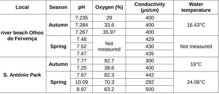
In the table below (Table 4) are the results of water analyses in situ (pre and post-hibernation seasons). At river beach Olhos de Fervença in the Autumn the ponds showed lower pH as well as lower conductivity related to the summer. This was also observed in S. António Park in two of the three ponds. Oxygen (%) values were lower in the Spring (post-hibernation season) compared to the winter in S. António Park.
46
Table 4. Abiotic parameters and the respective values in the ponds from the sampling sites between seasons.
Local Season pH Oxygen (%) Conductivity (µs/cm)
Water temperature
river beach Olhos de Fervença Autumn 7.235 29 400 16.43°C 7.284 33.6 400 7.267 35.97 400 Spring 7.48 Not measured 429 Not measured 7.52 430 7.47 435 S. António Park Autumn 7.77 92.7 300 16°C 7.25 38.6 400 Spring 7.87 82.3 442 24.06°C 10.09 70.3 292 8.97 63.2 500 2.9 Discussion
In this study was observed that the cultivable microbiota from the green-frog´s skin differs depending on the local, sex and season. The most frequent families were
Pseudomonadaceae, Enterobacteriaceae and Bacillaceae. This was also observed in
a study carried out by Assis et al 2016 with Phyllomedusa distincta sampled in two different sites in Brazil. Comparing genders, it was observed that females presented a greater number of bacterial families than males. Families like Oxalobacteraceae,
Paenibacillaceae, Planococcaceae, Shewanellaceae, Rhizobiaceae,
Sphingobacteriacea were only found in females. In males only the Deinococcaceae
family was found. Thus it is not expected that the difference we observed result in gender differential sensitivity to pathogens. Regarding diversity of isolates obtained for both sites, there wasn’t a great difference. This was similar when comparing the diversity of isolates between seasons.
Our results also revealed that some of the isolates presented antimicrobial activity. Taking into account both bacteria used as test lawns (A. salmonicida and
47
B.aquimaris) and the information on table 3, it was observed that the antimicrobial
activity was predominantly against B. aquimaris. Only Pseudomonas fluorescens obtained antimicrobial activity against both. P. fluerescens was isolated from a female from river beach Olhos de Fervença. In other studies, this bacterium showed antifungal (Srivastava, S.R., 2008) and antimicrobial activity evidenced by the production of secondary metabolites against some pathogenic bacteria strains (Trippe et al. 2013).
When regarding seosonality, according to our results, the antimicrobial activity was observed mainly in isolates obtained from the pre hibernation season and mostly from females. Only one male from S. António Park demonstrated this activity. Another important fact is that a slightly higher number of isolates with antimicrobial activity were retrieved from river beach Olhos de Fervença. The bacteria that showed this activity belong to Pseudomonadaceae, Enterobacteriaceae, Bacillaceae, Comamonadaceae and Paenibacillaceae families. Based on this it is possible to infer that the temperature and the organic matter could be a determining factor in antimicrobial activity of bacterial species. It is important to highlight that in the Autumn there were great quantities of organic matter in the soil and in water in both sites. In fact, the work carried out by Noaman et al., (2004) showed that variations on the carbon sources are capable of changing the production of antimicrobial compounds. This may indicate that seasonality might play an active role in the resistance to pathogens.
Overall our results allowed to perceive differences between the diversity of the cultivable microbiota from both sites and also within seasons. However, the most striking is that we have identified some bacteria with the ability to display antimicrobial activity, which is important if future local bioaugmentation strategies are intended to by implement. Another highlight is that seasonality might be an issue when considering amphibians’ resistance to pathogens. Indeed, some common bacterial strains collected in both seasons, displayed different antimicrobial activity between seasons, with most of the antimicrobial activity observed in Autumn. It is fundamental to keep these results in mind if future bioaugmentation strategies are needed, since the season for field implementation of the strategy might play a key role on its success.
48 2.10 References
Allentoft, M.E., O’Brien, J. 2010. Global amphibian declines, loss of genetic diversity and fitness: a review. Diversity, 2(1), 47-71.
Assis, A.B., Santos, C., Dutra, F.P., Motta, A.O., Costa, F.S., Navas, C.A., Magalhães, B.S., Barreto, C.C., 2016. Assessing antibacterial potential of components of Phyllomedusa distincta skin and its associated dermal microbiota. Journal of
Chemical Ecology, 42(2), 139-148.
Becker, M.H., Brucker, R.M., Schwantes, C.R., Harris, R.N., Minbiole, K.P.C., 2009. The bacterially produced metabolite violacein is associated with survival of amphibians infected with a lethal fungus. Applied and environmental microbiology,
75(21), 6635–6638.
Bletz, M.C., Loudon, A.H., Becker, M.H., Bell, S.C., Woodhams, D.C., Minbiole, K.P.C., & Harris, R.N. 2013. Mitigating amphibian chytridiomycosis with bioaugmentation: Characteristics of effective probiotics and strategies for their selection and use. Ecology Letters, 16(6), 807–820.
Costello, E.K., Lauber, C.L., Hamady, M., Fierer, N., Gordon, J.I., Knight, R. 2009. Bacterial community variation in human body habitats across space and time.
Science. 326(5960),1694–1697.
Culp, C.E., Falkinham, J.O., Belden, L.K., 2007. Identification of the natural bacterial microflora on the skin of eastern newts, bullfrog tadpoles and redback salamanders. Herpetologica, 63, 66–71.
Daum, J.M., Davis, L.R., Bigler, L., Woodhams, D.C., 2012. Hybrid advantage in skin peptide immune defenses of water frogs (Pelophylax esculentus) at risk from emerginf pathogens. Infection, Genetics and Evolution, 12, 1854-1864.
Grice, E.A., Segre, J.A., 2011. The skin microbiome. Nature Reviews
Microbiology, 9(4), 244-253.
Kouamé, N.G., Ofori-Boateng, C., Adum, G.B., Gourène, G., Rödel, M.O., 2015. The anuran fauna of a West African urban area. Amphibian & Reptile Conservation,
49
Kueneman, J.G., Parfrey, L.W., & Woodhams, D.C. 2014. The amphibian skin-associated microbiome across species, space and life history stages. Molecular
Ecology, 23(6) 1238–1250.
Lalitha, M.K. 2008. Manual on antimicrobial susceptibility testing. Indian Journal
of Medical Research, 88, 455-460.
Loureiro, A., Almeida, N.F., Carretero, M.A., Paulo, O.S. 2010. Atlas dos anfíbios e dos répteis de Portugal. Atlas Dos Anfíbios E Dos Répteis de Portugal, 126. Lozupone, C., Knight, R. 2007. Global patterns in bacterial diversity.
Proceedings of the National Academy of Sciences, 104(27), 11436–11440.
Noaman, N.H., Fattah, A., Khaleafa, M., Zaky, S.F., 2004. Factors affecting antimicrobial activity of Synechococcus leopoliensis. Microbiological research, 159 (4), 395-402.
Schenkel, M., 2012. The prevalence of the amphibian pathogen
Batrachochytrium dendrobatidis in permanent and temporary ponds. Master thesis.
Institute of Evolutionary Biology and Environmental studies. Universitat Zurich, 28 pp. SolaGasta Percursos Pedestres, (n.d.), accessed (08-2015)
http://solagasta.com/passeio-pedonal-praia-fluvial-dos-olhos-da-fervenca-cantanhede/
Srivastava, S.R., 2008. Antifungal activity of Pseudomonas fluorescens against different plant pathogenic fungi. The Internet Journal of Microbiology, 7(2), 1-7.
Stuart, S.N., Hoffmann, M., Chanson, J.S., Cox, N.A., Berridge, R.J., Ramani, P., Young, B.E. 2008. Threatened amphibians of the world, First Edition.
Trippe, K., McPhail, K., Armstrong, D., Azevedo, M., Banowetz, G., 2013.
Pseudomonas fluorescens SBW25 produces furanomycin, a non-pathogenic amino
acid with selective antimicrobial properties. BMC Microbiology,13(111), 1-10. Unlimited colours of Bairrada, Rota da Bairrada, (n.d.), accessed (08-2015) http://www.rotadabairrada.pt/irt/show/baixa-de-santo-antonio_pt_1629
Woodhams, D.C., Bosch, J., Briggs, C.J., Cashins, S., Davis, L.R., Lauer, A., Voyles, J. (2011). Mitigating amphibian disease: strategies to maintain wild populations and control chytridiomycosis. Frontiers in Zoology, 8(1), 1-8.
50
Woohams, D.C., Brandt, H., Baumgartner, S., Kielgast, J., Kϋpfer, E., Tobler, U., Davis, L.R., Scmidt, B.R., Bel, C., Hodel, S., Knight, R., Mckenzie, V., 2014. Interacting symbions and immunity in the amphibian skin mucosome predick disease risk and probiotic effectiveness. PLos ONE, 9(4), 1-13.
Zug, G.R.,Vitt, L.J., Caldwell, J.P., 2001. Herpetology: An Introductory Biology
of Amphibians and Reptiles. Retrieved from
51 Chapter III
In vivo assessment in Pelophylax perezi tadpoles of the antimicrobial potential
53
3. In vivo assessment in Pelophylax perezi tadpoles of the antimicrobial potential of bacterial isolates against the pathogenic agent Aeromonas
salmonicida
Abstract
Amphibian pathogens are among the major causes for the recent amphibian worldwide decline. In order to deal with this problem a global endeavor has been undertaken to find ways of controlling pathogenic agents which comprise fungus, bacteria and viruses. The bioaugmentation of amphibian symbiotic bacteria with pathogen specific antimicrobial activity is one of the most promising approaches. However, the majority of the studies have been focused in the fungus Batrachochytrium
dendrobatidis. Considering that, within bacteria, the genus Aeromonas is one of the
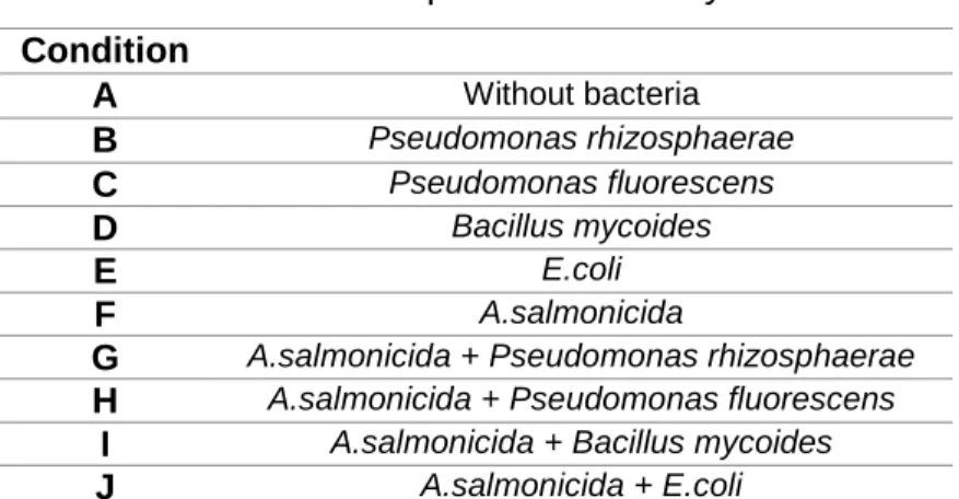
main responsible for most deaths in amphibians, our work aimed at testing, in vivo, the effect of the exposure of Pelophylax perezi tadpoles to both bacterial isolates with antimicrobial activity (Pseudomonas rhizosphaerae, Pseudomonas fluorescens,
Bacillus mycoides) and the pathogen Aeromonas salmonicida. Mortality, growth and
the activity of enzymes of the antioxidant defense system (total glutathione peroxidase, Se-dependent glutathione peroxidase, glutathione S-transferase and glutathione reductase), as well as lipid peroxidation were used as parameters to assess the condition of the animals after exposure to the bacteria. The results showed a 33% mortality among the tadpoles exposed solely to A. salmonicida while mortality in other conditions was negligible, being lower than 4%. Growth, enzymatic parameters and lipid peroxidation did not reveal any statistical significant difference. Nonetheless there was a global tendency for tadpoles exposed solely to P. rhizosphaerae, P. fluorescens,
and B. mycoides to present lower enzymatic activity when compared to organisms
exposed to A. salmonicida. Such was also verified when comparing the isolated exposure to the probiotics with the respective joint exposure with A. salmonicida. Our results suggest that the sole or joint exposure to A. salmonicida triggers the antioxidant
54
defense system and that the P. rhizosphaerae, P. fluorescens, and B. mycoides might have an important role in avoiding cellular damage caused by the pathogenic agent. Key words: tadpoles P.perezi, innate immune system, oxidative stress, antioxidant enzyme system, biomarkers
3.1 Introduction
Over the recent years amphibians’ populations of have been declining due to the interaction of numerous external factors (climate change, pollution, habitat change and invasive species (Stuart et al., 2004). According to a review made by Allentoft and O’Brien (2010) the genetic variation has been found, in various studies with amphibians, to be closely and positively correlated with fitness and adaptability. Such implies that a decrease in genetic variation leads to a loss of fitness. This fact leads to an increased need to preserve amphibian populations in whatever habitat they are found, ranging from pristine habitats to contaminated ones, and from rural to urban zones. Presently, one of the main responsible factors for amphibians’ declines are diseases caused by fungi, bacteria and ranavirus. Nonetheless, there are amphibian species that despite the presence of pathogens such as Batrachochytrium
dendrobatidis (Bd) fungus are able to survive (Chen and Robert, 2011; Hayes et al.
2010). One of such examples is the work carried out by Carver and coworkers (2010) where they studied the effect of the disease Chytridiomycosis in Litoria raniformis. They observed that shortly after inoculation with the fungus, individuals showed signs of infection being, however, able to fully recover after a few months. This type of study demonstrates that amphibians can even survive in the presence of the fungus responsible for Chytridiomycosis and quickly recover the infection, relying solely on their immune system. The immune system comprehends the set of all the organs and cells that interact coordinately with each other and are involved in protecting the organism against potential infections. (Abbas and Lichtman, 2011) The immune responses can be distinguished in innate and adaptive. Innate immunity is present since birth and it is the first line of defense against potential pathogen. Innate immunity
55
is non-specific and it generates a rapid response (within minutes or hours) to prevent the entry of foreign organisms through physical barriers such as skin and mucous membranes (Abbas et al. 2008). On the other hand, adaptive immunity develops when the organism is exposed to an antigen and may take days until the response is triggered. It´s characterized by its specificity and memory effect providing an increase of effectiveness of the response to repeated pathogen exposures. In amphibians, besides its innumerous functions such as thermoregulation and respiration, the skin assumes a major role in protecting the organism against exterior aggressions, namely pathogens. It contains chemical barriers that protect from potential infections. The granular glands produce antimicrobial peptides (AMP's) which are a good example of one of the innate immune responses. AMP’s can be very diverse, even within philogenically close species (Daum et al., 2012). The innate immune responses of the skin can be frequently reinforced by the action of skin bacteria (Lauer et al., 2007, 2008). One of such cases is the antifungal metabolite, violacein, produced by the bacterium Janthinobacterium lividum (Becker et al., 2009). It has been documented that the inoculation of this bacteria in salamanders increases their survival to infection by Bd. In fact, J. lividum has been used in recent studies (Muletz et al., 2012), as probiotic, through bioaugmentation. As Bletz et al (2013) defined it “…bioaugmentation is the augmentation of locally occurring protective bacteria to an individual or the environment with the purpose of altering the hosts’ microbial community structure to mitigate disease”.
In general, the immunity of organisms is put to the test when they are facing or exposed to xenobiotics since it becomes weakened. From a weakened immune system, infections might occur and with them alterations in the homeostasis. The effects begin at the cellular level but in most cases are detected later when already damage of tissues and organs occurred. Then it becomes more difficult to reverse the situation. One example of an effect is catalytic reactions that can generate overproduction of reactive oxygen species (ROS) (Ochsendorf, 1998). ROS are reactive molecules containing oxygen and they spontaneously react with other molecules. These reduction products of molecular oxygen (O2) include the superoxide
56
anion (O2-), hydrogen peroxide (H2O2) and hydroxyl radicals (OH-). On a cellular level
and in a controlled way, ROS are produced as the byproducts during the mitochondrial respiratory chain and functions in various physiological processes. Nonetheless, due to exogenous factors such as infections, chemicals, metals or UV radiation their production can increase and lead to protein oxidation, lipid peroxidation or DNA damage (Ochsendorf, 1998; Khansari et al. 2009). The control of ROS is associated with the antioxidant defense mechanisms from enzymatic origin. When an imbalance between antioxidant defenses and the production of free radicals occurs, where there is an ROS excess compare to antioxidant cell response, it is said that the organism is under oxidative stress (Khansari et al. 2008; Ferreira and Abreu, 2007; Apel and Hirt, 2004). To cope with this, living cells have enzymatic antioxidant defenses with enzymes such as glutathione peroxidase (GPX), glutathione reductase (GRED) and glutathione-S- transferase (GST), which functions as a coordinated system in ROS elimination. (Gillardin et al. 2009) and can be used as biomarkers.
Considering that one of the most recent ways to protect amphibians from diseases is through strengthening their immunity with probiotics, and that these should be adapted to each species (Becker et al., 2011), it is essential to search for adequate probiotics and assess their effectiveness. Furthermore, most of the endeavor for finding appropriate probiotics are related with Bd, leaving on the side other pathogens, namely bacteria, which may cause also severe health issues on amphibians. Within bacteria, the genus Aeromonas is one of the main responsibles for most deaths in amphibians. Taking this into account, the aim of this study was to determine the extent to which bacteria isolated from P. perezi with previously tested antimicrobial potential, could protect tadpoles from pathogens such as A. salmonicida, making use of antioxidant activity as an indicator of infection.