DNA METHYLATION PROFILE
AS A TOOL FOR LUNG CANCER
DIAGNOSIS & SUBTYPING
FRANCISCA OLIVEIRA DE MESQUITA DINIZ DISSERTAÇÃO DE MESTRADO APRESENTADA
AO INSTITUTO DE CIÊNCIAS BIOMÉDICAS ABEL SALAZAR DA UNIVERSIDADE DO PORTO EM
Francisca Oliveira de Mesquita Diniz
DNA METHYLATION PROFILE AS A TOOL FOR LUNG CANCER DIAGNOSIS
& SUBTYPING
Dissertação de Candidatura ao grau de Mestre em
Oncologia – Especialização em Oncologia
Molecular submetida ao Instituto de Ciências
Biomédicas de Abel Salazar da Universidade do
Porto.
Orientador
Professor Doutor Rui Manuel Ferreira Henrique
Professor Catedrático Convidado
Departamento de Patologia e Imunologia Molecular
Instituto de Ciências Biomédicas Abel Salazar
Universidade do Porto
Diretor do Serviço de Anatomia Patológica
Investigador Sénior do Grupo de Epigenética e
Biologia do Cancro
Centro de Investigação
Instituto Português de Oncologia – Porto
Coorientadora
Professora Doutora Carmen de Lurdes Fonseca
Jerónimo
Professora Associada convidada com Agregação
Departamento de Patologia e Imunologia Molecular
Instituto de Ciências Biomédicas Abel Salazar
Universidade do Porto
Investigadora Auxiliar e coordenadora do Grupo de
Epigenética e Biologia do Cancro
Centro de Investigação
“If you can’t fly, then run,
if you can’t run, then walk,
if you can’t walk, then crawl,
but whatever you do,
you have to keep moving forward.”
-Martin Luther King Jr.AGRADECIMENTOS
Seguir em frente quando estamos perante dificuldades torna-se muito mais fácil quando temos pessoas ao nosso lado dispostas a ajudar e a apoiar, é a elas que segue o meu agradecimento.
Antes de mais, ao Professor Rui Henrique e à Professora Carmen Jerónimo, meu orientador e coorientadora respetivamente, um obrigada por me terem recebido no Grupo de Epigenética e Biologia do Cancro, por todas as oportunidades, ensinamentos, horas dedicadas, críticas, conselhos e acima de tudo pela paciência. Obrigada por terem confiado em mim.
Ao Professor Manuel Teixeira, Diretor do Centro de Investigação do IPO-Porto, por ter autorizado a realização desta tese no Laboratório 3.
À Professora Berta Silva, Diretora do Mestrado em Oncologia, obrigada por me ter aceite no referido ciclo.
Agradeço ainda às Dras. Marta Soares e Ana Vítor Silva da Clinica de Pulmão, à Dra. Ana Luísa Cunha e à Técnica Sofia Paulino do Serviço de Anatomia Patológica, bem como ao Engº Luís Antunes do Serviço de Epidemiologia por toda a dedicação, paciência e contribuição para que a concretização desta dissertação.
Não poderia esquecer a Dona Luísa, amiga e sempre atenta ao que fazia falta no laboratório para que tudo corresse na normalidade e não nos faltasse nada. Obrigada pelos docinhos e pelos carinhos.
Ao João Carvalho, Inês Graça, Ana Luís e Sara Reis (a velha guarda), um sincero obrigado por terem estado sempre lá, atentos, dedicados e sempre com uma palavra de alento! Sem dúvida que com vocês aprendi muito mais do que poderia imaginar e nada teria sido igual sem o vosso apoio incondicional e os vossos conhecimentos. Além de tudo isto ainda me deram a vossa amizade e carinho que vou levar sempre no coração! Aos meus colegas do dia-a-dia na luta no laboratório: Salta (a amiga a quem vou dever notas de 5€ até ao fim, obrigada por me apoiares desde as coisas mais difíceis até às mais básicas, obrigada por tudo o que me transmitiste e acima de tudo pela paciência!), Soraia (tantas saudades do teu bullying), Catarina B. (obrigada pelas conversas, teorias, apoio, discussões e paciência), Lameirinhas (raspas lâminas como ninguém), Daniela (sempre pronta a mandar uma piada perversa com duplo sentido), Micaela (nunca esquecerei a tua voz de comando nem o apoio nos dias de maior angústia), Nuno (és e serás sempre insubstituível, nunca conheci ninguém como tu, obrigada por nunca falhares), Catarina Araújo (obrigada pela tua loucura e lado guna que me animou sempre), Diana (a minha amiga chique, grito histérico e mega sorrisão, obrigada pelos
conselhos e brincadeiras) David, Maria, Ângela e Ana laura (aos 4 mais novos desejo tudo de bom e que o ano de trabalho que se avizinha seja fácil de levar).
Eva e Maria João, minhas compinchas do mestrado, do laboratório e da vida! Com vocês partilhei 2 anos incríveis e sei que o próximo ano irá trazer coisas muito boas para as 3 e que vão ser vividas juntas, isso deixa-me extremamente feliz! Obrigada por tudo! Por estarem nos dias bons, nos dias menos bons, na saúde e literalmente na doença, de dia e de noite, nos sorrisos e nas lágrimas! Sois a melhor surpresa com que este mestrado me presenteou.
Tenho que agradecer também às minhas irmãs que trago comigo há 5 anos, desde o primeiro ano de licenciatura. Marta, Ju, Sara e Renata, a vocês é impossível enumerar tudo o que de bom me deram nestes 5 anos académicos, mas sei que sem o vosso apoio incondicional há muito que teria desistido. Obrigada pelas horas de gargalhadas, de discussões, de gritos de felicidade, pelos abraços e pelo vosso sorriso. Sei que por muito que nos desencontremos, acharemos sempre um caminho que nos traga de volta!
Aos meus colegas de licenciatura, Mário Gil e Vanessa, obrigada por não deixarem que a distância e mudanças alterassem a nossa amizade e por estarem sempre por perto. Agradeço ainda aos meus amigos de Monção, que felizmente por mais meses que passemos afastados nunca falhou a vossa amizade e os nossos reencontros mostram que os anos não passam.
Ao Lucas e Mateus, os meus irmãos, obrigada por nunca perderem o vosso lado de pestes e pelo carinho que conseguem demonstrar mesmo estando constantemente a chatear-vos a cabeça! Quero sempre que o nosso espirito de javardice e união se mantenha. Obrigada por me porem a sorrir nos dias em que a vontade para isso não era assim tanta.
Aos meus avós, Eduardo e Ju, um obrigada por todas as palavras, abraços, ensinamentos e acima de tudo um obrigada pela coragem que sempre me motivaram a ter. Um obrigada cheio de saudade à minha avó Ofélia, da qual guardo as seguintes palavras: “Não deixes morrer em ti a esperança e continua a gostar da vida”.
Por fim, mas os últimos são os primeiros, um profundo agradecimento aos meus pais. Sempre lutaram para me incutir os valores que achavam certos, motivaram-me a correr pelo que eu queria alcançar sem nunca atalhar pelos facilitismos e fizeram de tudo para ter tudo o que acharam necessário. Obrigada por me deixarem sonhar sem nunca cair na ilusão do mundo perfeito! Obrigada por me ampararem nas quedas e as que falharam me ajudarem a levantar É devido a vocês que sou como sou e que consegui chegar onde cheguei. Obrigada por terem dito sim mas também por terem dito não. Obrigada! Simplesmente isso.
This study was funded by a grant of the Research Centre of Portuguese Oncology Institute of Porto (CI-IPOP-74-2016).
vi
SUMMARY
BACKGROUND: Lung cancer (LCa) is the most common cancer in both men and
women, being responsible for more deaths than any other malignancy. It is well known the importance of early diagnosis of LCa and personalized therapy according to disease genomic characteristics. Thus, discrimination between the LCa subtypes becomes a key to reduce the mortality rate because allows for more specific treatments. LCa are broadly classified into two groups: small cell lung cancer (SCLC) and non-small cell lung cancer (NSCLC), the latter being divided into adenocarcinoma, squamous cell carcinoma (SCC) and large cell carcinoma (LCC), among other less frequent subtypes.
AIMS: The main goal of this dissertation was to evaluate the methylation profile of the
major LCa subtypes, with a panel of genes previously reported to be hypermethylated in LCa: APC, HOXA9, RARβ2, RASSF1A, TFPI2 and SHOX2. In particular, we aimed to discriminate adenocarcinoma, the most prevalent subtype, from the other LCa subtypes. Moreover, we evaluated the association between the gene-panel methylation levels and standard clinicopathological parameters as well as determined the prognostic value of the same gene-panel.
MATERIAL AND METHODS: Methylation levels of APC, RASSF1A, RARβ2, HOXA9,
SHOX2 and TFPI2 were assessed using real-time quantitative MSP in bisulfite-modified
DNA extracted from formalin-fixed paraffin embedded tissue samples from 152 LCa and 22 normal lung parenchyma (NL) from individuals with other neoplasias. Survival analyses were conducted to evaluate its prognostic value.
RESULTS: Methylation levels of APC, HOXA9, RARβ2 and RASSF1A discriminated the
major subtypes, NSCLC from SCLC (P < 0.001; P = 0.021; P < 0.001; P < 0.001; respectively). APC and RASSF1A distinguished SCC and Adenocarcinoma from SCLC (P < 0.001; P < 0.001; respectively), whereas. RARβ2 discriminated all subtypes of NSCLC from SCLC (Adenocarcinoma vs SCLC, P < 0.001; SCC vs SCLC, P < 0.001; LCC vs SCLC, P = 0.036). HOXA9 also differentiated Adenocarcinoma from SCLC (P < 0.001), and it was the only gene that discriminated Adenocarcinoma from SCC (P = 0.024).. Low
APC, HOXA9, RARβ2 and RASSF1A promoter methylation levels associated with poorer
disease specific survival, although not independently as it was dependent of poor tumor differentiation. Low RASSF1A promoter methylation levels also predicted poor disease-free survival in univariable analysis but due to its association with tumor differentiation, it did not retain independent prognostic significance in multivariable analysis.
vii CONCLUSIONS: Assessment of RARβ2 and HOXA9 promoter methylation levels using
qMSP is able to to discriminate among major LCa subtypes in tissue samples. The clinical usefulness of these biomarkers in plasma will be tested in the near future.
viii
RESUMO
INTRODUÇÃO: O cancro do pulmão é mais comum tanto em homens como em
mulheres, sendo, ainda, o principal responsável pela mortalidade associada a cancro. Está bem estabelecida a importância de um diagnóstico precoce de cancro do pulmão bem como a instituição de terapia personalizada, a qual é realizada de acordo com as características da neoplasia. Portanto, descriminar precocemente os principais subtipos de cancro de pulmão torna-se determinante para reduzir a taxa de mortalidade, uma vez que permite melhor especificar as estratégias terapêuticas. O cancro do pulmão é habitualmente classificado em dois grupos: carcinoma de pequenas células e carcinoma de não pequenas células. Este último subdivide-se em adenocarcinoma, carcinoma epidermoide e carcinoma de grandes células, para além de outros subtipos menos expressivos em termos de frequência.
OBJECTIVOS: O objetivo principal desta dissertação de mestrado foi avaliar os perfis de
metilação de diferentes subtipos de cancro do pulmão com um painel de genes previamente descritos na literatura - APC, HOXA9, RARβ2, RASSF1A, TFPI2 e SHOX2. Mais especificamente, pretendeu-se descriminar os adenocarcinomas, que representam o subtipo mais prevalente, dos restantes subtipos de cancro do pulmão. Adicionalmente, foi analisada a associação entre os níveis de metilação do painel de genes e as características clinico-patológicas, bem como o valor prognóstico.
MATERIAL E MÉTODOS: Os níveis de metilação de APC, RASSF1A, RARβ2, HOXA9,
SHOX2 e TFPI2 foram determinados através de PCR quantitativo de metilação em tempo
real, utilizando DNA modificado por bissulfito de sódio extraído de amostras de tecido fixado em formol e incluído em parafina de 152 cancros do pulmão e 22 amostras de parênquima pulmonar normal proveniente de indivíduos com outras neoplasias. A análise de sobrevivência foi realizada para avaliar o valor prognóstico dos genes do painel.
RESULTADOS: Os níveis de metilação dos genes APC, HOXA9, RARβ2 e RASSF1A
descriminaram os subtipos principais de cancro de pulmão (P < 0.001; P = 0.021; P < 0.001; P < 0.001; respetivamente). APC e RASSF1A diferenciaram os carcinomas epidermoides e adenocarcinomas dos carcinomas de pequenas células (P < 0.001; P < 0.001; respetivamente), enquanto que RARβ2 descriminou todos os subtipos pertencentes aos carcinomas de não pequenas células dos carcinomas de pequenas células (Adenocarcinoma vs Carcinoma de pequenas células, P < 0.001; Carcinoma epidermoide vs Carcinoma de pequenas células, P < 0.001; Carcinoma de grandes células vs Carcinoma de pequenas células, P = 0.036). HOXA9 também diferenciou os adenocarcinomas dos carcinomas de pequenas células (P < 0.001), sendo, ainda, o
ix único gene a descriminar os adenocarcinomas dos carcinomas epidermoides (P = 0.024). Por outro lado, SHOX2 e TFPI2 não mostraram diferenças estatisticamente significativas entre nenhum dos subtipos. Níveis baixos de metilação do promotor dos genes APC,
HOXA9, RARβ2 e RASSF1A associaram-se a pior sobrevivência específica de doença,
mas dependente do grau de diferenciação, enquanto que baixos níveis de metilação do gene RASSF1A se associaram a pior sobrevivência livre de doença, mas também dependente do grau de diferenciação.
CONCLUSÕES: A avaliação dos níveis de metilação dos promotores dos genes RARβ2
e HOXA9 podem ser úteis para descriminar os subtipos de cancro do pulmão em amostras de tecidos parafinado. A utilidade clínica destes genes como biomarcadores em plasma será avaliada num futuro próximo.
x
TABLE OF CONTENTS
LIST OF ABBREVIATIONS AND SYMBOLS ... xv
1. INTRODUCTION ... 6
1.1 Lung Cancer ... 2
1.1.1 Epidemiology and causes ... 2
1.1.1.1 Risk Factors ... 4
1.1.1.1.1 Cigarettes ... 4
1.1.1.1.2 Exposure to Other Carcinogens ... 4
1.1.1.1.3 Family History ... 5
1.1.2 Lung Cancer Subtypes ... 5
1.1.2.1 SCLC ... 6
1.1.2.2 NSCLC ... 6
1.1.2.2.1 Adenocarcinoma ... 6
1.1.2.2.2 Squamous Cell Carcinoma (SCC) ... 6
1.1.2.2.3 Large Cell Carcinoma (LCC) ... 7
1.1.3 Diagnosis ... 7 1.1.4 Staging ... 8 1.1.5 Prognosis ... 9 1.1.6 Treatment ...10 1.1.6.1 Surgery ...10 1.1.6.2 Radiotherapy (RT) ...10 1.1.6.3 Chemotherapy (ChT) ...11 1.1.6.4 Targeted Therapy...11 1.2 Epigenetics ...15 1.2.1 Epigenetic Mechanisms ...15 1.2.1.1 Non-coding RNAs ...16
1.2.1.2 Histone Post-translational Modifications and Variants ...16
1.2.1.3 DNA methylation ...16
1.2.2 DNA methylation in Lung Cancer ...18
1.2.2.1 Hypermethylated genes in Lung Cancer ...19
1.2.2.1.1 APC ...20
xi 1.2.2.1.3 RARβ2 ...21 1.2.2.1.4 RASSF1A ...21 1.2.2.1.5 SHOX2 ...22 1.2.2.1.6 TFPI2...22 2. AIMS ... 2
3. MATERIALS AND METHODS ...24
3.1 Study cohort – Patients and Samples ...26
3.2 DNA Extraction From Formalin-fixed Paraffin-embedded Tissues (FFPE) ...26
3.3 Bisulfite treatment of DNA, quantitative methylation- specific polymerase chain reaction (qMSP) ...26
3.4 Statistical analysis ...28
4. RESULTS ...30
4.1 Clinical Samples ...30
4.2 Assessment of aberrant promoter methylation levels in LCa and controls ...31
4.3 Association between quantitative promoter methylation and clinicopathological parameters ...32
4.4 Distribution of methylation levels according to major LCa subtypes ...32
4.5 Distribution of methylation levels according to LCa histological subtypes ...33
4.6 Survival analyses ...37
4.6.1 Disease- Specific Survival ...37
4.6.2 Disease-Free Survival ...42
5. DISCUSSION ...30
6. CONCLUSIONS AND FUTURE PERSPECTIVES ...46
7. REFERENCES ...52 APPENDICES...54 APPENDIX I ... II APPENDIX II ...III APPENDIX III ... IV APPENDIX IV ... V
xii
FIGURES INDEX
Figure 1 - (A) Estimated worldwide cancer incidence rates for both genders; (B)
Estimated worldwide cancer mortality rates for both genders. Adapted from Ferlay, 20123.
... 2
Figure 2 - Histological patterns of lung cancer. (A) Small cell lung cancer; (B)
Adenocarcinoma, (C) Squamous cell carcinoma; (D) Large cell carcinoma. ... 5
Figure 3 - Staging of lung cancer according to the TNM system13. ... 9 Figure 4 - Molecular diagnosis and treatment of lung cancer at different stages 10. ...10 Figure 5 - Proportion of known driver mutations in Non-small cell lung cancer(NSCLC)16.
...12
Figure 6 - Overview of molecular pathways and potential targets in Non-small cell lung
cancer (NSCLC)16. ...14 Figure 7 - Four distinct mechanisms of epigenetic regulation 19...15 Figure 8 - Conversion of cytosine to 5-methylcytosine by DNA methyltransferase (DNMT).
The methyl group (CH3) is transferred from S-adenosylmethionine (SAM) to 5-carbon
position of cytosine by DNMT25. ...17 Figure 9 - DNA methylation in normal and cancer cells. In normal cells, promoter Cpg
islands are unmethylated while in cancer cells, they have acquired aberrant DNA methylation, and consequently, transcriptional silencing. Adapted from Baylin, 2015 29. ..18 Figure 10 - Cancer is a result of the interaction between permanent genetic mutations
and dynamic epigenetic alterations. During cancer formation, a large number of epigenetic and genetic alterations lead to abnormal gene expression which evoke genome instability. Adapted from Mehta, 2015; Chen, 2014 10, 31. ...19 Figure 11 – DNA modification by sodium bisulfite. Following bisulfite conversion,
methylated cytosines remain unchanged, while unmethylated cytosines are deaminated to uracils. The level of DNA promoter hypermethylation its quantified by quantitative methylation-specific PCR (qMSP). ...27
Figure 12 - Boxplots of (A) RASSF1A, (B) RARβ2, (C) APC and (D) HOXA9 promoter
methylation levels between Lung Cancer (LCa) and normal lung (NL) samples. ...31
Figure 13 - Boxplots of the methylation levels of (A) APC, (B) RARβ2 and (C) RASSF1A
in the different stages (Mann Whitney Test, **P < 0.010; P*** < 0.01 ). ...32
Figure 14 – Boxplots of (A) RASSF1A, (B) RARβ2, (C) APC and (D) HOXA9 promoter
methylation levels between the major subtypes of Lung Cancer (LCa) samples. (NSCLC: Non-small cell lung cancer) (Mann Whitney Test, *P < 0.001; P*** < 0.01) ...33
Figure 15 – Boxplots of (A) RASSF1A, (B) RARβ2, (C) APC and (D) HOXA9 promoter
xiii cell carcinoma; Ade: Adenocarcinoma; LCC: Large cell carcinoma; SCLC: Small cell lung cancer) (Mann Whitney Test, *P < 0.001; P*** < 0.01) ...35
Figure 16 – Schematic representation of the association between genes promoter
methylation and discrimination of major and minor Lung Cancer (LCa) subtypes. (NSCLC: Non-small cell lung cancer; SCLC: small cell lung cancer) ...36
Figure 17 - Disease-Specific Survival according to (A) RASSF1A, (B) RARβ2, (C) APC
and (D) HOXA9 methylation levels. ...37
Figure 18 – Disease-Specific Survival according to (A) Differentiation grade, (B) Stage
and (C) Histological subtypes. (SCC: Squamous cell carcinoma; Ade: Adenocarcinoma; LCC: Large cell carcinoma; SCLC: Small cell lung cancer) ...38
Figure 19 - Disease-Free Survival according to (A) Differentiation grade, (B) RASSF1A
methylation level. ...42
Figure 20 - Boxplots of (A) TFPI2 and (B) SHOX2 promoter methylation levels between
Lung Cancer (LCa) and normal lung (NL) samples. ... IV
Figure 21 - Boxplots of (A) TFPI2 and (B) SHOX2 promoter methylation levels between
the major subtypes of Lung Cancer (LCa) samples. (NSCLC: Non-small cell lung cancer)V
Figure 22 - Boxplots of (A) TFPI2 and (B) SHOX2 promoter methylation levels between
subtypes of Lung Cancer (LCa) samples. (SCC: Squamous cell carcinoma; Ade: Adenocarcinoma; LCC: Large cell carcinoma; SCLC: Small cell lung cancer) ... VI
xiv
TABLES INDEX
Table 1 - Panel of genes hypermethylated in lung cancer. ...20 Table 2 - Primers sequences used and qMSP conditions for each of the tested genes. ..28 Table 3 - Clinical and Histopathological characteristics of patients with Lung cancer and
Normal pulmonary parenchyma. ...30
Table 4 - Kruskal Wallis and Mann Whitney tests analyze of APC, HOXA9, RARβ2,
RASSF1A, SHOX2 and TFPI2 promoter methylation levels between subtypes of Lung Cancer (LCa) samples. ...34
Table 5 - Distribution of promoter methylation levels of cancer-related genes in Lung
Cancer (LCa) samples measured by qMSP. ...34
Table 6 – Univariate Cox regression analyses assessing the potential of clinical and
epigenetic variables in the prediction of disease-specific survival for 152 LCa patients. ...39
Table 7 - Multivariate Cox regression analyses assessing the potential of clinical and
epigenetic variables in the prediction of disease-specific survival for 152 LCa patients. ...40
Table 8 - Multivariate Cox regression analyses assessing the potential of clinical and
epigenetic variables in the prediction of disease-specific survival for 152 LCa patients. ...40
Table 9 - Multivariate Cox regression analyses assessing the potential of clinical and
epigenetic variables in the prediction of disease-specific survival for 152 LCa patients. ...41
Table 10 - Multivariate Cox regression analyses assessing the potential of clinical and
epigenetic variables in the prediction of disease-specific survival for 152 LCa patients. ...41
Table 11 – Univariate Cox regression analyses assessing the potential of clinical and
epigenetic variables in the prediction of disease-free survival for 152 LCa patients. ...43
Table 12 - Multivariate Cox regression analyses assessing the potential of clinical and
xv
LIST OF ABBREVIATIONS AND SYMBOLS
ACTβ - β- Actina Ade - Adenocarcinoma
ALK - Anaplastic lymphoma kinase
APC - Adenomatous polyposis coli ChT - Chemotherapy CpG - Cytosine-phosphate-Guanine CT - Computerized topographies cTNM - Clinical TNM DFS - Disease-Free Survival DNMT - DNA methyltransferase DSS - Disease-Specific Survival
EGFR - Epidermal growth factor receptor
EML4 - Echinoderm microtubule-associated protein like-4
FFPE - Formalin-fixed paraffin-embedded
LCa - Lung Cancer
LCC - Large cell carcinoma
miRNA - microRNA
mRNA - messenger RNA
MSP - Methylation Specific PCR
NcRNA - Non-coding RNA
NL - Normal Lung
NLST - National Lung Screening Trial
NSCLC - Non-small cell lung cancer
PCR - Polymerase chain reaction
PET - Positron emission tomography
pTNM - Pathological TNM
xvi
RARβ2 - Retinoic acid receptor β2
RASSF1A - Ras association domain family 1 isoform A RT - Radiotherapy
SAM - S-adenosymethionine
SCC - Squamous cell carcinoma
SCLC - Small cell lung cancer
SHOX2 - Short stature homeobox 2 TCGA - The cancer genome atlas
TFPI2 - Tissue factor pathway inhibitor 2 TKI - Tyrosine kinase inhibitor
TSG - Tumor suppressor genes
2
1.1
Lung Cancer
1.1.1 Epidemiology and causes
Lung cancer (LCa) has been considered the most commonly diagnosed cancer in the world for several decades. As the leading cause of cancer related death in the world, LCa is currently a public health problem of enormous magnitude1. In 2012, LCa was
estimated to be the most common cancer worldwide (12.9% of total diagnosed cases) and the leading cause of cancer- related deaths worldwide (19.4% of total cancer cases). In fact it represents more than one-fifth of all cancer related deaths, which is higher than breast, colon and prostate cancer combined (Figure 1). Despite the incidence rate is lower in women compared to men, its remains the main cause of death by cancer for both genders2.
Figure 1 - (A) Estimated worldwide cancer incidence rates for both genders; (B) Estimated worldwide cancer mortality rates for both genders. Adapted from Ferlay, 20123.
3 Incidence and mortality rates of LCa in Europe are slightly different to those that characterize worldwide distribution. Regarding the incidence rate, is the fourth more incident, representing 11.9% of total diagnosed cases, more specifically the second more frequent in men (15.9%) and the third more common at women (7.4%). In terms of mortality it is considered the most frequent cause of cancer related deaths in Europe (one fifth of the total), being the most common cause of cancer death in men (26.1%), and the third in women (12.7%)4.
In Portugal, LCa is the fourth most frequent malignant neoplasia and is the second most mortal, following closely colorectal cancer (8.5%) 4.
Importantly in LCa, the mortality rate parallels the incidence rate mainly due to persistently low patient survival. Despite the development of clinical diagnosis techniques and treatment, the overall 5-year survival rate remains extremely low (10%). This poor outcome is attributable not only to the fact that almost two thirds of cases are diagnosed at advanced stages but also to the high rate of recurrence after surgical resection 5, 6.
LCa tends to be most incident in developed countries, especially in North America and Europe, and less common in developing countries, particularly in Africa and South America1.
At older age groups, both mortality and incidence rates continue to increase for both genders. However the increasing rates are decelerating more in men than in women. Regarding younger age groups, the rates of LCa are decreasing, for both genders, being more evident in men than women1.
The hypothesis that women might have a greater LCa risk than men with the same smoking habits has been suggested. Nevertheless, several other studies that compared the relative risk of a specific degree of smoking history for men and women demonstrate very similar risks. Interesting differences in LCa characteristics between men and women have been noted. First, women with LCa present a better prognosis than men. Second, estrogens may augment lung cancer risk. Third, among never smokers, women have higher percentage of adenocarcinomas and higher prevalence of EGFR mutations than men. These observations suggest that distinct gender differences in lung carcinogenesis might potentially be clinically important1.
Nonetheless, there are other factors considered to be possible risk factors, such as asbestos, pulmonary chronic disease, environmental pollution or family history7.
4 1.1.1.1 Risk Factors
Several risk factors contribute for development of LCa including tobacco smoking, asbestos, radon, environmental pollution or family history7.
1.1.1.1.1 Cigarettes
Cigarette smoking is by far the leading cause of LCa8. About 85% of LCa patients
presents a tobacco-smoking history and approximately 50% were former smokers9. The
risk increase with duration of smoking and the number of cigarettes smoked daily1.
Patients with a smoking history of at least 20 to 30 pack-years present a substantially increased risk to develop LCa. Smoking cessation is associated with a gradual reduction, however it does not reach that of a never smoker.9 Smoking confers an approximately
25-fold increased risk for lung cancer in current smokers1. Tobacco smoke is characterized
by a complexity of compounds that promote damage in lung cells and clearly contributes to the accumulation of genetic alterations in lung cancer8. Most of lung cancer cases in
men (85%) and nearly half of lung cancer cases in women are estimated as being the consequence of tobacco smoking7.
Passive exposure to cigarette is another risk factor that contributes to nearly 1% of all cases of LCa9. Passive smokers inhale a complex mixture of smoke, which is now
widely referred to as “environmental tobacco smoke”. Passive smoking is more weakly associated with LCa than is active smoking. This fact is due to the lower doses of carcinogens received by the nonsmoker compared with the smoker. Marriage to a smoker has been associated with about a 20% risk increase and exposure in the workplace has been associated with an increased risk of 24% to a twofold increase at the highest levels of exposure1.
1.1.1.1.2 Exposure to Other Carcinogens
Occupational exposure to lung carcinogens have been estimated to account for about 9% to 15% of LCa cases. Cigarette smoking potentiates the effect of some of the known occupational lung carcinogens1. Asbestos, is a well-established occupational
carcinogen which acts synergistically with smoke and increase the risk to LCa. Occupational exposure to asbestos leads to an estimated 4-fold higher risk for LCa9.
Specifically, a person who smokes and has been exposed to asbestos has a greater than 50-fold elevated risk for LCa than does a nonsmoker with no asbestos exposure1. Radon
5 1.1.1.1.3 Family History
A positive family history of lung cancer is a clinically useful risk indicator1. Patients
with a family history of early lung cancer (before 60 years old) accounts for an approximately 2.5-fold increased risk. Genetic susceptibility may be seen with rare autosomal dominant genes that explain only few cases of early-onset LCa. Contrarily, common genetic variants or polymorphisms are more likely to affect LCa risk8.
Nevertheless, as with smoking, not all who are exposed to these environmental factors go on to develop lung cancer1.
1.1.2 Lung Cancer Subtypes
Lung cancer may present multiple histologic types as classified by conventional light microscopy. There are two main histological groups: small cell lung cancer (SCLC) comprising approximately 20% of LCa cases and non-small cell lung cancer (NSCLC), which represents the remaining lung tumors (Figure 2). Histologically, NSCLC include three major histological subtypes: adenocarcinoma – the most prevalent form (40%) –, squamous cell carcinoma (25%) and large-cell carcinoma (10%)10.
Figure 2 - Histological patterns of lung cancer. (A) Small cell lung cancer; (B) Adenocarcinoma, (C)
6 1.1.2.1 SCLC
SCLC is one of the most aggressive and rapidly growing types of LCa. This subtype is characterized by a poor prognosis due to a propensity for early hematogenous dissemination7. Cigarette smoking has a strong connection with this type of cancer, being
98% of all SCLC cases caused by tobacco smoke. Clinically and biologically is considered different from NSCLC. Pathologic diagnosis can be challenging because of an abundance of necrotic tissue but is established by characteristic features such dense sheets of small cells with scant cytoplasm, finely granular nuclear chromatin, high degree of mitoses, necrosis and inconspicuous or absent nucleoli (Figure 2A)9. SCLC has a dismal
prognosis, with a 2 year survival rate of only 10% with metastatic disease and a 5 year survival rate of approximately of 25% with is no metastatic involvement. Younger age, female gender and surgery for limited disease are favorable features. Contrarily, continued smoking is a strong adverse prognostic factor. It is frequently identified by chest imaging, and more specifically, in lung parenchyma, that may spread along bronchi in a subepithelial and radial pattern, also involving lymphatic vessels7.
1.1.2.2 NSCLC
1.1.2.2.1 Adenocarcinoma
In the last two decades, adenocarcinoma incidence has been rising and it is now the most predominant histological subtype, surpassing squamous cell cancer9. This might
be due to the changes in the design and in the characteristics of manufactured cigarettes which might have increase the puff volume, causing a shift from more central deposition of tobacco smoke to more peripheral deposition. This is particularly relevant since this type of LCa usually originate in peripheral airways7. Moreover, malignant lesions in this region
may be present for a long time before symptoms manifestation, being mostly diagnosed in advanced stages. Histologically adenocarcinomas are characterized by glandular differentiation with mucin production (Figure 2B)11. Generally, this histological type is
diagnosed in women, non-smokers and in Asians. However, never-smokers and women are favorable prognostic factors. There are several subtypes of adenocarcinoma, however, the majority are histologically heterogeneous and thus classified as mixed7.
Compared with squamous cell, this subtype is prone to develop distant metastasis. Invasive adenocarcinoma represents nearly 90% of all cases of adenocarcinoma7, 9.
1.1.2.2.2 Squamous Cell Carcinoma (SCC)
Squamous cell carcinoma, also known as epidermoid carcinoma, represents the second most incident subtype7. Histologically it is characterized as a malignant epithelial
7 tumor that shows keratinization and/or intercellular bridges (Figure 2C)12. This type of LCa
grows commonly in central areas around major bronchi in a stratified or pseudo ductal arrangement9. Commonly is has a slow development, increasing the probability of finding
it in early stages compared to other types of LCa. However, SCC has a tendency to be locally aggressive, involving adjacent structures through direct invasion. This subtype is more common in men and smokers, when compared with other histological subtypes7.
1.1.2.2.3 Large Cell Carcinoma (LCC)
Large cell carcinomas have been classified as poorly differentiated carcinomas that lack any squamous or adenocarcinoma differentiation (Figure 2D). Gene expression profiling has shown evidence of epithelial-mesenchymal transition as a frequent finding in large cell carcinomas, reflecting their poor differentiation compared to other NSCLC. Only when additional staining is negative, unclear, or not available the diagnosis of large cell carcinoma is made. However, their incidence is decreasing as a reflection of alteration in the approach of in pathologists’ diagnostic which is mainly due to the introduction of immunohistochemistry for glandular and squamous markers. LCC lesions are typically localized on peripheral solid masses that are usually large, circumscribed, commonly with necrosis, but rarely with cavitation7. LCC, commonly has a rapid growth associated with a
vast capacity to spread. This subtype is often associated with an aggressive clinical course and poor survival rates, even when it is found in the setting of early-stage disease9. Classification as large cell carcinoma requires morphological and
immunohistochemical exclusion of other tumor types, as both cytological appearances can occur in other types of NSCLC7. Since this histologic subtype is often difficult to
accurately diagnose owing to an abundance of necrotic tissue and poor degree of differentiation, diagnosis requires an adequate tissue sampling. Most of LCC patients are smokers9.
1.1.3 Diagnosis
As LCa symptoms are similar to those of common several disease they are sometimes disregarded and the diagnosis is often delayed. There are several symptoms connected with the presence of LCa depending on the degree of tumor development11.
Some symptoms that should raise suspicion of LCa are coughing up blood (hemoptysis), chest and bone pain, breathing problems, weakness or loss of sensation in body parts. It is imperative, when this symptoms are detected, to determine whether these alterations are due LCa or other respiratory disease11, 12.
8 The majority of LCa patients have other tobacco-related cardiopulmonary diseases, therefore these overlapping symptoms often result in a late diagnosis of malignant disease9.
Moreover, at diagnosis, only 15% of patients with LCa are asymptomatic. Accurate diagnostic characterization of lung cancer is essential, since the status of mediastinal nodal metastases is crucial for determining prognosis, assessing resectability, and selecting the appropriate treatment strategy for primary LCa9.
Early stage LCa is often manifested as pulmonary nodules, defined as “rounded opacity, well or poorly defined, measuring up to 3 cm in diameter”. Pulmonary nodules may often be due to current or prior infection, although they also may be the manifestation of early cancer. National Lung Screening Trial (NLST) demonstrated that more than 95% of all detected nodules were false positives and noncancerous9.
Nowadays, the major detection tools are evaluation of clinical history, bronchoscopy (to allow evaluation of the extent of the disease in the tracheobronchial tree), blood tests joined with physical exams (to examine the general signs of health), chest x-ray (to evaluate the presence and size of tumors or abnormal fluid in the chest), computerized topographies (CT) scan (to examine the disease extent or the presence of pulmonary nodules) and biopsy (to allow tumor identification). However, the majority of diagnosis are made incidentally on a chest radiography9, 12.
1.1.4 Staging
When a tumoral mass is detected during diagnosis, LCa staging is essential for selection of the most appropriate treatment. Patients are staged according to the TNM classification for malignant tumors. This classification accounts the location and extension of the tumor, which might be organ confined or disseminated (lymph nodes, bones, liver and adrenal gland)12, 13.
The letter T describes the size and degree of locoregional invasion of the primary tumor. The letter N indicates the extent of regional lymph node involvement and the letter
M shows the presence of distant metastases (Figure 3)13.
TNM can be based on clinical diagnostic examinations (cTNM) or based on surgical/pathological material (pTNM). Clinical classification is based on the evidence acquired before treatment, including physical examination, imaging studies, laboratory tests and staging procedures (bronchoscopy for example). Pathological classification uses the evidence acquired before treatment, supplemented or modified by the additional evidence acquired during and after surgery (particularly from pathologic examination).
9 TNM is essential to treatment planning, evaluation of treatment outcomes and prognosis determination14.
The staging process is more efficient when several specific examinations are combined, as like blood tests, biopsies, surgical evaluation, CT scan or positron emission tomography (PET). Preoperative biopsies are less invasive and allows for an adequate sampling13. Furthermore, the recommended biopsy procedures for screen-detected
suspicious pulmonary nodules resulted in a low intervention rate for benign nodules15. CT
scan with contrast injection is the most requested staging technique since it allows visualization of metastasis in several organs. This exam has two major limitations namely the lack of ability to detect microscopic metastatic disease and the high rate of benign nodules’ detection. PET scan has a great sensitivity, allowing visualization of the metabolic activity of malignant disease. Moreover, it has the ability to characterize LCa nodular stage12, 13, 15.
Figure 3 - Staging of lung cancer according to the TNM system13. 1.1.5 Prognosis
This disease is often asymptomatic in early stages. Thus, at diagnosis most of the patients present locally advanced or metastatic disease and therefore a worse prognosis. Patients with LCa have a cure rate of only 16%, even in the most advanced Western health systems. The prognosis of LCa is highly dependent on disease stage, being never-smoking status and female sex favorable prognostic factors.9.
10 Nevertheless, the main criteria are performance status and disease extension at diagnosis (TNM stage), with the advanced stages displaying the worst prognosis7.
1.1.6 Treatment
LCa treatment depends on histopathological diagnosis, disease stage and patient’s general condition. There are several ways to treat LCa, including surgery, chemotherapy, radiation and targeted therapy (Figure 4)10.
Figure 4 - Molecular diagnosis and treatment of lung cancer at different stages 10.
1.1.6.1 Surgery
Tumor resection by surgery remains the best and the most successful treatment approach for patients with early stage disease (stage I and II and selected patients with stage IIIA), whose LCa are limited to the hemithorax and can be totally encompassed by excision. Tumors can be removed by anatomic segmentectomy, pneumonectomy or lobectomy. Lobectomy is currently the standard care that will result in complete resection of the tumor mass. However the great majority of patients present at diagnosis time inoperable tumors12.
1.1.6.2 Radiotherapy (RT)
Radiotherapy (RT) is performed in patients with resectable tumors that are medically unfit or refuse to undergo surgical resection. In these cases RT is used to control primary tumor growth and regional lymphatic dissemination. Therapeutic doses of radiation must be delivered to the target site, minimizing incidental irradiation of surrounding normal tissues. This process typically requires a planning CT scan with the
11 patient in treatment position. The radiation oncologist defines the target and surrounding normal tissues on the CT images using special treatment planning software. RT is also used as adjuvant therapy for patients with incomplete resection or node-positive disease and as palliative therapy, controlling symptoms and improving life quality 12.
1.1.6.3 Chemotherapy (ChT)
Chemotherapy has become the standard care for treating SCLC and unselected advanced NSCLC, and has also been advocated as an integral part of combined modality approaches to disease earlier stages. Initially it was used in patients with advanced metastatic disease as a palliative measure. Currently is used with curative intent alone or combined with others therapies. It was demonstrated that induction chemotherapy followed by RT prolongs the overall survival of patients with unresectable stage III disease compared with patients receiving RT alone. Therefore, chemotherapy has an emerging role in stage IlIA (N2) disease. The use of induction chemotherapy in patients (stage IIIA) alone or in conjunction with RT, results in a 5-year survival of 20 to 30 % compared with 5 to 10 % with surgery alone.9
1.1.6.4 Targeted Therapy
The identification of new potential biomarkers led to a novel strategy, named targeted therapy. In the last years has been improved mainly due to the information from molecular studies that identify specific alterations in groups of LCa patients. Contrarily to other LCa treatments, which act directly against cancer cells or tumor, immunotherapy, is a more sophisticated method that stimulates the patient’s immune system to target cancer cells. This therapy in the majority of the cases presents less severe side effects. Several agents that target various molecular pathways are being studied (Figure 5)16, 17.
Nowadays, the agents used for LCa treatment include: inhibitors and antibodies of epidermal growth factor receptor (EGFR); inhibitor of EML4-ALK inhibitors17.
12
Figure 5 - Proportion of known driver mutations in Non-small cell lung cancer(NSCLC)16.
EGFR
Mutations in tyrosine kinases receptors, such as epidermal growth factor receptor (EGFR) are well known cancer predictive biomarker. When EGFR is constitutively activated by mutations there are several inhibitors that can be used namely: gefitinib, erlotinib, lapatinib and cetuximab. EGFR-targeted inhibitors include monoclonal antibodies, that target EGFR extracellular domain, and tyrosine kinase inhibitors (TKIs), which are small molecules that inhibit intracellular tyrosine kinase activity of EGFR. The somatic mutations at the kinase domain of EGFR strongly correlates with sensitivity to
EGFR inhibitors, being observed in roughly 10-20% of cases of lung adenocarcinomas,
from patients of European descent and in roughly 50% of cases from patients of East Asian descent. These proportions can be explained to local smoking rates (areas with high smoking rates have lower rates of EGFR- mutated cancers). These mutations preferentially affect patients with adenocarcinoma subtype who never smoked, females and East Asian ethnicity. EGFR mutation is not only a predictive biomarker to EGFR tyrosine kinase inhibitors but also a prognostic factor. Therefore, the presence and the type of EGFR mutations is indicative which of patients will respond to therapy with EGFR inhibitors (Figure 6)7, 8, 12, 16, 17.
EML4-ALK
The inversion of two closely located genes on chromosome 2p, fusion of PTK echinoderm microtubule-associated protein like-4 (EML4) with anaplastic lymphoma kinase (ALK) yields the EML4-ALK fusion protein. The EML4 - ALK fused oncogene is
13 present in up to 3-7% of NSCLC and promotes malignant growth and proliferation. Similarly to EGFR alterations, ALK rearrangements are more likely to be seen in specific populations. Thus, young patients with adenocarcinoma subtype (mostly associated with an acinar pattern) who are light or never-smokers, males and frequent signet ring cells seen on histology are the main subset of patients with ALK alterations and benefit from treatment with the ALK inhibitor crizotinib. Clinical testing guidelines for ALK fusion detection in lung adenocarcinoma is already standard care. Moreover, immunohistochemistry is also sensible and specific tool for ALK rearrangements detection (Figure 6)7, 8, 12, 16, 17.
There are other potential biomarkers with therapeutic value, but without targeted therapies, yet (e.g. KRAS)16.
KRAS
KRAS mutations are the most common oncogenic driver alteration at the tyrosine kinase receptor pathway of lung adenocarcinomas in Caucasian populations. In fact a mutation rate of roughly 30% has been described in these population compared to only 10% in East Asian population. This mutation is associated with tobacco smoking which might explain this high percentage. KRAS mutation has been associated with poor prognosis, and importantly predicts chemotherapy and EGFR TKIs resistance. Although KRAS was one of the first described oncogenic drivers in NSCLC, effective targeting of this alteration remains a therapeutic challenge and no effective treatments for KRAS-mutant lung adenocarcinomas have been discovered so far. Direct RAS inhibition with salirasib was been proved unsuccessful; hence novel approaches are currently tested to inhibit downstream molecules in the RAS/RAF/MEK/ERK and PI3K/AKT/mTOR pathways (Figure 6)7, 8, 12, 16, 17.
Targeted therapy for SCC is now a major focus of research. Recent discoveries from the cancer genome atlas (TCGA) about the molecular pathology of SCC have identified several important signaling pathways (Figure 5). Although these pathways can be inhibited, clinically meaningful benefits were not achieved yet. Ongoing work should hopefully see the identification of targeted agents for SCC in the near future16.
14
15
1.2
Epigenetics
The term epigenetics derived from the Greek prefix epi- meaning “what stays beyond” –genetics. The original definition by Conrad Waddington (1941), epigenetics referred to all molecular pathways modulating the expression of a genotype into a particular phenotype. However, with the rapid growth of genetics, the meaning of the word has gradually narrowed. Epigenetics today is generally defined as ‘‘the study of heritable changes in gene function and that do not alter the primary DNA sequence’’.18
1.2.1 Epigenetic Mechanisms
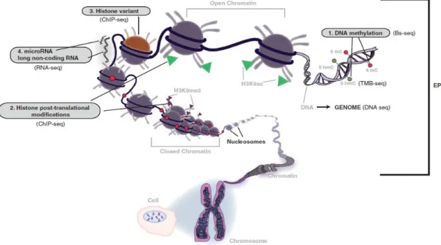
Epigenetic mechanisms can be grouped into at least four major types of modifications: DNA methylation, non-coding RNAs, histone post-translational modifications and histone variants (Figure 7).19 These mechanisms are essential for
normal development and maintenance of tissue-specific gene expression patterns in mammals. Abnormal epigenetic modifications were shown to contribute to common human diseases, including cancer 20. Indeed, deregulation of epigenetic mechanisms are
present in all types of tumors, contributing to its development and progression.21 The
reversible nature of epigenetic aberrations has led to the emergence of the promising field of epigenetic therapy.20
16 1.2.1.1 Non-coding RNAs
Non-coding RNAs (NcRNAs) are a class of RNA sequences that do not encode for proteins but are transcribed and biologically active. They are involved in a wide range of cellular functions, as chromosome dynamic control, splicing, RNA editing, translation inhibition and mRNA degradation. NcRNAs are composed by transcribed ultraconserved regions, small nucleolar RNAs, Piwi- interacting RNAs, large intergenic NcRNAs, long NcRNAs and microRNAs (miRNAs)22. MiRNAs are without doubt the best studied class.
They are small non-coding RNA molecules that can negatively regulate the expression of up to hundreds of messenger RNA (mRNA) targets23. In normal cells, microRNAs are
responsible for the fine-tuning of homeostatic gene expression and help to confer robustness to cellular processes, which is required for inducing and keeping cell fate decisions. MicroRNAs have also been implicated in the oncogenic transformation and their expression is altered at early stages of lung cancer10.
1.2.1.2 Histone Post-translational Modifications and Variants
In eukaryotic cells, chromatin is composed by DNA and histones, and it is in this context that transcription takes place. Histones are dynamic regulators of gene activity that undergo a wide variety of post-translational modifications influencing chromatin structure and recruitment of proteins complexes to DNA. Eight histones, one pair of each H2A, H2B, H3 and H4 constitute the basic unit of chromatin, the nucleosome. Histone H1 binds to the DNA between the nucleosomes. Post-translational modifications of histones are an epigenetic mechanism for the establishment and maintenance of gene activity, and consequently, regulate a wide range of cellular processes. The best characterized post-translational modifications are methylation, acetylation and phosphorylation24, 25
1.2.1.3 DNA methylation
DNA methylation is the best studied epigenetic modification being the major alteration that takes place during aging, embryogenesis and carcinogenesis26.
The DNA methylation consists in the addition of methyl group (CH3) to the 5’carbon
of a cytosine nucleotide preceding a guanine, originating 5-methylcytosine (5mC). This enzymatic addition is a normal process within cells25, 27. DNA methylation is catalyzed by a
series of sophisticated enzymes called DNA methyltransferases (DNMTs) that use S-adenosylmethionine (SAM) as a methyl donor group (Figure 8). Methylation in mammals primarily occurs in CpGs dinucleotides and only occasionally in non-CpG sites 17, 28, 29.
17
Figure 8 - Conversion of cytosine to 5-methylcytosine by DNA methyltransferase (DNMT). The methyl group
(CH3) is transferred from S-adenosylmethionine (SAM) to 5-carbon position of cytosine by DNMT25.
There are four known biologically active DNMTs in mammals: DNMT1, DNMT2, DNMT3a and DNMT3b.26 DNMIT1 is responsible for maintaining DNA methylation and
copies pre-existing methylation pattern onto the newly synthetized strand immediately after DNA replication. DNMT1’ function is to ensure that the methylation pattern of the parental cells is identically reproduced in each daughter cell. There is considerable evidence indicating an upregulation of DNMT1 in cancer. DNMT2 just appears to be involved in methylation of RNA and has shown only weak DNA methylation ability in vitro. DNMT3a and DNMT3b are the enzymes responsible for de novo methylation at CpG sites during embryogenesis targeting unmethylated CpG dinucleotides. Even though DNMT1 appear to be responsible for most of DNA-methylating capacity in cancer cells, specially at promoter regions, recent studies suggest an interaction between DNMT1 and DNMT3b to ensure propagation of methylation patterns during DNA replication in cancer cells21, 24-26
In normal mammalian cells, CpG islands are proximal to gene promoter regions (Figure 9). These regions are largely protected from DNA methylation and reside in restricted regions of open chromatin, or euchromatic states, which are favorable to gene transcription. In contrast, for most regions of the genome, such as gene bodies, repeat elements and pericentromeric regions of the genome, cytosines in CpG dinucleotides are methylated (Figure 8). This pattern of DNA methylation is common to the bulk of the human genome, which is packed as closed unfavorable for transcription29.
Global DNA hypomethylation occurs in cancer cells, which results in chromosomal instability and activation of proto-oncogenes. Concomitantly, abnormal methylation of gene promoter regions (hypermethylation) leads to tumor suppressor silencing (Figure 9). CpG islands, the major targets of DNA methyltransferases, are associated with the transcription start sites of almost half of human genes8. CG dinucleotides occur at a high
frequency in tumor suppressor genes (TSG) promoters, and these CpG islands are usually unmethylated or hypomethylated in normal cells, allowing the initiation of transcription. However, during malignant transformation, CpG islands became methylated
18 or hypermethylated, leading to repression of TSG transcription and potentiating oncogenesis30. Despite CpG islands cover approximately 1% of the total human genome,
they are present in >50% of human gene promoters which indicates their functional importance in transcriptional control24. Approximately 75% of all CpG dinucleotides in
normal cells are methylated in the human genome21.
Figure 9 - DNA methylation in normal and cancer cells. In normal cells, promoter Cpg islands are
unmethylated while in cancer cells, they have acquired aberrant DNA methylation, and consequently, transcriptional silencing. Adapted from Baylin, 2015 29.
The common occurrence of DNA hypermethylation in all types of cancer makes it an ideal biomarker, one that has been extensively investigated. DNA methylation is an inherently ideal substrate for cancer biomarker development for several key reasons. An advantage of DNA methylation over protein-based markers is that it is readily amplifiable and easily detectable using PCR-based approaches. Furthermore and, contrarily to cancer-specific DNA mutations, cancer-specific DNA hypermethylation occurs in defined regions, usually in or near the promoter of genes27. Moreover, the prevalence of CpG
methylation changes at literally hundreds of genes in a given tumor affords a vast number of possible tumor-specific targets for assay development. Finally, its association with gene silencing allows DNA methylation to serve as a substitute marker for gene expression, effectively providing a positive reading for negative expression21.
1.2.2 DNA methylation in Lung Cancer
Lung cancer develops through a multistage process involving permanent genetic alterations, dynamic epigenetic changes and environmental factors (Figure 10)10.
19
Figure 10 - Cancer is a result of the interaction between permanent genetic mutations and dynamic epigenetic
alterations. During cancer formation, a large number of epigenetic and genetic alterations lead to abnormal gene expression which evoke genome instability. Adapted from Mehta, 2015; Chen, 2014 10, 31.
DNA methylation plays a critical role in repressing gene activity of several TSG and maintaining genome stability. It has been demonstrated that two major changes in methylation status occur during carcinogenesis: regional promoter hypermethylation and genome wide hypomethylation. These methylation changes are critically associated with transcriptional silencing of the involved genes26.
It was suggested that LCa harbors a CpG island methylator phenotype, in other words a tumor phenotype characterized by widespread hypermethylation. This is not totally surprising, given the well-known upregulation of DNMTs in NSCLC23.
DNA methylation plays an important role in the etiology of LCa, therefore it might have a potential value as diagnostic and prognostic biomarkers. Expanding our understanding of how epigenetic events contribute to the genesis of LCa and how they can be translated into clinical relevant biomarkers and therapeutic targets will enhance our capacity to manage LCa patients and consequently reduce the heavy global burden of this critical disease23. Research on epigenetic has provided new insights of early cancer
development and progression, allowing increased knowledge of early stages of the disease and therapeutic interventions24.
1.2.2.1 Hypermethylated genes in Lung Cancer
Currently, there are many genes described as hypermethylated in LCa. Some of the most studied in this context and more informative include: APC, HOXA9, RARβ2,
20
Table 1 - Panel of genes hypermethylated in lung cancer.
Gene Locus Gene function References
APC 5q22.2
Adenomatous polyposis coli: TSG that acts as a negative regulator of Wnt and also is involved in cell migration and adhesion, transcriptional activation, and apoptosis
33
HOXA9 7p15-7p14.2
HOX genes encode transcription factors
that play essential roles in regulation of embryonic morphogenesis in animals
34
RARΒ2 9p24.2 Retinoic acid receptor beta is involved in
cell growth and differentiation.
5
RASSF1A 3p21.31
Ras association (RalGDS/AF-6) domain family member 1 is a putative TSG involved in apoptosis and cell cycle control
35
SHOX2 3q25.32
Homeobox family gene is involved in gene transcription with putative involvement in cell growth and differentiation
36, 37
TFPI2 7q22 TFPI2 decreases activation of
metalloproteinases
38, 39
1.2.2.1.1 APC
The adenomatous polyposis coli (APC) gene encodes for a cytoplasmic protein involved in cell signaling through Wnt pathway which plays an important role in cell-cycle regulation and apoptosis (Table 1)40, 41. The APC binds to β-catenin, axin and glycogen
synthase kinase 3β to form a large protein complex, in which β-catenin is phosphorylated and broken down, resulting in negative regulation of the Wnt signaling pathway40. An
impaired function of APC is often attributable to mutations within the coding sequence of the gene. This in turn leads to lack of degradation and nuclear accumulation of β-catenin which acts as a transcriptional activator, causing loss of cell growth control41. In addition,
APC is involved in cell motility through its association with microtubules and it also
stimulate guanine nucleotide exchange factor40. APC is considered a tumor suppressor
gene and high APC promoter methylation is significantly associated with a decrease in survival at LCa. Therefore APC is promise a biomarker of biologically aggressive NSCLC42.
21 1.2.2.1.2 HOXA9
HOX genes encode transcription factors that are critical in the regulation of
embryonic morphogenesis in animals (Table 1)43. HOX proteins are essential switches of
development stage-specific and cell-specific gene regulation. Thus, HOX proteins are key determinants of cell identity and potential targets during tumorigenesis44. Most of the
HOXA promoters contain highly dense CpG islands, and its methylation is integral to the
control of HOXA9 gene expression34. In LCa, HOXA9 displays higher methylation levels in
tumor tissues than normal tissues. Therefore, detection of aberrant HOXA9 gene hypermethylation might be useful as biomarker for the early diagnosis of primary LCa45.
1.2.2.1.3 RARβ2
Retinoic acid is known to interact with nuclear retinoic acid receptors and retinoic X receptors. Both receptors have three subtypes (alpha, beta and gamma) which have distinct functions46. Receptors of the RAR family are differentially expressed during
development and in adults life. There is strong evidence that RARβ plays a central role in epithelial cells growth regulation and in tumorigenesis47. The RARβ2 gene has two
different promoters and transcripts which are produced by alternative splicing. Most human cells express RARβ2 as predominant form. This isoform plays a central role in mediation of growth inhibition of different types of cancer cells and is responsible for coding vitamin A nuclear receptor which is required for normal cell growth and differentiation (Table 1)48-50. RARβ2 expression is not only lost or reduced in a large
percentage of LCa patients but also in a people with high risk to development LCa. Approximately 40% of NSCLC present loss or reduced RARβ246, 48.It was also described
that RARβ2 hypermethylation might be associated with short recurrence-free survival in never-smokers adenocarcinoma’s patients 46.
1.2.2.1.4 RASSF1A
Ras association domain family 1 isoform A (RASSF1A) is a tumor suppressor
gene whose inactivation is implicated in the development of many human cancers (Table 1). It is termed RASSF1A because the protein contains a putative Ras association domain35. The RASSF1A protein, encoded by one of the 8 splicing isoforms, termed 1A to
1H, is expressed in all normal human tissues, and carries several domains mediating protein-protein interactions with multiple partners51. RASSF1A modulates a broad range of
essential cellular functions for normal growth control, such cell motility, invasion, cell cycle and apoptosis, regulation of microtubules and maintenance of genomic stability35. Besides
this, it was also suggested that RASSF1A plays a role in tumor cell adhesion and motility51. This gene appears to suffer frequent transcriptional inactivation in tumor cells
22 due to aberrant promoter methylation35. It has been reported that RASSF1A gene is
frequently inactivated in primary LCa by the de novo methylation of CpG islands in the promoter region52. Due to this, RASSF1A represents an important potential diagnostic
target35.
1.2.2.1.5 SHOX2
The human Short Stature Homeobox 2 (SHOX2) has been identified as highly homologous to the short stature homeobox gene SHOX (Table 1). Homeobox genes code for proteins harboring specific DNA-binding homeodomains (homeoproteins), which play fundamental roles in vertebrate development and differentiation by acting as transcriptional regulators. SHOX2 is a known regulator of chondrocyte hypertrophy and act in skeleton development and embryogenic pattern formation36, 37. Genomic gain of
chromosome 3q involving the SHOX2 gene has been recognized as one of the most prevalent and significant chromosomal rearrangements in LCa36. SHOX2
hypermethylation was shown to be a useful biomarker for detecting SCC and SCLC with high specificity and sensitiviy36, 53. An in vitro diagnostic test for SHOX2 methylation has
recently become commercially available in Europe, and it was demonstrated that it helped pathologists in the diagnosis of LCa with sensitivity of 68% and 95% of specificity54.
1.2.2.1.6 TFPI2
The human Tissue Factor Pathway Inhibitor 2 (TFPI2) is a potential inhibitor of the plasmin within the extracellular matrix. Degradation of this protein was strongly associated with the progression of LCa (Table 1)38. TFPI2 is synthesized and secreted by endothelial,
mesenchymal and epithelial cells, monocytes/macrophages and the syncytiotrophoblast39.
TFPI2 decreases activation of metalloproteinases (MMP1, MMP3, MMP9 and MMP13)
which inhibit plasmin and trypsin leading to a reduction of tumor invasion and metastasis39, 55. Downregulation of TFPI2 promote migration and invasion of LCa lines.
Thus TFPI2 is considered a TSG in LCa and aberrant TFPI2 promoter hypermethylation may be a valuable prognostic marker38.