1 Doi:10.1016/j.micron.2008.05.008
Chromosome-specific microdissected centromecic sequences of C. cricetus
display a different physical location in the related species P. eremicus
Sandra Louzada a, Ana Pac¸o a, Svatava Kubickova b, Filomena Adega a, Henrique Guedes-Pinto a, Jiri Rubes b, Raquel Chaves a,*
a
Institute for Biotechnology and Bioengineering, Centre of Genetics and Biotechnology, University of Tra´s-os-Montes and Alto Douro (IBB/CGB-UTAD), Apdo 1013, 5001-801 Vila Real, Portugal
b
Veterinary Research Institute, Hudcova 70, 621 32 Brno, Czech Republic
Abstract
Constitutive heterochromatin comprises a substantial fraction of the eukaryotic genomes and is mainly composed of tandemly arrayed satellite DNAs (satDNA).
These repetitive sequences
represent a very dynamic and fast evolving component of genomes. In the present work we report the isolation of Cricetus cricetus (CCR, Cricetidae, Rodentia) centromeric
repetitive sequences from
chromosome 4 (CCR4/10sat), using the laser microdissection and laser pressure catapulting procedure, followed by DOP-PCR amplification and labelling. Physical mapping by
fluorescent in situ hybridisation of these sequences onto C. cricetus and another member of Cricetidae,
Peromyscus eremicus, displayed quite
interesting patterns. Namely, the centromeric sequences showed to be present in another C. cricetus
chromosome (CCR10) besides CCR4. Moreover, these almost chromosome-specific sequences revealed to be present in the P. eremicus genome, and most interestingly, displaying a ubiquitous scattered distribution throughout this karyotype. Finally and in both species, a co-localisation of CCR4/10sat with constitutive heterochromatin was found, either by classical C-banding or C-banding
2 sequential to in situ endonuclease
restriction.
The presence of these orthologous sequences in both genomes is suggestive of a phylogenetic proximity. Furthermore, the existence of common repetitive DNA sequences with a different chromosomal
location foresees the occurrence of an extensive process of karyotype restructuring somehow related with intragenomic movements of these repetitive sequences during the evolutionary process of C. cricetus and
P. eremicus species.
Keywords: Satellite DNA, Rodentia, Orthologous sequences, Constitutive heterochromatin, Genome evolutionary restructuring
Introduction
A substantial proportion of the higher eukaryotes genome consists of constitutive heterochromatin (CH),preferentially found in (peri)centromeric regions (see Corridini et al 2007; Rossi et al. 2007), although telomeric and interstitial positions have been also described in different species (see Adega et
al. 2007; Meles et al. 2007). This genomic
fraction is mainly composed by highly repetitive sequences of satellite DNA (SatDNA) (Jonh 1988; Chaves et al. 2004b), organized into long and uninterrupted tandem
arrays of more or less well defined repeat units (Charlesworth et al. 1994).
In a general way, eukaryotic SatDNA sequences are characterized by a highly dynamic molecular behaviour, promoted by concerted evolution, which leads to rapid change between repeat sequences of different species, throughout sequence alteration (promoted by nucleotide substitutions), amplification of new variants during speciation, and intragenomic movements (Ugarkovic and Plohl 2002; Hamilton et al. 1992). In this way, SatDNA forms the most
3 rapidly evolving compartment of the genome,
making these sequences valuable evolutionary markers (see Saffery et al. 1999). This characteristic pattern of occurrence allows that some taxonomic groups enclose specific SatDNA sequences, being these sometimes species-specific (Jobse 1995; Nijman and Lenstra 2001). Moreover, it is also recognized that different SatDNA families can coexist in the same genome, forming a SatDNA library (see Hamilton et al. 1992). According with the model proposed by Fry and Salser (1977), related species share a collection of SatDNA and the existence of species-specific satellite profiles is explained as a consequence of fluctuations in the copy number of SatDNA within a library. In some taxa, however, it’s observed that the evolution of SatDNA families proceeds in a very slowly fashion way (Mravinac et al. 2002; Li and Kirby 2003; Cafasso 2003), meaning that species separated by several million years may also share orthologous repetitive sequences. These few cases of repetitive sequences
conservation indicate a complex behaviour of this genome fraction. The molecular analysis of repetitive sequences and their physical mapping in chromosomes of different species, have shown the importance of these analysis in measuring species phylogenetic relationships, while also clarifying important aspects of both repetitive sequence and genome evolution (see Lander et al. 2001; Ugarkovic and Plohl 2002).
Given the high dynamic of SatDNA, it is believed that this repetitive sequences play an important role in the mammal genome evolution by promoting chromosomal rearrangements (see Wichman et al. 1991; Reig et al. 1992; Schluter et al. 1997; Slamovits et al. 2001). In accordance, several works discuss the involvement of CH in the occurrence of chromosomal evolution, suggesting that these regions are considered as hotspots that preferentially enable structural chromosome rearrangements (Yunis and Yasmineh 1971; Peacock et al. 1982; John 1988; Chaves et al. 2004b). Recent
4 studies focused on molecular characterization
of the breakpoint regions (see Garagna et al. 2001; Li et al. 2002; Locke et al. 2003; Kehrer-Sawatzki et al. 2005; Ruiz-Herrera et
al. 2006) have demonstrated that the location
of evolutionary breakpoint regions is coincident with the location of regions rich in repetitive sequences.
The C-banding technique is extremely useful for identification of CH in chromosomes, however the location of CH determined by this technique, and the distribution of SatDNA sequences ascertained by in situ hybridization, are often, but not always, coincident within the chromosomes (reviewed by Jonh 1988). The in situ restriction endonuclease (RE) digestion with sequential C-banding technique proved to be very useful in the understanding of the mechanisms involved in the CH evolution in different genomes (reviewed by Gosálvez et al. 1997). Moreover this technique can also reveal CH bands not evidenced by conventional C-banding, denominated cryptic C-bands (see
Chaves et al. 2004b; Adega et al. 2005; Adega et al. 2007), whose location can be coincident with the SatDNA distribution revealed by in situ hybridization.
The two studied species, the common hamster
Cricetus cricetus (CCR), and the cactus
mouse Peromyscus eremicus (PER), belong to the Cricetidae family (Order Rodentia) and display diploid numbers of 22 and 48 chromosomes, respectively. C. cricetus enclose a nearly meta/submetacentric karyotype, whose CH seems to be greatly located at the (peri)centromeric regions, exhibiting the majority of the chromosomes two very large blocks at this location (see Gamperl et al. 1976; Paço et al. submitted). The P. eremicus comprise a very distinct karyotype organization, with only submetacentric chromosomes. This karyotype also displays great amounts of CH, especially located at the (peri)centromeric regions, being the p arms of the majority of chromosomes almost entirely heterochromatic (see Pathak et
5
submitted). In both species the CH was
already characterized by in situ RE digestion with sequential C-banding, being identified several cryptic C-bands (Paço et al.
submitted).
In the present work we report the isolation of CCR centromeric repetitive sequences using the laser microdessection and laser pressure
catapulting procedure. The in situ
hybridization of this probe onto C. cricetus and P. eremicus display a different physical distribution. The existence of common repetitive DNA sequences with different chromosomal location in these related genomes is discussed.
Materials and methods
Chromosome preparations
Metaphase chromosomal spreads were prepared from a fibroblast cell line of the rodent species Cricetus cricetus (CCR) and
Peromyscus eremicus (PER), both part of the
cell and tissue collection housed at the Department of Sistematics and Evolution, Muséum National d’Histoire Naturelle (MNHN). Standard cell culture from both rodent species was performed according to described by Chaves et al. (2004a) in order to prepare fixed chromosome spreads. The nomenclature of C. cricetus (2n=22) and P.
eremicus (2n=48) chromosomes is according
to Gamperl et al. (1976) and Com. Comm. Stand. Chromos. Peromyscus (1977), respectively.
GTD-banding
Air dried slides were aged at 65ºC for 5 h or overnight and then submitted to standard G-banding procedures with trypsin (Sumner et al. 1971). As the chromosome slides proceeded sequentially to C-banding they were fixed with formaldehyde. Briefly, dry slides were placed in 1PBS solution (25 min) followed by fixation in 3% formaldehyde (Sigma)/1PBS
6 (room temperature) for 20 min. After, slides
were dehydrated for 5 min in 70%, 90% and 100% chilled ethanol and air dried. DAPI was used for staining (instead of routine Giemsa) in order to obtain a better contrast (Chaves et al. 2002). The inversion of DAPI colour in Adobe Photoshop revealed the chromosome G-banding (GTD-G-banding, G-bands by trypsin with DAPI) permitting its identification.
In Situ RE digestion
Air dried slides were aged at 65ºC for 6 h and then were submitted to in situ RE digestion. The five restriction enzymes used (AluI, BamHI, DraI, PslI and RsaI) were diluted in buffers indicated by the manufacter (Invitrogen Life Technologies), and final concentrations of 30 U per 100 µl were obtained. A total of 100 µl of each of these solutions were placed on slides and afterwards covered with coverslips. The slides were incubated in a moist chamber for 16h at 37ºC. Control slides were also prepared according with the mentioned procedures but they were incubated only with
buffer. Slides were then washed in distilled water and air-dried. Once these slides proceeded to C-banding techniques they were fixed with formaldehyde, as described above for GTD-banding. Ultimately the slides were stained with DAPI (the inversion of the DAPI colour revealed the RE-banding). The residual bands obtained after the endonuclease digestion were suitable for chromosome identification.
CBP-banding sequential to G-bands or RE-bands
The C-banding technique was performed sequentially to G-banding or RE-banding, being performed after distaining the slides. CBP-banding [C-bands by Barium Hydroxide using Propidium Iodide (PI)] was done according the standard procedure of Sumner (1972) but with PI as counterstaining. Briefly, the slides were submitted to routine C-banding with classical treatment times reduced, approximately to half: Hydrocholic Acid (0.1M) 20 min, Barium Hydroxide (5%
7 solution) 7 min and 2× Saline Solution Citrate
(2×SSC: 0.3mol/lNaCl, 0.03 mol/l Sodium Citrate) at 60ºC for only 40 min. The slides where then counterstained with PI (1,5µl/ml).
Microdissection, preparation of DNA probes and fluorescent in situ hybridization
The PALM MicroLaser system (P.A.L.M. GmbH, Bernried, Germany) was used for chromosome dissection and collection. The referred system consists of a 337-nm nitrogen laser coupled to the light path of an inverted microscope (Olympus) and focused through an oil immersion objective (100x magnification), with high numerical aperture to yield a spot size of less than 1 µm in diameter. About 10 chromosome centromeres from C. cricetus chromosome number 4 were microdissected and catapulted by a single laser pulse directly into the cap of a PCR tube, to which 2 µl PCR oil had been applied. The microdissected material was then dissolved in 20 µl 10mmol/l Tris-HCl pH 8,8 in the cap, that was placed in the respective tube and submitted to
centrifugation. Probes were generated and labelled with digoxigenin-11-d-UTP (Roche, Molecular Biochemicals) after DOP-PCR amplification, as described by Kubickova et al. (2002). Hybridization was carried out in moist chamber at 37ºC overnight, and the most stringent post-hybridization wash was 2× SSC at 37ºC, allowing sequences with more than 85% similarity to remain hybridized. Digoxigenin-labeled probes were detected with anti-digoxigenin (Roche, Molecular Biochemicals).
Chromosome observation
Chromosomes were observed with a Zeiss Axioplan 2 Imaging microscope, coupled to an Axiocam digital camera with AxioVision software (version Rel. 4.5 – Zeiss). Digitized photos were prepared for printing in Adobe Photoshop (version 5.0); contrast and color optimization were the functions used and affected the whole of the image equally.
8
Results and Discussion
In the present work we report the isolation of
C. cricetus centromeric repetitive sequences
from chromosome 4, using the laser microdissection and laser pressure catapulting procedure. The in situ hybridization of this probe onto C. cricetus and P. eremicus chromosomes show that these sequences are present in the genome of this two species, suggesting their existence in a common ancestor and thus these sequence variants can be considered as orthologous sequences. Although, they display a very different chromosome location, as it can be observed in Fig. 1 and Fig. 2. With few exceptions, the genomic distribution pattern of the CCR4cent sequence in the two genomes is co-localized with the distribution of CH, evidenced by classic C-banding or C-banding after in situ RE digestion. Moreover, in P. eremicus chromosomes, it is also important to refer that this sequence is more frequently co-localized with cryptic C-bands, what proves the importance of in situ RE digestion and
sequential C-banding technique in the analysis of physical distribution of this type of sequences.
Fluorescence in situ hybridization onto CCR metaphases
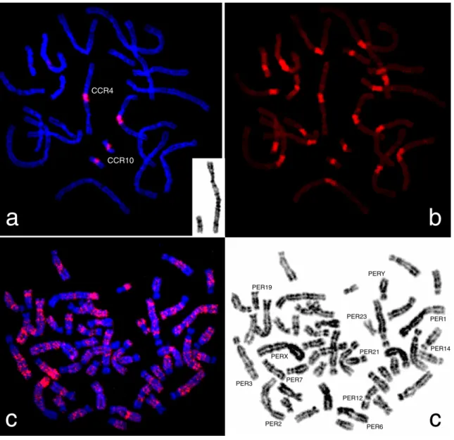
After the hybridization of the microdissection obtained probe, CCR4cent, onto C. cricetus chromosome preparations, it was observed a strong hybridization signal on (peri)centromeric region of CCR4 and CCR10 chromosomes (Fig. 1). This interesting feature suggests the existence of repetitive sequences with high homology in these two chromosomes (more than 85%). Moreover, this hybridization pattern also reveals a co-localization of the CCR4cent sequences with (peri)centromeric CH, evidenced by classic C-banding. The present results indicate a certain chromosome specificity of these sequences, making this the first report on chromosome-specific sequences in C. cricetus (as far as we know). A possible explanation for the origin of chromosome-specific sequences results from
9 the occurrence of mutations in the ancestral
sequence, followed by independentl amplification in the descendent repeat units in different chromosomes (Fátyol et al. 1994). Other chromosome-specific sequences have been described in different rodent species, namely in Mus musculus (Boyle and Ward, 1992), Rattus norvegicus (Essers et al. 1995),
Cricetulus griseus (Fátyol et al. 1994) and Mesocricetus auratus (Yamada et al. 2006),
being the last two mentioned species from the same family and subfamily (Cricetidae, Cricetinae) of C. cricetus.
Fluorescence in situ hybridization onto PER metaphases observe
The chromosomal distribution of the CCR4cent probe in the genome from the same family member, P. eremicus, revealed interesting results. No hybridization signal was detected in (peri)centromeric regions. It was observed an interspersed hybridization pattern in almost all P. eremicus chromosomes (Fig. 2), except for PER17, PER20, PER21 and
PER22 with complete absence of hybridization signal (Fig. 3). Moreover, when karyotypes were built, a banding like pattern was observed commonly in each of the homologues. In Fig. 3 is presented a detailed analysis of CCR4cent hybridization pattern in P. eremicus
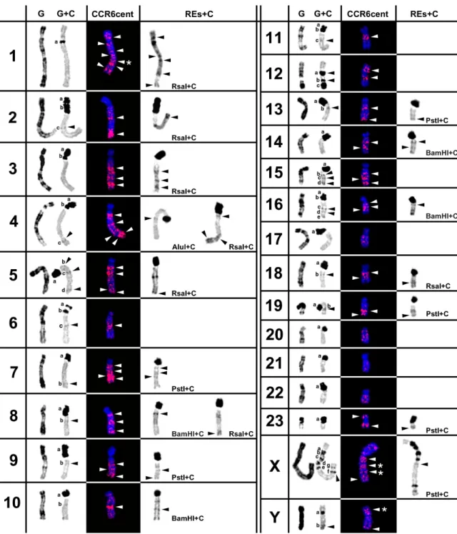
chromosomes. In the left column of this figure, it is possible to observe the G- and C-banding of each chromosome pair, which are the controls for the subsequent comparative analysis of the different RE actions. In the other columns, CCR4cent and REs+C, is presented the hybridization pattern of CCR4cent probe (white arrowheads) and the action of several REs in P. eremicus CH, respectively. The analysis of the present results allows to verify that the genomic distribution of the CCR4cent sequences in this species chromosomes is co-localize with the distribution of CH, revealed by classic C-banding or C-C-banding sequential to in situ RE digestion (Fig. 3). A more complete analysis demonstrated that in some chromosomes, PER6, PER11, PER12, PER15 and PERY,
10 these sequences are only co-localized with CH
bands revealed by classic C-banding (black arrowheads in control chromosomes column). For example, the CCR4cent signals observed in PER12 chromosome corresponded to a and
b C-bands revealed by classic C-banding (Fig.
3). Nevertheless in other chromosomes, PER1, PER3, PER10, PER14 and PER23, this probe only co-localizes with cryptic C-bands (black arrowheads in RES+C column), evidenced by the didestion withseven REs, namely AluI, ApaI, BamHI, DraI, HaeIII, PstI and RsaI. It should be mentioned that the hybridization signals corresponding to cryptic C-, however in this figure is only presented the action of one RE for each corresponding C-band (e.g. in PER3 the CCR4cent signal closer to the centromere corresponds to a cryptic C-band revealed by the seven REs used, though is only presented the corresponding band revealed by RsaI). At last, concerning chromosomes PER2, PER4, PER5, PER7, PER8, PER9, PER13, PER16, PER18, PER19 and PERX, the obtained hybridization signal corresponds to
C-bands evidenced by classical C-banding and C-banding after in situ RE digestion, with the previously mentioned REs.. In some specific chromosomes, PER1 and sex chromosomes (bands marked with an *), the CCR4cent signal did not correspond to any cryptic C-band neither to any C-C-band observed in control chromosomes. The high correspondence between the chromosome location of the isolated sequences, and P. eremicus CH, allows the suggestion that the non-corresponding hybridization signal identified (neither with classical C-band nor cryptic bands) must be CH sequences as well. The enlargement of the restriction panel, using other REs with sequential C-banding, possibly will reveal more cryptic C-bands that could be co-localize with these signals.
Comparative analysis of CCR4cent physical distribution in genome of the two Cricetidae species
C. cricetus and P. eremicus belong to the
11 different subfamilies, Cricetinae and
Neotominae, respectively. The presence of the same repetitive sequences in the genomes of these related species imply their existence, at least, in a common ancestor of the two subfamilies. Fossil records and molecular data suggest the origin of Cricetinae and Neotominae subfamilies to be in the middle Miocene (McKenna and Bell 1997; Baskin 1989; Neumann et al. 2006), meaning that this sequences ages at least from this epoch. To determine weather these sequences are older, or if they are present in other Cricetidae subfamilies, we would have to enlarge our study to other rodent species.
The CCR4cent orthologous sequences present however, a different chromosome location in the two species. According to the results obtained and regarding the parsimony rules, it’s proposed that these sequences had originally a (peri)centromeric position, as can be observed in C. cricetus chromosomes, and later assumed an interspersed pattern/location as it can be observed in P. eremicus
chromosomes. This fact can be explained (1) as the result of karyotype restructuring after the radiation of these species, as weel (2) as the consequence of intragenomic movements. Regarding the first hypothesis, it is assumed that P.eremicus karyotype evolution results mainly from pericentromeric inversions (as well from heterochomatic additions and deletions) (Deaven et al. 1977; Hamilton et al. 1992), which could promote the repositioning of these sequences from the cetromere to other chromosomal positions. On the other hand, regarding C. cricetus karyotype evolution it is thought that it involved Robertsonian translocations (Gamperl et al. 1976), what is consistent with the manteinance of these sequences in a (peri)centromeric position. Another possible scenario is related with the occurrence of intragenomic movements by means of recombinational events, such as rolling circle amplification. During the rolling circle amplification it is suggested that extrachromosomal circles originated by intra strand recombination between repeat units of
12 the same array, undergo rolling circle
amplification followed by its incorporation in original genome position or in a new location (Walsh 1987; Hamilton et al. 1992). The occurrence of this process can explain how CCR4cent sequences assumed the chromosomal location present in P. eremicus genome. Wichman et al. (1991) postulated that rapidly evolving repetitive sequences, promote chromosomal rearrangements by means of their intragenomic movements among nonhomologous chromosomes and between different chromosomal fields. Following this reasoning line, it can be proposed that the pericentric inversions occurred during P.
eremicus karyotype restructuring can also be
the consequence of the presence and the high dynamics of the repetitive sequences.
A similar study was performed in the rodent
Ctenomys by Rossi et al. (1995) regarding
RPCS (Repetitive PvuII Ctenomys Sequence). They observed that Ctenomys species possessing karyotypes closer to the hypothesized ancestral have pericentromeric
heterochromatin (containing RPCS), whereas more derived karyotypes show also interstitial and full arm localization. In this way, can be also proposed that P. eremicus has a more derivative karyotype than C. cricetus,
originated by a larger number of complex chromosomal rearrangements. The construction of comparative maps between these species and other rodents, will certainly clarify the previous idea.
Conclusions and General Remarks
The present work demonstrated that centromeric repetitive sequences isolated from
C. cricetus chromosome 4 using laser
microdissection procedure, are also present in the genome of the related species, P. eremicus. This implies their existence in a common ancestor, being considered the orthology of these variant sequences. Moreover, if repetitive sequences form the most rapidly evolving compartment of the genome, being verified that close species often have non-orthologous satellite DNAs at homologous chromosomal
13 locations (Csink and Henikoff 1998), the
presence of this sequence in the both genomes suggests a phylogenetic proximity between the two species. The different physical distribution of the CCR4cent sequences, a (peri)centromeric location in C. cricetus and a noncentromeric location in P. eremicus, suggests that this last condition could have result from an extensive process of karyotype restructuring and occurrence of intragenomic movements during the evolution of these sequence.
Laser microdissection and laser pressure catapulting followed by DOP-PCR amplification along with labelling, revealed to be a very effective procedure for the isolation of repetitive sequences, allowing its study in different species genomes.
The detailed molecular analysis of these sequences and the extension of its search to other related genomes, will certainly shed light on evolutionary history of the these repetitive sequences and simultaneously, contribute to
the clarification of the phylogenetic relationships of the species sharing them.
References
Adega F, Chaves R and Guedes-Pinto H (2005) Chromosome Restriction Enzyme Digestion in Domestic Pig (Sus scrofa) Constitutive heterochromatin arrangement. Genes Genet. Syst. 80: 49-56.
Adega F, Chaves R and Guedes-Pinto H (2007) Constitutive heterochromatin characterization of White-lipped and Collared peccaries (Tayassuidae). Journal of Genetics 86:19-26. Ananiev EV, Phillips RL and Rines HW (1998)
Complex structure of knob DNA on maize chromosome 9. Retrotransposon invasion into heterochromatin. Genetics 149: 2025-2037. Charlesworth B, Sniegowski P and Stephan W (1994)
The evolutionary dynamics of repetitive DNA in eukaryotes. Nature 371: 215-220.
Chaves R, Adega F, Santos S, Guedes-Pinto H and Heslop-Harrinson JS (2002) In situ hybridization and chromosome banding in mammalian species. Cytogenet Genome Res
96: 113-116.
Chaves R, Frönicke L, Guedes-Pinto H and Wienberg J (2004a) Multidirectional chromosome painting between the Hirola antelope (Damaliscus
14
hunteri, Alcelaphini, Bovidae), sheep and human. Chromosome Research 12: 495-503. Chaves R, Santos S and Guedes-Pinto H (2004b)
Comparative analysis (Hippotragini versus caprini, Bovidae) of X-chromosome’s constitutive heterochromatin by in situ restriction endonuclease digestion: X-chromosome constitutive heterochromatin evolution. Genetica 121: 315-325.
Corradini N, Rossi F, Giordano E, Caizzi R, Vern F and Dimitri P (2007) Drosophila melanogaster as a model for studying protein-encoding genes that are resident in constitutive heterochromatin. Heredity 98: 3-12.
Deaven LL, Vidal-Rioja L, Jett JH and Hsu TC (1977) Chromosomes of Peromyscus (rodentia, cricetidae). VI. The genomic size. Cytogenet Cell Genet. 19(5):241-9.
Dimitri P, Corradini N, Rossi F and Verní F (2004) The paradoxo of functional heterocromatin. BioEssays 27: 28-41.
Dimitri P, Verní F, Mei E, Rossi F and Corradini N (2005) Transposable elements as artisans of the heterochromatic genome. Cytogenet Genome Res 110: 165–172.
Duffey P A (1972) Chromosome variation in Peromyscus: a new mechanism. Science 176, 1333-1334.
Gamperl R, Vistorin G and Rosenkranz W (1976) A comparative analysis of the karyotypes of Cricetus cricetus and Cricetulus griseus. Chromosoma 55: 259-265.
Gosálvez J, López-Fernández C, Goyanes R and Mezzanotte V (1997) Chromosome differentiation using nucleases: an overview, pp. 23–49 in: Chromosomes Today, Vol. 12, edited by N. Henriques-Gil, J.S. Parker & M.J. Puertas. Chapman & Hall, London.
Hsu TC and Arrighi FE (1966) Chromosomal evolution in the genus Peromyscus (Cricetidae,Rodentia). Cytogenetics 5: 355-359.
Jobse C, Buntjer JB, Haagsma N, Breukelman HJ, Beintema JJ and Lenstra JA (1995) Evolution and recombination of bovine DNA repeats. Journal of Molecular Evolution 41:277-283. John B (1988) The biology of heterochromatin. In:
Heterochromatin (Verma, R.S., ed.). Cambridge, Cambridge University Press.
Matthey R (1952) Chromosomes des Muridae (Microtinae et Cricetinae). Chromosoma 5: 113-138.
Meles S, Adega F, Guedes-Pinto H and Chaves R (2007) The karyotype and sex chromosomes of Praomys tullbergi (Muridae, Rodentia): A detailed characterization. Micron (published online) doi:10.1016/j.micron.2007.07.002.
15
Nijman IJ and Lenstra JA (2001) Mutation and recombination in cattle satellite DNA: a feedback model for the evolution of satellite DNA repeats. J. Mol Evol 52: 361-371.
Paço A, Adega F, Guedes-Pinto H and Chaves R (2007) Chromosome Restriction Enzyme Digestion in
Cricetus cricetus, Peromyscus eremicus
(Cricetidae) and Praomys tullbergi (Muridae) Rodentia species: insights into chromosome evolution. Journal of Genetics (submitted). Pathak S, Hsu TC and Arrighi FE (1973) Chromosomes
of Peromyscus (Rodentia, Cricetidae). IV. The role of heterochromatin in karyotypic evolution. Cytogenet Cell Genet 12:315–326. Peacock WJ, Dennis ES and Gerlach WL (1982) DNA
sequence changes and speciation. Prog Clin Biol Res 96: 123-142.
Pieczarka JC, Nagamachi CY, Muniz JAPC, Barros RMS and Mattevi MS (1998) Analysis of constitutive heterochromatin of Aotus (Cebidae, Primates) by restriction enzyme and fluorochrome bands. Chromosome Res. 6: 77– 83.
Reig OA, Massarini AL, Ortells MO, Barros MA, Tiranti SI and Dyzenchauz FJ (1992) New karyotypes and C-banding of subterranean rodents of the genus Ctenomys (Caviomorpha, Octodontidae) from Argentina. Mammalia 56: 603-623.
Rossi F, Moschetti R, Caizzi R, Corradini N and Dimitri P (2007) Cytogenetic and Molecular Characterization of Heterochromatin Gene Models in Drosophila melanogaster. Genetics
175: 595–607.
Schluter D, Price T, Mooers A and Ludwig D (1997) Likelihood of ancestor states in adaptative radiation. Evolution 51: 1699-1711.
Slamovits CH, Cook JA, Lessa EP and Rossi MS (2001) Recurrent amplifications and delections of satellite DNA accompanied chromosomal diversification in South American tucos-tucos (genus Ctenomys, Rodentia: Octodontidae): a phylogenetic approach: Mol Biol Evol 18: 1708-1719.
Slamovits CH and Rossi MS (2002) Satellite DNA: Agent of Chromosomal Evolution in Mammals. A review. Mastozoologia Neotropical / J. Neotrop. Mammal 9(2): 297-308.
Sumner AT, Evans HJ and Buckland RA (1971) New technique for distinguishing between human chromosomes. Nature 232:31-32.
Sumner AT (1972) A simple technique for demonstrating centromeric heterochromatin. Exp. Cell Res. 75: 304-306.
Sumner AT (2003) Chromosomes Organization and Function. Blackwell Publishing Company. United Kingdom. P. 287.
16
Wichman HA, Payne CT, Ryder OA, Hamilton MJ, Maltbie M and Baker RJ (1991) Genomic distribution of heterochromatic sequences in equids: Implications to rapid chromosomal evolution. Journal of Heredity 82:369-377.
Yunis JJ and Yasmineh WG (1971) Heterochromatin, satellite DNA, and cell function. Structural DNA of eucaryotes may support and protect genes and aid in speciation. Science 174: 1200-1209.
17 Fig. 1. Representative in situ hybridisation of CCR4/10sat sequences from Cricetus cricetus
(CCR) onto C. cricetus chromosomes. CCR4 and CCR10 were DAPI inverted for chromosome identification (a). The same metaphase after C-banding (b). In situ hybridisation of CCR4/10sat onto Peromyscus eremicus chromosomes (c). The same metaphase was DAPI inverted for chromosome identification, where some of the most interesting chromosomes are identified (d).
18 Fig. 2. Table showing the in situ hybridisation pattern of CCR4/10sat in Peromyscus eremicus
chromosomes. G- and C-banding of each P. eremicus chromosomes are shown in the left column. The letters (a–g) represent the C-bands according to order of appearance in each chromosome. In the other columns it is possible to observe the hybridisation pattern of the CCR4/10sat probe and the constitutive heterochromatin bands produced by in situ restriction endonuclease digestion followed by banding (RE + banding). The black arrowheads indicate classical and cryptic C-bands that co-localize with CCR4/10sat sequences. The white arrowheads evidence the CCR4/10sat signal in P. eremicus chromosomes.