Universidade de Aveiro
Universidade NOVA de Lisboa
2017
Departamento de Ciências Médicas
Faculdade de Ciências Médicas
Roberto Alexandre
dos Santos Dias
Caracterização do papel da Gαo na neuritogénese:
um destaque para o complexo Gαo-Proteina
Precursora de Amilóide
Characterization of Gαo role on neuritogenesis: a
focus on the Gαo-Amyloid Precursor Protein
complex
Tese apresentada à Universidade de Aveiro para cumprimento dos requisitos necessários à obtenção do grau de Doutor em Biomedicina, realizada sob a orientação científica da Doutora Sandra Isabel Moreira Pinto Vieira, Professora Auxiliar Convidada do Departamento de Ciências Médicas da Universidade de Aveiro e da Doutora Odete Abreu Beirão da Cruz e Silva, Professora Auxiliar Convidada com Agregação do Departamento de Ciências Médicas da Universidade de Aveiro
Este trabalho é financiado por Fundos FEDER através do Programa
Operacional Factores de
Competitividade – COMPETE e por Fundos Nacionais através da FCT – Fundação para a Ciência e a Tecnologia no âmbito do projeto «PTDC/SAU-NMC/111980/2009»; por Fundos da FCT e do programa POPH/FSE no âmbito da bolsa individual «SFRH/BD/90996/2012»; e pelo Instituto de Biomedicina – iBiMED «UID/BIM/04501/2013».
o júri
presidente Doutor João Carlos Matias Celestino Rocha
professor Catedrático do Departamento de Química da Universidade de Aveiro
Doutor Carlos Jorge Alves Miranda Bandeira Duarte
professor Associado com Agregação do Departamento de Ciências da Vida da Universidade de Coimbra
Doutora Fernanda Cristina Gomes de Sousa Marques
investigadora Auxiliar do Instituto de Investigação em Ciências da Vida e Saúde da Universidade do Minho
Doutora Sandra Maria da Costa Tavares Rebelo
professora Auxiliar Convidada do Departamento de Ciências Médicas da Universidade de Aveiro
Doutora Sandra Isabel Moreira Pinto Vieira
agradecimentos À professora Sandra Vieira e à professora Odete da Cruz e Silva pela orientação e apoio durante a elaboração deste trabalho
À Joana Rocha, Regina Cerqueira e Bruno Gonçalves, sem os quais este trabalho não teria sido possível.
Aos meus colegas do CBC e iBiMED que fizeram parte deste percurso, em especial à Ana Marote, Catarina Pinho, Filipa Martins, Joana Oliveira, Joana Serrano, Liliana Carvalho, Luisa Bastos, Mariana Santos, Marlene Marafona, Patricia Tenreiro e Soraia Martins.
À Juliana, Joana, Miguel e Tiago. Ao Hugo, João, Marco e Fred.
Ao Joni, João, Ana Isabel, Denise e Patricia.
To Hideo Kojima, Hidetaka Miyazaki, Neil Gaiman, Andrzej Sapkowski, Stephen King, and many others that, without knowing, made this journey so much easier.
À minha família pelo apoio que sempre me deram.
palavras-chave diferenciação neuronal, fosforilação, STAT3, ERK1/2, proteassoma, lisossoma, degradação de proteínas, ImageJ, células SH-SY5Y
resumo Gαo é a subunidade Gα mais abundante no cérebro, no entanto, as suas funções especificas ainda estão longe de serem claras. Estudos das vias de sinalização moduladas pela Gαo têm exposto potenciais papéis para a Gαo no
desenvolvimento do sistema nervoso, especialmente em neuritogénese. A caracterização do interactoma da Gαo também tem sido crucial para uma melhor compreensão das funções desta proteína. Uma das proteínas interatoras da Gαo é a proteina precursora de amiloide (APP), uma proteina que se encontra
envolvida em várias funções fisiológicas, como sobrevivência celular, migração neuronal, e diferenciação neuronal. APP também é mais conhecida pelo seu envolvimento da Doença de Alzheimer (AD). APP liga-se e ativa a Gαo, uma interação que tem sido associada com migração neuronal e AD. No entanto, até agora, não existem estudos publicados que investiguem a interação APP-Gαo na neuritogénese. O principal objetivo deste trabalho foi então caracterizar o papel da Gαo na neuritogénese através do foco na investigação dos efeitos
neuritogénico do complexo Gαo-APP.
Primeiro, através do uso de células de neuroblastoma SH-SY5Y, estudámos o impacto da fosforilação da serina 655 (S655) da APP na interação APP-Gαo. Através do uso de dois mutantes da APP que mimetizam o estado fosforilado e desfosforilado da S655, SE e SA APP respetivamente, demonstrámos que a fosforilação da S655 aumenta a eficiência da APP em ligar e ativar a Gαo. Além disso, apresentamos provas de que a APP modula os efeitos neuritogénicos da Gαo num mecanismo fosfo-dependente. Neste mecanismo neuritogénico, a sinalização da STAT3 e ERK1/2 exibiram uma ativação sequencial, com a STAT3 participando na formação de novos processos e a ERK1/2 na elongação dos mesmos. Apresentamos ainda dados que suportam um papel da APP-Gαo na dendritogénese em culturas neuronais primárias.
A segunda parte deste trabalho focou-se na investigação de mecanismos
envolvidos no controlo dos níveis proteicos celulares da APP e Gαo. Identificámos o lisossoma como um novo processo pelo qual a Gαo é degradada em
consequência da sobre expressão da SA APP. Também mostramos provas de que este mecanismo pode fazer parte de autofagia mediada por chaperonas, através do qual a sinalização da APP-Gαo poderá estar a ser regulada. Finalmente, devido ao nosso interesse em estudar diferenciação neuronal e à falta de ferramentas para este estudo em imagens de contraste de fase, criámos o NeuronRead, uma macro do ImageJ capaz de analisar de forma
semiautomática imagens neuronais de contraste de fase e fluorescência. NeuronRead foi extensivamente validado, e usado para monitorizar a diferenciação de células SH-SY5Y após modulação da atividade da Gαo. Com este trabalho contribuímos com novos dados que ajudam na compreensão da função e regulação do complexo Gαo-APP, e disponibilizamos para a
keywords neuronal differentiation, phosphorylation, STAT3, ERK1/2, proteasome, lysosome, protein degradation, ImageJ, SH-SY5Y cells
abstract Gαo is the most abundant Gα subunit present in the brain, however, its specific functions are still far from clear. Studies of the signaling pathways modulated by Gαo have uncovered potential roles for Gαo in the development of the nervous system, especially in neuritogenesis. The characterization of Gαo interactome has also been crucial for the better understanding of this protein’s functions. One of the Gαo interacting proteins is the amyloid precursor protein (APP), a protein that is involved in several physiological functions, such as cell survival, neuronal migration, and neuronal differentiation. APP is also best known for its involvement in Alzheimer’s Disease (AD). APP binds and activates Gαo, an interplay that was associated with neuronal migration and AD. However, so far, no published study has investigated the effects of the APP-Gαo interaction on neuritogenesis. The main goal of this work was thus to characterize Gαo role on neuritogenesis by focusing the research on the neuritogenic effects of the Gαo-APP complex. First, by using SH-SY5Y neuroblastoma cells, we studied the impact of APP serine 655 (S655) phosphorylation on the APP-Gαo interaction. Through the use of two APP mutants mimicking the phosphorylated and dephosphorylated state of S655, SE and SA APP respectively, we have demonstrated that S655
phosphorylation increases APP efficiency to bind and activate Gαo. Moreover, we present evidence that APP modulates Gαo neuritogenic effects in a phospho-dependent mechanism. STAT3 and ERK1/2 signaling displayed a sequential activation on this neuritogenic mechanism, with STAT3 being mainly involved in the formation of new processes, while ERK1/2 was more involved in neuritic elongation. We also present data supporting a role for the APP-Gαo complex on dendritogenesis in rat primary neuronal cultures.
The second part of this work focused on unraveling the mechanisms involved in the control of APP and Gαo cellular protein levels. We identified the lysosome as a new pathway by which Gαo is degraded, as an effect of SA APP overexpression. We also provide evidence that this degradation mechanism might be part of chaperone-mediated autophagy, through which APP-Gαo signaling might be regulated.
Finally, due to our interest in studying neuronal differentiation and a lack of reliable tools to analyze phase contrast images, we developed NeuronRead, an ImageJ macro capable of semi-automated analysis of both phase contrast and
fluorescence neuronal images. NeuronRead was extensively validated and used to monitor SH-SY5Y differentiation upon modulation of Gαo activity.
With this work, we delivered new data that advances knowledge on the function and regulation of the Gαo-APP complex in a neuronal context, and provided the scientific community with a new tool for the study of neuronal differentiation.
Table of contents
Table of Figures ... 5
Table of Supplementary Material ... 7
Abbreviations ... 9
A. General Introduction and Aims ... 13
A1. Neuronal Differentiation ... 15
A1.1. Neuritogenesis and Acquisition of Neuronal Polarity ... 15
A1.2. Cytoskeleton remodeling during neuronal differentiation ... 17
A1.3. Axon specification ... 19
A1.4. Dendritic vs Axonal structure ... 20
A1.5. Synaptogenesis ... 21
A1.6. Signaling during neuronal differentiation ... 21
A2. The Other G protein (Go) ... 26
A2.1. Gαo genetics ... 27
A2.2. Gαo expression and distribution ... 27
A2.3. Gαo signaling in the brain ... 30
A3. The Amyloid Precursor Protein ... 41
A3.1. APP processing ... 42
A3.2. APP trafficking ... 44
A3.3. APP phosphorylation ... 45
A3.4. APP as a neuritogenic protein ... 47
A4. The APP‐Gαo complex ... 50
A4.1. APP‐Gαo interaction ... 50
A4.2. APP‐Gαo physiological function ... 52
B. Results ... 85
B1. The APP‐Gαo interaction is modulated by APP S655 phosphorylation and impacts neuritogenesis via STAT3 and ERK activation... 87
B1.1. Abstract ... 88
B1.2. Introduction ... 89
B1.3. Materials and Methods ... 91
B1.4. Results ... 95
B1.5. Discussion ... 107
B1.6. References ... 112
B1.7. Supplementary Material ... 121
B2. Regulation of Gαo and APP protein levels ... 125
B2.1. Abstract ... 126
B2.2. Introduction ... 127
B2.3. Materials and Methods ... 130
B2.4. Results ... 133
B2.5. Discussion ... 144
B2.6. References ... 148
B2.7. Supplementary Material ... 156
B3. NeuronRead, a semi‐automated tool for morphometric analysis of phase contrast and fluorescence neuronal images ... 157
B3.1. Abstract ... 158
B3.2. Introduction ... 159
B3.3. Materials and Methods ... 161
B3.4. Results ... 168
B3.6. References ... 181
B3.7. Supplementary Material ... 187
C. General Discussion ... 197
C1. Main findings and future work ... 199
C2. Potential role of APP‐Gαo signaling during brain development ... 205
C3. Conclusion ... 208
Table of Figures
Figure A1.1. Stages of Neuronal Differentiation ... 16
Figure A1.2. Growth cone structure ... 18
Figure A1.3. Different factors that influence axon specification in vivo ... 20
Figure A1.4. Signaling in neuronal differentiation ... 25
Figure A2.1. Gαo expression in the different regions of the human nervous system ... 28
Figure A2.2. CB1R‐Gαo‐STAT3 signaling in neurite outgrowth ... 31
Figure A2.3. Wnt‐Gαo signaling in synaptogenesis ... 38
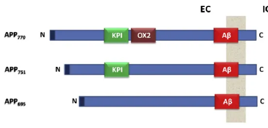
Figure A3.1. Different isoforms of APP ... 41
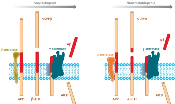
Figure A3.2. APP processing ... 42
Figure A3.3. APP trafficking ... 45
Figure B1.1. APP‐Gαo interaction and Gαo activation are potentiated by APP S655 phosphorylation ... 96
Figure B1.2. Gαo:APP functionally cooperate in neuritogenesis ... 97
Figure B1.3. APP and Gαo modulation of the STAT3 signaling pathway ... 100
Figure B1.4. Gαo:APP morphological and signaling effects after 24h of transfection ... 102
Figure B1.5. Gαo:APP neuritogenic effects in primary neurons ... 104
Figure B1.6. STAT3 and ERK1/2 signaling on APP‐Gαo effects in primary neurons. ... 106
Figure B2.1. APP and Gαo distribution in SH‐SY5Y cells ... 133
Figure B2.2. Effects of APP and Gαo on each other’s protein levels ... 134
Figure B2.3. Effects of proteasome inhibition on Gαo protein levels ... 135
Figure B2.4. Gαo and APP colocalization in SH‐SY5Y cells ... 136
Figure B2.5. APP and Gαo degradation in lysosomes ... 137
Figure B2.6. Presence of lysosomal‐targeting motifs in the Gαo sequence ... 138
Figure B2.7. Gαo and Hsc70 co‐localize in SH‐SY5Y cells ... 139
Figure B2.8. Impact of PTX treatment on Gαo and APP protein levels ... 141
Figure B2.9. Effect of proteasome inhibition on PTX treatment ... 143
Figure B3.1. Processing workflow of the NeuronRead macro ... 163
Figure B3. 2. Details of the NeuronRead workflow, applied to Phase Contrast Images ... 164
Figure B3.3.NeuronRead versus manual detection of neuronal cell bodies areas ... 169
Table of Supplementary Material
Supplementary Figure B1.1. Morphometric analysis of SH‐SY5Y cells transfected with the different
APP‐GFPs for 6h ... 121
Supplementary Figure B1.2. JAK2‐STAT3 inhibition ... 121
Supplementary Figure B1. 3. EGFR inhibition in SH‐SY5Y cells ... 122
Supplementary Figure B1.4. Confirmation of the neuritic nature in 4 DIV neurons ... 122
Supplementary Figure B1.5. Impact of EGFR inhibition on APP‐Gαo effects in primary neurons . 123 Supplementary Figure B2.1. Effect of proteasome inhibition on PTX treatment ... 156
Supplementary Figure B3.1. Comparison between NeuronRead analyses of phase contrast images and fluorescence images ... 187
Supplementary Figure B3.2. Analysis of rat cortical primary neuronal cultures ... 187
Supplementary Figure B3.3. Testing NeuronRead robustness and sensitivity to noises ... 188
Supplementary Figure B3.4. Differentiated SH‐SY5Y cells analyzed with NeuronRead ... 189
Abbreviations
5‐HT Serotonin
AD Alzheimer's disease
ADF Actin depolymerizing factor ADP Adenosine diphosphate
AICD APP intracellular C‐terminal domain ANOVA Analysis of variance
AP Adaptor protein
APLP Amyloid precursor‐like protein APP Amyloid precursor protein APPL β‐amyloid‐like protein Arp Actin‐related protein Aβ Amyloid β‐peptide
BACE β‐site APP‐cleaving enzyme BCA Bicinchoninic acid
BDNF Brain‐derived neurotrophic factor BSA Bovine serum albumin
CA Constitutively active
CaMKII calcium‐calmodulin‐dependent protein kinase II cAMP cyclic adenosine monophosphate
CB1R Type 1 cannabinoid receptor cdc42 Cell division control protein 42 CMA Chaperone‐mediated autophagy CQ Chloroquine
CREB cAMP response element binding protein CTF C‐terminal fragment
DAPI 4',6‐diamidino‐2‐phenylindole DIV Days in vitro
DN Dominant negative DoG Difference of Gaussians ECL Enhanced chemiluminescence
ERK Extracellular signal‐regulated kinase FAD Familial Alzheimer's Disease
FBS Fetal bovine serum FC Fold change
FGF Fibroblast growth factor Fluor Fluorescence
GAP GTPase‐accelerating protein GAP43 Growth‐associated protein 43 GDI Guanosine dissociation inhibitor GDP Guanosine diphosphate
GEF Guanine nucleotide exchange factor GFP Green fluorescent protein
GNAO1 Gαo gene
GPCR G protein‐coupled receptor GR Golgi region
GRIN G protein‐regulated inducer of neurite outgrowth GSK3 Glycogen Synthase Kinase 3
GTP Guanosine triphosphate holoAPP Full length APP
Hsc70 Heat shock cognate protein 70 Hsp90 Heat shock protein 90
ICC Immunocytochemistry IGF Insulin‐like growth factor IL‐6 interleukin‐6
JAK Janus kinase
JNK c‐Jun N‐terminal kinase KO Knockout
KPI Kunitz‐type protease inhibitor Lac Lactacystin
LAMP2 Lysosome‐associated membrane protein 2 LTD Long‐term depression
LTP Long‐term potentiation
M1AChR Muscarinic acetylcholine receptor M1 MAP Microtubule‐associated protein MAPK Mitogen‐activated protein kinase MCH Melanin‐concentrating hormone MEK MAPK/ERK kinase
MIS Müllerian inhibiting substance NGF Nerve growth factor
NMJ Neuromuscular junction NT Neurotrophin
O/N Overnight
ORL Opioid receptor‐like p75NTR Neurotrophic receptor p75
PACAP Pituitary adenylate cyclase‐activating peptide PAGE Polyacrylamide gel electrophoresis
pAPP Phosphorylated APP
PAR Protease‐activated receptor PBS Phosphate buffered saline PDBu Phorbol 12,13‐dibutyrate
PDK1 phosphoinositide‐dependent kinase 1 PhC Phase contrast
PI3K Phosphatidylinositol 3‐kinase
PIP3 phosphatidylinositol (3,4,5)‐tri‐phosphate PKA Protein kinase A
PKC Protein kinase C PM Plasma membrane
PSD‐95 Postsynaptic density protein 95 PSI Proteasome inhibitor I
PTX Pertussis toxin RA Retinoic acid
RGS Regulator of G‐protein signaling ROI Region of interest
SDS Sodium dodecyl sulfate SE Structuring element SEM Standard error of the mean
STAT Signal transducer and activator of transcription TAG Transient axonal glycoprotein
TBS‐T Tris‐buffered saline with Tween 20 Tf Transfection
TGFβ Transforming growth factor β TGN Trans‐Golgi network
Trk Tyrosine receptor kinase WB Western blot
A. General Introduction
and Aims
A1. Neuronal Differentiation
A1. Neuronal Differentiation
The brain is a complex organ made of different types of highly specialized cells. The main “unit” of the brain is the neuron, a cell with a very characteristic morphology. The neuron is composed of a long process called the axon, that can grow for longer than 1 meter, and several shorter but highly branched processes called dendrites. The mechanism by which neurons acquire this morphology has been the subject of intense study in neurosciences. From the initial morphological changes that undifferentiated cells suffer when they start to form new processes to the formation of synapses between mature neurons, and all the signaling pathways underlying these different steps in neuronal differentiation, these are mechanisms that researchers have explored to better understand how neurons work. Understanding neuronal differentiation has also shed light on other mechanisms, such as neuronal regeneration, that can prove essential to the understanding and treatment of several neuropathologies.
A1.1. Neuritogenesis and Acquisition of Neuronal Polarity
Early embryonic neurons are spherical cells, so the first step of neuronal differentiation involves the formation of membrane extensions that will become neurites. This step is designated by neuritogenesis, but is also usually called neurite outgrowth or neurite initiation/extension, and is accompanied by an extensive reorganization of the cytoskeleton [1, 2]. After the initial formation of neurites, these have to differentiate into axons and dendrites, in a phenomenon called neuronal polarization [3, 4]. Both neuritogenesis and neuronal polarization are highly dynamic mechanisms and must occur in tandem so that neuronal differentiation is properly developed. Earlier research has established the different developmental stages that cultured hippocampal neurons go through (Figure A1.1) [5]. In stage 1, which in vitro occurs during the first hours after plating, cells extend their membranes around them, creating the lamellipodium. This is a filamentous actin (F‐actin) structure that makes cells strongly adhere to the cell plates. Several small finger‐like F‐actin processes start to appear at the edge of the membrane, named filopodia. In stage 2, that occurs throughout the first day of differentiation, the filopodia start to enlarge, giving rise to several neurites. At this point, all neurites are virtually indistinguishable, with each one having the potential to further elongate and develop into the axon. These two first stages encompass the bulk of neuritogenesis. Stage 3 sees the beginning of neuronal polarity, with one neurite starting to grow
significantly faster than the rest (5‐10x faster), eventually becoming the axon. Stage 4 occurs 2‐3 days after the initial axon growth, usually around 4 days after cells plating, and it is characterized by the growth and branching of the remaining neurites that will make the dendritic tree. At this time, the axon continues to elongate, although to a slightly lower rate (but still at least 5x faster than dendrites). Stage 5 is the maturation of the neuron, characterized by the formation dendritic spines, where cell‐to‐cell contacts are made in the form of synapses [6]. Several other studies have been published describing neuronal differentiation both in vitro and in vivo, and looking at different types of neurons. Neuritogenesis and neuronal polarization occur roughly the same in cortical neurons in vivo, with a few differences [4, 7, 8]. For example, excitatory cortical neurons start their differentiation in the cortical ventricular zone of the developing embryo by forming several neurites, thus becoming a multipolar cell. One of these neurites suffers elongation and becomes a trailing process, that further develops into the axon, while another neurite becomes the leading process, defining the neuron’s first dendrite. The remaining neurites suffer a retraction, thus turning the cell into a bipolar neuron. At this stage these neurons migrate through the cortical plate into the marginal zone. That is why the future dendrite is called the leading process (the process that “guides” the migration) whereas the future axon is called the trailing process (the process that follows behind the migratory neuron) [9–11]. Upon reaching the marginal zone of the cortical plane, the neuron matures, with the leading process suffering further elongation and ramification to become the dendritic tree.
Figure A1.1. Stages of Neuronal Differentiation. Stage 1: Formation of Lamellipodia; Stage 2: Neurite outgrowth; Stage 3: Axon specification and elongation; Stage 4: Dendritic growth; Stage 5: Neuronal maturation and synaptogenesis. Image adapted from [2].
A1. Neuronal Differentiation
A1.2. Cytoskeleton remodeling during neuronal differentiation
A1.2.1. Lamellipodia and Filopodia formation
Neuritogenesis starts with the assembly of actin filaments on the edge of the differentiating neuron (Stage 1). These actin filaments, a result of actin polymerization, form two distinct structures: the lamellipodium, a sheet‐like extension of the plasma membrane all around the cell; and the filopodia, several thin protrusions, comprising bundles of actin filaments, that arise from the lamellipodium [12, 13]. The exact mechanism that leads to the formation of filopodia it is still not completely clear. Some studies describe the formation of branches of actin filaments within the lamellipodium by association of proteins to the filaments barbed end (the fast‐growing end, or plus‐ end). Subsequent recruitment of fascin to the barbed ends culminates in the bundling of different actin filaments and the filopodia formation [1, 14, 15]. This is called the convergent model. The de
novo nucleation model, or tip nucleation model, describes filopodia formation as actin filaments
present in the lamellipodium that are nucleated by formin proteins, thus growing and protruding out and that are later crosslinked together by fascin [16, 17]. The presence of common players (e.g. fascin) between both models indicates that the real formation of filopodia might comprise a mix of both mechanisms [1, 17]. As evidenced by both models, actin dynamics are the main force behind neurite initiation and elongation. Continuous polymerization/depolymerization of actin filaments is required for the elongation of the filopodia, in a “treadmilling” mechanism [14]. In this mechanism, there is an exchange of actin subunits (globular actin, G‐actin) between the pointed end (minus end) and the barber end (plus end) of the actin filament. Polymerization occurs at the barber end (addition of G‐actin), while depolymerization occurs at the pointed end (removal of G‐ actin) [2, 14]. This mechanism allows the actin filament to “push” against the plasma membrane and thus elongate the filopodia [18].
A1.2.2. Neurite stabilization and maturation
Filopodia are highly dynamic structures, suffering continuous formation and retraction. To stabilize these processes and form neurites (Stage 2), it is required the involvement of another component of the cell cytoskeleton, the microtubules [1, 19]. The microtubule subunit is a heterodimer of two types of tubulin, α and β. Of the several known α and β subunits, β‐III tubulin is the only isoform specific to neurons and its expression is increased during neurite outgrowth [1, 20, 21]. After the formation of the filopodia, microtubules formed at the centrosome start to extend into the
filopodia. This extension occurs either by re‐distribution of stable microtubules into the actin filopodia or through polymerization of new microtubules [1, 19, 22]. The stabilization of the microtubules and maturation of the first neurite normally results in the commitment of this neurite to axon specification [23, 24].
Lamellipodia and filopodia remain present at the extremity of the growing neurite, in a structure called growth cone. This structure is present in all neurites, but it is especially dynamic and active in the first neurite, contributing to its faster extension and eventual differentiation into the neuronal axon (Figure A1.2) [4, 25]. Filopodia are also formed during dendritic and axonal growth, and, if matured, give rise to dendritic and axonal branches, an essential step in neuronal differentiation that allows a single neuron to make contact with thousands of other cells [26].
A1.2.3. Control of Actin and Microtubules Dynamics
Several proteins are involved in the control of actin polymerization/depolymerization, and in the control of microtubules transport and stabilization. The Arp2/3 complex is one of the main factors involved in actin nucleation (assembly of actin monomers) [15], being essential in the formation of actin filaments, and plays a role in the formation of the lamellipodia [1, 3, 12]. Its exact role on neurite outgrowth it is still not completely clear, though, with different studies pointing to either a role of Arp2/3 in neurite formation [27, 28], or Arp2/3 inhibition having no impact on the formation of filopodia [29]. Cofilin I and ADF (actin depolymerizing factor) are two members of the cofilin
Figure A1.2. Growth cone structure. Actin (Green) forms lamellipodia and filopodia at the extremity of the growth cone, while microtubules (red) extend through the neurite into the growth cone to stabilize it. Adapted from [3].
A1. Neuronal Differentiation
family abundantly present in the growth cone. They act by binding to the pointed end of the actin filaments and thus promoting the depolymerization of actin, driving neurite elongation [3, 30]. Other proteins that influence actin dynamics include WAVE, Ena/VASP, and profilin [1, 3].
The main regulators of microtubules dynamics are the MAPs (microtubule‐associated proteins). MAP1b promotes microtubule nucleation, polymerization and stabilization, and is believed to be a bridge between actin and microtubules, regulating both neurite elongation and branching [1, 26, 31]. MAP2 also stabilizes microtubules but has the additional function of binding to actin and participate in the formation of the actin bundles [22, 26]. MAP2 and MAP tau are also specially interesting because of their differentially localization in mature neurons, with MAP2 being specific to the dendrites, and MAP tau being enriched in the axon, highlighting possible specific functions for the different MAPs during neuronal polarization [22, 32].
A1.3. Axon specification
As previously mentioned, an early neuron starts its differentiation by protruding several equivalent neurites (Stage 1‐2). However, one of these neurites at one point starts to elongate much faster than the remaining ones, and eventual becomes the axon (Stage 3‐4) [5]. The extensive reorganization the cytoskeleton suffers during neuronal differentiation is especially evident in the growth cone of the future axon, with a high degree of actin instability and the stabilization of microtubules being key in this mechanism [24, 33, 34], however there is still little information about what triggers one neurite to elongate in detriment of the rest. One model explaining the beginning of neuronal polarization is the “Touch & Go” model. In this model, cell‐to‐cell interactions between the pioneering axon of a pyramidal neuron and the neurite of a multipolar cell triggers the cytoskeleton remodeling in the neurite of the latter that leads to its elongation into an axon, a mechanism dependent on the cell‐adhesion molecule transient axonal glycoprotein 1 (TAG1) (the signaling pathways underling this mechanism will be discussed further ahead) [4, 8, 35]. Cell‐cell interactions mediated by N‐cadherin are also important to trigger axonal specification, since knockdown of this protein disrupts the efficient transformation of a multipolar cell into a bipolar cell [4, 36].
Several extracellular cues are also involved in axon specification and growth, such as the brain‐ derived neurotrophic factor (BDNF), neurotrophin 3 (NT3), Reelin, transforming growth factor (TGFβ), insulin‐like growth factor 1 (IGF1), semaphorins and Wnts [4, 6, 8]. Prevailing theories
suggest that these factors might act in an autocrine or paracrine way not only to trigger axon specification but to also maintain its elongation (Figure A1.3) [6, 8]. Differential distribution of these factors in vivo, such is the case of TGFβ, could also help explain the axon specification on different areas of the developing brain [4]. The gradient of neurotrophic factors existing in the medium could also explain why only one neurite develops into an axon. Accumulation of these factors on the growing axon location could mean a lack of stimuli on the other neurites, resulting in an inhibition of their growth [37].
The activation of different intracellular signaling pathways also regulates axon initiation and extension, but these will be discussed in a later section.
A1.4. Dendritic vs Axonal structure
Though at first glance dendrites are just shorter and more ramified versions of the axon, there are key structural differences between both type of neuronal processes. As mentioned above, after filopodia elongation through actin remodeling, microtubules invade the filopodia to mature it into a neurite. While in the axon the microtubules are densely packed and are uniaxially orientated (their minus end is always facing the cell body and their plus end is always facing the growth cone, Figure A1.2), in the dendrites microtubules have both orientations [23]. While is not completely clear why these differences arise, MAP2 and tau different localizations to the dendrites and axon, respectively, could play a role in the microtubule orientation [24].
Figure A1.3. Different factors that influence axon specification in vivo. Neurotrophins released by neuronal cells act in a paracrine and autocrine to activate intracellular signaling pathways (Blue). Similar signaling pathways are activated by cell‐to‐cell contacts (purple). Gradients of extracellular molecules drive both axonal polarization as well as neuronal migration. Adapted from [8].
A1. Neuronal Differentiation
A1.5. Synaptogenesis
Neuronal differentiation culminates with the formation of cell‐cell contacts defined as synapses (Stage 5). Synapses are the main place of data transmission between neurons and are one of the most dynamic structures of the adult brain [38]. A synapse consists of a presynaptic terminal (axonal side), a postsynaptic terminal (dendritic side) and a synaptic cleft separating both [39] (this defines the chemical synapses, electrical synapses will not be explored here). There are reports of two ways neurons establish synapses[40]. In the postsynaptic spine hypothesis, filopodia developed in the dendrites establish contact with presynaptic neurons, which triggers the filopodia maturation into dendritic spines and consequently the formation of the synapse [26, 40]. In the presynaptic hypothesis, it is the axon who initiates the synapse formation. The axon continues to grow until it reaches near a postsynaptic terminal. At this point, a signal terminates the axonal growth cone elongation and starts its maturation into a presynaptic terminal[40]. This signal might be mediated by collapsins [41], with the activation of the Wnt‐7a signaling playing a part in the maturation of the presynaptic terminal[42]. The differences in both models could relate to the synaptogenesis in different types of neurons. Moreover, synaptogenesis in the adult brain seems to be regulated by different mechanisms, related to neuronal activity [43].
A1.6. Signaling during neuronal differentiation
While neuronal differentiation is ultimately a result of an extreme reorganization of the cell cytoskeleton, there are several signaling pathways that control this remodeling. From extracellular cues, like BDNF, to intracellular proteins, such as the Rho family or the MAPK/ERK pathway, there are a lot of factors that intervene in the different steps, from the initial filopodia formation to the acquisition of the neuronal polarity and formation of synapses.
A1.6.1. Brain Derived Neurotrophic Factor (BDNF)
BDNF is a neurotrophin produced and secreted by neurons that has been for a long time implicated in the promotion of several aspects of neuronal differentiation [44]. Upon secretion, it can bind to the Tyrosine receptor kinase B (TrkB), as well as to the neurotrophic receptor p75 (p75NTR). Recent experiments show that BDNF induces axonal growth in in vitro conditions, while in vivo its role seems to be more on promoting the branching of the elongating axon [45]. Studies with Xenopus showed that expressing a dominant negative TrkB on retinal ganglion cells did not affect the ability
of the axons to reach their target, but significantly altered the growth cone morphology and impaired the formation of axonal branches [45, 46]. The axonal branching promoted by BDNF relies upon the activation of the ERK1/2 signaling pathway [47]. BDNF is able to influence correct wiring of the brain not only by extending new axonal branches, but also by pruning unnecessary ones through the activation of the p75NTR [47, 48]. BDNF also modulates the dendritic tree morphology, especially in aiding the formation of dendritic spines, and consequently synapses. BDNF activation of TrkB promotes dendritic filopodia motility in a PI3K‐dependent way [45, 47]. BDNF also stimulates the increase of PSD‐95 in dendritic filopodia [26, 45], thus promoting the maturation of the postsynaptic terminal.
BDNF role in neuronal differentiation is also highlighted by its common use as a neurotrophic factor in the differention of SH‐SY5Y neuroblastoma cells [49–52]. Pre‐incubation of these cells with retinoic acid leads to the expression of TrkB, with follow‐up treatment with BDNF resulting in the differentiation of SH‐SY5Y cells into neuron‐like cells, expressing neuronal markers such as MAP2 and tau [49]. BNDF effects on these cells are mediated by activation of both the ERK1/2 and PI3K signaling pathways [51], which will be discussed ahead.
A1.6.2. Rho small GTPases
The three main members of the Rho protein family are RhoA, Cdc42 and Rac1. These are small GTPases that act as molecular switches in several signaling pathways. All three have been associated with neuronal differentiation: Cdc42 and Rac1 have mainly a positive role in neurite outgrowth, while RhoA has negative role [53]. Cdc42 knockout in mice leads to the development of smaller brains, with a reduced number of axons, that results in death at birth, thus highlighting a fundamental role of Cdc42 in axon specification [54, 55]. Cdc42 modulates the actin cytoskeleton, promoting the formation and elongation of filopodia [56]. Rac1 activation promotes axonal branching and formation of dendrites, while its inactivation leads to a decrease in the number of primary dendrites, and also affects axon growth and guidance [54]. Interestingly, Rac1 activation has to be tightly controlled during neuronal differentiation, since some experiments showed that expressing a constitutively active (CA) form of Rac1 or a dominant negative Rac1 both resulted in a decrease in neurite outgrowth [54, 57]. Similarly, a cyclic activation of Cdc42 is required for it to promote neuronal polarization [58]. RhoA seems to act as a limiting factor in neuronal differentiation [54]. Activating RhoA in hippocampal neurons and neuronal models, such as PC12, inhibits the growth of small processes or even promotes the retraction of neurites, respectively,
A1. Neuronal Differentiation
while its inactivation greatly enhances neurite outgrowth [4, 53]. It has been hypothesized that RhoA role in vivo is to control axon elongation and neuronal polarization by inhibiting the formation of extra axons [4]. This is supported by the evidence that RhoA activity is higher in growth cones of smaller neurites when compared to the axonal growth cone [59].
While these opposing roles between Cdc42/Rac1 and RhoA have been well established, normal neuronal differentiation is a result of a coordinated interplay between the three proteins, with defects in any of them seriously affecting the proper formation of axons and dendrites [53].
A1.6.3. PI3K / Akt pathway
The phosphatidylinositol 3‐kinase (PI3K) and Akt signaling can be activated by different membrane receptors during neuronal differentiation, including TrkA and TrkB, as well as G‐protein coupled receptors [1, 60, 61]. Activation of PI3K leads to the increase of PIP3 (phosphatidylinositol (3,4,5)‐ tri‐phosphate), which in turn promotes Akt phosphorylation via phosphoinositide‐dependent kinase (PDK1). For instance, BDNF signaling through TrkB increases filopodia motility and its number in hippocampal dendrites through the activation of PI3K signaling [1], while the neuritogenic effects of the Nerve Growth Factor (NGF) in dorsal root ganglion neurons involves the activation of a TrkA‐ PI3K‐Akt signaling [60]. In both cases, PI3K/Akt translates its effects to the cell cytoskeleton by mediating the activation of the Rho GTPases proteins. Indeed, a possible positive signaling mechanism mediating neuronal polarization has been described involving PI3K, Cdc42 and Rac1 [8]. Signaling of BDNF through PI3K‐Akt has also been shown to be involved in regulating the complexity of the dendritic tree as well as the formation of dendritic spines, with chronic inhibition of PI3K resulting in a decrease in the formation of dendritic spines and filopodia [62]. Interestingly, this study also showed a cooperation between PI3K‐Akt and MAPK signaling in regulating dendritic morphology.
Besides directly inducing neurite formation, the PI3K/Akt signaling also promotes differentiation by inhibiting the GSK3 signaling [63]. Activation of PI3K/Akt in NGF signaling results in the phosphorylation of GSK3β, thus inactivating it, and this inactivation is essential in the promotion of NGF neuritogenic effects [64]. Moreover, activation of the 5‐HT1A GPCR potentiates NGF neuritogenic output in a signaling dependent on PI3K and Akt activation [61]. Taking together, these different reports indicate that PI3K/Akt seems to be one of the main signaling pathways where several extracellular cues converge to induce neuronal differentiation.
A1.6.4. ERK1/2 pathway
The extracellular regulated kinases 1 and 2 (ERK1/2) are part of the main pathway of the MAPK (mitogen‐activated protein kinase) signaling. Different receptors translate their intracellular signaling through the activation of small GTPases of the Ras family. These activate a kinase signaling cascade that starts with the Raf kinases, which in turn activate the MAPK kinases (MEK1/2) and culminates in the activation of ERK1/2. Several reports have put ERK1/2 has a main player in neuronal development [65]. Treatment with different neurotrophic factors, usually NGF and BDNF, lead to neuritic outgrowth in vitro as a result of increased ERK1/2 activity [51, 66–68]. In PC12 cells, ERK1/2 is especially important, being able to induce different outcomes depending on the duration of its signaling. Sustained activation, as the one induced by NGF, results in neurite outgrowth, while temporary activation leads to cell proliferation, normally as a result of EGF incubation [68]. ERK1/2 seems to be especially involved in the axon specification mechanism. Studies involving the Rit GTPase showed that its activation led to axonal elongation in detriment of dendritic growth, and inhibition of ERK activation blocked Rit effects [69]. Another study also showed that ERK2 phosphorylation of Par3 modulated neuronal polarization[70].
In vivo, knockout studies have been essential not only to identify potential ERK functions, but also to differentiate between ERK1 and ERK2. ERK2 deletion results in a 50% reduction in axonal length, as well as a reduction in dendritic branching on mice cortex [71]. ERK2 deletion also led to embryonic cell death, contrary to ERK1 knockout, revealing a crucial role for this isoform on normal development[72].
A1.6.5. STAT3 pathway
The signal transducer and activator of transcription 3 (STAT3) is a transcriptional factor involved in innumerous cellular functions, as well as a key factor in the genesis of different cancer types. STAT3 is activated by phosphorylation by tyrosine kinase signals, with its canonical activator being the Janus kinase 2 (JAK2), but being also activated by the Src kinase or directly phosphorylated by tyrosine kinase receptors such as the Epidermal Growth Factor Receptor (EGFR) [73–75]. Although STAT3 function has been mainly studied in a context of carcinogenesis, a possible role in neuronal differentiation has been unveiled through the last years. Treatment of PC12‐E2 cells with interleukin‐6 (IL‐6) induces neuronal‐like morphological changes similar to the incubation of these cells with NGF. However, while NGF treatment is accompanied by a substantial increase in ERK1/2 activation, IL‐6 leads to STAT3 activation[76]. Moreover, blocking STAT3 activity, but not blocking
A1. Neuronal Differentiation
of ERK1/2, significantly decreased IL‐6 induced neurite outgrowth, showing that STAT3 activation in these cells is sufficient to induce neuronal‐like differentiation [77]. Notwithstanding these results, the STAT3 role on neuronal differentiation in vivo is still not clear. In the adult nervous system, STAT3 has been implicated in the organism response to neuronal injury, being activated in cases of brain ischemia and spinal cord injury [78, 79]. STAT3 is also important in synaptic plasticity, for its activation in the postsynaptic terminal is required for long‐term depression (LTD) to take place [80]. Interestingly, activation of STAT3 by the Src kinase also leads to neurite outgrowth in Neuro‐2A cells, in a signaling mechanism mediated by Gαo [81]. This pathway will be explored in more detail in the following section.
Over the years, several other players in neuronal differentiation have been uncovered. A detailed scheme of the main signaling pathways involved in neuritogenesis and neuronal polarization is displayed on figure A1.4, adapted from [8].
Figure A1.4. Signaling in neuronal differentiation. The different signaling pathways described here have to cross‐ talk with each other to successfully induce neuronal differentiation. Adapted from [8].
A2. The Other G protein (Go)
Heterotrimeric G proteins are one of the main components of intracellular signal transduction. They consist of three subunits, the α, β, and γ, with the latter two usually tightly bound together into the βγ complex. G proteins are divided according to their alpha subunit into 4 major families: Gs, Gi/o, G12/13, and Gq/11 [82]. One of the most intriguing G proteins is one of the members of the Gi/o family, the Go protein. Its alpha (α) subunit, Gαo, got its name due to being discovered after both Gs and Gi: Gs was named for being able to stimulate the adenylate cyclase activity, while Gi inhibits it. Since the new G protein had no specific function or attribute, it was named the “other” G protein [83, 84].
Similar to the small G proteins, heterotrimeric G proteins work as molecular switches of intracellular signal transduction. When inactive, Gαo is bound to GDP (guanosine diphosphate), and forms a trimeric complex with the βγ subunit. To be activated, Gαo must release the GDP and bind GTP (guanosine triphosphate). This is promoted by the binding of Guanine Nucleotide Exchange Factors (GEFs) to the G protein, with the most common GEFs being the G protein‐coupled receptors (GPCRs). The activation of a GPCR by an extracellular ligand results in a conformational change that allows the binding of the receptor to the G protein. This in turn causes a second conformational change on the α subunit that results in the exchange of GDP for GTP, and the separation of the α and βγ subunits. At this point the G protein is active, and both the α and βγ subunits interact with downstream effectors to modulate different signaling pathways. What characterizes the G proteins as molecular switches is the intrinsic GTPase activity that the α subunits possess (as well as the small G proteins). Thus, G proteins only stay active for short periods of time, after which the GTP is hydrolyzed to GDP, the α and βγ subunits reconnect, and Go returns to its inactive state. Members of the Regulators of G‐protein Signaling (RGS) family can bind to the α subunit and drastically increase the rate of GTP hydrolysis, thus terminating the G protein signaling faster. RGS proteins are also known as GTPase‐accelerating proteins (GAPs), however this term is more commonly used to proteins that interact and regulate small monomeric G proteins, such as Ras and Rho family proteins [85–87]. As a member of the Gi/o family, Gαo is also inhibited by the Pertussis Toxin (PTX). PTX ADP‐ribosylates the cysteine located four residues from the carboxyl terminus of Gαo, thus blocking Gαo interaction with its activators [88].
A2. The Other G Protein
Although it has been intensively studied for the last 3 decades, Gαo specific function in the human organism is still not completely clear. Some data regarding Gαo physiological function has come out from a few published knockout reports. General knockout of Gαo in mice results in viable animals but that have a lifespan of only 7 weeks. The animals are small, hyperactive, hyperalgesic and have severe motor control impairments, exhibiting a turning behavior that result in the mice going around in circles for long periods of time. At a cellular level, there was a decrease in the ability of opioid receptors to inhibit Ca2+ channel currents in dorsal root ganglion cells, indicating a role for Gαo in translating the intracellular signaling of these receptors [89].
Since Gαo is the most expressed Gα subunit in the brain, accounting for around 1% of the total membrane protein [90], most of the studies have been devoted to try uncover the Gαo role in the brain physiology.
A2.1. Gαo genetics
The Gαo protein is highly conserved across several species, including human, rat, bovine, fly, nematode, among others, sharing over 80% identity between proteins of the different species [90]. In humans, the Gαo gene (GNAO1) is located on chromosome 16, comprising over 100 kb and containing 11 exons [91]. Analysis of the GNAO1 gene detected that exon 7 and exon 8 are duplicated, and mechanisms of alternative splicing give rise to two different isoforms, Gαo1 (aka GαoB) containing the exons 7A and 8A, and Gαo2 (aka GαoB) containing the exons 7B and 8B [91– 93]. These isoforms are almost identical, with differences appearing only in 20 amino acids of the last portion of the protein (C‐terminal), and since this region is essential to receptor and effector binding, Gαo1 and Gαo2 could have different functions in the brain [91, 94].
A third isoform, Gαo3, has been identified as a result of a posttranslational modification, where an asparagine at residue 346 of Gαo1 is converted through deamidation into an aspartate [95, 96]. However, there is still no description of this modification occurring in human Gαo.
A2.2. Gαo expression and distribution
Although Gαo can be found a little all over the human body, it is greatly enriched in the brain [97]. Initial immunohistochemical studies in rat brain showed that Gαo is mainly present in neuropil (regions with abundance of dendrites and axons, and consequently rich in synapses) and absent
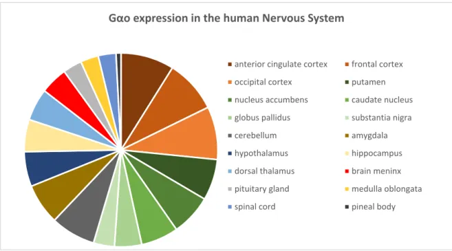
from cell bodies [98]. The study also detected a differential distribution of Gαo along the rat central nervous system: Gαo is enriched in cerebral cortex, especially in the molecular layer (layer 1), in neuropil of the hippocampal formation, striatum, subtstantia nigra pars reticulate, molecular layer of the cerebellum, substantia gelatinosa of the spinal cord, and posterior pituitary [98]. Current data retrieved from the database Expression Atlas shows that Gαo also has a differential expression in the human nervous system, being enriched in the cerebral cortex and the basal ganglia (putamen, nucleus accumbens, caudate nucleus, globus pallidus and substantia nigra) (Figure A2.1) [97, 99– 102].
At a cellular level, mouse Gαo is located in striatal neurons, cortical neurons, cerebellar granular cells, as well as striatal glial cells, cerebral cortex and colliculi glial cells. At a subcellular level, neuronal Gαo is present on the plasma membrane, mainly at cell‐cell contacts, and in neurite arborization. It is also present at low levels in the cytoplasm and is absent from the nucleus [103]. During neuronal development, Gαo is especially present on the growth cones of elongating neurites [104]. In glial cells, Go is present throughout the cell in low levels, with specially strong staining around the nucleus [103].
Gαo expression in the human Nervous System
anterior cingulate cortex frontal cortex
occipital cortex putamen
nucleus accumbens caudate nucleus
globus pallidus substantia nigra
cerebellum amygdala
hypothalamus hippocampus
dorsal thalamus brain meninx
pituitary gland medulla oblongata
spinal cord pineal body
Figure A2.1. Gαo expression in the different regions of the human nervous system. Data was retrieved from four studies present in the Expression Atlas database, and the values were normalized to each study. The studies analyzed were The FANTOM5 project, The Human Protein Atlas and two studies from the Genotype‐Tissue Expression (GTEx) Project [97, 99–102].
A2. The Other G Protein
Gαo expression suffers distinct variations during neuronal development. Initial studies showed that differentiation of neuroblastoma cells (NG 108‐15 and N1E‐115 cells) induced different expression profiles on both Gαo isoforms: Gαo1 expression was either absent (N1E‐115) or very low (NG108‐ 15) in undifferentiated cells, with differentiation greatly increasing its protein levels; Gαo2 was already present in undifferentiated cells and its protein levels did not change substantially during differentiation [105, 106]. However, analysis of primary cultures of matured neurons showed that Gαo2 was almost absent [106], indicating that neuronal differentiation is accompanied by an increase in Gαo1 and a decrease in Gαo2 protein levels. Moreover, analysis of Gαo metabolism showed a significant increase in the protein half‐life with differentiation, being around 28h in undifferentiated neuroblastoma cells, 58h in differentiated cells, and 154h in primary culture of cerebellar granule cells [107]. Adding the results that showed that mRNA levels on cerebellar cortex of mice did not suffer significant alterations during cerebellum development [108], the increase in Gαo levels with differentiation could be a result of a decrease in Gαo degradation rather than an increase in Gαo gene expression.
Interestingly, while differentiation of PC12 cell also correlates with an increase of Gαo levels [109], differentiation of the neuroblastoma cell line SH‐SY5Y with retinoic acid (RA) gave opposing results, with Gαo levels suffering a slight decrease, although not significant [110]. Of note, the study did not distinguish between Gαo1 and Gαo2. Nevertheless, this could indicate that distinct differentiation mechanisms on different cell types affect Gαo expression differently.
Rat primary neuronal cultures have also evidenced an increase in Gαo expression during the differentiation of mesencephalon and hypothalamus neurons, with Gαo levels being barely detectable for the first 2 days in vitro, but rapidly increasing after 4 days and stabilizing 2‐3 weeks after plating [111]. This increase in Gαo levels was associated with a significant increase of Gαo presence in neuronal processes, dendrites and axons. Also, in the case of mesencephalon neurons, increasing the cell density also resulted in an increase in Gαo levels, which could be an effect of the increase in cell‐cell contacts [111]. A study using rat brain extracts also showed that Gαo protein levels not only increases during development, but continues to increase for several days after birth [112].
A2.3. Gαo signaling in the brain
As mentioned before, although intensively studied, Gαo role in the brain is still not completely clear. The discovery of the receptors that activate intracellular signaling through Gαo, as well as its downstream effectors have helped to establish Gαo signaling pathways, as well as deciphering potential functions of Gαo, particularly in neuritogenesis [90, 113, 114].
A2.3.1. Necdin
Necdin is a neuronal protein highly expressed on post‐mitotic neurons, where it functions by blocking cell cycle progression, thus maintaining the neurons in the G0 phase of cell cycle [115]. It is also expressed during brain development, opening a potential role in regulating neuronal differentiation. A recent study by Ghil’s group as identified Necdin has an interactor of Gαo [116]. Using co‐immunoprecipitation assays followed by quantitative western‐blot, the authors showed that Necdin interacted preferentially with the activated Gαo, thus putting Necdin as a downstream effector of Gαo. Overexpression of a constitutively active (CA) form of Gαo with Necdin enhanced the Necdin‐induced blocking of cell proliferation, while co‐transfection of Necdin with either wild‐ type or Gαo CA increased of the number of cells with neurites (this work stablishes a neurite as being a process longer than the cell body length). Furthermore, Gαo activation of Necdin signaling was promoted by activation of the type 1 Cannabinoid Receptor (CB1R), and culminated on the inhibition of the transcription factor E2F1.
A2.3.2. Src-STAT3 pathway
As mentioned above, the STAT3 pathway is a prominent player in brain development and function [77, 80], and while JAK2 is known as STAT3’s canonical activator, some signaling pathways involve STAT3 activation by the Src kinase [73]. Initial studies using NIH‐3T3 fibroblasts showed that overexpressing a Gαo CA resulted in proliferation and neoplastic transformation of these cells [117, 118]. This transformation was accompanied by an increase in STAT3 activity, with no alterations in ERK1/2 activity. Moreover, the Gαo‐induced transformation was a result of STAT3 phosphorylation by the Src kinase rather than by JAK2. The authors had already hypothesized a possible role for the Gαo‐Src‐STAT3 pathway in differentiation by stating that mechanisms that induce NIH‐3T3 transformation sometimes translate to other cell types as differentiation mechanisms, with their follow‐up work supporting this statement. Using Neuro2A cells as a model, the research shows that
A2. The Other G Protein
stimulating the CB1R significantly increases the number of cells with neurites (this work stablishes a neurite as being a process at least 2x longer than the cell body diameter), a mechanism mediated by the activation of the Gαo‐Src‐STAT3 pathway [81, 119]. CB1R activation causes Gαo to bind to Rap1GAP, a Rap1 negative regulator protein. This binding results in the targeting of Rap1GAP to proteasomal degradation, thus eliminating the blockage upon Rap1 activity [119, 120]. Rap1 activates Ral, which in turn activates the Src kinase, culminating in STAT3 phosphorylation. Besides phosphorylating STAT3 directly, the study also showed that Src kinase can activate STAT3 via an alternate pathway, where it activates Rac1‐c‐Jun N‐terminal kinase (JNK) signaling. Activation of both signaling pathways by CB1R‐Gαo are essential in inducing neurite outgrowth (Figure A2.2). Interestingly, although G protein effectors tend to bind with
more affinity to the activated forms of the Gα subunits, Rap1GAP binds preferentially to the wild‐type form of Gαo when compared to the Gαo CA. Since stimulation of the CB1R leads to the activation of Gαo, it is unexpected that this mechanism would lead to the binding of Gαo to Rap1GAP. The authors try to explain these events as a possible sequential mechanism [119]. Go activation by CB1R leads to the separation of the Gα and Gβγ subunits. This separation allows Rap1GAP to bind to Gαo, since Rap1GAP binds to the same region as the βγ subunit, through the GoLoco motif. The binding of Gαo to Rap1GAP is initially weak, but it is strengthened when GTP is hydrolyzed to GDP. At this point, Rap1GAP would act as a guanosine nucleotide dissociation inhibitor (GDI), maintaining Gαo in its inactivation state until Rap1GAP could be targeted to degradation. Although no direct evidences are shown to support this theory, the idea that Gαo proper function relies on an activation/deactivation cycle is supported by similar mechanisms described in small G proteins, as the aforementioned Cdc42 [58].
An interplay between Gαo and the Src kinase has also been described downstream of Reelin [121]. As mentioned before, Reelin is an extracellular factor known to participate in neuronal polarization, as well as in the control of neuronal migration [8, 122]. Treatment of primary cultures of
Figure A2.2. CB1R-Gαo-STAT3 signaling in neurite outgrowth. Image reproduced from [81].
hippocampal neurons with Reelin increased both the total neuritic length as well as neuritic branching. Treatment with PTX blocked Reelin neuritogenic effects on hippocampal neurons, while knockdown of Gαo did the same in F11 cells [121], thus demonstrating that Reelin activates an intracellular pathway dependent on Gαo. By trying to fully comprehend the complete signaling pathway involved in Reelin effects, the authors identified the Src kinase as a player in this signaling. Co‐immunoprecipitation assays showed that Src and Gαo interact with each other, and that this interaction is strengthened by treatment with Reelin. Gαo activation in this pathway was accompanied by an increased activation of JNK, while Akt and GSK3β (other common players in Reelin signaling) were unaffected. Interestingly, activation of Src and JNK was also seen in the cannabinoid‐induced signaling [81], which could implicate some cross talk between both pathways. However, the authors did not check for STAT3 activity, so is not certain that the interaction of Gαo‐ Src in Reelin‐treated cells leads to same outcome that in cannabinoid‐treated cells. Also, surprisingly, in this study Src seems to be acting upstream Gαo in the Reelin signaling pathway, rather than downstream as seen before. This was demonstrated by showing that inhibiting Gαo with PTX did not significantly inhibited Src kinase. Combining both studies [81, 121], one could hypothesize that Gαo and Src kinase could be involved in a positive loop, where Src activates Gαo, which in turn activates a signaling pathway (Rap1‐Ral) that further activates Src. Such positive loop has already been described as an important mechanism of neuronal polarization, where activation of PI3K by Trk receptors leads to the activation of a cascade involving Cdc42Par‐complexRac1 that feedbacks into further activation of PI3K (Figure A1.4) [8], opening the possibility for Gαo‐Src also participating in a similar process. Nevertheless, further research is required to better understand the inner works of Gαo‐Src signaling
A2.3.3. GAP-43
Although G proteins are mostly known to be activated by GPCRs, one of the first known activators of Gαo was the growth associated protein 43 (GAP‐43 aka neuromodulin) [104]. GAP‐43 is protein highly enriched in neurite growth cones and is commonly used as marker for neuronal differentiation [123, 124]. The presence of both GAP‐43 and Gαo in growth cones raised the question if these proteins could functionally interact in the regulation of the growth cone dynamics. Indeed, GAP‐43 can bind to Gαo and stimulate the exchange of GDP for GTP, thus acting as a GPCR‐ like protein [104, 125]. The Gαo‐activating sequence of GAP‐43 was then shown to be able to induce neurite outgrowth in N1E‐115 cells, an effect that was mimicked by mastoparan (an activator of
A2. The Other G Protein
Gi/o) and inhibited by PTX, thus showing that Gαo plays a role in mediating GAP‐43 neuritogenic function. The effect of GAP‐43 interaction with Gαo is not always the same, however, since in dorsal root ganglia neurons this interaction leads to the collapse of the growth cones [126], indicating that the outcome of GAP‐43‐Gαo interaction might depend upon the cellular environment in which it occurs, and also that a tight control is required to achieve successful neurite outgrowth.
There are some contradictory reports regarding the mechanism by which GAP‐43 interacts with and activates Gαo. The initial report showed that treatment with PTX did not alter the ability for GAP‐ 43 to activate Gαo [125], contrary to what happens normally to GPCRs, which could mean that GAP‐ 43 binds to a different region of Gαo. However, later studies showed that effects mediated by GAP‐ 43 are sensible to PTX treatment [126]. The authors explain these differences as a possible result of GAP‐43 being overabundant in the initial experiments, thus somehow being able to bypass PTX inhibition, or that the GAP‐43 peptides used in the later experiments are more susceptible to PTX action than the full‐length protein [126]. The interaction between GAP‐43 and Gαo is also affected by palmitoylation of Gαo, a reversible post‐translational modification that facilitates the attachment of Gαo to the cell membrane [127]. GAP‐43 ability to activate Gαo is greatly increased when Gαo is depalmitoylated [128]. The authors point to the fact that activation of G proteins by GPCRs results in the increase of Gαo depalmitoylation, indicating that GAP‐43 could function as an intracellular potentiator of GPCR signaling. Indeed, some studies have already showed that GAP‐43 is able to modulate GPCR‐Gαo signaling. GAP‐43 and the muscarinic M2 receptor can synergistically activate Gαo in vitro, while injection of GAP‐43 in Xenopus laevis oocytes significantly increased GPCR response to agonist stimulation [129]. More recently, a study showed that an α7 nicotinic receptor could modulate neurite outgrowth by interaction with a protein complex containing Gαo, GAP‐43 and GRIN1 (another Gαo interactor that will be discussed further ahead) [130], not only adding evidence that GAP‐43 is a potential intracellular positive modulator of GPCR‐Gαo signaling, but also that this signaling is important in regulating neuritogenesis.
A2.3.4. ERK1/2 pathway
One of the main signaling pathways at the center of Gαo activity is the ERK1/2 signaling. Gαo was first described to activate ERK1/2 in CHO cells [131]. In these cells, stimulation of the muscarinic acetylcholine receptor M1 (M1AChR) and the platelet‐activating factor receptor (PAFR) resulted in activation of ERK (note that this work only checked for the activation of p44 MAPK aka ERK1). This effect was blocked by treatment with PTX and rescued by the expression of a PTX‐insensitive Gαo,
thus demonstrating that Gαo activity was required for ERK activation. Gαo activation of ERK was done by a non‐canonical pathway, since Ras was not involved but the protein kinase C (PKC) was required [131]. Further work confirmed this, with PKC inactivation blocking ERK activation by Gαo [132]. The pathway by which Gαo activates ERK was further resolved, with PI3K and B‐Raf linking Gαo‐PKC to ERK1/2. It was also shown that Gαo activation by GPCRs could lead to the modulation of signaling activated by other receptors, such as the Epidermal growth factor receptor (EGFR), since expressing Gαo CA was not enough to activate ERK, but significantly potentiated ERK activation by EGFR [132]. The Gαo‐PKC‐ERK pathway has still not been seen in a neuronal setting, however, PKC‐ERK signaling has already been strongly associated with neurite outgrowth [133, 134], so Gαo participation on the mediation of these effects should not be excluded.
Neuronal activation of ERK1/2 by Gαo seems to be associated with a variety of functions. In Neuro2A cells, expression of Gαo CA significantly increases the number of cells with neurites, accompanied by an increase in ERK1/2 activation [135]. This activation of ERK1/2 was mediated by a small GTPase, Rit, with transfection of a dominant negative form of Rit blocking Gαo neuritogenic effects and decreasing ERK1/2 phosphorylation. Another study also showed that Gαo neuritogenic roles in Neuro2A are possibly translated via ERK1/2 activation [136]. Focusing on the study of RGS8, the authors showed that this protein inhibits Gαo, producing several effects: it blocked the ability to Gαo activate Necdin, reduced the formation of neurites induced by Gαo transfection, and blocked ERK1/2 activation induced by a protease‐activated receptor (PAR1)/Gαo signal. Of note, while these results show that Gαo has a neuritogenic effect, and it is able to activate ERK1/2, these events were evaluated in separated, so it is not clear if ERK1/2 activation induced by the PAR1/Gαo signaling can produce neuritogenic effects.
In SH‐SY5Y cells, Gαo might also potentially induce neurite outgrowth via ERK1/2 [137]. Treatment of cells with melanin‐concentrating hormone (MCH) led to an increase in the number of neurites per cell, as well as an increase in their length. This effect was accompanied by an increase in ERK1/2 phosphorylation, and was significantly decreased when cells were treated with PD98059, an inhibitor of MEK. MCH induction of ERK1/2 activation was blocked by treatment with PTX, indicating that the MCH receptor is coupled to either Go or Gi. Since no specific experiments were performed to differentiate between Gαo and Gαi, further studies are required to confirm the potential MCH‐ Gαo‐ERK1/2 pathway in neurite outgrowth.
One of Gαo functions in the brain seems to be in the modulation of nociception[89], with ERK1/2 potentially playing a role in this function. Knockouts of Gαo in mice resulted in animals that suffered


![Figure A2.2. CB1R-Gαo-STAT3 signaling in neurite outgrowth. Image reproduced from [81]](https://thumb-eu.123doks.com/thumbv2/123dok_br/15913033.1092916/45.892.540.794.411.955/figure-gαo-stat-signaling-neurite-outgrowth-image-reproduced.webp)
![Figure A2.3. Wnt-Gαo signaling in synaptogenesis. Image reproduced from [159].](https://thumb-eu.123doks.com/thumbv2/123dok_br/15913033.1092916/52.892.209.639.385.690/figure-a-wnt-gαo-signaling-synaptogenesis-image-reproduced.webp)
![Figure A3.3. APP trafficking. Reproduced from [200].](https://thumb-eu.123doks.com/thumbv2/123dok_br/15913033.1092916/59.892.211.704.442.817/figure-a-app-trafficking-reproduced-from.webp)