Determination of the activity of BNIP
derivative compounds incorporated into
liposomes against pancreatic tumor cells
Dissertação orientada pela Professora Doutora Anabela Cordeiro e co-orientada pela Doutora Raquel T. Lima
Vanessa Maia Duarte
Dissertação do 2º Ciclo de Estudos Conducente ao Grau de Mestre em Tecnologia Farmacêutica
II
DE ACORDO COM A LEGISLAÇÃO EM VIGOR, NÃO É PERMITIDA A REPRODUÇÃO DE QUALQUER PARTE DESTA DISSERTAÇÃO
O autor:
III
Acknowledgements
First and foremost I would like to thank my supervisor, Professor Anabela Cordeiro da Silva for giving me the opportunity to do my thesis in this field. For all the confidence that she had in me and for all the support throughout this thesis.
I would also like to thank Professor Helena Vasconcelos for being so trustworthy. For all the indispensable help on the biological section of the thesis and also for all the support throughout this past year, in every respect.
To my co-supervisor Doctor Raquel T. Lima for all the support, availability and encouragement throughout this whole period (even on maternity leave). For all the friendly advices and positive outcome that helped my motivation tremendously .
I would like to thank Doctor Sofia Costa Lima, for all the unconditional support, patience, ability and availability to teach. For the constant readiness to teach how to become scientists, to truly understand the "why" of things whilst simultaneously allowing develop ability to critique.
Very special thanks to Diana Sousa, for all the enormous help, availability and friendship. For all the words of support, all the smiles and all the optimism that the project was going to go well.
To Rita Vidal, I could sit here and write a page just for her. I would like to thank her for everything, not just this year, but also throughout the Masters. For listening, for lifting me up when times were really black over these past 2 years. She was always there. My time in Porto would not have been the same without her. We have made a friendship for life.
To Paulo Raimundo, because moving to Porto was a decision that affected him too, and even so he was always present. He always made it his priority to support me unconditionally on many levels. I want to thank him for, like always, being able to bring the best out of me.
To Èlia Oliveira, my manager at Women Secret. I thank her enormously for all her understanding and support. For always "letting me off" when my tiredness would turn into lack of patience. For all the days off she gave me before exams, for allowing me to dedicate 100% to my thesis these past few months.
IV
A big thank you to my family, without whom, I would have never given that huge leap to do my thesis. A huge thank you for always being there, for believing in me, for never allowing me to give up on my ambitions, and mostly for all the love and comfort you give me every day.
Last but by no means least, I would like to dedicate my Master thesis in loving memory of my Mum. There is no one in this world I would love more to share my happiness and sadness in my projects than with her.
V
Abstract
The use of nanotechnology in medical chemistry and more specifically in drug delivery is set to spread rapidly. Among several promising new drug-delivery systems, liposomes, phospholipid vesicles with a bilayered membrane structure, represent an advanced approach to deliver active molecules to the site of action, and control their release at a predetermined rate and time point. Their interest lies in their composition, which makes them biocompatible and biodegradable. They have been widely used as pharmaceutical carriers, in particular for treatment of cancer.
Pancreatic cancer is the fifth most common cause of cancer death worldwide, leading to estimated 227,000 deaths per year. Over the past decade, gemcitabine has been considered the reference treatment in advanced pancreatic cancer with a response rate of 23.8% in pancreatic cancer patients. This relatively low response means that there is still an urgent need for new and more efficient therapies.
In particular, two BNIP derivatives, BNIPDaoct and BNIPDaoxoct (which have been used in the present study), were previously shown to have good in vitro cytotoxicity against BxPC-3 cells (pancreatic cancer) and MDA-MB-231 cells (breast cancer). However, they also presented undesirable physico-chemical properties such as a poor aqueous solubility. This poor aqueous solubility of the drug also restricts in vivo studies, since a drug is commonly dissolved in aqueous media for in vivo parenteral administrations.
Therefore, the main aims of the present work were to: i) confirm the cytotoxic activity of two BNIP derivatives towards a pancreatic cancer cell line (BxPC-3), when compared to conventional compounds such as doxorubicin and gemcitabine, ii) further study the mechanism of action of the most potent compounds; iii) incorporate the most potent compound into liposomes to overcome its lack of aqueous solubility.
The effect of the compounds/drugs in the in vitro growth of BxPC-3 cells was determined by the Sulforhodamine B assay. The IC50 value for each compound/drug was determined and used to select the most active compounds to further study their mechanism of action. From the compounds studied, BNIPDaoct was found to be the most potent (IC50 of 1.68 ± 0.08 μM), compared to BNIPDaoxoct (6.13 ± 0.08 μM), doxorubicin (4.55 ± 0.6 μM) or with gemcitabine (34.8 ± 3.9 μM). None of these BNIP derivatives studied caused alterations in the BxPC-3 cell cycle profile. BNIPDaoct was found to induce apoptosis in BxPC-3 cells.
Incorporation of BNIPDaoct into liposomes maintained the cytotoxicity activity of the compound on BxPC-3 cells, as revealed by the IC50 of 1.48 ± 0.06 μM.
VI
In conclusion, BNIP compounds showed promising action against the BxPC-3 pancreatic cancer cell line, and this effect was maintained followingincorporation in liposomes. Future work will need to be carried out in order to continue these studies in other cell lines and animal models and further verify the mechanism of action of this compound.
VII
Resumo
O uso da nanotecnologia na química medicinal e mais especificamente no direcionamento de fármacos está cada vez mais em ascensão. Entre vários sistemas de direcionamento de fármacos, os lipossomas, vesículas com dupla membrana fosfolípida, representam uma tecnologia avançada para entregar moléculas ativas no local de ação e controlar a sua libertação num tempo e concentração predeterminados. O seu interesse recai sobretudo na sua composição, que os torna biocompatíveis e biodegradáveis. Os lipossomas têm sindo frequentemente usados como vetores de fármacos, em particular no tratamento do cancro.
O cancro pancreático é a quinto cancro mais mortal no mundo, resultando em 227.000 mortes estimadas por ano. Ao longo da última década, a gemcitabina tem sido considerada como o tratamento de referência em cancro pancreático avançado com uma taxa de resposta de 23,8% em pacientes com cancro pancreático. Esta baixa resposta evidencia a urgente necessidade de encontrar novas e eficientes terapias.
Em particular, dois compostos derivados de BNIPs, BNIPDaoct e BNIPDaoxoct (que foram utilizados no presente estudo), mostraram previamente possuir uma boa citotoxicidade em células BxPC-3 (cancro pancreático) e células MDA.MB-231 (cancro da mama). No entanto, estes compostos apresentam algumas propriedades físico-químicas indesejáveis como a sua fraca solubilidade em água. Esta fraca solubilidade em água restringe os estudos in vivo, uma vez que um fármaco é frequentemente dissolvido em meio aquoso para administrações parentais in vivo.
Desta forma, os objetivos principais do presente trabalho foram: i) confirmar a atividade citotóxica dos dois derivados de BNIPs numa linha celular de cancro pancreático (BxPC-3), quando comparada com compostos convencionais, tais como a doxorubicina e a gemcitabina; ii) estudar o mecanismo de ação dos compostos mais potentes; iii) incorporar o composto mais potente em liposomas para ultrapassar a falta de solubilidade aquosa.
O efeito dos compostos/fármacos no crescimento in vitro das células BxPC-3 foi determinado pelo método da Sulforrodamina B. O valor do IC50 para cada composto/fármaco foi determinado e utilizado para selecionar os compostos com maior actividade, posteriormente estudar o seu mecanismo de ação.
Dos compostos estudados, BNIPDaoct foi o composto mais potente (IC50 de 1.68 ± 0.08 μM), comparado com BNIPDaoxoct (6.13 ± 0.08 μM), doxorubicina (4.55 ± 0.06 μM) ou com a gemcitabina (34.8 ± 3.9 μM). Nenhum dos compostos derivados de BNIPs
VIII
estudados causou alterações no perfil de ciclo celular das células BxPC-3 mas BNIPDaoct mostrou induzir apoptose nestas mesmas células.
A incorporação de BNIPDaoct em liposomas manteve a actividade citotóxica do composto livre nas células BxPC-3, como demonstra o IC50 1.48 ± 0.06 μM.
Em conclusão, os compostos derivados de BNIPs mostraram uma ação promissora contra a linha celular pancreática BxPC-3, e este efeito foi mantido aquando da incorporação do composto mais potente em lipossomas. Será necessária prosseguir estes estudos noutras linhas celulares e em modelos animais para, posteriormente se verificar o mecanismo de ação deste composto.
Palavras chave: cancro pancreático; células BxPC-3; lipossomas; derivados de
IX
Table of Content
Acknowledgements ... III Abstract ... V Resumo ... VII List of figures ... XI List of Tables ... XII Abbreviations ... XIII1- Introduction ... - 1 -
1.1- Overview ... - 1 -
1.2- Liposomes ... - 3 -
1.2.1- Basic Properties of liposomes ... - 3 -
1.2.2- Liposome Classification ... - 4 -
1.2.3- Liposome-cell interactions and drug/compound cytosolic delivery ... - 5 -
1.2.4- Liposomes therapeutic application ... - 7 -
1.3- Pancreatic Cancer ... - 9 -
1.4- Conventional versus new therapies ... - 11 -
1.5- Bisnaphthalimidopropyl (BNIP) derivative compounds as potential antitumor compounds ... - 14 -
2- Aims ... - 17 -
3- Materials and Methods ... - 18 -
3.1- Compounds ... - 18 -
3.2- Cell Culture ... - 18 -
3.3- In vitro cell growth inhibitory activity ... - 19 -
3.4- Cell treatments for cell cycle and apoptosis analysis ... - 20 -
3.4.1- Analysis of cell cycle profile by flow cytometry ... - 20 -
3.4.2- Analysis of apoptosis by flow cytometry ... - 21 -
3.4.3- Protein expression analysis by Western blot ... - 21 -
3.5- Liposome preparation ... - 22 -
3.6- Characterization of liposomes ... - 22 -
3.6.1- Size, polydispersity index and zeta potential ... - 22 -
3.6.2- Transmission Electron Microscopy ... - 23 -
3.6.3- Encapsulation efficiency... - 23 -
3.7- Statistical Analysis ... - 23 -
X
4.1- Effect of the compounds/drugs in the growth of BxPC-3 cells ... - 24 -
4.2- Effect of BNIPDaoxoct and BNIPDaoct in viable cell number ... - 27 -
4.3- Effect of BNIPDaoct and BNIPDaoxoct in BxPC-3 cell cycle profile ... - 28 -
4.4- Effect of BNIPDaoct and BNIPDaoxoct in apoptotic cell death ... - 29 -
4.5- Effect of BNIPDaoct and BNIPDaoxoct in protein expression levels ... - 30 -
4.6- Physicochemical characteristics of empty and BNIPDaoct-loaded liposomes .... - 31 -
4.7- Effect of BNIPDaoct-loaded liposomes on the in vitro growth of BxPC3 cells ... - 33 -
5- Conclusions ... - 35 -
XI
List of figures
Figure 1 - Different uptake pathways for cytosolic delivery mediated by NanoDDS. ... - 6 - Figure 2 – Global view of pancreas ... - 10 - Figure 3 - Chemical structures of bisnaphtalimides. ... - 15 - Figure 4- Basic structure of BNIP-derivate compounds showing the point of diversity between different compounds. ... - 15 - Figure 5 - Representative scheme of the 96-well plate used for SRB assay. ... - 20 - Figure 6- Effect of the compounds/drugs studied in BxPC-3 cell growth.. ... - 24 - Figure 7 - Effect of BNIPDaoxoct and BNIPDaoct in BxPC3 viable cell number……… - 27 - Figure 8 - Cell cycle profile of BxPC3 cells treated with BNIPDaoxoct and BNIPDaoct - 29 - Figure 9 - Expression of BCl-2 in BxPC-3 cells following treatment with BNIPDaoxoct and BNIPDaoct ... - 31 - Figure 10 - Size distributions for BNIPDaoct-loaded liposomes. ... - 32 - Figure 11 - Transmission electron micrographs (TEM) of the liposome formulations. ... - 32 - Figure 12 - Effect of the empty liposomes in BxPC-3 cells. ... - 33 -
XII
List of Tables
Table 1 – Different criteria used for the liposome classification. ... - 5 -
Table 2 - Clinically approved liposomal therapeutic formulations. ... - 9 -
Table 3 - Summary of conventional therapies to treat pancreatic cancer ... - 12 -
Table 4 - Summary of targeted therapies to treat pancreatic cancer ... - 13 -
Table 5 - Chemical structure of the compounds/drugs used in this study ... - 18 -
Table 6 - IC50 values of the studied compounds/drugs ... - 25 -
Table 7 - Percentage of BxPC3 apoptotic cells following treatment with BNIPDaoxoct and BNIPDaoct ... - 30 -
Table 8 - Size, PDI, zeta-potential and encapsulation efficiency of empty and BNIPDaoct-liposomes ... - 32 -
XIII
Abbreviations
BNIP – Bisnaphthalimidopropyl
BNIPDaoct – Bisnaphthalimidopropyl Diaminoctano
BNIPDaoxoct – Bisnaphthalimidopropyl Diaminooxaoctane Chol - Cholesterol
DMSO - Dimethyl Sulfoxide
EDTA - Ethylenediamine tetraacetic acid E.E. - Encapsulation Efficiency
FBS - Fetal Bovine Serum PBS - Phosphate-buffered saline
NanoDDs- Nano Drug Delivery Systems PC -Phosphatidylcholine
PE- Phosphatidylethanolamine PEG - Polyethylene glycol
SDS-PAGE - Sodium dodecyl sulfate polyacrylamide gel electrophoresis TEM - Transmission electron microscope
- 1 -
1- Introduction
1.1- Overview
Nanotechnology is currently considered to be the technology of the future. This type of approach can be further developed to be applied in different fields such as communications, engineering, physics, robotics, chemistry, biology and medicine (1). In the field of pharmaceutical industry, delivery systems are of particular interest since they may have a positive impact in the treatment of several diseases, such as cancer, metabolic and infectious diseases, inflammation and autoimmune disorders (2).
But what makes nanotechnology so interesting for pharmaceutical industry and which new strategies are emerging from it? Nanotechnology takes advantages of Nano Drug Delivery systems (NanoDDs) to change the pharmacological properties of conventional drugs (in order to improve their therapeutic outcome).
Several NanoDDs are already available, which include micelles, nanoemulsions, different polymeric and metallic nanoparticles, nanocapsules, nanogels, liposomes, solid lipid nanoparticles, quantum dots, dendrimers, lipoproteins, nanotubes, nanofibers, polymer therapeutics and nanodevices (3). Several examples of NanoDDs exist in the market or are already in clinical trials such as Doxil ® (a liposomal formulation used in ovarian and breast cancer treatment), Genexol-PM (micelles used in the breast cancer treatment ) or Nanobase ® (solid-lipid particles used in Hepatitis C) (4-6).
In particular, nanoparticles are defined as particles sized below 1 m and they may consist of different biodegradable materials like natural or synthetic polymers, lipids or phospholipids, even metals. The drug/compound to be incorporated in these particles can be integrated in the matrix, adsorbed or attached to the particle surface (7). Depending on the particle size, surface and relative hydrophobicity, nanoparticles may adsorb preferentially to a target organ or tissue. The goal of this targeted delivery is to transport proper amounts of drugs to the desirable sites (such as tumors) in a controlled manner, while minimizing unwanted side effects of the drugs on healthy tissues (8) .
The failure of a significant number of new bioactive agents to reach a clinic application may be related to their poor physicochemical properties such as lack of blood solubility, metabolic stability or bioavailability (9). These drawbacks can be overcome using a NanoDDS that can act by modification of the pharmacokinetic profile, alteration of the bioactive agent tissue distribution profile, enhancement of its intracellular penetration and
- 2 -
protection from degradation (2). With a nanotechnological approach it is possible to overcome these critical drawbacks, allowing the tailoring of the active drug for maximal efficacy. For example, poorly soluble drugs like paclitaxel, cyclosporine or amphotericin B showed increased dissolution rate and absorption in the gastrointestinal tract when formulated as nanosuspensions. Another main advantage of NanoDDs is their ability to cross membrane barriers, particularly in the central nervous system (CNS) and in the gastrointestinal tract (10). The ability of drugs/compounds, by using nanoengineered technologies, to efficiently cross the blood-brain barrier (BBB) in animal models will have a significant impact in the area of brain research.
However, nano-scaled particles also present some disadvantages, including difficult production, storage and administration due to a physical instability phenomena (e.g. aggregation).
In recent years, “intelligent NanoDDs” have been developed which may sense and respond directly to pathophysiological conditions (1). This new class of "intelligent therapeutics" refers to intelligent and responsive delivery systems designed to perform various functions such as detection, isolation and/or release of therapeutic agents. To accomplish these requirements, it is necessary to interface synthetic and hybrid materials with dynamic biological systems at micro- and nano-length scale. One example of “intelligent therapeutics” is the stimuli-responsive drug delivery (11). This particular NanoDDs is based on a concept in which a drug is delivered at a suitable rate in response to a stimuli or a "trigger". Diseases or even different stages of tumors may cause an alteration in some parameters of the body. A large variety of polymers/copolymers has been synthesized to respond to physical (e.g. temperature, pH and light), chemical (e.g. various "signaling" molecules) or biological stimuli (e.g. enzymes). Stimuli-responsive polymers undergo dramatic and abrupt physical and chemical changes in response to external stimuli (12).
Among several promising new drug-delivery systems, liposomes represent an advanced technology to deliver active molecules to the site of action, and currently several formulations are in clinical use (13). Their interest lies in their composition, which makes them biocompatible and biodegradable. Also, as therapeutic tools they possess great potential to effectively deliver bioactive agents to the site of action and to control their release at a predetermined rate and time, although their biocompatibility represents their main advantage (2, 14, 15).
- 3 -
1.2- Liposomes
1.2.1- Basic Properties of liposomes
Liposomes are microscopic vesicular colloidal particles composed of self-assembled amphiphilic molecules (phospholipids) arranged in one or several concentric lipid bilayers surrounding aqueous compartments (16). The hydrophilic moieties or polar portions (head) of phospholipids are oriented towards the extra-vesicular solution and inter-lamellar aqueous spaces, while the hydrophobic chains or nonpolar tails form the bilayer (17).
The first breakthrough in this field came in 1965 with the work of Bangham, Standish and Watkins who developed a system for ion diffusion through lipid membranes featuring a system of artificial phospholipid vesicles. These “entities” were, 3 years later, given the name of liposomes (18). Allison and Gregoriadis proposed the use of liposomes for the first time, in 1972, as NanoDDs. This was based on the structural versatility of liposomes as well as on their biocompatible, biodegradable and non-immunogenic nature. Also, liposomes presented the possibility of encapsulating different kind of molecules. The encapsulation of hydrophilic substances occurs at the internal aqueous space of liposomes, while hydrophobic substances are incorporated into their lipid bilayer (19, 20). The liposome lipid bilayer provides a remarkable permeability barrier, forming an internal compartment and protecting its content. Drugs encapsulated within this lipid bilayer are, therefore, protected from extra-liposomal reactions that could affect the efficacy of the drug, such as enzymatic degradation or drug modification (15).
To understand how liposomes can be used to improve the performance of the incorporated drug, it may be useful to consider the following basic features:
Direction - Liposomes can deliver a drug to the intended site of action in the body, thus enhancing its therapeutics efficacy. Liposomes may also direct a drug away from body sites that are particularly sensitive to the toxic action of the drug (site-avoidance delivery) (21).
Duration - Liposomes can slowly release the encapsulated drug over time. Such a controlled released process can be exploited to maintain therapeutic (but non-toxic) drug levels in the bloodstream, or at the local administration site, for certain periods of time. Thus, an increased duration of action and a decreased frequency of administration are beneficial consequences (22).
- 4 -
Protection - Drugs encapsulated into liposomes, are protected against the action of detrimental factors present in host cells (e.g. degradative enzymes). On the other hand, the patient can be protected against detrimental toxic effects of drugs (15).
Internalization - Liposomes can be taken up by target cells through different mechanisms promoting the intracellular delivery of the drug which in its "free" form, due to unfavorable physicochemical characteristics, would not be able to enter cells (23).
Amplification – If the agent incorporated is an antigen, liposomes can act as immunological adjuvants in vaccine formulations (20).
These features for using liposomes are not mutually exclusive. Indeed, often the successful application of liposomes is based on the combination of two or even more of these conditions (22).
1.2.2- Liposome Classification
There are different criteria for the classification of liposomes. The most common are based on: 1) structural parameters; 2) preparation methods (lamellarity and size) ; 3) composition and 4) in vivo application (24, 25). These are summarized on Table 1 (22, 26, 27).
- 5 -
Table 1 – Different criteria used for liposome classification. Adapted from (22)
1.2.3- Liposome-cell interactions and drug/compound cytosolic delivery
Despite all the advances in the field of liposomes applied to drug delivery, the molecular mechanism of liposome cell-interaction has not been not completely elucidated yet (28). Liposomes may have to cross physiological barriers in order to deliver the drug/compound. This requires their interaction with cellular membranes by several mechanisms including: extracellular release, membrane absorption and fusion, and endocytosis (29).
The adsorption of liposomes onto the cell surface may be either specific or non-specific. Liposomes may remain bound to the outer cell membrane and then destabilize, releasing their entrapped bioactive agent. The subsequent diffusion of the bioactive agent over the plasma membrane is possible for molecules that are able to cross the plasma membrane. Alternatively, the surface absorbed liposomes may fuse with cell membrane, delivering their contents directly to the cytosol (29).
Nevertheless, endocytosis is the main mechanism by which all eukaryotic cells actively internalize large molecular complexes, including NanoDDS (30). This consists in the formation of a cell membrane invagination that engulfs the extracellular particles with the
Structural Parameters (lamellarity and size)
• Multilamellar large vesicles (MLV); > 0.5 μm • Unilamellar vesicles (UV); all size range • Small unilamellar vesicles (SUV); 20 - 100 nm • Large unilamellar vesicles (LUV); > 100 nm
Preparation Method
• Reverse-phase evaporation (REV) • Freeze / thawing (FAT)
• Extrusion methods (VET) • French press (FPV) • Fusion (FUV)
• Dehydration-rehydration (DRV)
Composition and Application
• Conventional liposomes (Non-modified Surface)
• Long-circulating liposomes (Modified Surface; sterically stabilised) • Targeted liposomes (Modified Surface; Reactivity to specific sites) • Cationic liposomes (Modified Surface)
- 6 -
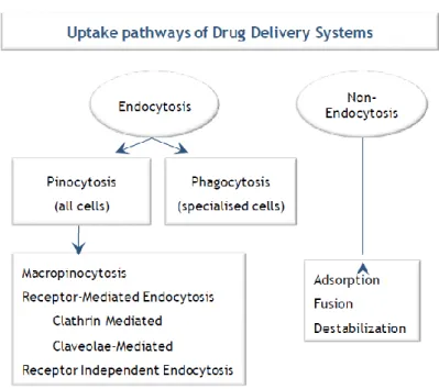
surrounding fluid, forming an intracellular membrane-bound vesicle or endosome (31). Different pathways may be related to endocytosis including: i) phagocytosis, which is exclusive of specialized cells called phagocytes (comprising macrophages, monocytes, dendritic cells and granulocytes) and ii) pinocytosis, which is common to all cell types (Figure 1). Pinocytosis can be further divided into: macropinocytosis, clathrin-mediated or caveolae-mediated endocytosis and clathrin/caveolae independent endocytosis (32). It is believed that such a diversity of mechanisms is used by the cells to accomplish different tasks and might have an effect on the intracellular trafficking of liposomes and thereby on the success of drug delivery(30) .
Figure 1 - Different uptake pathways for compound cytosolic delivery mediated by NanoDDS. Adapted from (32)
The specific endocytic mechanism, by which NanoDDs are taken up by the cell, depends on several factors such as the size, presence (and type) of targeting ligand and cell type (31). Usually, NanoDDs internalization is mediated by clathrin and involves the binding of a ligand (attached to the internalizing particle) to a specific cell surface receptor. Thus, through a sequence of events, which include the formation of clathrin-coated vesicles and their fusion into endosomes, the endocytosed NanoDDs will end up mainly in the lysosomal compartment (30-32). However, NanoDDs internalization may also occur via caveolae-mediated endocytosis. In this case, small, hydrophobic membrane micro-domains (rich in cholesterol) glycosphingolipids and the protein caveolin-1 form
flask-- 7 flask--
shape pits called caveolae. The uptake of extracellular particles is also believed to be specifically mediated via receptors in caveolae (33).
1.2.4- Liposomes therapeutic application
Liposomes have been extensively investigated as NanoDDS for application in several areas, from cancer chemotherapy, enzymatic and antimicrobial therapy, vaccine formulation, diagnostics and topical therapy (34).
1.2.4.1 Liposomes in gene delivery
The ability of encapsulating DNA into liposomes was demonstrated in the 1970's. A decade later incorporation of DNA into cationic liposomes was shown to increase the efficiency of gene transfection into cells. In previous studies, cationic lipids were synthesized and showed improvements in gene transfer and tolerability of cellular properties, as well as the development of new properties to more efficiently encapsulate DNA in liposomes (35).
1.2.4.2 Liposomes as vaccine adjuvants
Liposomes were long ago shown to be effective immunological adjuvants for protein and peptide antigens. They can induce both humoral and cellular immune responses towards the liposomal incorporated antigens (36). A strategy to increase the effect of a vaccine is to specifically deliver the antigen in the target organ.
The combination of viral proteins in the membrane of liposomes (virosomes) provides the opportunity to explore the targeting and membrane fusogenic properties of viral proteins. This property bypasses the disadvantage of degradation by liposomal content before reaching the cytoplasm, since virosomes are used as the virus itself, introducing the material into the cytoplasm (37) .
- 8 - 1.2.4.3 Liposomes in cancer therapy
Although a drug targeting strategy can potentially improve the clinical efficacy of therapeutics used in the treatment of many diseases, most targeting research has been focused in cancer drugs (38, 39). The high morbidity and mortality among cancer patients evidently justifies this focus on tumor-targeted drug delivery systems.
Cancer is a leading cause of disease and death worldwide. In total, 7.6 million cancer deaths occurred in 2008, accounting for approximately 13% of all cell deaths (40).
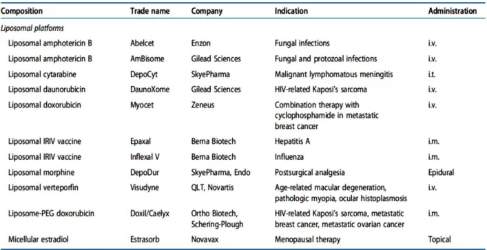
The concept of the “enhanced permeability and retention” (EPR effect) in solid tumors is one of the few tumor-specific characteristics that is becoming a gold standard in the delivery of antitumor drugs (41). It is now a well-established phenomenon that, under certain circumstances (such as inflammation or hypoxia commonly observed in tumors), the endothelial lining in the blood vessel wall becomes more permeable than in the normal state (42). As a result, in such areas, large molecules and even relatively certain particles (10 to 500 nm in size), can leave the vascular bed and accumulate inside the interstitial space. Such spontaneous accumulation or "passive" targeting, is known as the EPR effect. This works particularly well in tumors due to the lack of lymphatic drainage (41). Long-circulating liposomes (with a modified surface usually by polyethylenoglicol - PEG) can be used to treat various tumors, because these can circulate for an extended time and benefit from the EPR effect. Usually, drug-containing liposomes are small enough (60 to 150 nm) to extravasate from blood into the interstitial space of the tumor (43, 44). Table 2 lists some liposomal products that have been approved in the past 15 years. In particular Doxil® (liposomal doxorubicin) was the first liposomal drug formulation approved by FDA (in 1995) for the treatment of AIDS- associated Kaposi sarcoma (45).
- 9 -
Table 2 - Clinically approved liposomal therapeutic formulations. Adapted from (46)
1.3- Pancreatic Cancer
The pancreas is a large gland, with approximately 15.2 cm long and shaped like a leaf, part of the digestive system, involved in the production of insulin and other hormones involved in digestion. The part of the pancreas which produces the digestive juices is called the exocrine pancreas, while the part responsible for the production of hormones (including insulin), is called the endocrine pancreas (47) (Figure 2).
Pancreatic cancer is the fifth most common cause of cancer deaths worldwide, leading to estimated 227,000 deaths per year (48). Pancreatic cancer arises from the morphologically and genetically clearly defined precursor lesions through a step-wise accumulation of genetic alterations. These precursor lesions include pancreatic intraepithelial neoplasia, the intraductal papillary mucinous neoplasm and the mucinous cystic neoplasm (49).
Data from 2003 to 2007 have shown that mortality associated to pancreatic cancer has increased by 0.1% per year in women and by 0.7% per year in men. For this type of cancer, the 1 year relative survival rate is 26% while the 5 year relative survival rate is only 6% (50). The lethal nature of pancreatic cancer is due to its rapid dissemination to distant organs and lymphatic system. In addition, another major cause of treatment failure is intrinsic or extrinsic (acquired) drug resistance, which is frequently observed in
- 10 -
pancreatic cancer cells and results in resistance to known chemotherapeutic agents. The most prominent pancreatic cancer risk factors are: smoking, alcohol, diet/obesity, family history, hereditary pancreatitis, chronic pancreatitis and diabetes mellitus. (51). Usually, symptoms in patients diagnosed with pancreatic cancer do not develop until it is either unresectable or metastatic, rendering it difficult to cure (52). Pancreatic resection currently remains the only chance to cure patients, with a 5-year overall survival rate between 7% and 34% compared to a median survival of 3-11 months for unresected cancer patients (53).
Figure 2 – Global view of pancreas
The head of the pancreas is on the right side of the abdomen where the stomach is attached to the first part of the small intestine (the duodenum). The tail of the pancreas - its narrowest part - extends to the left side of
- 11 -
1.4- Conventional versus new therapies
In pancreatic cancer, the low survival rate of patients points towards an increased need for novel strategies to treat this disease (52). Despite advances in conventional therapies (chemotherapy and radiotherapy) and in targeted therapies which have proved successful in certain cancers (breast, lung, colorectal and melanoma), the survival of pancreatic cancer patients has not substantially improved in the past 30 years (55). At any stage, pancreatic cancer has a very poor response to systemic treatments (56). Several molecular mechanisms have been proposed to be responsible for resistance of pancreatic cancer cells to current treatments. These range from activation of anti-apoptotic signaling pathways) to the role of the stromal cell compartment in drug uptake and activation of alternative escape pathways (57). Indeed, recent data suggests a critical role of the tumor microenvironment in the development and maintenance of resistance to therapies (58). Cancer related inflammation is one of the main features of the tumor microenvironment. The connection between inflammation and cancer is now accepted as enabling some of the most relevant characteristics of cancer (59).
Therapeutic options for patients with pancreatic cancer include external beam radiotherapy alone, combined chemo-radiotherapy (CRT), and single-agent or combination chemotherapy. A summary of conventional drugs used for pancreatic cancer treatment can be found in Table 3. Conventional radiotherapy plus 5-fluorouracil (5-FU) has been associated with a median survival rate of 10-11 months. Over the past decade, gemcitabine has been considered to be the reference treatment in advanced pancreatic cancer. After its approval, a number of randomized controlled trials have been conducted combining several cytotoxic and targeted agents with gemcitabine. However, none of these studies showed a clinically significant survival benefit, compared with treatment with gemcitabine as single agent (60).
- 12 -
Table 3 - Summary of conventional therapies to treat pancreatic cancer
Novel targeted agents have received extensive attention because of the relative insensitivity of pancreatic cancer to conventional therapy (Table 4). Recent attention has been focusing in the inhibition of epidermal growth factor receptor (EGFR) since it is overexpressed in several tumors, and is often linked to poor prognosis. In particular, erlotinib, an EGFR tyrosine-kinase inhibitor, has been demonstrated to have cytotoxicity activity in diverse pancreatic cancer cell lines (77). The use of erlotinib in combination with gemcitabine in chemotherapy-naive patients with locally, advanced, unresectable or metastatic pancreatic carcinoma had been approved in many studies where positive results of phase II trials about erlotinib and gemcitabine have been published (78).
Therapy Compound Autor
Single-agent regimen Gemcitabine (61) S-1 (62) Iritotecan (63) Binary Combination
Gemcitabine and cisplatin (64)
Gemcitabine and S-1
(65)
Gemcitabine oxaliplatin (66)
Gemcitabine and glufosfamide (67)
Gemcitabine and etoposide (67, 68)
Gemcitabine and docetaxel (69)
Capecitabine and doxetaxel (70)
5-fluorouracil and paclitaxel (71)
Iritotecan and oxaliplatin (72)
Gemcitabine and curcumi (73)
Triple or more combination
FOLFOX (combination of 5-fluorouracil, leucovorin and oxaliplatin)
(74)
FOLFIRI (5-fluorouracil, leucovorin and irinotecan) (75)
GEMOXEL (gemcitabine, oxaliplatin
and capecitabine )
- 13 -
Another group of novel targeted agents are monoclonal antibodies. An example of a monoclonal antibody used in the treatment of pancreatic cancer is the inhibitor of cyclooxygenase-2. Cyclooxygenase-2 is found overexpressed in 45 to 75% of pancreatic cancers and was identified as being associated with tumorigenesis, chemoresistance, increased invasion and promotion of angiogenesis in pancreatic cancer (79). Also, the combination of celecoxib (a selective oral cyclooxygenase-2 inhibitor) with gemcitabine and irinotecan to treat advanced pancreatic cancer has been shown that 20% of the patients had a partial response and 80% of the patients had a stable response with a median response rate of 9 months. The median overall survival was 18 months of the patients achieving 1-year survival and 20% achieving 2-year survival (79).
There are several other options of targeted drugs to treat pancreatic cancer such as LY293111, lentinan, PX-12, tipifarnib (80-83) .
Table 4 - Summary of targeted therapies to treat pancreatic cancer
Targeted Therapy Compound Autor
Tyrosine kinase Inhibitors
Erlotinib (77) Gefitinib (84) Sutinib (85) Masitinib (86) Monoclonal antibodies Nimotuzumab/cetuximab (87) Inhibitor of cyclooxygenase-2 (79) Gastrin antagonist (88) Other targeted drugs LY293111 (80) Lentinan (81) Personalized peptide vaccination (89) PX-12 (82) Tipifarnib (83)
Recently, the combination of gemcitabine with a new taxane, nab-paclitaxel (90), or FOLFIRINOX (oxaliplatin, irinotecan, fluorouracil, and leucovorin) (91) was able to provide a survival improvement over monotherapy with gemcitabine. However there is still an urgent need for new and more efficient therapies.
- 14 -
One group of compounds which has shown promising potential as anticancer agents is the Bisnaphthalimidopropyl (BNIP) polyamine derivatives (92).
1.5- Bisnaphthalimidopropyl (BNIP) derivative compounds as
potential antitumor compounds
Napththalimides (originated in the 1970s) and bis-naphthlimido compounds (developed in the early 1990s) are able to intercalate DNA by the planar aromatic rings interfering between the base pairs of the double helix, which distorts the DNA backbone. These compounds were shown to have potential anticancer activity, particularly in breast cancer MCF-7 cells and in vivo on four human solid tumor xenografts, MX-1 mammary carcinoma, CX-1 and DLD-2 colon adenocarcinomas, and LX-1 lung carcinoma (93-95). Bisnaphthalimides are composed of two chromophore units (naphthalimido rings) linked together by linker chains containing at least one or two amine groups. Previous studies using the mononaphthalimide series have proven that this amino group is essential for the cytotoxic activity of these compounds (94).This difference from naphthlamides (one or two amine group in the linker chain) lead to improvements in the DNA binding capacity, and, therefore, the overall therapeutic capacity of these compounds as anticancer agents (96). However, bis-naphthalimides, composed by two naphatalimide rings covalently attached at the end of a linker chain containing two nitrogen atoms, were shown to be highly insoluble in aqueous solutions, making their potential development into therapeutic agents difficult (94, 97). To overcome this problem, the synthesis of bisnaphthalimidopropyl polyamine (BNIP) derivative compounds has been reported (Figure 3) (39). The synthesis of these compounds was based on the theory that the increase of the linker chain between two naphtalimido rings connected to three carbon propyl groups would enhance the aqueous solubility of the compound. Indeed, at that time, the newly synthesized BNIP derivative compounds - BNIPPutrescine, BNIPSpermidine and BNIPSpermine showed increased solubility and induced promising toxic effects in cancer cells (93, 98).
- 15 -
Figure 3 - Chemical structures of bisnaphtalimides. Adapted from (93)
The next generation of BNIPs arose based on the modification of the linker chain, maintaining the primary goal to create potential anti-proliferative compounds and to increase their solubility. For that, compounds were prepared with variations in the number of carbons and insertion of heteroatoms (nitrogen and/or oxygen), cyclohexane ring(s) and /or benzene ring(s), (Figure 4)
.
It has also been reported that with longer alkyl chains, the two naphatalimido rings do not tend to stack on top of each other by π-π interactions between the aromatic rings and hence favor aqueous solubility (99). The cytotoxicity of this new generation of BNIP derivatives was confirmed in a variety of human cancer cell lines, including leukaemia, lung, colon, prostate and breast cancer cells (93, 100, 101).Figure 4- Basic structure of BNIP-derivate compounds showing the point of diversity between different compounds. Adapted from (99).
In particular, two BNIP derivatives, BNIPDaoct and BNIPDaoxoct (which have been used in the present study) (102) (103) were previously shown to have good in vitro cytotoxicity against BxPC-3 (pancreatic cancer) and MDA-MB-231 (breast cancer) cells (92, 104).
- 16 -
However, they also presented undesirable physico-chemical properties: they presented a very poor aqueous solubility. This poor aqueous solubility of the drug also restricts in vivo investigations, since a drug is commonly dissolved in aqueous media for in vivo parenteral administrations. It was previously reported that BNIPDaoct loaded into polymeric sell-assemblies resulted in an aqueous formulation successfully prepared with a drug loading efficiency of 30% (92).
Another strategy to overcome this drawback of poor aqueous solubility is the use of liposomes. As previously described, liposomes have great potential in the release rate control of drugs by appropriate modifications to the carrier itself, without altering the structure of the original drugs. Thus, it is possible that by encapsulating/incorporating the BNIP compounds into liposomes, the lack of aqueous solubility of these compounds may be overcome.
- 17 -
2- Aims
New treatments are urgent for pancreatic cancer. In this context, the primary objective of this project was to evaluate the efficacy and mechanism of action of two BNIP derivatives, BNIPDaoxoct and BNIPDaoct in a pancreatic cancer cell line. The most active compound was then incorporate into liposomes to overcome its poor aqueous solubility.
The specific aims of this thesis were to:
1) Evaluate the cytotoxic activity of two free BNIPs and of two standard drugs in the pancreatic cell line (BxPC-3) and study their effect in:
Cell number and viability; Cell cycle profile;
Apoptosis;
Protein expression levels.
2) Incorporate the most active BNIP compound in a liposomal formulation and confirm its cytotoxic activity in the pancreatic cell line (BxPC-3), by performing the following:
Preparation of liposomes by hydration of the lipid film and extrusion method; Physicochemical characterization of the liposomal formulations
Evaluation of the cytotoxic activity of the liposomal formulation in a pancreatic cell line (BxPC-3).
- 18 -
3- Materials and Methods
3.1- Compounds
Bisnaphthalimidopropyl derivate compounds were kindly provided by Professor Paul Kong (Robert Gordon University, Aberdeen, UK), doxorubicin was purchased from Sigma-Aldrich and gemcitabine was kindly provided from Dr. Mehdi Ouaissi (Marseille,
France). Stock solutions of all compounds (in DMSO) were stored at -20 ºC. BNIPDaoct was prepared in a stock concentration of 20 mM, BNIPDaoxoct of 79 mM, doxorubicin of 2.2 mM and gemcitabine of 152 mM.
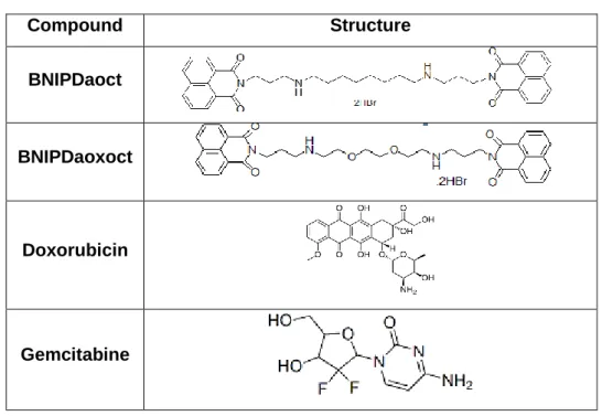
Table 5 - Chemical structure of the compounds/drugs used in this study
Compound Structure BNIPDaoct BNIPDaoxoct Doxorubicin Gemcitabine 3.2- Cell Culture
The human pancreatic adenocarcinoma cell line, BxPC-3 (American Type Culture Collection, USA) was maintained at 37 ºC in a humidified atmosphere with 5% CO2. Cells were grown in RPMI-1640 medium with ultraglutamine I (Lonza, Belgium) supplemented with 5% or 10% heat inactivated fetal bovine serum (FBS, Gibco USA). When the cells reached approximately 80% confluence (or when starting experiments), cells were
- 19 -
trypsinized. For this, cell medium was removed and cells were washed with PBS (Gibco, USA). Cells were trypsinized with TrypLE™ Express (Gibco, USA) for 10 min at 37 ºC. Thereafter, trypsin was inactivated with 5% serum-containing RPMI-1640 medium. The cell suspension was then centrifuged at 172 x g in aHigh speed 350-r centrifuge (Mpw, Poland) and further resuspended in RPMI-1640 medium containing 5% or 10% FBS, according to the experiments. After counting the number of viable cells, dilutions were made to obtain appropriate cell densities. Cell number and viability was routinely analyzed with trypan blue exclusion assay. For this, cell suspension was diluted 1:1 with 0.2 % trypan blue (Sigma-Aldrich, UK) and cells were counted under a TS100 inverted microscope (Nikon, USA) using a Neubauer chamber (hemocytometer).
3.3- In vitro cell growth inhibitory activity
The effect of the compounds (BNIPDaoct and BNIPDaoxoct), drugs (doxorubicin and gemcitabine) or liposomal formulations in the in vitro growth of BxPC-3 cells was evaluated using the Sulforhodamine B assay (SRB) (105, 106). Appropriate dilutions of compound stocks were prepared in medium supplemented with 5% FBS. BNIPDaoct stock was diluted in a concentration range from 10 to 0.625 μM. BNIPDaoxoct stock and doxorubicin were diluted in a concentration range from 20 to 1.25 μM. Gemcitabine stock was diluted in a concentration range from 40 to 2.5 μM. Doxorubicin was used as a positive control of cytotoxicity. The effect of the vehicle solvent, DMSO, on cell growth was also evaluated by treating cells with the maximum concentration of the vehicle used in each experiment (0.05% for BNIPDaoct, 0.02% for BNIPDaoxoct, 0.83% for doxorubicin and 0.01% for gemcitabine).
Briefly, the SRB assay was carried out by plating 1x10⁴ cells/well in 96-well plates. After 24 h incubation, cells were treated for 48 h with five serial dilutions of each compound (Figure 6). Following 48 h incubation, cells were fixed in situ with 50 μL ice-cold 50% trichloroacetic acid (TCA; VWR, Belgium) and incubated on ice for 1 h. Medium was discarded and the plates were washed five times with water and air-dried. A volume of 100 μL of 0.4% SRB (w/v; (in 1% acetic acid) was added to each well and plates were further incubated for 30 min at room temperature. Unbound dye was then removed by washing three times with 1% acetic acid and the plates were then air-dried. The bound SRB was subsequently solubilized with 200 μL of 10 mM Tris-base pH 10.5 (Sigma-Aldrich, USA) for 15 min at room temperature. Absorbance was measured at 510 nm in a microplate reader (Synergy HT- Biotek). The IC50 value, defined as the drug concentration that inhibits 50% of growth in relation to untreated cells, was then determined for each
- 20 -
compound/drug/formulation by linear regression analysis using the following equation: % viable cells = (Absorbance of concentration tested / Absorbance of Blank)*100.
Figure 5 - Representative scheme of the 96-well plate used for SRB assay.
3.4- Cell treatments for cell cycle and apoptosis analysis
BxPC-3 cells were plated in 6-well plates at a final density of 3x10⁵ cells/well and incubated at 37 ºC for 24 h. Cells were then treated with medium only (Blank), with BNIPDaoxoct [at its IC50 concentration (6.13 μM) or BNIPDaoct at half of its IC50 concentration (0.84 μM)], or with DMSO (control, corresponding to the maximum concentration used). Following 48 h incubation, cell number and viability were assessed as previously described and cells were further processed according to the following experiments.
3.4.1- Analysis of cell cycle profile by flow cytometry
Following 48h treatment, cells were trypsinized as previously described but not discarding the removed cell medium, which was used to inactivate trypsin. Following centrifugation of this cell suspension, cell pellets were fixed in ice-cold 70% ethanol for at least 12 h. Prior to analysis, pellets were re-suspended in PBS containing 0.1 mg/mL RNase A and 5
- 21 -
μg/mL propidium iodide solution (Sigma-Aldrich, USA). Cellular DNA content was analyzed by flow cytometry using a FACSCalibur (BD Biosciences, USA) plotting at least 15,000 events per sample. The analysis of cell cycle distribution was subsequently performed using the FlowJo 7.6.5 Software (Tree Star, Inc., Ashland, USA) after cell debris and aggregates exclusion.
3.4.2- Analysis of apoptosis by flow cytometry
Following 48 h treatment, cells were trypsinized as previously described but not discarding the removed cell medium, which was used to inactivate the trypsin. Following centrifugation of this cell suspension, analysis of apoptosis was carried out using the “Human Annexin V-FITC/PI apoptosis kit” (Bender MedSystems GmbH, Vienna, Austria), according to manufacturer instructions. Briefly, cell pellets were resuspended in 400 µL of binding buffer (previously diluted in water). To 195 µL of this cell suspension, were added 5 µL of Annexin V-FITC and incubated for 10 min in the dark at room temperature. Then, 10 µl of propidium iodide were added to this suspension and further incubated for 2 min of incubation on ice. Annexin V-FITC /PI staining was then analyzed by flow cytometry using a FACSCalibur (BD Biosciences, USA) plotting at least 15,000 events. Samples which were not incubated with annexin V-FITC and propidium iodide were also analyzed to assess autofluorescence presented in each sample. The data was analyzed using the FlowJo 7.6.5 software (Tree Star, Inc.).
3.4.3- Protein expression analysis by Western blot
For protein expression analysis cells were lysed in Winman's Buffer (1% NP-40, 0.1 M Tris-HCl pH 8.0, 0.15 M NaCl and 5 mM EDTA) complemented with EDTA-free protease inhibitor cocktail (Roche) for 30 min (with agitation) at 4ºC. Quantification of the total protein content was carried out with the “DC Bio-Rad Protein Assay” (Bio-Rad) according to the manufacturer's instructions and protein expression was analyzed by Western blot. Briefly, the same amount of protein (20 g) was loaded onto a 12% Bis-Tris gel and subjected to SDS-PAGE electrophoresis. Proteins were transferred electrophoretically to a Hybond C extra nitrocellulose membrane (GE Healthcare) and incubated with Bcl-2 antibody (1:100; Dako). Membranes were then incubated with the secondary antibody HRP-conjugated (Santa Cruz Biotecnhology): anti-mouse-IgG-HRP (1:2000). The signal
- 22 -
was detected with the ECL Western Blotting Detection Reagents (GE Healthcare) exposed to Hyperfilm ECL (GE Healthcare) and revealed using developer and fixer (Sigma).
3.5- Liposome preparation
Appropriate amounts of phosphatidylcholine in chloroform (PC) phosphatidylethanolamine-distearoyl methoxypolyethylene glycol conjugate (PEG-PE) and cholesterol (Chol) (all from Sigma, USA) were combined in a molar ratio of 60:30:5. Then, BNIPDaoct was added to a final theoretical concentration of 50 μM to the phospholipids mixture in chloroform in a glass vial. The solvent was evaporated in a heating plate (37ºC) under a stream of the inert gas, nitrogen for 30 min to form the lipid film. The lipid film was hydrated with of PBS to obtain a final lipid concentration of 5 mg/mL. Liposomes were then extruded sequentially using a LiposoFast LF-50 extruder (Avestin, Germany), through polycarbonate filters ranging from 0.4 to 0.2 μm in pore size. All raw materials were stored at -20ºC except chloroform and liposomes which were stored at 4ºC until further use.
3.6- Characterization of liposomes
3.6.1- Size, polydispersity index and zeta potential
The samples were prepared by 1:10 dilution in distilled water. Size, polydispersity index and zeta potential determinations were performed by dynamic light scattering (DLS) methods using a Zetasizer Nano ZS (Malvern Instruments, Malvern, UK) with a detection angle of 173 º. Measurements were made in triplicate at 25 ºC.
- 23 -
3.6.2- Transmission Electron Microscopy
The morphology of prepared liposomes was observed by transmission electron microscopy (TEM) (JeOL JEM-1400, Tokyo, Japan). About 20 μL of liposome formulation was placed on carbon coated copper grids. Following removal of the excess of liposome formulation, the sample was stained with an aqueous solution of 3% uranyl acetate for 1 min. The excess was again removed and samples were then observed in an electron microscope (JeOL JEM-1400, Tokyo, Japan) at the accelerating voltage of 60 kV.
3.6.3- Encapsulation efficiency
The encapsulation efficiency of BNIPDaoct in liposomes was determined by quantification of encapsulated drug, after the disruption of liposomes with 0.01%Triton. BNIPDaoct has intrinsic fluorescent properties, exhibiting one peak in the emission spectra, at 390 nm, after excitation at 350 nm in a FluoroMax-4 spectrofluorometer (Horiba Scientific, France). Using standard solutions of BNIPDaoct, linear calibration curves were elaborated to determine, by interpolation, the drug concentration.
3.7- Statistical Analysis
Statistical analysis was performed using Student’s t-test. Values were considered significant when p < 0.5.
- 24 -
4- Results and Discussion
4.1- Effect of the compounds/drugs in the growth of BxPC-3 cells
The effect of the four compounds/drugs in the in vitro growth of BxPC-3 cells was analyzed following 48 h incubation using the SRB assay. This assay, which is frequently carried out with cells cultured in 5% FBS medium, measures the amount of protein in the cells, therefore allowing the indirect analysis of cell number. Results (Figure 6) indicate that all the compounds/drugs studied inhibited BxPC-3 cell growth. In addition, the effect of DMSO was also analyzed at the highest concentration used but showed no cytotoxicity towards these cells (data not shown).
Figure 6- Effect of the compounds/drugs studied in BxPC-3 cell growth. Cells were treated for 48h with (A) BNIPDaoct, (B) BNIPDaoxoct, (C) doxorubicin or (D) gemcitabine. Results are the mean ± SD of three
- 25 -
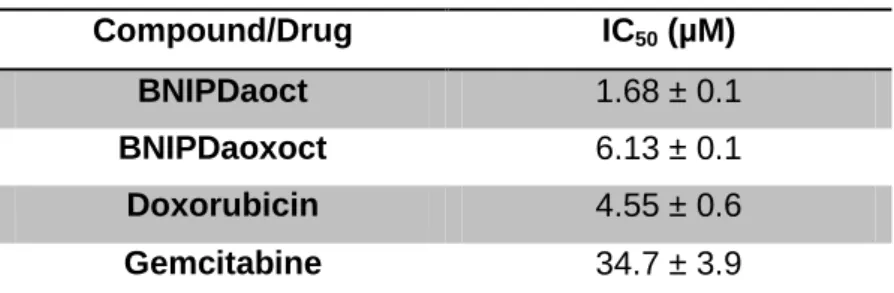
Analyzing the IC50 concentrations obtained for each of the four compounds/drugs studied (Figure 6 and Table 6) it was possible to observe that BNIPDaoct was the most potent one, exhibiting an IC50 of 1.68 ± 0.1 μM, while BNIPDaoxoct and doxorubicin presented IC50 values of 6.13 ± 0.1 and 4.55 ± 0.6 μM, respectively. Gemcitabine presented an IC50 of 34.8 ± 3.9 μM.
Table 6 - IC50 values of the studied compounds/drugs
Compound/Drug IC50 (µM)
BNIPDaoct 1.68 ± 0.1
BNIPDaoxoct 6.13 ± 0.1
Doxorubicin 4.55 ± 0.6
Gemcitabine 34.7 ± 3.9
The data is the mean ± SD of three experiments carried out independently.
BNIPs have been previously described to decrease tumor cells viability, namely in colorectal adenocarcinoma (Caco-2) and breast adenocarcinoma (MCF 7) cells (102, 107). In addition, it was previously reported that alterations in the linker chain between the two naphthalimidopropyl groups in the BNIP compounds, besides affecting their solubility, may enhance their biological activity in breast and colon cancer cell models (99, 104). BNIPDaoct has been previously synthesized from the lead compound BNIPSpd (spermidine derivative) by increasing the length of the alkyl central chain. On the other hand, another derivative, BNIPDaoxoct was synthesized modifying BNIPDaoct structure by inserting two atoms of oxygen. It was previously reported that with the longer alkyl chain, the two naphthalimido rings do not tend to stack on top of each other by π-π interactions between the aromatic rings and hence favor aqueous solubility(99).
In the present work the in vitro growth inhibitory effect of these two compounds, BNIPDaoxoct and BNIPDaoct, was studied in the pancreatic cancer cell line BxPC-3, and compared to currently used drugs (gemcitabine and doxorubicin). Results clearly indicated that from the two BNIP derivatives, BNIPDaoct was the most active compound with an IC50 of 1.68 ± 0.1 μM following 48 h treatment (compared to IC50 6.13 ± 0.1 μM of BNIPDaoxoct). Therefore, the removal of a nitrogen atom does not appear to substantially affect the cytotoxic properties of this compound (102). Also, the cytotoxic activity of the BNIP derivative compounds depends on the length of the polyamine linker chains and also, the number of heteroatoms in the molecules (98). However, in this pancreatic cancer
- 26 -
cell line, the introduction of oxygen atoms in BNIPDaoct structure (originating BNIPDaoxoct) tends to decrease the cytotoxic activity when compared to BNIPDaoct. Indeed, BNIPDaoxoct showed an IC50 of 6.13 ± 0.1 μM in comparison to IC50 of 1.68 ± 0.1 μM of BNIPDaoct. In a previous study, it was reported that the presence of bisnaphthalimidopropyl functionality and a linker chain of <C12 are essential in achieving high DNA-binding interactions and cytotoxic properties and that the introduction of oxygen atoms in the linker chain did not significantly affect either cytotoxicity or DNA-binding capacities (104). So, we can suggest that BNIPDaoct has higher DNA-binding interactions which increase its cytotoxicity activity. To confirm this it would be interesting to study DNA-binding capacity, for example by thermal denaturation studies.
The IC50 of other conventional cytotoxic drugs, such as gemcitabine and doxorubicin has also been determined in this work. The determined IC50 for gemcitabine, the standard drug use for pancreatic cancer therapy, was 34.8 ± 3.9 μM. This value is higher than what has been previously published by other authors for this particular cell line (8.5 ± 0.2 μM). (92). In another study, an IC50 of 80 ± 14.14 nM has been determined (108) and other authors have found an IC50 of 14.0 ± 2.1 nM (109). These differences found between our results and the ones published in different works, are probably due to the different methodologies used to evaluate the cytotoxicity and the different times of treatment of cells with gemcitabine. In addition, the high value for the IC50 of gemcitabine determined in this thesis might be related to a problem of the stock solution of gemcitabine, which may not be stable and may have lost its cytotoxic properties. Regarding doxorubicin, the other conventional drug used in this work, an IC50 of 4.55 ± 0.06 μM was determined, which was similar to the IC50 of 2.2 μM previously determined by others in another human pancreatic cell line, L.3.6 (110).
Based on these results, BNIPDaoct and BNIPDaoxoct were selected for further studies to understand their mechanism of action in BxPC-3 cells.
- 27 -
4.2- Effect of BNIPDaoxoct and BNIPDaoct in viable cell number
The effect of BNIPDaoxoct and BNIPDaoct, at their determined IC50, was directly analyzed in BxPC-3 viable cell number by counting cells with the trypan blue exclusion assay, following 48 h of cellular treatment. Results (Figure 8) showed that, as expected, treatment with the IC50 concentration of BNIPDaxoct caused a reduction in the number of viable cells to 55%. However, treatment with the IC50 concentration of BNIPDaoct resulted in a higher decrease in the number of viable cells than it was expected (to 25%). Therefore it was decided to reduce the BNIPDaoct concentration to 0.84 μM, corresponding to half IC50 concentration and analyse its effect. Results showed that half of IC50 concentration caused a reduction in the number of viable cells to approximately 60% (Figure 7).
Figure 7 - Effect of BNIPDaoxoct and BNIPDaoct in BxPC3 viable cell number. Cells were treated for 48 h with BNIPDaoxoct (6.13 μM) and BNIPDaoct (1.68 and 0.84 μM respectively). Results are presented as percentage of viable cells in relation to blank cells and are mean ± SD of three independent experiments.
Intrigued by the fact that effect of BNIPDaoct was stronger when analyzed with the trypan blue assay than when analysed with the SRB assay, it was decided to verify if the percentage of FBS in the cell culture medium was affecting the results. This was based on the fact that SRB assay had been carried out with cells in 5% FBS supplemented medium (as indicated in the publication where the assay was described, in order to avoid interference of proteins from the FBS in the assay (106)). However, the trypan blue assay was carried out with cells supplemented with 10% of FBS medium. Therefore, the effect of
0 20 40 60 80 100 120 Blank DMSO BNIPDaoxoct DMSO BNIPDaoct IC50 BNIPDaoxoct IC50 BNIP Daoct 1/2 IC50 BNIPDaoct V i a b l e C e l l s ( % o f B l a n k )
- 28 -
BNIPDaoct was further analysed with the SRB assay performed with cells in 10% FBS supplemented medium. Under 10% FBS conditions, the SRB assay provided an IC50 concentration for BNIPDaoct of 0.94 μM (result referring to one experiment only).
The enormous difference obtained in the IC50 concentrations when the SRB assay was performed with 5% or 10% FBS (which is not frequently found for other compounds) also suggests that this compound’s effect will vary immensely with the amount of growth factors available for the cells. This is suggestive of a mechanism of action that is dependent on growth factors availability.
Nevertheless, since the SRB result at 10% refers to one experiment only, it would be interesting to repeat the experiment to confirm this hypothesis.
4.3- Effect of BNIPDaoct and BNIPDaoxoct in BxPC-3 cell cycle profile
To gain insight into the mechanism of action of the compounds (BNIPDaoct and BNIPDaoxoct) their effect on the cell cycle profile of BxPC-3 cells was analyzed by flow cytometry.
Results on Figure 8 show that 48 h cell treatment with BNIPDaoxoct (6.13 μM) caused no major alterations in the cell cycle profile of BxPC-3 cells. On the other hand, treatment with 0.84 μM BNIPDaoct caused an increase in the percentage of cells in S-phase, as well as slight decrease in G2 phase. Regarding treatment with BNIPDaoct (1.68 μM), its effect was not assessed since the number of cells following treatment was not enough for analysis. Treatment with the vehicle (DMSO) seemed to increase slightly the G2/M phase when compared to the blank, but this effect was not statistically significant.
- 29 -
Figure 8 - Cell cycle profile of BxPC3 cells treated with BNIPDaoxoct (6.13 μM) and BNIPDaoct (0.84 μM) for 48 h. Results are the mean ± SD of 5 independent experiments
Although in the case of BNIPDaoct results showed an increase in the percentage of cells in S-phase, as well as a decrease in G2 phase, the profiles were not significantly different. This is not in agreement with previous findings, in which other BNIP derivatives were described to influence cell cycle profile (111).
4.4- Effect of BNIPDaoct and BNIPDaoxoct in apoptotic cell death
Since BNIPDaoct has been previously described by other authors as inducing apoptosis in this particular pancreatic cancer cell line (92), it was decided to confirm the previously described results. Therefore, flow cytometry analysis using Annexin-V-FITC/PI (which is specific for apoptosis) was carried out 48 h following cellular treatment with BNIPDaoct. In addition, the same analysis was also carried out for BNIPDaoxoct. Results (Table 7) showed that the DMSO control of both compounds did not induce apoptosis, since no alterations were observed when compared to blank cells. On the other hand, when cells were treated with BNIPDaoxoct a slight increase (even though not statistically significant difference) of apoptosis was observed when compared with untreated cells. When analyzing the effect of BNIPDaoct, no alteration was observed with 0.84 μM (half of IC50 concentration according to the SRB assay performed with 10% FBS). However a high and statistically significant increase in the levels of apoptosis was observed when cells were
- 30 -
treated with 1.68 μM of BNIPDaoct (p<0.01). Indeed, the levels of apoptotic cells increased from 14.5 ± 6.0 % to 82.0 ± 8.1 % (Table 7)
Table 7 - Percentage of BxPC-3 apoptotic cells following 48 h treatment with BNIPDaoxoct and BNIPDaoct Apoptotic cells (%) Blank 14.4 ± 3.8 DMSO BNIPDaoxoct 14.6 ± 3.9 DMSO BNIPDaoct 14.2 ± 3.7 BNIP Daoxoct (6.13 μM) 21.8 ± 5.6 BNIP Daoct (0.84 μM) 14.5 ± 6.0 BNIP Daoct (1.68 μM) 82.0 ± 8.1 *
The results are the mean ± SD of 3 independent experiments. (* P< 0.01 between treatments and their DMSO control)
Results from flow cytometry analysis using Annexin-V-FITC/PI showed a slight increase in apoptosis, although not statistically significant, following treatment with 6.13 μM of BNIPDaoxoct (IC50) in relation to the blank or DMSO control. When analyzing the effect of BNIPDaoct, no alteration was observed with 0.84 μM (1/2 of IC50 concentration determined with 5% FBS). However, it was observed a statistically significant high increase in the levels of apoptosis, following treatment with 1.68 μM (IC50 concentration at 5% FBS). Indeed, the levels of apoptotic cell death increased from 14.5 ± 6.0 % to 82.0 ± 8.1 % of apoptotic cells (P<0.01). This was in agreement with previous studies in which BNIPDaoct treatment in BxPC-3 cells had induced apoptotic cell death (92). The same has been observed for BNIPDaCHM in the non-small lung cancer cell line NCI-H460 (111).
4.5- Effect of BNIPDaoct and BNIPDaoxoct in protein expression levels
The effect of the compounds BNIPDaoxoct and BNIPDaoct was analysed in the expression of the anti-apoptotic protein Bcl-2 in BxPC-3 cells. Preliminary results (from only one experiment) seem to indicate that both BNIP derivatives decreased the levels of Bcl-2 protein in BxPC-3 cells (Figure 9).