THE ANTIMICROBIAL EFFECT OF RED WINE ON Bacillus Cereus IN SIMULATED GASTROINTESTINAL CONDITIONS
Thesis presented to Escola Superior de Biotecnologia of the Universidade Católica Portuguesa to fulfill the requirements of Master of Science degree in Microbiology
by
Miguel José Santos Vaz
THE ANTIMICROBIAL EFFECT OF RED WINE ON Bacillus cereus IN SIMULATED GASTROINTESTINAL CONDITIONS
Thesis presented to Escola Superior de Biotecnologia of the Universidade Católica Portuguesa to fulfill the requirements of Master of Science degree in Microbiology
by
Miguel José Santos Vaz
under the supervision of
José António Gomes Couto and Timothy Alun Hogg
i
Resumo
Diversos estudos têm vindo a descrever uma panóplia de efeitos benéficos na saúde humana, potencialmente atribuíveis ao consumo de vinho, incluindo efeito anti-oxidante, anti-carcinogénico, anti-inflamatório, anti-cardiovascular, assim como propriedades antimicrobianas. Este estudo foi conduzido com o objectivo de avaliar a actividade antimicrobiana do vinho sobre Bacillus cereus, células vegetativas e esporos. Os resultados apresentados neste trabalho indicam claramente, via testes in vitro, que o vinho inactiva com eficácia as células vegetativas das duas estirpes de B. cereus utilizadas. O vinho tinto inactivou as células vegetativas em fase estacionária, atingindo-se números de colónias não detectáveis (< 500 CFU mL-1), em menos de 10 s de exposição. Como tal, os ensaios de inactivação subsequentes foram efectuados com vinho diluído com água (diluição de 1:4 e 1:8). O vinho diluído 1:4 causou uma redução de 4.5 ciclos logarítmicos nas contagens de células viáveis, em 20 s de ensaio. No entanto, os esporos de B. cereus apresentaram uma elevada resistência à exposição directa ao vinho, com reduções nas contagens inferiores a 1.0 ciclo logarítmico, em 3 h. A influência de componentes do vinho (etanol, ácidos orgânicos, baixo pH e compostos fenólicos) também foi contemplada neste estudo, em células vegetativas. A combinação de ácidos orgânicos e etanol resultou numa actuação sinergética, que provocou padrões de inibição de viabilidade celular similares aos do vinho. Os compostos fenólicos testados não causaram inactivação das células (nas concentrações utilizadas). Relativamente aos resultados obtidos em condições gástricas simuladas, em contexto de refeição simulada, podemos concluir que o consumo de vinho ao longo de uma refeição pode diminuir consideravelmente o número de células de B. cereus que poderá persistir no tracto gastrointestinal. O queijo fresco pasteurizado conferiu maior protecção às células do B. cereus, quando comparado com a matriz arroz com frango. Nesta investigação também foi avaliado o comportamento de esporos de B. cereus quando submetidos a condições gastrointestinais na presença e na ausência de vinho. A presença de vinho inibe a multiplicação das células resultantes da germinação de esporos no fluido intestinal sintético, dando origem a contagens totais (células vegetativas e esporos) de B. cereus mais baixas do que na ausência de vinho. Esta tese gerou resultados que indicam que o consumo de vinho durante uma refeição conduz à redução do número de células viáveis de B. cereus no tracto gastrointestinal, assim como à diminuição do impacto da eventual germinação de esporos que pode ocorrer no intestino, reduzindo, consequentemente, o risco de infecção que o referido patogénico pode causar.
ii
Abstract
Several studies describe the burgeoning health benefits of red wine consumption, including anti-oxidative, anti-carcinogenic, anti-inflammatory, anti-cardiovascular and antimicrobial properties. This study aimed to evaluate the antimicrobial activity of wine against Bacillus cereus vegetative cells and spores. The results of this work clearly show, via in vitro tests, that wine exerts a strong inactivation effect against vegetative cells of two B. cereus strains. The red wine tested inactivated B. cereus stationary phase vegetative cells to undetectable numbers (< 500 CFU mL-1) in less than 10 s. Thus, further inactivation assays were carried out with wine diluted with water (1:4 and 1:8). Wine diluted 1:4 caused a reduction of 4.5 log cycles on viable cell counts, in 20 s. Nevertheless, B. cereus spores were found to be highly resistant to the wine exposure, with decreases in the counts lower than 1.0 log cycles, after 3 h. The influence of wine components (ethanol, organic acids, low pH and phenolic compounds) was investigated on vegetative cells. Organic acids, when combined with ethanol, acted synergistically and conduced to a similar inhibition pattern as that of wine. The wine phenolic compounds assayed displayed no activity against the vegetative cells at the concentrations studied. Regarding data obtained in simulated gastric conditions, in a simulated meal context, we can conclude that the ingestion of wine during a meal diminishes considerably the number of B. cereus cells persisting in the alimentary tract. Pasteurized fresh cheese was found to be more protective to the cells than the chicken-rice matrix. We also evaluated the behavior of B. cereus spores under gastrointestinal conditions. In a consumption-like scenario, the treatment SGF (synthetic gastric fluid)-SIF (synthetic intestinal fluid) +Food+Wine, when compared to the system SGF-fluid)-SIF+Food+Water, led to lower total counts of B. cereus in the intestine, showing that wine inhibits the multiplication of the cells obtained from the germination of spores.
This work provides evidence that drinking wine with meals leads to a reduction of the number of viable cells of B. cereus and reduces the impact of the germination of spores that may occur in the small intestine, thus lowering the risk of infection the aforementioned pathogen may cause.
iii
Acknowledgements
The work described in this thesis was carried out in the Microbiology of Wine Laboratory, at Escola Superior de Biotecnologia, Católica University.
I would like to deeply thank my supervisory team of Dr. José António Gomes Couto and Dr. Timothy Alun Hogg, for their outstanding knowledge, encouragement, guidance and constructive advice throughout this thesis which have contributed to the success of this study.
I am grateful to my colleagues for all the help and advice. You have been great colleagues and wonderful friends.
iv
Contents
RESUMO……… i ABSTRACT………... ii ACKNOWLEDGEMENTS………... iii CONTENTS………... iv ABBREVIATIONS……….... viLIST OF FIGURES………... vii
1. INTRODUCTION………. 1
1.1. B. cereus taxonomy………... 1
1.1.1. Bacillus genus……….. 1
1.1.2. B. cereus group………... 1
1.1.3. B. cereus sensu stricto……….……….. 2
1.2. B. cereus spores……….. 4
1.2.1. Bacterial spores general considerations and B. cereus spore structure……… 4
1.2.2. B. cereus spore properties……… 5
1.2.3. Spore formation and germination of B. cereus spores………. 6
1.2.4. Using B. cereus as a probiotic……….. 7
1.3. B. cereus ecological niches and food spoilage……….. 8
1.4. B. cereus human infections……….. 9
1.4.1. B. cereus nongastrointestinal infections……….. 10
1.4.2. B. cereus gastrointestinal infections……… 10
1.4.2.1. B. cereus gastrointestinal infections: Outbreak reports………. 10
1.4.2.2. B. cereus gastrointestinal infections: Emetic food-borne disease and detection of cereulide……… 11
1.4.2.3. B. cereus gastrointestinal infections: Diarrhoeal food-borne disease and detection of cytotoxins……….. 13
1.5. Gastrointestinal barrier……….. 15
1.6. Wine……… 17
1.6.1. Biochemical composition of wine and actual global wine consumption demand…………. 17
1.6.2. Red wine consumption and its health benefits………... 18
1.6.2.1. Cardiovascular disease prevention………... 19
1.6.2.2. Cancer prevention……… 20
1.6.2.3. Antimicrobial properties………... 20
1.7. Aim of the research………. 23
2. MATERIALS AND METHODS……….. 24
2.1. Bacterial strains and growth medium………. 24
v
2.3. Wine……….. 24
2.4. Wine components……… 24
2.5. Behavior of B. cereus vegetative cells and spores in wine and wine components………. 25
2.6. Synthetic gastric and intestinal fluids preparation……… 25
2.7. Behavior of B. cereus vegetative cells in simulated gastric medium………. 26
2.8. Behavior of B. cereus spores considering simulated gastrointestinal passage……….. 26
2.9. Data analysis……….. 26
3. RESULTS AND DISCUSSION………... 27
3.1. Behavior of B. cereus vegetative cells and spores in wine and wine components………. 27
3.2. Behavior of B. cereus vegetative cells in simulated gastric medium………. 33
3.3. Behavior of B. cereus spores during the simulated gastrointestinal passage………. 35
4. CONCLUSIONS………... 41
5. FUTURE WORK……….. 43
vi
Abbreviations
ATCC American Type Culture Collection
aw Water activity
B. Bacillus bp Base pair(s)
CFU Colony Forming Units
DNA Deoxyribonucleic acid
DPA Dipicolinic acid
EFSA European Food Safety Authority
EU European Union
Fig. Figure
g gram
GIT Gastrointestinal tract
h Hour(s) M (106), molar m Milli (10-3), meter Mb Megabase MHL Million Hectolitres min Minute(s)
PCR Polymerase Chain Reaction
RNA Ribonucleic Acid
rpm Revolution per minute
s Second(s)
SGF Synthetic/Simulated Gastric Fluid SIF Synthetic/Simulated Intestinal Fluid
TSA Tryptone Soy Agar
TSB Tryptone Soy Broth
UK United Kingdom
USA United States of America
vol. Volume
v/v Volume/volume
w/v Mass/volume
vii
List of Figures
Fig. 1. Microscopic images of B. cereus cells. Bars, 5 µm. (A) Vegetative cells aggregating in stationary phase; (B) aggregated cells forming spores; (C) spores present in foam. Images adapted from de Vries et al. (2004)……….……….
4
Fig. 2. Transmission electron micrograph of a sporulating culture of the emetic B. cereus
strain F4810/72. Adapted from Shaheen (2009)……… 5
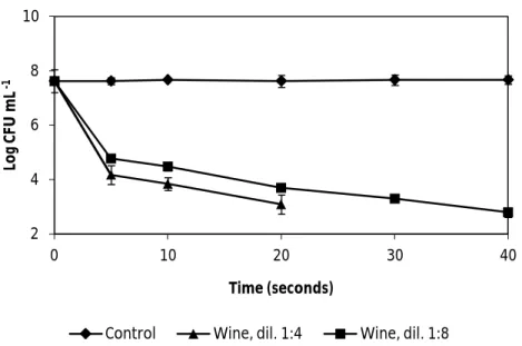
Fig. 3. The effect of wine, dilutions 1:4 and 1:8, on the vegetative cells of B.cereus ATCC 11778. Error bars represent the standard deviation of the mean of three
replications………. 28
Fig. 4. The effect of wine, dilutions 1:4 and 1:8, on the vegetative cells of B. cereus ATCC 14579. Error bars represent the standard deviation of the mean of three
replications………. 28
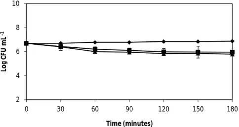
Fig. 5. The effect of wine on the spore counts of B. cereus ATCC 11778 and ATCC 14579. Error bars represent the standard deviation of the mean of three
replications………. 29
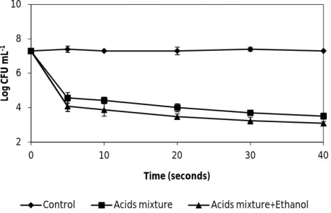
Fig. 6. The effect of wine organic acids mixed (5.5 g L-1 tartaric acid, 0.5 g L-1 acetic acid, 2 g L-1 lactic acid and 0.5 g L-1 citric acid) with and without ethanol 13% (v/v) , diluted 1:4, on the vegetative cells of B. cereus ATCC 14579. Both solutions of organic acids were adjusted to pH 3.3. Error bars represent the standard deviation of the mean of three replications………... 30
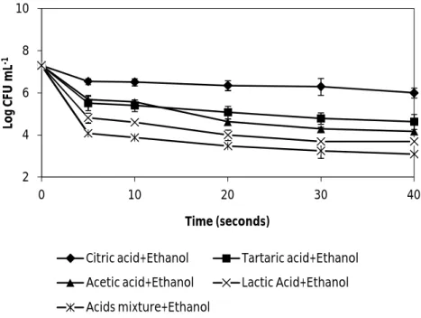
Fig. 7. The effect of wine organic acids, mixed and separated (5.5 g L-1 tartaric acid, 0.5 g L -1
acetic acid, 2 g L-1 lactic acid and 0.5 g L-1 citric acid) supplemented with ethanol 13% (v/v) , diluted 1:4, on the vegetative cells of B. cereus ATCC 14579. All solutions were adjusted to pH 3.3. Error bars represent the standard deviation of the
mean of three replications……… 31
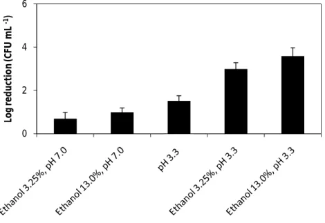
Fig. 8. The effect of ethanol 3.25% and 13% (v/v) at pH 7.0 and pH 3.3, and the effect of low pH (pH 3.3), on the vegetative cells of B. cereus ATCC 14579, after 7 min. Error bars represent the standard deviation of the mean of three
replications………... 32
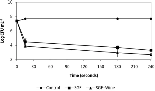
Fig. 9. The effect of synthetic gastric fluid (SGF) and the effect of SGF combined with wine on the vegetative cells of B. cereus ATCC 14579. Error bars represent the standard
viii
deviation of the mean of three replications………... 34
Fig. 10. The effect of different treatments on the vegetative cells of B. cereus ATCC 14579, in a model stomach system. Chicken-rice baby meal was the food matrix used. Error bars represent the standard deviation of the mean of three replications………... 35
Fig. 11. The effect of different treatments on the vegetative cells of B. cereus ATCC 14579, in a model stomach system. Pasteurized fresh cheese was the food matrix used. Error bars represent the standard deviation of the mean of three replications………... 35
Fig. 12. The effect of different treatments on the spore counts of B. cereus ATCC 14579, considering passage from simulated gastric fluid (SGF) to simulated intestinal fluid (SIF). Chicken-rice baby meal was the food matrix used. Error bars represent the standard deviation of the mean of three replications……….. 36
Fig. 13. The effect of different treatments on the total counts of B. cereus ATCC 14579, considering the passage from simulated gastric fluid (SGF) to simulated intestinal fluid (SIF). Chicken-rice baby meal was the food matrix used. Error bars represent the standard deviation of the mean of three replications……… 37
Fig. 14. The effect of different treatments on the spore counts of B. cereus ATCC 14579, considering the passage from simulated gastric fluid (SGF) to simulated intestinal fluid (SIF). Pasteurized fresh cheese was the food matrix used. Error bars represent the standard deviation of the mean of three replications……… 37
Fig. 15. The effect of different treatments on the total counts of B. cereus ATCC 14579, considering the passage from simulated gastric fluid (SGF) to simulated intestinal fluid (SIF). Pasteurized fresh cheese was the food matrix used. Error bars represent the standard deviation of the mean of three replications……… 38
1
1. Introduction
1.1. B. cereus taxonomy
1.1.1. Bacillus genus
The genus Bacillus includes members that demonstrate a wide range of diversity from physiology and ecological niche to DNA sequence and gene regulation. The bacteria in this genus belong to the family Bacillaceae, phylum Firmicutes. The Bacillus genus comprises species distributed ubiquitously in the environment, being commonly isolated from soil, air, water and dust (Harwood, 1989; Drobniewski, 1993). The bacteria found in this genus are aerobic or facultative anaerobic, Gram positive endospore-forming rod shaped organisms (Harwood, 1989; Drobniewski, 1993). The species important for human health are species belonging mostly to two groups, B. cereus and B. subtilis, both members of Bacillus RNA group 1 (Stackebrandt and Swiderski, 2002).
The vegetative cells range from approximately 0.5 by 1.2 to 2.5 by 10 µm in length and most Bacillus species grow optimally at temperatures from 25 °C to 37 °C. Several thermophilic and psychrophilic species exists which can grow at temperature up to 75 °C and down to 3 °C, respectively (Drobniewski, 1993). Many bacilli produce extracellular hydrolytic enzymes capable of breaking down polymers such as polysaccharides, proteins or peptides and nucleic acids allowing the bacteria to use the monomers/oligomers as carbons sources and electron donors (Maidgan, 2003). Some species in the Bacillus genus are responsible for the production of several antibiotics, such as bacitracin, polymyxin and cirulin. (Maidgan, 2003) Traditionally they are classified as low GC gram positive bacteria, however their GC level range from 32% to 69% (Drobniewski, 1993; Maidgan, 2003).
1.1.2. B. cereus group
The B. cereus group, a very homogeneous cluster within the Bacillus genus, comprises B. cereus, B.anthracis, B. thuringiensis, B. weihenstephanensis, B. mycoides and B. pseudomycoides (Drobniewski, 1993; Lechner et al., 1998; Nakamura, 1998). These six species are genetically closely related, but with regard to pathogenicity they differ; B. cereus – food poisoning, systemic infections; B. thuringiensis – insect pathogen and B.anthracis – the causative agent of anthrax. Recent data have proposed that B. cereus sensu stricto, B. anthracis, and B. thuringiensis should be considered as one species designated B. cereus sensu lato (Helgason et al., 2000; Drobniewski, 1993). Nevertheless, other studies indicate sufficient genetic discrimination between B. anthracis, B. cereus and B. thuringiensis (Keim et al., 1997) and the genetic differentiation of B. cereus and B. thuringiensis (Cherif et al., 2003). Thus, at the moment there is no consensus as to whether these bacteria should be considered separate species or specialized variants of a single species. Despite these arguments, members of the B. cereus group retain their status as separate species due to their varying and distinctive pathogenic features (Priest et al., 2004). In general, species of the B. cereus group have a
2
low G+C content of DNA (35%) (Drobniewski, 1993), hydrolyze lecithin and do not ferment mannitol to acid (Maidgan, 2003). Formerly, these species were classified as distinct because of the great relevance of their phenotypical differences, which formed the basis for their classification. These three species differ in virulence, which is encoded by genes located on plasmids recognized as mobile genetic elements (Van der Auwera et al., 2007). These include the cry gene encoding δ-endotoxins of B. thuringiensis, pXO1 plasmid carrying genes for the anthrax toxin complex and the pXO2 encoding the poly-γ-D-glutamic acid capsule of B. anthracis as well as the positive regulator of the virulence factor AtxA located on pXO1 (Arnesen et al., 2008). The B. cereus emetic toxin genes (ces) are also present on a large plasmid (Hoton et al., 2005; Rasko et al., 2007). B. anthracis is capable of capsule formation and the production of toxins that lead to carbuncles in animals and in humans, causing the disease known as anthrax (Mock and Fouet, 2001). The species B. thuringiensis is an insect pathogen which produces insecticidal δ-endotoxins during sporulation and is commercially used for crop protection (Drobniewski, 1993). The species B. mycoides and B. pseudomycoides are phenotypically distinguishable from the species B. cereus sensu stricto by their rhizoidal colony shape and whole cell fatty acid composition (Nakamura, 1998). B. weihenstephanensis is the psychrotolerant species within the B. cereus group, characterized by the ability to grow aerobically at 7 ºC or lower in agitated liquid culture but not at 43 ºC, possessing a signature sequences in the major cold shock gene cspA and in the 16 rDNA sequence (Lechner et al., 1998). Finally, B. cereus lacks those characteristics and can cause food contamination. Later, however, despite these phenotypic differences, comparison of their 16S rRNA nucleotide sequences revealed less than 1% divergence between them (Ash et al., 1991), but a cutoff of 3% divergence is recommended as a conservative criterion for demarcating species, supporting the suggestion as a single species. Therefore, the sequences of 16S rDNA and the phenotypical traits are some of the main factors that give rise to the phylogenetic discussions about the classification of these species. The main diagnostic features of B. cereus sensu lato are their ability to hydrolyze lecithin and an inability to ferment mannitol.
1.1.3. B. cereus sensu stricto
B. cereus is a Gram-positive, motile, spore forming, aerobic rod-shaped bacteria, generally 0.9 to 1.2 µm by 2 to 4 µm and the spore is ellipsoidal, central or paracentral, rarely distending the sporangia (Granum, 1994). It is ubiquitous in nature and is the most frequently isolated soil bacterium, also contaminating agricultural products, playing a major role in spoilage of food products (Kramer and Gilbert, 1989). It may also be present in a diversity of foods such as pasta, rice, dairy products, dried foodstuffs, vegetables, fruit, seafood, meat and poultry (Schoeni and Wong, 2005). The organism has also been identified in cocoa bean fermentations (Ardhana and Fleet, 2003) and cocoa powders (Te Giffel et al., 1997; Ardhana and Fleet, 2003). Optimum growth occurs between 28 and 37 °C although the temperature range is between ≤ 5 and 55 °C (Forsythe, 2000). Growth may occur over a wide range of pH, from 4.1–9.3 and at salt concentrations of up to 7.5% (Clavel et al., 2004). The minimum water activity (aw) for growth is 0.93 (Forsythe, 2000). Under optimum conditions generation time for B. cereus may be as short as 18–27 min, however at temperatures around 5 °C this increases
3
substantially (Choma et al., 2000). It is also possible for B. cereus to adhere to the surface of stainless steel, a material commonly used within the food industry. Once attached, cells may form a biofilm, which confers a number of benefits to the cells with regards to survival in unfavourable conditions. The process of biofilm formation has been described by Wijman et al. (2007) and involves attachment of the organism to a surface within a matrix of exopolymeric substances.
As has been previously discussed, B. cereus is genetically linked to other organisms in the B. cereus group. However, the genetic characteristics, virulence factors and growth/survival characteristics of organisms within the species B. cereus can vary considerably (Carlin et al., 2006). Nevertheless, emetic strains appear to share a distinct cluster of characteristics. There is also evidence to link emetic B. cereus virulence with other toxin producers in the B. cereus group (Ehling-Schulz et al., 2006). Carlin et al. (2006) demonstrated that when compared to diarrhoeal and non-toxigenic strains, emetic strains were unable to grow below 10 °C, but were able to grow at 48 °C. On average the spores from emetic strains were more heat resistant at 90 °C but showed reduced germination potential, especially at lower temperatures. No differences were observed at growth over the range 24–37 °C or at pH 5, 7 or 8. This confirms the special risk involved from emetic strains in foods which are kept warm after cooking, but perhaps not in refrigerated foods. Ehling-Schulz et al. (2006) comment on the close relations between emetic strains in contrast to the diversity discovered among diarrhoeal isolates.
The method for the enumeration of B. cereus in foods has been standardized by the International Organization for Standardization (ISO, 2004). The method is based on growth on mannitol egg yolk polymyxin (MYP) agar. Other plating media are commonly used for the isolation, detection and enumeration of B. cereus from foods, including PEMBA (polymyxinpyruvate-egg yolk-mannitol-bromthymol blue-agar) (Holbrook and Anderson, 1980; Mossel et al., 1967). In addition to selective compounds like polymyxin, these media utilize the bacterium’s lecithinase production (egg-yolk reaction giving precipitate zones) and lack of mannitol fermentation. A thorough description of these media is found in Kramer and Gilbert (1989). More recently, chromogenic media have been developed for several food pathogens, including B. cereus (for instance Cereus–Ident-Agar from heipha Dr Müller GmbH, and chromogenic B. cereus Agar from Oxoid Ltd). B. cereus ATCC 14579 is the type strain of B. cereus (Fig.1), being an environmental isolate and harboring the three enterotoxins, but not the emetic toxin (Ivanova et al., 2003).
4
Fig.1. Microscopic images of B. cereus cells. Bars, 5 µm .(A) Vegetative cells aggregating in stationary phase; (B) aggregated cells forming spores; (C) spores present in foam. Images adapted from de Vries et al. (2004).
1.2. B. cereus spores
1.2.1. Bacterial spores general considerations and B. cereus spore structure
Bacterial spores are described as the ultimate survival vehicles, and have astonished scientists for over a century. Spore-formation is a very successful survival and dispersal strategy: spores are encountered virtually everywhere, and may persist for long periods as they are difficult to destroy. Considering these characteristics, it is not hard to imagine that spores cause major problems in settings where sterility and hygiene are factors of extreme importance, such as the medicare and food-industry. Consequently, bacterial spores are of prime interest from both fundamental and applied perspectives. Moreover, several spore-formers have pathogenic properties, extending the occurrence of their spores from a hygiene issue to a health issue. In this thesis, such a pathogenic spore-former is introduced in a food context: B. cereus.
Bacterial spores are highly specialized, differentiated cell types, designed for the survival of adverse conditions. They are formed inside the bacterial cell and hence called endospores. All Bacillus species can form heat stable endospores (Harwood, 1989). The bacterial endospore is a resting, dormant, tough, non reproductive structure and it is the most resistant living structure known (Atrih and Foster, 1999). Endospores formed by Bacillus and related aerobic endospore-forming Firmicutes are a strategy to survive during unfavourable conditions. The structures of the mature spore are: 1. The core which is the analog of the vegetative cell protoplast as it contains DNA, ribosomes, tRNA and a high concentration of dipicolinic acid (DPA) and of Ca+2 (Setlow, 2006). It contains only 25–50% of water, i.e. the content of free water is extremely low such that the macromolecular movement is greatly restricted (Cowan et al., 2003). 2. The germ cell wall is composed of peptidoglycan identical to that of the vegetative cell (Setlow, 2006). 3. The cortex consists of a peptidoglycan which is different from the vegetative cell peptidoglycan (Atrih and Foster, 1999). The cortex is essential for the formation of a dormant spore and for the reduction of its water content (Andersson, 1998).4. The spore coat complex consisting of several layers of different proteins, mostly spore-specific and important for resistance towards chemicals and lytic enzymes (Setlow, 2006). 5. The exosporium which is a loose fitting balloon-like structure (Yan et al., 2007). The B. cereus exosporium contains more than 20 proteins
5
(Todd et al., 2003), amino and neutral polysaccharides, lipids and ash (Matz et al., 1970). Alanine racemase protein is a major component of the exosporium of B. cereus spores (Yan et al., 2007). It converts reversibly L-alanine to D-alanine (Todd et al., 2003). The structures of the spores of the B. cereus emetic strain F4810/72 are displayed in Fig. 2. The exosporium of the Bacillus spores is surrounded by a hair like external protein layer. It has been a suggested that these proteins are tightly absorbed on spore surface after the cell lyses or are included between the coat and the exosporium (Todd et al., 2003). B. cereus spores are covered with appendages not present in many other Bacillus species (Arnesen et al., 2008). The appendages consist of protein as the main part together with a small amount of carbohydrate and lipid (Arnesen et al., 2008). There was a large difference in the protein profiles of the appendages of different strains of B. cereus (Granum, 2007) and a variation in the surface characteristics of B. cereus spores between strains (Tauveron et al., 2006).
Fig. 2. Transmission electron micrograph of a sporulating culture of the emetic B. cereus strain F4810/72. Adapted from Shaheen (2009).
1.2.2. B. cereus spore properties
Diverse properties reported for the spores of B. cereus make them a problem for the food industry. B. cereus spores are highly resistant to adverse conditions such as heat, dehydration, desiccation, starvation, ionizing radiation, mechanical abrasion, hydrolytic enzymes, extreme pH values, antibiotics, disinfectants and cleaning agents (Setlow, 2000). The spores of B. cereus are hydrophobic and adhere to the processing equipment and subsequently form biofilm (Andersson et al., 1995; Peng et al., 2002). The resistance properties reported for the spores of B. cereus are also a problem for human health. B. cereus spores are highly resistant to acidity in a range of media simulating the conditions in the human stomach after food ingestion. The decrease in the spore counts was less than 1.5 log CFU mL-1 after 6 h of incubation at pH 1 and 1.5 (Clavel et al., 2004). The conditions prevailing during the sporulation: the temperature (Gonzales et al., 1999) and the composition of the sporulation medium (de Vries et al., 2004) affect the properties of the formed spores. The heat resistance of the B. cereus spores increases with the increase in sporulation temperature (Gonzalez et al., 1999). B. cereus spores showed higher survival at 90 ºC when the spores were produced at 37 ºC as compared
6
to 15-20 ºC (Gounina-Allouane et al., 2008). Spores of B. cereus have extreme metabolic dormancy with respiratory activity of low as 10-4 of the maximum rate for vegetative cells metabolizing substrate (Andersson, 1998). Several components are important for the resistance properties of the spores. Dipicolinic acid (pyridine-2,6-dicarboxylic acid) is responsible for the reduction of the spore core water content during sporulation and for the UV photochemistry of the spore DNA. This molecule comprises ~5 to 20% of the dry weight of Bacillus spores. It is chelated with divalent cations, mainly Ca+2 (Setlow, 2007). The small acid-soluble proteins (SASP) in the spore core play an important role of spore resistance. SASP proteins (α, β) represent 5-10% of the total core protein which is sufficient to saturate and protect the spore DNA (Setlow, 2007) especially against UV radiation. SASP also play a role in the osmoresistance of spores (Tovar-Rojo et al., 2003).
1.2.3. Spore formation and germination of B. cereus spores
The spore formation, also called sporulation, involves asymmetric cell division with a copy of the genome partitioned into each of the sister cells. The smaller cell develops into the mature endospore and the mother cell contributes to the differentiation process of the endospore and then autolyses releasing the mature spore into the environment (Henriques and Moran, 2007). It takes approximately 6 h for the process of spore formation of B. cereus to complete (Henriques and Moran, 2007). Spore germination involves a series of rapid degradative reactions, leading to dismantlement of the unique spore structure and loss of spore dormancy and resistance. The subsequent steps that lead to cell-enlargement and cell-division are termed outgrowth, which is considered a separate process, distinct from germination (Campbell and Leon, 1958). Germination can be enhanced by several treatments, including heat-treatments, time, and certain chemicals (Keynan and Evenchick, 1969). This enhancement is called activation (Foster and Johnstone, 1990), and the underlying mechanisms have not been resolved yet. Germination is a non-log-linear event. Some spores form vegetative cells within 2 h, others only after many hours or even days. For 12 B. cereus strains tested, 2 out of the 10 strains did not germinate and the maximum spore germination was obtained after 100 min with no additional germination was observed up to 160-200 min (Broussolle et al., 2008). Germination occurs without need for synthesising any new macromolecules and all the needs are present in the mature dormant spores (Moir, 2006). In the process of germination, substances acting as germinants permeate the outer coat and cortex layers of the spores and interact with receptors located in the inner spore membrane (Moir, 2006). Then compounds such as monovalent cations (H+, Na+, K+), divalent cations (Ca+2, Mg+2, Mn+2) and DPA are released from the spore core (Moir, 2006). The germ cell wall becomes the bacterial cell wall when the spore germinates. The release of Ca-DPA triggers the hydrolysis of the spores peptidoglycan cortex by activating the cortex lytic enzymes (Moir, 2006). Hydrolysis of the peptidoglycan is required for germination and outgrowth of the spores (Atrih and Foster, 1999). The spore core rapidly takes up water so that the core water content rises to that in the protoplast of growing cells and the macromolecular motion and enzyme activity in the core are restored. In B. cereus L-alanine and the purine ribonucleoside inosine are effective germination-promoting compounds (Gounina-Allouance et al., 2008) and D-alanine is an effective inhibitor of
L-7
alanine-induced germination. The most rapid germination of B. cereus spores was observed in a mixture of 0.1 mM L-alanine and 0.1 mM inosine. B. cereus spores failed to germinate in minimum salts medium with glucose plus yeast extract in 0.1 mM inosine (Warren and Gould, 1968). Other germinants have also been identified, like L-phenylalanine, L-glutamine, a mixture of L-asparagine, glucose, fructose and K+. Some mammalian cells like Caco-2 cells were reported to induce germination of enterotoxigenic B. cereus spores whereas HEp-2 cells did not trigger germination (Wijnands et al., 2007). A number of germination receptors are present in the spores of B. cereus. B. cereus type strain ATCC 14579 spore may contain seven functional receptors (Hoornstra et al., 2006). gerP-encoded protein of B. cereus is believed to be important in establishing a coat that is permeable to germinants. Variability of response to inosine or to L-alanine was observed between spores of B. cereus. Some strains can germinate at low germinant concentration (i.e. 0.05 mmol L-1) (Broussolle et al., 2008). Mild preheating activates spores to germinate, in the presence of germination permissive environment. Optimal heating temperature depends on the sporulation temperature. B. cereus spores that were formed at 37 ºC require 80-90 ºC heat shock for activation, whereas those formed at room temperature, need only heat shock of 70-75 ºC (Becker et al., 2005). Gamma-radiation, reducing agents such as thioglycolate or mercaptoethanol and oxidizing agents also may activate the spores (Andersson, 1998).
1.2.4. Using B. cereus as a probiotic
Probiotics are live microbial feed supplements which beneficially affect the host by improving its intestinal microbial balance. The potential benefits that are claimed include improved efficiency of feed, protection against infectious disease (Duc et al. 2004; Fuller, 1989), enhancement of the host immune responses or inhibition of tumor growth in animal models (Spinosa et al., 2000). Among the bacteria used as probiotics, bacteria belonging to the genus Bacillus represent a peculiar situation. Unlike other bacteria, B. subtilis, B. cereus, B. clausii (Spinosa et al., 2000) and B. coagulans are given orally as spores, not as vegetative form Bacillus, even if they are suspected (for example in the case of B. natto). The following three basic mechanisms have been proposed for how orally ingested nonindigenous bacteria can have a probiotic effect in a host: (i) immunomodulation (that is, stimulation of the GALT-gut-associated lymphoid tissue) (e.g., induction of cytokines), (ii) competitive exclusion of gastrointestinal pathogens (e.g., competition for adhesion sites), and (iii) secretion of antimicrobial compounds which suppress the growth of harmful bacteria (Duc et al., 2004). Since the mammalian intestine is an anaerobic environment and Bacillus spp. are preferentially aerobic, germination and outgrowth of spores in the intestine seem difficult to attain. Moreover, the capacity to survive the lytic action of bile salts, one of the criteria used to select potentially probiotic strains, is highly uncommon in nonenteric microorganisms (Spinosa et al., 2000). Although recent evidence suggests that Bacillus spores do germinate in the gastrointestinal tract, it remains unclear which form, cell, spores, or both, is actually responsible for the competitive exclusion and probiotic effects. B. cereus belongs to the few species of Bacillus genus that are used as probiotics (Barbosa et al., 2005). Probiotic B. cereus are
8
used as animal feed supplements (Schierack et al., 2009), for aquaculture (Ravi et al., 2007) and for human (Duc et al., 2004).
1.3. B. cereus ecological niches and food spoilage
B. cereus has a ubiquitous presence in nature and can be isolated from a wide variety of environmental samples (Granum, 2007; Kramer and Gilbert, 1989). It can be found in many types of soils, sediments, dust and plants (Kramer and Gilbert, 1989; von Stetten et al., 1999; Schoeni and Wong, 2005). Spores may be passively spread and thus found also outside natural habitats. It is believed that B. cereus sensu lato exists in soil as spores, and germinates and grows when brought in contact with organic matter or an insect or animal host. Interest in the ecology of this bacterium spurred a study showing that B. cereus could germinate, grow and sporulate in soil, thus demonstrating a saprophytic life cycle (Vilain et al., 2006). Furthermore, a multicellular phenotype with a filamentous mode of growth was observed and suggested to be a means of translocation through soil (Vilain et al., 2006). A multicellular, filamentous mode of growth has also been observed in the gut of insects. The intestines of insects were suggested as a habitat for B. cereus when sporeforming bacteria, later identified as B. cereus, were isolated from guts of different soil-dwelling arthropod species, where the bacteria appear to exist in symbiosis with their invertebrate host. The role of the insect gut microbial communities as a natural niche for part of the B. cereus life cycle is further discussed by Jensen et al. (2003), and it is also suggested that the existence of different morphological modes used by B. cereus, such as the filamentous mode, may be adaptations to different life cycles like the ‘normal’ cycle of life as a symbiont or the more infrequent pathogenic life cycle with rapid growth. B. cereus has been reported to be present in stools of healthy humans at varying levels (Kramer and Gilbert, 1989; Jensen et al., 2003). Its ubiquitous low level presence in environments, feed and foods would ensure B. cereus a transient presence in the mammalian gut (Kramer and Gilbert, 1989). However, genomic data from the B. cereus type strain ATCC 14579 and from B. anthracis suggested that their metabolic capacity is more adapted to the use of proteins as a nutrient source than carbohydrates, and furthermore that genes for establishment within a host were conserved (Ivanova et al., 2003). Another nuance to the scenario is a recent genomic and phenotypic comparison between B. cereus strains ATCC 14579 and ATCC 10987 which revealed that ATCC 14579 actually has the capacity to metabolize a larger number of carbohydrates than what was initially believed based on genomic analysis alone (Mols et al., 2007). These data suggest that in addition to a full life cycle in soil, where it is richly present, B. cereus is also adapted to a lifestyle in a host, as a pathogen or perhaps as a part of intestinal flora, as well as to growth in foods. The possible adaptation of B. cereus to the environment of the animal gut could be the basis of their proposed probiotic effect. Being present in so many environments, it is expected that B. cereus should also be found in water; however, there are not many data on the presence of B. cereus in water sources, and standard methods for the detection from water are not available. Norwegian surface waters were investigated for presence of B. cereus spores, and cytotoxic strains were isolated from several rivers (Østensvik et
9
al., 2004). This suggests the possibility that the water supply may be a means by which B. cereus enters the food processing chain.
Growth of unwanted bacteria can cause enormous expenses for food industry, as this may lead to food spoilage. B. cereus can be isolated from a wide range of different foods and food ingredients, including rice, dairy products, spices, dried foods and vegetables (Kramer and Gilbert, 1989). Cross-contamination can distribute spores or cells to other foods, such as meat products (Gilbert and Kramer, 1986; Granum, 2007). At harvest, B. cereus cells or spores may accompany plant material into food production areas and establish on food-processing equipment. Food spoilage caused by B. cereus occurs mainly in dairy industry, thereby for instance shortening the shelf-life of milk. B. cereus is present in soil, on cattle feed and in cattle faeces and is thus ubiquitously present in the dairy farm environment. From these sources raw milk can be easily contaminated with B. cereus, as its spores germinate more easily in milk than spores from other bacilli (Wilkinson and Davies, 1973). Spores and vegetative B. cereus cells present in food products can attach to processing equipment and form biofilms. Biofilms are multicellular complexes embedded in a matrix of exopolysaccharides that grow attached to a surface. Cells embedded in a biofilm are more resistant to cleaning agents and other anti-microbial substances, making them difficult to eradicate from processing equipment (Peng et al., 2002). Biofilms in processing equipment are a continuous source of contamination for food products by detachment of cells and spores from the biofilm. Biofilm formation may also cause economic losses by causing equipment failure (Kumar and Prasad, 2006). Modern large-scale food production technology, with extended use of refrigeration as a means of conservation, has created a cold niche well suited for bacteria that are not very competitive, but that can survive heat treatment and also grow at low temperatures. In addition to dairy products, lightly heat-treated foods with extended refrigerated storage also represent a new and favourable environment for B. cereus group species. Considering the ubiquitous presence of B. cereus, its resilient spores, and the non-fastidious nature of this microorganism, no type of food with pH > 4.8 (Gilbert and Kramer, 1986) can be excluded as a possible vehicle or as representing a risk of food spoilage or food-borne disease. Failure by consumers to follow basic food preparation rules, i.e. slow or inadequate cooling, storage at ambient temperature or prolonged heat-keeping at < 60 ºC, may allow growth of B. cereus and it happens often in cases of food-borne disease. B. cereus strains isolated from food spoilage incidents generally do not produce cereulide. In contrast, enterotoxin producing strains are commonly isolated from food. Recently, however two psychrotrophic strains were also shown to produce cereulide (Thorsen et al., 2006). Therefore, advanced knowledge is needed about B. cereus diversity, behavior and pathogenic capacity in order to allow for better control of this pathogen in foods and in food production environments.
1.4. B. cereus human infections
B. cereus and related species can cause two forms of food poisoning, in addition to several forms of nongastrointestinal disease.
10 1.4.1. B. cereus nongastrointestinal infections
B. cereus can give rise to a number of local and systemic clinical infections. B. cereus spores can be found within hospital environments and as such may contaminate dressings, intravenous catheters and linens (Drobniewski, 1993). Local infections from B. cereus may develop in post surgical situations, traumatic wounds, burns and also in the eye. Ocular infections may take the form of keratitis, endophthalmitis and panophthalmitis (Gigantelli et al., 1991). It is usually the case that such infections will arise in immunocompromised patients and also in those engaged in intravenous drug use via contamination of the drug or injection equipment. However, the introduction of a foreign body into the eye and contaminated contact lens solutions has also been implicated in infections (Pinna et al., 2001). Systemic disease may take the form of bacteraemia or septicaemia, bacterial endocarditis, respiratory or central nervous system infections. Drobniewski (1993) provides a thorough review of reported cases, which includes incidences of meningitis, pneumonia and endocarditis. It has been speculated that bacterial toxins and enzymes are the key virulence factors in B. cereus infections. Beecher et al. (1995a) concluded that ocular virulence was multifactoral and that the toxin hemolysin BL was only partly responsible for ocular infections.
1.4.2. B. cereus gastrointestinal infections
1.4.2.1. B. cereus gastrointestinal infections: Outbreak reports
Two distinct food-borne disease types, emetic and diarrhoeal, are associated with B. cereus. Both are generally mild and self-limiting, although more serious and even lethal cases have occurred (Granum, 1994; Lund et al., 2000).
B. cereus is an important cause of food-borne disease worldwide (Clavel et al., 2007; Granum, 2007), although it is probably highly under-reported in official lists of food-borne disease causes. In the European Union, Bacillus species (including non-cereus) were reported to be responsible for 1.4% of food-borne outbreaks in 2005 (Anonymous, 2006). In the years 1992–2006, 45 outbreaks of gastroenteritis attributed to Bacillus spp. in England and Wales were reported to the Health Protection Agency Centre for Infections (www.hpa.org.uk/infections/topics_az/bacillus/fp/fpdata.htm). Between 1993 and 1998 in the Netherlands, B. cereus accounted for 12% of food-borne disease outbreaks where a causative agent was identified (Schmidt, 2001). Several factors contribute to the number of food-borne B. cereus disease being largely under-reported. It is a consequence of the generally short and mild course of disease, which does not motivate the patient to seek medical attention. Furthermore, when diagnosed, the disease is not reportable. In addition, cases and/or outbreaks may not always be attributed to B. cereus, because the symptoms of the emetic disease are not easily distinguished from those caused by S. aureus intoxication, and the B. cereus diarrhoeal disease shows the same symptoms as C. perfringens type A food poisoning. The number of cases of B. cereus food-borne disease is reportedly increasing in industrialized countries (Gilbert and Kramer, 1986; Kotiranta et al., 2000). However, as the surveillance systems for food-borne disease differ
11
between countries, it is difficult to compare data and obtain true incidence estimates. Examples of cases and outbreaks are well described in several publications (Gilbert and Kramer, 1986; Kramer and Gilbert, 1989; Granum, 2007). Somewhat different distribution between countries is observed for the emetic and diarrhoeal diseases, which could partly be a reflection of the association of the two types of disease with different food vehicles: in Japan and the UK, the emetic disease dominates (Gilbert and Kramer, 1986; Shinagawa et al., 1995), while in Northern Europe and North America, the diarrhoeal disease seems more prevalent (Kotiranta et al., 2000). At least part of the difference in disease pattern is probably due to different eating habits, but it is difficult to document whether the distribution is truly different and not a result of reporting differences.
1.4.2.2. B. cereus gastrointestinal infections: Emetic food-borne disease and detection of cereulide
The emetic food poisoning was identified in the UK in the 1970s and was frequently associated with the consumption of cooked rice dishes (McElroy et al., 1999). The emetic form of illness is characterised by nausea, vomiting, abdominal cramping and malaise (Granum, 1997). Symptoms may appear between 0.5 and 6 hours after consumption of the contaminated food (Arnesen et al., 2008). The illness is usually self limiting, with recovery occurring within 24 h. The rapid onset of the symptoms is due to the ingestion of a preformed toxin, cereulide. Cereulide is a small, cyclic, heat stable dodecadepsipeptide, identified and named by Agata et al. (1995). Ehling-Schulz et al. (2005) hypothesised that its chemical structure was (D-O-Leu-D-Ala-L-O-Val-L-Val)3 and demonstrated that it was in fact produced by a nonribosomal peptide synthetase (NRPS). This hypothesis was based on the characteristics of the toxin and its synthesis in the late exponential and stationary phase of the organism’s growth (Häggblom et al., 2002). Ehling-Schulz et al. (2006) have confirmed the chemical structure as above and further sequenced the cereulide synthetase (ces) gene cluster and placed it on a 208 kb megaplasmid (now named pCER270 – Rasko et al., 2007). This has many similarities to the pXO1-like plasmids implicated in B. anthracis toxin production (Hoton et al., 2005). This similarity provides further evidence of the close relationship between the various members of the B. cereus group. The genetic regions flanking the ces gene also have a high homology to the virulence plasmids of other B. cereus, anthracis and thuringiensis. There is also evidence to suggest that the ability to produce emetic toxin is restricted to a single evolutionary lineage, owing to very low diversity among emetic isolates (Ehling-Schulz et al., 2005). In contrast, considerable diversity has been found among diarrhoeal isolated and other non-emetic strains. Cereulide acts as a K+ ionophore through mitochondrial membranes and interferes with oxidative phosphorylation. The effects on mitochondria have been demonstrated on boar spermatozoa and rat liver cells, with a stimulation of swelling and respiration of the mitochondria observed (Mikkola et al., 1999). Mahler et al. (1997) report on the death of a boy from liver failure associated with impaired function of liver mitochondria due to food-borne cereulide. A similar case has also been reported more recently by Dierick et al. (2005), in which a 7 year old girl died from (among other symptoms) liver failure, 13 h after consuming a pasta dish containing > 108 CFU g-1 B. cereus. Paananen et al. (2002) have also demonstrated that cereulide can
12
inhibit human natural killer (NK) and killer T cells and suggest that it may have immunomodulating properties. For illness to occur the B. cereus must be able to produce the toxin within the contaminated food prior to consumption. Kramer and Gilbert (1989) have reviewed earlier studies of the levels of B. cereus present in foods causing both forms of illness. A total of 107 cases of emetic poisoning were identified with the number of the organism present in the food ranging from 103–1010 CFU g-1. Granum (1997) found counts between 200 and 109 CFU g-1 in incriminated foods and concluded that although foods with a level of > 103 CFU g-1 could not be considered safe. The real infective dose was above 105 CFU g-1. While this level is generally accepted, Häggblom et al. (2002) warn that food poisoning risk cannot be evaluated on microbial load alone and that toxin production should also be measured. Many foodstuffs have been implicated in emetic food poisoning cases including beef, poultry, pasta, infant formula, milk and cream (Granum and Lund, 1997; Schoeni and Wong, 2005). By far the most common cause of the disease, however, is cooked rice dishes. Kramer and Gilbert (1989) review a number of studies, which found between 10 and 100% of raw and cooked rice samples to be contaminated with B. cereus. Cooked rice provides a model for the formation of cereulide within foods. B. cereus spores, present in the rice, are heat activated during the cooking or cooling process and can multiply and produce toxin if the rice is held at room temperature. Finlay et al. (2002) found that toxin was produced by emetic B. cereus in cooked rice at 15–30 °C. Toxin production was significantly greater between 15 °C than 20 or 30 °C. Re-heating of the rice is usually insufficient to inactivate the toxin. Pasta has also been implemented and Rajkovic et al. (2006) found that penne pasta and potato puree provided a better substrate for cereulide production than rice when incubated at 28 °C. At lower temperatures background flora was found to be a determining factor, aeration and agitation were also found to reduce toxin production. Finlay et al. (2000), in addition to their work on cereulide production in cooked rice, have shown that toxin production in skimmed milk is significantly greater at 12 and 15 °C than at 30 °C. This was in agreement with their later study (2002) which showed toxin production on solid laboratory media was greatest at 12 °C. However, Häggblom et al. (2002) suggest that 21 °C was the optimum temperature for toxin production. Agata et al. (2002) also found that toxin production in cooked rice was greatest at the higher temperature of 35 °C. Toxin was also produced at 20 and 30 °C, but not in such quantities. All studies largely agree that the minimum and maximum limits for growth and toxin production are 10 and 37 °C respectively (Finlay et al., 2000). Although B. cereus is frequently isolated from soil, agricultural products and milk, Altayar and Sutherland (2005) have found that emetic isolates are rare. Of the 271 isolates from soil, animal faeces, raw and processed vegetables 45.8% were found to be B. cereus. Of these isolates only 4 were found to produce emetic toxin. This is in contrast to other research which showed that 44% of B. cereus isolated from rice paddies in Bangladesh were emetic toxin producing isolates (Ueda and Kuwabara, 1993), suggesting that rice paddies provide a selective environment for emetic isolates. Svensson et al. (2006) examined 5668 isolates obtained from dairies and dairy farms. No emetic strains were found in milk at the farm during the cow’s outdoor grazing period. Up to 3.8% of milk and environment isolates were found to be emetic while the cow’s grazed in indoor stalls. In total, 0.05% of isolates from dairies were found to produce emetic toxin although there was evidence for 1 silo in particular having a significant emetic flora.
13
Rhesus monkey was used as the experimental animal to demonstrate that the heat stable toxin (cereulide) produced by B. cereus isolates from emetic outbreaks was associated with the emetic syndrome (Melling et al., 1976). The monkey feeding test and ligated rabbit ileal loop (LRIL) methods were used with success to show that the factors responsible for the vomiting and the diarrhoeal illnesses were distinct. The LRIL and the vascular permeability reaction (VPR) methods were found to be of value for studying the diarrhoeal toxin but not the vomiting factor (Turnbull et al., 1979). A quantitative chemical assay was introduced by Häggblom et al. (2002) based on liquid chromatography followed by ion trap mass spectrometry (HPLCMS). This method allows measuring the exact contents of the molecule cereulide in the B. cereus biomass as well as in food or samples from environmental origins. A rapid sperm bioassay was developed in 2004. It allows detecting the toxicity of B. cereus bacterial extract in a short period of time (Andersson et al., 2004). Research on the emetic toxin was hampered by the fact that rodents are insensitive to the orally given toxin (Yokoyama et al., 1999). Therefore primates were needed for each test until Hughes et al. (1988) developed an in vitro assay based on the vacuolisation of the human larynx carcinoma cells (HEp-2 cells). These authors tested samples connected to food poisoning and cultured isolates connected to food poisoning in rice. They noticed that the extracts obtained from some isolates caused vacuoles in the HEp-2 cells. Sakurai et al. 1994 observed that the vacuoles formed in the HEp-2 cells were swollen mitochondria. Agata et al. (1995) extracted and purified the factor which causes the vacuolation in HEp-2 cells from the culture supernatant of B. cereus strain NC7401 connected to a case of emetic syndrome food poisoning and named the toxin cereulide. Shinagawa et al. (1995) and Agata et al. (1995) found that the factor causing vacuolation of HEp-2 cells also caused vomiting when fed to rhesus monkey (Macaca mulatta) and the house musk shrew (Suncus murinus) and concluded that this was the emetic toxin. Cereulide was chemically synthesized by Isobe et al. (1995) and shown to possess the same emetic and pathogenic activities. Andersson et al. (1998) developed a bioassay based on loss of the motility of boar spermatozoa upon 24 h exposure to the toxin. Finlay et al. (1999) developed the metabolic staining assay MTT using as an indicator 3-(4,5-dimethylthiazol-2-yl)-2,5-diphenyltetrazolium bromide, a water soluble yellow tetrazolium salt. This salt was converted to an insoluble purple formazan (MTT) in HEp-2 cells but not in cells exposed to the emetic toxin. This allowed detecting the cytotoxicity of cereulide towards the HEp-2 cells.
1.4.2.3. B. cereus gastrointestinal infections: Diarrhoeal food-borne disease and detection of cytotoxins
Perhaps the first description of the diarrhoeal form of the illness comes from Hauge (1950, 1955) in 1950s. The latter study covers an outbreak of diarrhoeal illness, involving around 600 patients in 4 Norwegian hospitals. Hauge (1955) found that cornstarch used in the preparation of a vanilla sauce contained 104 B. cereus spores g-1. Incubation of sterile vanilla sauce with a solution containing approximately 104 cells g-1 of the B. cereus isolate led to the presence of 107 cells mL-1 in the sauce. After consumption of a portion of the sauce, diarrhoea and abdominal pain lasting 8 h were reported. The mechanism of infection differs from the emetic illness. In the case of diarrhoeal food poisoning
14
bacterial spores and/or vegetative cells are consumed, which survive the acidic conditions within the stomach and germinate within the small intestine (Wijnands et al., 2006). It is here that enterotoxins are produced, causing illness (Drobniewski, 1993). Symptoms can include diarrhoea, abdominal pain and rectal tenesmus, with nausea, vomiting and fever less frequently reported (Hauge, 1955; Granum and Lund, 1997). The illness has an incubation period of between 8 and 16 h and symptoms may last for 12 to 24 h. Many foods have been implicated with the diarrhoeal syndrome including cooked and raw meats, poultry, fish, vegetables, dairy products, desserts and sauces (Schoeni and Wong, 2005). It appears that B. cereus is able to produce a number of different enterotoxins capable of causing the diarrhoeal syndrome, although identification of the toxins themselves attracts some debate. The first toxin to be discovered was a three component haemolysin–HBL (Beecher and Macmillan, 1990). In addition to haemolytic activity, this toxin was found to be dermonecrotic, cause fluid accumulation in ligated rabbit illial loops (Beecher et al., 1995b) and be cytotoxic to Chinese hamster ovary cells. HBL has been suggested as the primary virulence factor in the diarrhoeal syndrome (Granum and Lund, 1997) although this has been frequently questioned due to the wider presence of NHE genes among B. cereus isolates (Moravek et al., 2006). A non-haemolytic, three protein enterotoxin has also been identified – NHE (Lund and Granum, 1996). The 3 components of NHE (Nhe A, B and C) show some similarities to those of HBL and each is required in specific concentration (in a ratio of 10:10:1) for cytotoxic activity (Lindbäck, et al. 2004). NheB has been found to be the binding component for the enterotoxin complex. Other possible enterotoxins such as Cytotoxins CytK-1 and CytK-2 (Lund et al., 2000), Enterotoxin T (Guinebretière et al., 2002) and Enterotoxin FM (Moravek et al., 2006) have been suggested as virulence factors for the illness, however, further research is needed. In addition, several other haemolysins and enzymes have been described as potential contributors to the diarrhoeal disease (Arnesen et al., 2008). It has been demonstrated that preformed enterotoxins are not able to pass through the human digestive system and that live cells may not survive transit through the stomach (Shinagawa et al., 1991). However, Clavel et al. (2004) have shown that vegetative cells may pass through the stomach if the pH is raised sufficiently by the presence of foodstuffs. The degree of survival was found to depend on the pH and the type of food consumed. In extreme conditions (pH > 5.0) B. cereus may even be able to grow during gastric transit (Clavel et al., 2004). Wijnands et al. (2009) comment that up to 26% of vegetative cells ingested may survive the gastric passage under normal conditions. The enterotoxins are thought to be produced during the late exponential phase of growth (Kramer and Gilbert, 1989) so it is necessary for spores to germinate within the human small intestine. Recently, Wijnands et al. (2007) demonstrated that germination of B. cereus was induced by Caco-2 cells, a human cell line which mimics the epithelial cells of the small intestine. This study also demonstrated that the spores were able to adhere to the intestinal cells at a rate of around 1%. Andersson et al. (1998) had previously discussed the possibility of spore adhesion to epithelial cells providing an additional virulence mechanism. Four out of ten strains tested produced spores able to adhere to human epithelial cells. One strain used was involved with an outbreak of diarrhoeal food poisoning where symptoms were more severe and longer lasting than would usually be observed. As with attachment to food and processing equipment surfaces, the hydrophobicity of the spores is a contributing factor in the adhesion mechanism. Minnaard et al. (2001) have also discussed the effect
15
of exocellular factors on human intestinal epithelial cells, including the loss of plasma membrane asymmetry of mitochondrial activity.
B. cereus is expected to be present in different foods and raw materials, and thus detection of the bacterium is not always the main issue for food safety purposes. Instead, ability to detect the possibly harmful strains, or their toxic products, is the highly desired goal. As cereulide and the three cytotoxins Hbl, Nhe and CytK are the main known virulence factors in B. cereus food-borne disease, focus has been on their detection. Antibodies have been produced for the three-component toxins Nhe and Hbl (Dietrich et al., 1999), and two antibody-based detection kits targeting these toxins are commercially available (Buchanan and Schultz, 1994; Day et al., 1994). The BCET-RPLA kit (Oxoid Ltd., UK) is a semi-quantitative assay detecting, by reversed antibody agglutination, the L2 component of Hbl in foods and in cultures of B. cereus (Beecher and Wong, 1994a). The sensitivity of the test is reported to be 2 ng mL-1 test extract. The TECRA-BDE kit (Tecra International Pty Ltd., Australia) detects the NheA component of the Nhe toxin by an enzyme-linked immunosorbent assay (ELISA) sandwich test (Beecher and Wong, 1994a). The sensitivity reported by the manufacturer is 41 ng mL-1 prepared sample, and the kit is intended for use on foods and environmental samples. Neither of the kits will confirm the presence of biologically active toxin, because only one of each of the three-component toxins is detected. For the third and more recently described toxin CytK, there is at present no commercially available detection kit. For nonspecific detection and characterization of B. cereus enterotoxins, different laboratory animal and tissue culture assays have been employed. Among the tests involving live animals are the rabbit ileal loop (RIL) test, performed by injection of B. cereus cultures or extracts into ligated rabbit intestinal loops followed by observation of fluid accumulation, the guinea pig skin reaction, and the vascular permeability assay (Kramer and Gilbert, 1989). The use of tissue culture assays for detecting B. cereus enterotoxins has been shown to correlate well with results from traditional methods, and represent a convenient alternative for screening purposes (Gilbert and Kramer, 1984; Thompson et al., 1984; Shinagawa et al., 1991). The cell culture lines used include CHO cells (Buchanan and Schultz, 1994), McCoy cells (Fletcher and Logan, 1999), Caco-2 cells (Hardy et al., 2001) and Vero cells (Lund and Granum, 1996; Dietrich et al., 1999). For specific detection of the genes encoding the B. cereus toxins Hbl, Nhe and CytK, several PCR schemes, including multiplex PCR, have been developed (Mäntynen and Lindström, 1998; Yang et al., 2005). Considering the wide distribution of cytotoxin genes among B. cereus strains (Mäntynen and Lindström, 1998; Ehling-Schulz et al., 2005), the use of PCR techniques to identify diarrhoeal strains is of little use for practical food safety purposes, because detection of a toxin gene does not reveal the level of toxin production and thus cannot predict the potential pathogenicity of a particular B. cereus strain.
1.5. Gastrointestinal barrier
The mammalian gastrointestinal tract (GIT) and its accessory organs form an essential organ system with intriguing biology. It is composed of 11 main organs: the mouth, pharpharynx, oesophagus, stomach, intestine, colon, anus, and accessory organs which are the salivary glands, liver, gall