Different Schematic Eyes and their Accuracy to the
in vivo
Eye: A Quantitative Comparison Study
*Marina Storani de Almeida and ‡Luis Alberto Carvalho * Universidade Federal de S˜ao Carlos (UFSCar)
‡Grupo de ´Optica, IFSC-USP-Instituto de F´ısica de S˜ao Carlos, Av. Dr. Carlos Botelho, 1465,
13560-250, S˜ao Carlos, SP, Brazil
Received on 4 December, 2006
Current ophthalmic technology allows the manipulation of eye components, such as anterior cornea and lens, of the human eye with a considerable precision and customization. This technology opens up the possibility of exploiting some characteristics of the eye in order to improve the methods of correcting optical aberrations. Moreover, product development and research for the eye-care professional has reached very high standards, since there is nowadays software available to design and simulate practically any mechanical or optical char-acteristic of the product, even before it is thrown into production line. Although quite similar in the general form, different human eye models simulate the image formation by considering different property combinations in the constitutive elements of the eye structure (such as refraction index and surface curvatures), producing retinal images that resemble very closely those of the biological eye. Using optical design software, we have implemented a simulation of 5 well-known schematic eyes available in the literature. These models were the Helmholtz-Laurance, Gullstrand, Emsley, Greivenkamp and Liou & Brennan. The optical performance of these different models was compared using different quantitative optical quality parameters. The model of Liou and Brennan, contains features of the biological eye that were not considered in previous models, as the distribution of a gradient refraction index and a decentered pupil. Furthermore, it has great reliability since it takes into account the mean value of empirical measurements of thein vivoeye in order to define size and parameters such as anterior and posterior curvature of cornea, lens, axial length, etc. Comparisons between the MTF (Modula-tion Transfer Func(Modula-tion), spot diagrams and ray fan showed the difference in image quality between eye models, and the Strehl Ratio was also used as a parameter of comparison. A careful comparison between the different models showed that the first four schematic eyes have better optical quality than what is expected for the general and healthy emmetropicin vivoeye. Liou and Brennan schematic eye is the one that most closely resembles the
in vivobiological eye. Therefore, in applications, such as research or product development for customized vi-sion correction, which must consider optical properties intrinsic to the biological eye, we recommend this latter model; for applications that do not require refraction-limited performance, most of the other models should be a good approximation.
Keywords: Eye model; Schematic Eye; Ray tracing; Optical aberrations
I. INTRODUCTION
The interest in the physiological aspects and optical prop-erties of the human in vivo eye, and how they relate to vi-sual acuity, come from very ancient times. Historically, af-ter Gauss (1841) established the basic laws that govern im-age formation properties, many theoretical models have been proposed. In the late 19th century, Helmholtz undertook a very thorough study on this subject, and published the now famous collection Helmholtz Treatise on Physiological Op-tics[1]. This model was later modified by Laurance and became known as the Helmholtz-Laurance model[2], which contains all optical surfaces found in the biological eye. Al-though this model designates refractive indices to eye com-ponents that not necessarily correspond to true measured val-ues, its overall properties have a close resemblance to those of the human eye. Afterwards, the Swedish Ophthalmologist Allvar Gullstrand (1862-1930) conducted important research in the field of physiology and in 1911 received the Nobel Prize for his work regarding the eye as an optical element [3]. While Gullstrand’s simplified schematic eye treats the cornea as a single refracting surface, just as the previous model from Helmholtz-Laurence, in Gullstrand’s non-simplified model the cornea is considered to have two surfaces, which
guar-antees a perfect image formation at the retina. Although it simplifies the cornea, the vitreous and the aqueous humor, this model is especially suitable for the computation of intraocular lens (IOL) power, as it also contains the anterior and posterior surface of the crystalline lens.
eye, which are described by a set of Zernike Polynomials [21-22]. There is also the double-pass technique [23] which can be used to measure even higher frequency aberrations associated with scatter. Together these techniques and instrumentation form a collection of tools that aid the eye-care professional in providing the best diagnostic and treatment available to their patients. Although these eye models are of great use to the development of such techniques, current eye models are lim-ited since they do not consider all the complexity of the visual system, once the properties of each component are approxi-mations based on the biological eye. Hence, the ongoing in-terest in coming up with better eye models, which is essen-tial to guide the refraction correction methods already in use, improve them and even allow the development of new ones. In 1997 Liou & Brennan have proposed an interesting model [24], which is, to our knowledge, the closest to anatomical, biometric and optical data as compared to the physiological eye. Their objective was to develop a model that could be used to predict visual performance under normal and altered conditions of the eye, using empirical values of ocular para-meters.
All of the models above have their own merit on describing the optical and physiological properties of the human eye. The present work should serve as a guide for precise quantitative comparison between some of the schematic eyes available in literature. Depending upon the application/research that will be implemented, one or other eye should have more desirable features than the other, varying from an extremely simple and approximate eye model to more sophisticated and anatomi-cally correct ones.
II. MATERIAL AND METHODS
The quantitative comparison between the five eye mod-els considered here was based on simulations implemented using the Zemax software package [25]. This software was chosen taking into account the authors’ previous expe-rience and the availability of this tool in our research lab-oratory. However, there are other software such as Code-V (www.opticalres.com) and OSLO (www.lambdares.com) where the current analysis can also be undertaken. In Fig. 1 we list the layouts of the models analyzed.
Observing the layouts we notice that the Helmholtz-Laurance and the Gullstrand models are very similar, having little differences in corneal radius of curvature, size and index of refraction. Actually, the Gullstrand model is a slight im-provement of the Helmholtz-Laurance eye model. The Em-sley eye is the simplest model, as it considers the entire eye as composed of just one material. On the other hand, the Gullstrand and Liou & Brennan models are the most complex ones, as they take into account some aspects of the physiolog-ical eye, such as the shape of the posterior cornea, to evaluate the ray tracing. Besides considering the gradient variation of the refraction index, Liou & Brennan model considers decen-tration and tilt of the pupil that are present in the biological eye for an entrance pupil diameter of 4 mm. Recent research into pupil geometric center has revealed that this feature changes
with change in illumination and pupil size [26-28], although this was not taken into account in our simulations. The first four models presented on Table 1 were proposed for paraxial rays with no restriction on the entrance pupil diameter (EPD) value. However, the Liou &Brennan model [24] was proposed for an EPD of 4 mm and a distance between the eye and the object of approximately 44 cm. Other aperture values would certainly modify the shape and the refraction index of the crystalline lens. Even though, we conducted the simulation using the four aperture values considered for the other models using the same properties for the crystalline lens. As men-tioned before, certain optical engineering functions were used for performance analysis and comparison between the pre-sented models. Among them the Modulation Transfer Func-tion (MTF), the Spot Diagram, the Ray Fan and the Strehl Ratio, which are described in detail in the following section.
III. RESULTS
1. Modulation Transfer Function
The sharpness and contrast of an imaging system or of a component of the system may be characterized by a parameter called the Modulation Transfer Function (MTF), also known as spatial frequency response. The MTF curve has different meanings according to the corresponding frequency.Its height at frequencies of 1.5 cycles/degree represents the contrast-behavior of the optical system [29]. It is known from expe-rience that a good lens system should perform over 95% at this frequency for both sagittal and tangential directions, and values worse than 90% represents a bad performance.
Frequencies in the gap of 3 to 12 or higher cycles/degree represent the sharpness-ability of a lens. MTF readings taken at 12 cycles/degree indicate how good a lens can transmit very fine structures. For an optimal quality based on the human eye, the lens should perform over 50% at 6 cycles/degree [29]. Perceived image sharpness is more closely related to the spa-tial frequency where MTF is 50% (0.5), where contrast has dropped by half. Typical 50% MTF frequencies are in the vicinity of 12 to 24 cycles/degree for individual components and often as low as 9 cycles/degree for entire imaging systems [29].
All the tangential and sagital MTF curves obtained for all 5 models are presented in Fig. 2, and they are organized in in-creasing values of EPD. The tangential and the sagittal modu-lation transfer functions were plotted to illustrate the influence of decentration and tilt of the pupil considered by the Liou & Brennan model.
2. Spot diagram
aper-Model
(year of proposal)
Layout Structural parameters
Helmholtz-Laurance (1909)
Surface Radius (mm)
Thickness (mm)
Refraction index
1 8.0 3.6 1.333
2 10.0 3.6 1.450
3 -6.0 15.18 1.333
Gullstrand (1911)
Surface Radius (mm)
Thickness (mm)
Refraction index
1 7.8 3.6 1.336
2 10.0 3.6 1.413
3 -6.0 16.97 1.336
Emsley (1946)
Surface Radius (mm)
Thickness (mm)
Refraction index
1 5.55 22.22 1.3333
Schwiegerling (1995)
Surf. Radius (mm)
Thick. (mm)
Asph. Refraction index
1 7.8 0.55 0.75 1.3771
2 6.5 3.05 0.75 1.3374
3 11.03 4.0 -3.30 1.42
4 -5.72 16.60 -1.17 1.336
Liou and Brennan (1997)
Surface Radius (mm)
Thickness (mm)
Asphericity Refraction index (555nm)
1 7.77 0.50 -0.18 1.376
2 6.40 3.16 -0.60 1.336
3 12.40 1.59 -0.94 Grad A
4 Infinity 2.43 —– Grad P
5 -8.10 16.27 0.96 1.336
GradA=1.368+0.049057·z−0.015427·z
2−0
.001978·r
2 GradP= 1
.407−0.006605·z
2− 0.001978·r
2
n(λ) =n(0.555µm) +0.0512−0.1455·λ+0.0961·λ
EPD Tangential MTF Sagittal MTF
2mm
4mm
6mm
8mm
FIG.2. Modulation Transfer Function for the five models, labeled as: ,
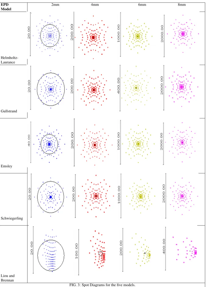
EPD Model
2mm 4mm 6mm 8mm
Helmholtz-Laurance
Gullstrand
Emsley
Schwiegerling
Liou and Brennan
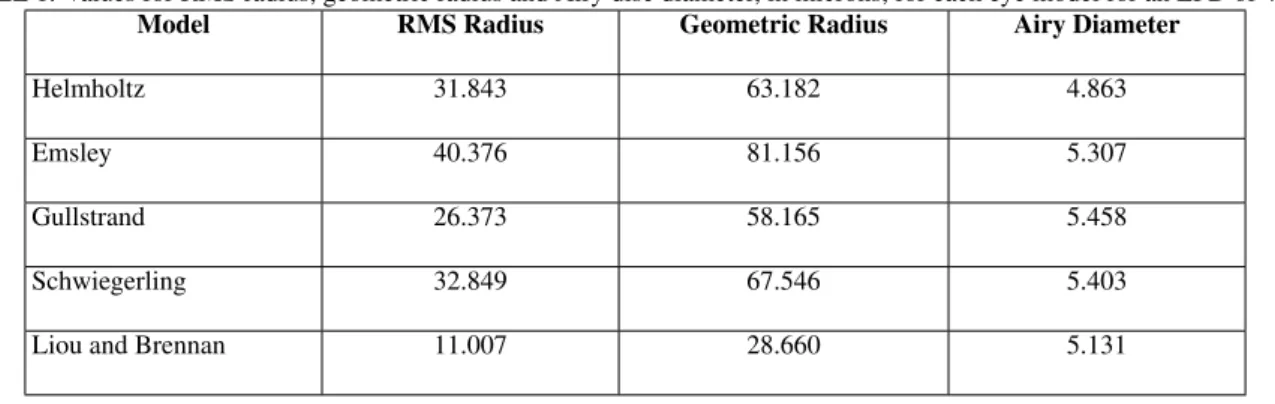
TABLE 1. Values for RMS radius, geometric radius and Airy disc diameter, in microns, for each eye model for an EPD of 4 mm.
Model RMS Radius Geometric Radius Airy Diameter
Helmholtz 31.843 63.182 4.863
Emsley 40.376 81.156 5.307
Gullstrand 26.373 58.165 5.458
Schwiegerling 32.849 67.546 5.403
Liou and Brennan 11.007 28.660 5.131
ture is small, diffraction effects may be more important than lens aberrations in limiting the sharpness of images. When light passes through a small circular aperture, a diffraction pattern is formed consisting of a bright central circle, also known as theairy disk, and low contrast peripheral concen-tric rings [30]. When the eye focuses on a point object, the image formed at the retina will be a circle with the size of the airy disk, even in the ideal case of aberration absence. The airy disk diameter is given by equation 1.
D=2.44λ
d (1)
Whereλis the wavelength andd is is the diameter of the system aperture. Since the airy disk diameter is proportional to the diameter of system aperture, the effects of diffraction increase as the EPD decreases. Wave-front aberrations are the limiting factor in image quality at large apertures and diffrac-tion sets the limit at small apertures. The construcdiffrac-tion of spot diagrams is one of the methods for visualizing the effect of aberrations have on image quality. The distribution of ray in-tersections depends on the aberrations of the system. For our analysis, a square grid pattern was selected at the EPD for ray tracing, where the principle ray is always at the center of the pattern. The airy disk is shown as a black circle for each spot diagram (see Fig. 3).
The root mean square (RMS) spot radius is another measure of quality that relies on the spot diagram. It is computed as the RMS of all distances between each marginal ray intersection (xi,yi)with the image plane and a reference point (xo,yo), gen-erated by the chief ray intersection. The RMS spot radius is given by
Rrms= n
∑
i=1 q
(xi−x0)2+ (yi−y0)2
n (2)
Wherenis the total number of rays considered.
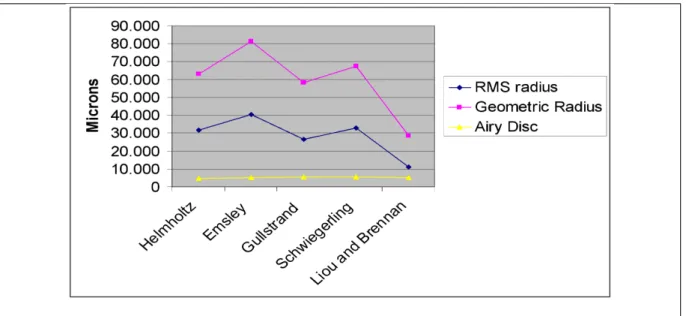
Another value obtained from the spot diagram is the geo-metric radius, which just encloses the outermost ray pierce in the pattern relative to the reference point. The values obtained for RMS radius, geometric radius and the airy disk diameter obtained for each model for an EPD of 4mm are presented in micrometers on Table 1 and Fig. 4.
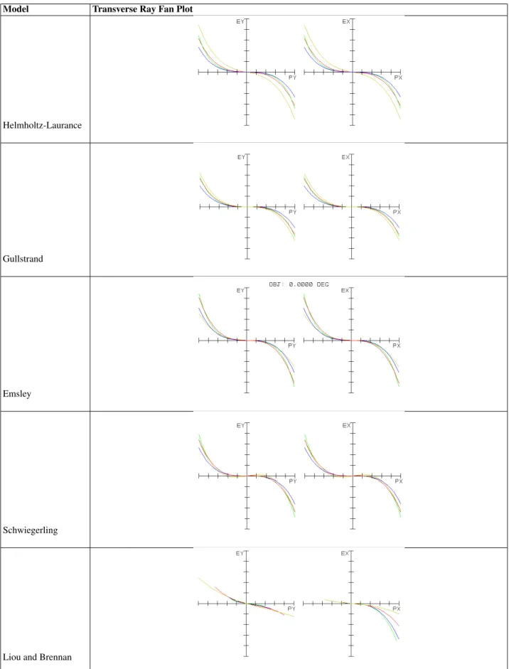
3. Ray Fan
Another technique of analysis is the ray fan plot, which de-scribes the relative distances of the rays that hit the image plane. This diagnostic plot is obtained from an off-axis ray launched along the EPD. The chief ray is the one that pierces exactly at the center of the EPD, and its height on the image plane is the reference for the others rays. The height that rays hit the EPD is normalized and placed at the horizontal axis, while the relative distance of the rays that arrive at the image plane is placed on the vertical axis.
The shape of the ray fan plot depends upon the type and magnitude of the aberrations present in the system. An ideal system would convert all the rays to a unique point on the image plane, and its corresponding ray fan would be a line exactly over the abscissa.
4. Strehl ratio
The Strehl ratio is defined as the ratio of the central in-tensity in the aberrated pattern to the central inin-tensity in the un-aberrated pattern. Unless a design for an imaging system approaches diffraction-limited performance, there is no point in looking at Strehl values [25]. The Strehl ratio ranges from 0 (for systems having significant aberration) to 1 (for perfect systems). In Table 5 and Fig. 5 it is shown the Strehl ratio values for each eye model.
IV. DISCUSSION
The five eye models presented here were simulated using the Zemax software package, based on parameters for each eye model. The retinal images were analyzed by conventional optical design test functions. Comparisons between the MTF, spot diagram and ray fan showed the difference in image qual-ity for the considered models. The Strehl ratio was also used for comparison.
FIG.4. RMS radius, geometric radius and Airy disc for all models based on values from Table 1.
TABLE 5: The Strehl ratio for each model and EPD considered in the analysis. EPD
Model
2mm 4mm 6mm 8mm
Helmholtz 0.795 0.028 0.011 0.039
Emsley 0.826 0.033 0.015 0.013
Gullstrand 0.938 0.066 0.017 0.043
Schwiegerling 0.961 0.065 0.019 0.014
Liou and Brennan 0.820 0.158 0.045 0.038
15 cycles/degrees [31], Liou & Brennan schematic eye is the one that most closely resembles thein vivohuman eye. It is interesting to note that the tangential and sagittal MTFs of the Liou &Brennan model are the only ones that are different for all the considered apertures, something that does not happen for the other models. The reason for this difference between the tangential and the sagittal MTF on the Liou and Brennan model is that, although the object is on-axis, it is not rotation-ally symmetric about the axis of the eye (it considers decen-tration and tilt of the pupil that are present in the biological eye), which means that the MTF will not be rotationally sym-metrical. Another point to be considered is that in general the MTF curves get worse as the aperture is increased, since the models furnish increasing spherical aberration as the aperture increases. A good measure that would consider all meridians at once is the volume MTF [32], which was not implemented at this time.
From Fig. 3 we observe that all the spot diagrams differ from the ideal case, which in the ideal case would be just a point in the image plane. The Airy disk indicates the occur-rence of the diffraction effect on the images. Spot diagrams that are more compact indicate less aberration. ¿From the spot diagrams, it is easy to see that the Liou & Brennan model has the best performance for an entrance pupil diameter of 4 mm. Analyzing the ray fan plots in Fig. 4, we observe an evolu-tion from the Helmholtz to the Liou and Brennan schematic eye, i.e., as abscissa values increase the image size values do not increase as much as in the other models, demonstrating
that the Liou & Brennan model contains less spherical aber-ration . All of the models are good at the central portion of the image plane, at the intersection of the principal ray, but, as expected, marginal rays are the most aberrated. The Gull-strand and Emsley ray fans are the most similar ones, but ob-serving them very carefully we see that the plot for the Gull-strand model has a greater length on the abscissa axis. The Schwiegerling model plot looks like if the system has a spher-ical aberration that is compensated by other aberrations.
Finally, the Strehl ratio was computed for all models (Table 5 and Fig. 5). It may be seen that the Strehl ratio decreases as the EPD increases. This decreasing tendency is slower for the Liou and Brennan model, which means that this model is closer to the diffraction limit for a greater range of EPDs. It is not recommended to calculate the Strehl ratio when the MTF plot is distant from the diffraction limit, as its values will be so small that can simply be ignored. However, its values are shown on Fig. 5 in order to illustrate the rate of growing aberrations for the models considered, and are also a factor of comparison. Although the Liou & Brennan model does not have the greatest Strehl ratio for an EPD of 2 mm, this is the model that has the greatest values of Strehl ratio for the apertures of 4 and 6 mm (it is important to remember here that this model was proposed for an EPD of 4 mm).
Model Transverse Ray Fan Plot
Helmholtz-Laurance
Gullstrand
Emsley
Schwiegerling
Liou and Brennan
FIG. 6. Strehl ratio values for different pupil sizes for each eye model.
have the best MTF, but is the one that most closely approxi-mates to thein vivohuman eye. The other optical design test functions (Spot Diagram, Ray Fan and Strehl ratio) showed the best results for the Liou and Brennan schematic eye. An-other type of comparison, which was not carried out here, but that would certainly add value to the other types of optical quality parameters used, is the Zernike Polynomial fitting of the wavefront for each eye model. A comparison of each Zernike coefficient would clearly and quantitatively indicate
the benefits of each model in terms of individual aberrations. In general terms we may claim that the Liou and Brennan’s schematic eye would be the best choice for applications which require eye models that are optically and physically more sim-ilar to the biological eye. Otherwise, most of the other simpli-fied model eyes should be sufficient for applications which re-quire only an approximation of the optical and physical prop-erties of the eye.
[1] H. von HelmholtzHelmholtz’s treatise on physiological optics
Edited by J. P. C. Southall (2000).
[2] M. Alpern,The eyes and vision.In: Handbook of Optics (1978). [3] A. H. Tunnacliffe,Introduction to Visual Optics.(1989). [4] L. S. Pedrotti and F. L. Pedrotti,Optics and Vision(1998). [5] D. J. Schanzlin and J. R. Robin,Corneal topography:
measur-ing and modifymeasur-ing the cornea.(1992).
[6] D. R. Sanders and D. D. Koch,An atlas of corneal topography.
(1993).
[7] S. D. Klyce, Invest. Ophthalmol. Vis. Sci.25, 1426 (1984). [8] D. D. Koch, G. N. Foulks, C. T. Moran, and J. S. Wakil, Refract.
Corneal Surg.5, 424 (1989).
[9] J. Wang, D. A. Rice, and S. D. Klyce, Refract. Corneal Surg.5, 379 (1989).
[10] R. J. Mammone, M. Gersten, D. J. Gormley, R. S. Koplin, and V. L. Lubkin, IEEE Trans. Biomed. Eng.37, 66 (1990). [11] P. P. van Saarloos and I. J. Constable. Optom. Vis. Sci.68, 960
(1991).
[12] R. B. Mandell, Contact Lens Assoc. Ophthalmol. J. 18, 267 (1992).
[13] L. Carvalho, S. A. Tonissi, and J. C. Castro, J. Cataract Refrac. Surg.25, 821 (1999).
[14] L. A. Carvalho, M. Stefani, A. C. Rom˜ao, L. Carvalho, J. C. Castro, S. Tonissi, P. Schor, and W. Chamon, J. Cataract Refrac. Surg.28, 2006 (2002).
[15] J. E. Greivenkamp, J. Schwiegerling, J. M. Miller, and M. D. Mellinger, Am. J. Ophthalmol.120, 227 (1995).
[16] J. Liang, B. Grimm, S. Goelz, and J. F. Bille, J. Opt. Soc. Am. A Opt. Image Sci. Vis.11, 1949 (1994).
[17] L. A. Carvalho, A. C. Rom˜ao, M. Stefani, L. A. Carvalho, J. C. Castro, F. Yasuoka, F. Scannavino J´unior, J. Santos, P. Schor, and W. Chamon. Arq. Bras. Oftalmol.66, 261 (2003). [18] L. A. V. Carvalho and J. C. Castro, Braz. J. Phys. 33, 140
(2003).
[19] L. A. Carvalho, J. Biomed. Inform.37, 1 (2004).
[20] L. A. Carvalho, J. Castro, W. Chamon, and P. Schor. J. Refract. Surg.22, 954 (2006).
[21] M. Born and E. Wolf,Principles of Optics: Electromagnetic Theory of Propagation, Interference and Diffraction of Light.
7th ed. (1999).
[22] L. A. Carvalho, Proceedings of SPIE5772, 181 (2005). [23] F. Diaz-Douton, A. Benito, J. Pujol, M. Arjona, J. L. Guell, and
(1997).
[25] J. M. Geary, Introduction to lens design with practical ZEMAX°Rexamples.(2002).
[26] G. Walsh, Ophthalmic. Physiol. Opt.8, 178 (1988).
[27] M. A. Wilson, M. C. W. Campbell, and P. Simonet, Optom. Vis. Sci.69, 129 (1992).
[28] L. A. Carvalho and A. C. Rom˜ao, Revista de F´ısica e Instrumentac¸˜ao16, 24 (2003).
[29] N. Koren, Understanding image sharpness part 1: In-troduction to resolution and MTF curves. Available on: ¡http://www.normankoren.com¿, accessed in 12 mar. 2006. [30] M. M. Sternheim and J. W. Kane, General Physics.2nd ed.
(1991).