Rev Bras Cardiol Invasiva. 2014;22(4):382-5
© 2014 Sociedade Brasileira de Hemodinâmica e Cardiologia Intervencionista. Published by Elsevier Editora Ltda. All rights reserved.
Percutaneous Occlusion of Left Atrial
Appendage and Patent Foramen Ovale in the
Same Procedure Avoiding Transeptal Puncture
Francisco Chamié
1, Daniel Chamié
2, Luiz Carlos do Nascimento Simões
3, Ênio Guérios
4, João Carlos Tress
5ABSTRACT
Left atrial appendage occlusion has been successfully em-ployed to prevent embolic events in patients with atrial fibrillation as an alternative to oral anticoagulation. Left atrial access through the patent foramen ovale or ostium secundum atrial septal defect has been discouraged due to the fear that entering the septum in a higher position through the foramen would prevent adequate device posi-tioning. In this manuscript we report a case in which the left atrial appendage and the foramen ovale were sequen-tially occluded avoiding transseptal puncture, making the procedure simpler and faster.
DESCRIPTORS: Atrial appendage. Atrial ibrillation. Cardiac catheterization. Foramen ovale, patent.
1 Hospital Federal dos Servidores do Estado, Rio de Janeiro, RJ, Brazil. 2 Instituto Dante Pazzanese de Cardiologia, São Paulo, SP, Brazil. 3 Intercat – Cardiologia Intervencionista, Rio de Janeiro, RJ, Brazil. 4 Concept − Centro de Cardiopatias Congênitas e Estruturais do Paraná,
Curitiba, PR, Brazil.
5 Hospital de Clínicas de Niterói, Niterói, RJ, Brazil.
Correspondence to:Francisco Chamié. Rua Real Grandeza, 108, salas 223-224 − Botafogo − CEP: 22281-034 − Rio de Janeiro, RJ, Brazil E-mail: [email protected]
Received on: 9/11/2014 • Accepted on: 11/20/2014
RESUMO
Oclusão Percutânea do Apêndice Atrial Esquerdo e do Forame Oval Patente no Mesmo Procedimento
sem Necessidade de Punção Transeptal
A oclusão do apêndice atrial esquerdo tem sido realizada com sucesso para a prevenção de fenômenos embólicos em pa-cientes com ibrilação atrial, como alternativa à anticoagulação oral. O acesso atrial, através de forame oval ou comunicação interatrial tipo ostium secundum, tem sido evitado em função da crença de que o posicionamento do dispositivo é diicul-tado pela disposição mais alta do forame no septo interatrial. Neste manuscrito, relatamos um caso em que foram ocluídos, sequencialmente, o apêndice atrial esquerdo e o forame oval sem a necessidade de punção transeptal, que simpliicou e tornou mais seguro o procedimento.
DESCRITORES: Apêndice atrial. Fibrilação atrial. Cateterismo cardíaco. Forame oval patente.
P
ercutaneous occlusion of the left atrial append-age (LAA) has been used as an alternative to oral anticoagulation in patients for whom this strategy is not safe.1-6 It is a routinely performed procedure,including in Brazil.7-9
Patent foramen ovale (PFO) is located more crani-ally in the septum, and it is believed that its use as an access route for LAA occlusion hinders the cor-rect positioning of the delivery sheath. In such cases, traditional transeptal puncture has been the preferred method, which increases the complexity, time, and potential complications of the procedure. In this study, a case of simultaneous occlusion of PFO and LAA, without the need for transeptal puncture, is reported.
CASE REPORT
The patient was a 66-year-old female, referred for percutaneous closure of the LAA. She had an episode of cryptogenic ischemic stroke when she was young and had chronic coronary disease, having had acute myocardial infarction episodes at the ages of 32 and 33 years. She had undergone coronary artery bypass graft surgery twice, in 1983 and 2000.
She had atrial ibrillation for one year, and was treated with propafenone. In May of 2014, she had a new episode of ischemic stroke; oral anticoagulation with rivaroxaban was prescribed, and was interrupted after conjunctival hemorrhage. At that time, she was referred to LAA occlusion.
Chamié et al. Percutaneous Occlusion of LAA and PFO Rev Bras Cardiol Invasiva.
2014;22(4):382-5
383
The procedure was performed under general anes-thesia, monitored by transesophageal echocardiography (TEE). Intravenous heparin was administered at a dose of 5,000 IU. An IV dose of 2 g of cephalexin was also administered.
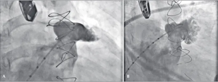
The PFO was crossed using a multipurpose catheter, subsequently replaced by 5F pigtail catheter positioned in the LAA. Injections were performed at 20° in the right anterior oblique projection, with cranial and caudal angulation to identify the LAA; the target-region was measured for prosthesis implantation, as well as the LAA ostium. The appendage was bilobulated. The target-region measured 17 mm and the ostium, 24 mm (Figure 1).
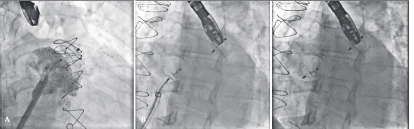
Subsequently, a 0.035”/ 260 cm super-stiff guide wire was introduced inside the LAA and, while main-taining the guide wire in position, the pigtail catheter was removed, being substituted by the Torq Vue® 13 F
long sheath with double curvature structure (St. Jude Medical, Plymouth, United States), positioning it coaxi-ally along the appendage axis. A 24 mm Amplatzer®
Cardiac Plug (AGA Medical Corporation, Golden Valley, United States) was introduced through it, releasing the lobe in the target-region. With the prosthesis lobe well-adhered to the walls of the LAA, the disc was external-ized, perfectly occluding the LAA entrance (Figure 2A). After the adequate prosthesis position was conirmed through the TEE and new atriographies, the device was released, maintaining the long sheath in the left atrium. Subsequently, a 25 mm Amplatzer® PFO Occluder (AGA
Medical Corporation, Golden Valley, United States) was introduced, connected to the Amplatzer® Cardiac Plug
delivery cable, using the long sheath. Implantation was performed as usual, aiming to occlude the PFO (Figure 2B). After the adequate position of the second
prosthesis was conirmed by TEE, the sheath was removed and hemostasis was performed by manual compression. A small femoral arteriovenous istula was detected on the day after the implantation at the venipuncture site, which was submitted to suture for its occlusion.
The patient was discharged in good status, with both prostheses well positioned and occluding their respective defects (Figures 2C and 3).
DISCUSSION
The use of the foramen ovale as the access route for LAA occlusion has been discouraged due to the fear that it’s more cranial position in the interatrial septum would hinder access to the LAA and prevent adequate device positioning inside it. As a result, a more caudal transeptal puncture, more adequate for this purpose, has been the preferred method. This technique increases the luoroscopy time and adds a small degree of risk to the procedure, which should be lower with a more experienced surgeon.10
The viability of LAA occlusion through septal de-fects (ASD or PFO) was demonstrated by Koermendy et al.,11 who reported the advantages of its use in 96%
of selected patients. The absence of transeptal puncture prevented the complications related to this technique and reduced the overall luoroscopy time.
The complication rate of transeptal puncture is low, but includes perforation of heart chambers or the aorta, pericardial effusion followed by tamponade, systemic or cerebral thromboembolism, and coronary or cerebral air embolism, which are all potentially severe situations that have a negative impact on the procedure outcome.10,12-18 There are also cases in which
Figure 1 – Angiography of the left atrial appendage. In A, right anterior oblique view with cranial tilt shows the appendage anatomy, with emphasis on the ostium and target region. In B, right anterior oblique view with caudal tilt demonstrates more clearly the trabecular portion of the left atrial appendage.
Chamié et al.
Percutaneous Occlusion of LAA and PFO
Rev Bras Cardiol Invasiva. 2014;22(4):382-5
384
the transeptal puncture cannot be performed and the procedure has to be abandoned.
Another advantage of avoiding the transeptal puncture is not to create an iatrogenic septal defect, as itoccurs in ablations or in mitral valve repair in approximately 5% of cases. These defects tend to spontaneously close in over 80% of cases after 18 months.19 Although they
have little or no hemodynamic importance, these small iatrogenic defects can be mediators of paradoxical embolism episodes.
The PFO or ASD facilitate and allow for a safe access to the left atrium. PFOs with long tunnels can greatly hinder the access to the LAA; in such cases, if there is dificulty in the atrial appendage catheterization, the traditional transeptal puncture should be performed. In the present case, the occlusion of the PFO and the LAA was indicated based on the patient’s previous
episode of paradoxical embolism, and the occlusion of the LAA was indicated by the complications reported after the use of oral anticoagulation for chronic atrial ibrillation.
CONCLUSION
The occlusion of the LAA through the PFO was simple, fast and safe. This can become an excellent choice of access route in cases with communication between the atria (ASD or PFO), simplifying the pro-cedure and reducing its complications.
CONFLICT OF INTERESTS
The authors declare no conlicts of interest.
FUNDING SOURCES
None declared.
Figure 2 – Prosthesis implantation. In A, injection performed through the long sheath shows that the prosthesis is adequately positioned and that the disk completely obstructs the atrial appendage ostium. In B, fully released left atrial appendage prosthesis and the second prosthesis, of the foramen ovale, implanted in the atrial septum, but still attached to the delivery cable. In C, at the end of the procedure, luoroscopy shows the two released prostheses and in good position.
Figure 3 – Three-dimensional transesophageal echocardiography performed at the end of the procedure shows in A, the prosthesis of the atrial ap-pendage and in B, the foramen ovale prosthesis, both correctly implanted.
A B C
Chamié et al. Percutaneous Occlusion of LAA and PFO Rev Bras Cardiol Invasiva.
2014;22(4):382-5
385
REFERENCES
1. Sievert H, Lesh MD, Trepels T, Omran H, Bartorelli A, Della Bella P, et al. Percutaneous left atrial appendage transcatheter occlusion to prevent stroke in high-risk patients with atrial ibrillation: early clinical experience. Circulation. 2002;105(16): 1887-9.
2. Nietlispach F, Krause R, Khattab A, Gloekler S, Schmid M, Wenaweser P, et al. Ad hoc percutaneous left atrial appendage closure. J Invasive Cardiol. 2013;25(12):683-6.
3. Nietlispach F, Gloekler S, Krause R, Shakir S, Schmid M, Khattab AA, et al. Amplatzer left atrial appendage occlu-sion: single center 10-year experience. Catheter Cardiovasc Interv.2013;82(2):283-9.
4. Holmes DR Jr, Schwartz RS. Left atrial appendage occlusion eliminates the need for warfarin. Circulation. 2009;120(19): 1919-26.
5. Holmes DR, Reddy VY, Turi ZG, Doshi SK, Sievert H, Buch-binder M, et al. Percutaneous closure of the left atrial ap-pendage versus warfarin therapy for prevention of stroke in patients with atrial ibrillation: a randomised non-inferiority trial. Lancet. 2009;374(9689):534-42.
6. Holmes DR Jr, Fountain R. Stroke prevention in atrial ibril-lation: WATCHMAN versus warfarin. Expert Rev Cardiovasc Ther. 2009;7(7):727-9.
7. Armaganijan LSR, Pedra SF, Moreira DA, Braga SLN, Feres F, et al. Experiência inicial com o novo amplatzer cardiac plug para oclusão percutânea do apêndice atrial esquerdo. Rev Bras Cardiol Invasiva. 2011;19(1):14-23.
8. Guerios EE, Schmid M, Gloekler S, Khattab AA, Wenaweser PM, Windecker S, et al. Left atrial appendage closure with the Amplatzer cardiac plug in patients with atrial ibrillation. Arq Bras Cardiol. 2012;98(6):528-36.
9. Montenegro MJ, Quintella EF, Damonte A, Sabino HC, Za-jdenverg R, Laufer GP, et al. Percutaneous occlusion of left atrial appendage with the Amplatzer Cardiac PlugTM in atrial ibrillation. Arq Bras Cardiol. 2012;98(2):143-50.
10. Lew AS, Harper RW, Federman J, Anderson ST, Pitt A. Recent experience with transeptal catheterization. Cathet Cardiovasc Diagn. 1983;9(6):601-9.
11. Koermendy D, Nietlispach F, Shakir S, Gloekler S, Wenaweser P, Windecker S, et al. Amplatzer left atrial appendage occlusion through a patent foramen ovale. Catheter Cardiovasc Interv. 2014;84(7):1190-6.
12. Adrouny ZA, Sutherland DW, Griswold HE, Ritzmann LW. Complications with transseptal left heart catheterization. Am Heart J. 1963;65:327-33.
13. Braunwald E. Cooperative study on cardiac catheterization: transseptal left heart catheterization. Circulation. 1968;37(5 Suppl):III74-9.
14. Henderson M. Transseptal left atrial catheterization [letter]. Cathet Cardiovasc Diagn. 1990;21(1):63.
15. Libanoff AJ, Silver AW. Complications of transseptal left heart catheterization. Am J Cardiol. 1965;16(3):390-3.
16. Nixon PG, Ikram H. Left heart catheterization with special reference of the transseptal method. Br Heart J. 1966;28(6): 835-41.
17. Lindeneg O, Hansen AT. Complications in transseptal left heart catheterization. Acta Med Scand. 1966;180(4):395-9. 18. Singleton RT, Scherlis L. Transseptal catheterization of the left
heart: observations in 56 patients. Am Heart J. 1960;60(6): 879-85.