Comparative analysis of the anterior and posterior length and deflection
angle of the cranial base, in individuals with facial Pattern I, II and III
Guilherme Thiesen1, Guilherme Pletsch2, Michella Dinah Zastrow3, Caio Vinicius Martins do Valle4, Karyna Martins do Valle-Corotti5, Mayara Paim Patel6, Paulo Cesar Rodrigues Conti7
How to cite this article: Thiesen G, Pletsch G, Zastrow MD, Valle CVM, Valle-Corotti KM, Patel MP, Conti PCR. Comparative analysis of the anterior and posterior length and delection angle of the cranial base, in individuals with facial Pattern I, II and III. Dental Press J Orthod. 2013 Jan-Feb; 18(1):69-75.
Submitted: September 22, 2009 - Revised and accepted: January 20, 2011 » The author reports no commercial, proprietary or inancial interest in the prod-ucts or companies described in this article.
Contact address: Guilherme Thiesen Av. Madre Benvenuta, 1285, Santa Mônica CEP: 88.035-001 – Florianópolis / SC – Brazil E-mail: [email protected] 1 MSc in Orthodontics and Facial Orthopedics, PUCRS. Professor of Orthodontics,
UNISUL and UNIASSELVI.
2 Specialist in Orthodontics, UNISUL.
3 MSc in Radiology, UFSC. Professor of Radiology and Stomatology, UNISUL. 4 MSc in Orthodontics, Bauru School of Dentistry – São Paulo University
(FOB-USP). Coordinator of the Specialization Course in Orthodontics, UNIASSELVI. PhD Student in Oral Rehabilitation, FOB-USP.
5 MSc and PhD in Orthodontics, FOB-USP. Associate Professor, Department of
Orthodontics, São Paulo City University (UNICID). Professor of the MSc Program in Orthodontics, UNICID.
6 MSc and PhD in Orthodontics, FOB-USP. Professor of the Specialization Course in
Orthodontics, Dentistry and Health Catarinense Institute.
7 Head Professor of the Prosthesis Department, FOB-USP. Honorary member of the Ibero
Latin American Academy of Craniomandibular Dysfunction and Orofacial Pain.
Objective:This study evaluated the variations in the anterior cranial base (S-N), posterior cranial base (S-Ba) and delection of the cranial base (SNBa) among three diferent facial patterns (Pattern I, II and III). Method: A sample of 60 lateral cephalometric radiographs of Brazilian Caucasian patients, both genders, between 8 and 17 years of age was selected. The sample was divided into 3 groups (Pattern I, II and III) of 20 individuals each. The inclusion criteria for each group were the ANB angle, Wits appraisal and the facial proile angle (G’.Sn.Pg’). To compare the mean values obtained from (SNBa, S-N, S-Ba) each group measures, the ANOVA test and Schefé’s Post-Hoc test were applied.
Results and Conclusions: There was no statistically signiicant diference for the delection angle of the cranial base among the diferent facial patterns (Patterns I, II and III). There was no signiicant diference for the measures of the anterior and posterior cranial base between the facial Patterns I and II. The mean values for S-Ba were lower in facial Pattern III with statistically signiicant diference. The mean values of S-N in the facial Pattern III were also reduced, but without showing statistically signiicant diference. This trend of lower values in the cranial base measurements would explain the maxillary deiciency and/or mandibular prognathism features that characterize the facial Pattern III.
Keywords: Cranial base. Orthodontics. Maxillofacial development. Face.
Objetivo:o presente estudo avaliou as variações da base craniana anterior (S-N), base craniana posterior (S-Ba), e ângulo de delexão da base do crânio (SNBa) entre três diferentes padrões faciais (Padrão I, II e III). Métodos: selecio-nou-se uma amostra de 60 telerradiograias em norma lateral de pacientes brasileiros leucodermas, de ambos os sexos, com idades entre 8 anos e 17 anos. A amostra foi dividida em três grupos (Padrão I, II e III), sendo cada grupo consti-tuído de 20 indivíduos. Os critérios de seleção dos indivíduos para cada grupo basearam-se nos valores de ANB, Wits e ângulo do contorno facial (Gl.Sn.Pg’). Para observar se houve diferença nos valores médios de SNBa, S-N e S-Ba entre os diferentes grupos, utilizou-se a Análise de Variância One Way - ANOVA, seguida de testes post-hoc de Schefé.
Resultados e Conclusões: não houve diferença estatisticamente signiicativa na delexão da base do crânio entre os diferentes padrões faciais (Padrão I, II e III). Também não houve diferença signiicativa nos valores da base anterior e posterior do crânio entre o Padrão I e o Padrão II. Os valores médios de S-Ba apresentaram-se reduzidos no Padrão III, com diferença estatisticamente signiicativa. Os valores médios de S-N também se apresentaram reduzidos no Padrão III, embora sem diferença estatisticamente signiicativa. Essa tendência a valores reduzidos da base do crânio poderia explicar a deiciência maxilar e/ou prognatismo mandibular, características que podem estar presentes no Padrão III.
INTRODUCTION
The cranial base has been the subject of numerous studies.1,4 It is a special interest region in orthodontics, once its growth and development are interrelated to the face, directly inluencing the growth of the maxilla and mandible and, consequently, the establishment of their anteroposterior relationship.
The cranial base is composed of diferent bones (sphenoid, ethmoid, frontal, parietal, temporal, and occipital) interconnected by synchondrosis.11 It can also be divided into anterior base (S-N) and posterior base (S-Ba or S-Ar).29
Initially, during intrauterine life, the cranial base is practically flat. But gradually it suffers deflection, increasing its angulation, due to the growth of the brain.9 According to Björk,4 the cranial base devel-ops mainly from the chondrocranium, and its shape, during development, may vary considerably. There is a flattening tendency until birth, which changes during the first years of life, gradually flexing until approximately ten years old, when normally its final shape is reached. According to Bishara,3 the cranial base reaches 87% of its adult size at two years of age, 90% at 5 years and 98% at 15 years. Also, according to Moore and Lavelle,19 the cranial base reaches about 90% of its total size of about five years of age, and from this age on it can be considered stable. Therefore, its development is fast during the first years of life, fol-lowed by a decelerated growth. According to Moy-ers,21 the growth of the cranial base is mainly in the anterior-inferior direction, influencing the growth of the maxilla and mandible, being its main growth sites the spheno-occipital, the sphenoethmoidal, the sphe-noidal inter-and intraoccipital synchondrosis.
Brodie5 emphasized the importance of understand-ing the growth of the cranial base for orthodontists, since the successful treatment of malocclusions depends, largely, of the entire craniofacial growth. Therefore, or-thodontists were gradually understanding that the facial skeleton, in which the teeth and alveolar process are in-serted, is closely related with the cranial base, with the nasomaxillary portion connected to its anterior region, and the jaw, to its posterior region. For this reason, any changes that occur between the anterior and posterior cranial base (e.g., changes in the length and angle be-tween them) may generate signiicant results in the re-lationships of the facial parts.15
Ricketts23 stated that the cranial base has an impor-tant inluence over the total facial prognathism and the establishment of the jaws anteroposterior relationship. Moyers21 reported that the growth of the cranial base has a direct efect on the positioning of the mandible and the middle region of the face, and as this base is the most stable of all parts of the craniofacial skeleton, it is less afected by external inluences (orthodontic treat-ment, for example). In 1993, Enlow9 mentioned the cranial base as the template over which the face devel-ops. So what happens in the cranial base directly afects the structure, the angles, the size and positioning of the various parts of the face. According to Enlow,9 the opening of the cranial base angle causes a retrusive efect on the mandible, and its closure, a protrusive efect.
Therefore, the deflection angle and the size of the cranial base have been considered as potential causes of skeletal Class II malocclusions, where an increased length of the anterior cranial base would be associ-ated with an anterior displacement of the maxilla24 and a increased cranial base angle would be correlated with a higher degree of mandibular retrusion. How-ever, when Ngan, Byczek and Scheick22 and Varella28 studied the early morphological characteristics of the Class II malocclusion, they found a normal configu-ration and bending of the cranial base.
Guyer et al13 compared cephalometric radiographs of Class III patients with Class I patients. They found a shorter posterior cranial base in subjects with Class III, and no signiicant diference for the angle of the cra-nial base. On the other hand, Marquez18 using cepha-lometric radiographs of 30 patients with mandibular prognathism and comparing them with a control group, observed that the anterior portion and the delection angle of the cranial base are smaller in Class III patients. Sanborn25 compared 42 Class III subjects with a control group (35 individuals), and observed a lower S-N value in the Class III group. The author also found statistically signiicant correlation between the slope of cranial base and the Class I, II and III malocclusions, being that the SNBa angle is sharper in Class III malocclusion.
showed that the delection of cranial base have no fun-damental importance in determining the malocclusion, since the mandibular size was signiicantly diferent in the diferent malocclusions.
According to Moyers,21 a pattern is a set of re-straining rules acting to preserve the integration of the parts under various conditions. In this way, it was suggested that the face morphogenetic patterns and the maxillomandibular growth should be considered with the same connotation. In 1907, Angle2 said that the orthodontist would be able to classify malocclu-sion by facial evaluation only. According to Capeloz-za Filho,6 a facial pattern is the “management of facial configuration throughout time”, and once the facial morphology is defined, the individual is diagnosed as having particular facial pattern with all its relevant features, therefore allowing the understanding of the malocclusion and its prognosis. One must understand that the study of some variables is not enough to de-termine the morphological facial pattern of the in-dividual. However, these may be aggregated to the characteristics pertinent to a specific facial pattern. Therefore, the objective of this study was to compare the angular (SNBa) and linear (S-N and S-Ba) mea-surements of the cranial base in subjects with differ-ent facial patterns (Pattern I, II and III).
MATERIAL AND METHODS
For this study, sixty cephalometric radiographs from Brazilian individuals aged between 8 and 17 years, of both genders, were selected., All subjects were or have been under orthodontic treatment at the Clinic of Orthodontics, University of Southern Santa Catarina - UNISUL. The sample was subdivided into three groups (Pattern I group; Pattern II group and Pattern III group), each group being composed of twen-ty (20) cephalometric radiographs.
The selection criterion for the age group rang-ing from 8 to 17 years of age was based on the age at which the cranial base has already reached the growth peak and also its final morphology. From this age on it continues to grow, but in reduced pro-portions and without changing its configuration. On the other hand, during this same period, the different facial patterns characteristics are developed and confirmed. The sample’s mean age was 12 years and 4 months, and for each group as follows.
» Pattern I – 12 years and 10 months. » Pattern II – 13 years and 1 month. » Pattern III – 11 years and 2 months.
The lateral cephalometric radiographs, part of the ini-tial orthodontic records of each individual, were previ-ously obtained at the same radiological service, pertaining to UNISUL. Kodak® radiographic ilms, size 18 x 24 cm were used. The radiographs were processed by the radio-logical service using an Imaging Corp® (All-Pro), model All-Pro in a proper darkroom, using a total processing time of 2 minutes. No correction was performed for the linear magniication of the radiographic images (approxi-mately 7% compared to the median plane). All cepha-lometric tracings were manually performed by the same previously calibrated investigator, using black pencil 0.3 mm HB. The comparative analysis between groups was performed by means of angular and linear measure-ments obtained from the cephalometric radiographs, with scale of 0.50 degrees and 0.5 mm. These measurements allowed the assessment of the cranial base morphology and also its relations with the maxilla and the mandible. The cephalometric points, planes and angles used in this study were the following:
» S (sella): Situated at the midsagittal region of the sphenoid bone center. The point should be marked at the sella turcica’s geometric center. » Ba (basion): Located at the most inferior point
on the anterior margin of the foramen magnum, in the sagittal plane.
» N (nasion): Located in the most anterior region of the frontonasal suture (suture between the frontal and nasal bone).
» A-point (subspinale): Located in deepest region of the premaxilla’s anterior curvature.
» B-point (supramentale): Located at the deepest point of the anterior curvature of the alveolar process of the mandible.
» G’ (soft tissue glabella): Located at the soft tissue glabella.
» Sn (subnasale).
» Pg’ (soft tissue pogonion). » S-Ba (posterior cranial base). » S-N (anterior cranial base).
» SNBa: Expresses the degree of deflection of the cranial base.
Figure 1 - Standard cephalogram used in the study. 1) posterior cranial base;
2) anterior cranial base; 3) cranial base angle.
» G’.Sn.Pg’ (angle of facial convexity): Expresses the maxilla-mandibular anteroposterior rela-tionship in soft tissue profile.
» Wits: Expresses linearly, the anteroposterior relationship between the maxilla and the man-dible.
The Figure 1 shows a standard cephalogram used in this study.
For examiner calibration, twenty radiographs of the sample were selected masked (to avoid any biased analy-sis) and analyzed for a second time, respecting a 10 days interval. From these measurements, the Kappa test was applied. To calculate the random error, Dahlberg’s for-mula was used.
To determine the inclusion of individuals in their respective group each facial pattern characteristics (Pat-tern I, II or III) was taken into account, as recommend-ed by Capelozza Filho6 as well as the facial convexity angle (G’.Sn.Pg’) according to Suguino et al.27 In ad-dition, each individual’s ANB angle and Wits appraisal were analyzed. The mean values for ANB, Wits and G’.Sn.Pg’ of each group were as follows (Table 1):
» Pattern I group: ANB = 3.25°, Wits = 0.82 mm and G’.Sn.Pg’ = 166.8°.
» Pattern II group: ANB = 8.53°, Wits = 5.26 mm and G’.Sn.Pg’ = 158.27°.
» Pattern III group: ANB = - 1.5°, Wits = - 6.2 mm and G’.Sn.Pg’ = 174.3°.
Once the groups were delimited, the measurements of the cranial base (SNBa, S-N and S-Ba) were assessed for the whole sample. The study data were then present-ed as mean and standard deviation for the three groups. To assess if there was diference in the SNBa, S-N and S-Ba analyses between diferent facial patterns, we used the one-way analysis of variance (ANOVA) followed by Schefé post-hoc tests.
RESULTS
From the analysis, Kappa test and percent agreement were applied (Table 2), observing that the examiner was able to conduct all analyses of this research. For the random error, no signiicant value was found for repre-sentative of error for angular and linear measurements, with the greatest measurement diference found being of 0.5 ° and 0.5 mm, respectively.
The mean value found for the cranial base delection angle (SNBa) for Pattern I individuals was 131.7°, with a
G’
N
2
S
3
Ba
B
Pg’ Sn A
1
standard deviation of 4.5°. In Pattern II group, the aver-age value of SNBa was 132.8°, with a standard deviation of 4.9°. For Pattern III group, the average value of SNBa was 132.5°, with a standard deviation of 3.2°. There was no statistically signiicant diference for the SNBa value in the diferent facial patterns assessed, using the one-way analysis of variance (ANOVA) (Table 2).
In the evaluation of the anterior cranial base (S-N, the mean value found for Pattern I group was 69.4 mm, with a standard deviation of 2.3 mm. In Pattern II group, the mean value of S-N was slightly higher, presenting with 70.4 mm and a standard deviation of 4.7 mm. Although there was no statistically signiicant diference, the Pattern III group had the lowest mean value for the anterior cra-nial base, which was 67.1 mm (SD = 4.1 mm) (Table 3).
Ater performing the ANOVA test, it was found sta-tistically signiicant diference between of the groups only for the posterior cranial base (S-Ba) mean values (Table 4). Post-hoc Schefé tests were conducted to locate the signiicant diferences between the diferent patterns. Diferences were detected to the value of S-Ba between Pattern III and Pattern I groups (P = 0.007), and between Pattern III and Pattern II groups (P = 0.001).
DISCUSSION
The human skull, especially its base, has always aroused the interest of many scientists, such as anthropologists and orthodontists. Current orthodontics is no longer restricted to dental arches and their occlusion. Its constant evolu-tion has enabled a better understanding of the craniofacial growth and development, thus obtaining an integrated view of the cranium, face, TMJ and dental occlusion.
Coben7 analyzing the integration of the craniofacial skeleton variants, emphasized that, when evaluating in-dividual craniofacial skeletal patterns, a greater perspec-tive on the etiology of malocclusions is found. This is because not all Class II and III malocclusions can be ex-plained based on the mandibular and/or maxillary size. When considering the relationship of the cranial base with the dentofacial complex, one concludes that the factors combination is complex, with a great array of adjustments, and the integration of these factors deter-mines the facial harmony or disharmony.
In this study, the size of the posterior cranial base, in Pattern III, showed a statistically signiicant diference (reduced when compared to Pattern I and II). This may help explain the prognathism that occurs in this facial type. A reduced posterior cranial base generates a more anterior position of the glenoid fossa of the temporal bone, where the mandibular condyles are articulated with the cranial base.9 Being this joint in a more anterior position, the ramus and, consequently, the entire jaw, will also be more anteriorly positioned, leading to man-dibular prognathism.4 Enlow,9 in one of his papers on the relationship between the cranial base and the jaws, states that individuals with a cranial base of reduced size have a tendency to a more brachycephalic head shape. As a result on the face, it is found a relatively retrusive na-somaxillary complex and a more anterior positioned jaw, resulting in a greater tendency for a prognathic proile. However, Enlow9 himself states that most individuals presents structural characteristics that compensate these morphogenetic trends of facial pattern (in this case, a smaller jaw or a larger maxilla, for example). Thus, these features can compensate in a greater or lesser degree, a structural disharmony presents in cranial base, where the individual can present at least reasonable facial propor-tions. However, if these compensatory characteristics do not occur, or if it is insuicient, the cranial base intrinsic morphogenetic tendencies will be expressed with great severity and gravity on the face of the individual.
Group Pattern I Pattern II Pattern III
ANB 3.25° 8.53° -1.5°
Wits 0.82 mm 5.26 mm - 6.2 mm
G’.Sn.Pg’ 166.8° 158.27° 174.3°
Table 1 - Mean values for ANB, Wits and G’.Sn.Pg’ for the three studied facial patterns.
Table 3 - Mean and standard deviation values for the diferent measurements performed in the three studied facial patterns.
Table 2 - Kappa test and percentage agreement values for intraexaminer
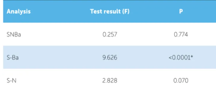
cali-bration. Table 4 -for the diferent facial patterns. ANOVA (F) results and respective signiicance probability performed
* Statistically signiicant. * Statistically signiicant.
Analysis Kappa Values P Percent
agreement
SNBa 1.000 <0.0001* 100%
G’.Sn.Pg’ 0.928 <0.0001* 95%
S-Ba 0.935 <0.0001* 95%
S-N 1.000 <0.0001* 100%
Wits 1.000 <0.0001* 100%
ANB 1.000 <0.0001* 100%
Group Pattern I Pattern II Pattern III
SNBa 131.7 ± 4.5 132.8 ± 4.9 132.5 ± 3.2
S-Ba 46.0 ± 2.7 46.8 ± 2.9 42.5 ± 3.0
S-N 69.4 ± 2.3 70.4 ± 4.7 67.1 ± 4.1
Analysis Test result (F) P
SNBa 0.257 0.774
S-Ba 9.626 <0.0001*
In this study, Pattern III also presented diference when compared to Pattern I and II, for the mean val-ues of the anterior cranial base length. Although there was no statistically signiicant diference, this value was reduced in Pattern III group. This result, like the re-duced posterior base, can help explain the concave fa-cial proile in Pattern III. This is because the nasomax-illary complex develops over this region.5,7,9,21 Thus, a reduced length of anterior cranial base can result a retrusive positioning of the whole nasomaxillary com-plex. In the facial proile, this is expressed as a ten-dency to poor middle-third of the face. Björk,4 when studying the human prognathism, stated that a short-ening of the anterior cranial base is accompanied by an increase of facial prognathism if the other structures involved remain unchanged. This is consistent with the results of this study. However, the shortening of the cranial base is not the prime factor for a facial Pat-tern III, since it can present other important etiologic factors. Nevertheless this shortening is usually present in Pattern III, contributing in a greater or lesser de-gree, to the characteristic proile of this group.
According to Weidenreich,30 a sharper SNBa angle is usually related to a more brachycephalic skull and great-er mandibular prognathism. Enlow9 found a sharper SNBa angle, a short middle-third of the face and man-dibular ramus with a more anterior orientation in sub-jects with Angle’s Class III malocclusion, when com-pared to a control group. The same authors found, in subjects with Class II malocclusion, a less sharp SNBa angle, with a base of greater length when compared to a control group (Class I).
According to Björk,4 a reduced SNBa angle and shortening of the cranial base, would some of the facial prognathism causes. He also asserts that the opposite is true, where an increase in the angle of the cranial base, as well as a greater length base, would be responsible for a more retrognathic facial pattern. However, the dif-ference between the anteroposterior positioning of the maxilla and mandible is partly due to variation in the size of the jaws, and partly due to variation in the length and lexure angle of the cranial base, which are associ-ated with the both jaws.
The angle of the cranial base (SNBa) did not difer between the diferent groups. This result is consistent with what Freitas12 found on his studies. There were also no statistically signiicant diferences between the
linear dimensions of the cranial base (S-N and S-Ba) between Pattern I and Pattern II. But these results do not conirm what part of the literature describes about the subject.1,4,8,9,12,15
This study investigated the differences in the de-flection angle of the cranial base, as well as in the length of its anterior (S-N) and posterior (S-Ba) por-tions in different facial patterns (Pattern I, II and III). However, it is important to keep in mind that the morphogenetic facial pattern is formed by a series of specific characteristics of each face type. And each of these characteristics alone does not define a facial pat-tern. It also possible to have compensations in max-illomandibular structures. These compensations can act minimizing an abnormal morphological pattern of the cranial base.
The fact that these values (SNBa for the three groups and S-N and S-Ba for Pattern I and II) did not show sig-niicant diferences can be explained by such morpho-logical compensation existing in the studied subjects. For example, when comparing Pattern I and II individ-uals, they can present values for SNBa, S-N and S-Ba, within the normal range. The diference in these facial patterns may have been caused by a smaller mandible and / or a larger maxilla in the Pattern II individual.
Therefore, it would be interesting to conduct further studies on the topic, including larger speciicities be-tween diferent facial patterns (separating the Pattern III caused by maxillary deiciency from the Pattern III caused by mandibular excess; separating the Pattern II caused by maxillary excess from the Pattern II caused by mandibular deiciency, for example), thus distinguish-ing the main etiological factor for each facial pattern.
1. Anderson D, Popovich F. Relation of cranial base lexure to cranial form and mandibular position. Am J Phys Anthropol. 1983;61(2):181-7.
2. Angle EH. Treatment of malocclusion of the teeth. Philadelphia: The S. S. With Dental Manufacturing; 1907.
3. Bishara SE. Ortodontia. São Paulo: Ed. Santos; 2004.
4. Björk A. Cranial base development. Am J Orthod. 1955;41(3):198-225. 5. Brodie AG Jr. The behavior of the cranial base and its components as revealed
by serial cephalometric reontgenograms. Angle Orthod. 1955;25(3):148-160. 6. Capelozza Filho L. Diagnóstico em Ortodontia. Maringá: Dental Press; 2004. 7. Coben SE. The integration of facial skeletal variants. Am J Othod.
1955;41(6):407-34.
8. Dhopatkar A, Bhatia S, Rock P. An investigation into the relationship between the cranial base angle and malocclusion. Angle Orthod. 2002;72(5):456-63. 9. Enlow DH. Crescimento facial. 3ª ed. São Paulo: Artes Médicas; 1993. 10. Faltin Jr. K. A individualização do diagnóstico e conseqüentes opções de tratamento. In: Grupo brasileiro de Professores de Ortodontia e Odontopediatria. São Paulo; 1997. p. 166-72.
11. Ferner AG, Staubesand RT. Sobotta Atlas de Anatomia Humana. Rio de Janeiro: Guanabara Koogan; 1983.
12. Freitas JC. Inluência da base craniana nas más oclusões [dissertação]. Rio de Janeiro (RJ): Universidade Federal do Rio de Janeiro; 1983.
13. Guyer EC, Ellis EE 3rd, McNamara JA Jr, Behrents RG. Components of Class III maloclusion in juveniles and adolescents. Angle Orthod. 1986;56(1):7-30. 14. Jacobson A, Evans WG, Preston CB, Sadowsky PL. Mandibular prognathism. Am
J Orthod. 1974; 66(2):140-71.
15. Kasai K, Moro T, Kanazawa E, Iwasawa T. Relationship between cranial base and maxilofacial morphology. Eur J Orthod. 1995;17(5):403-10.
16. Klocke A, Nanda RS, Kahl-Nieke B. Skeletal Class II paterns in the primary dentition. Am J Orthod Dentofacial Orthop. 2002;121(6):596-601. 17. Lanza P, Santos Pinto A, Bolini PDA. Estudo cefalométrico do crescimento e
lexão da base do crânio humano do nascimento aos seis meses de idade. Rev Dental Press Ortod Ortop Facial. 2003;7(2):33-9.
REFERENCES
18. Marquez IM. Avaliação do padrão facial, preparo ortodôntico e capacidade do tratamento cirúrgico em pacientes Classe III com prognatismo mandibular [tese]. Bauru (SP): Faculdade de Odontologia de Bauru; 1993.
19. Moore WJ, Lavelle CLB. Growth of the facial skeleton in the hominoidea. New York: Academic; 1974.
20. Mouakeh M. Cephalometric evaluation of craniofacial pattern of Syrian children with Class III maloclusion. Am J Orthod Dentofacial Orthop. 2001;119(6):640-9. 21. Moyers RE. Ortodontia. 4ª ed. Rio de Janeiro: Guanabara Koogan; 1991. 22. Ngan PW, Byczek E, Scheick J. Longitudinal evaluation of growth changes in
Class II division 1 subjects. Semin Orthod. 1997;3(4):222-31.
23. Ricketts RM. Planning treatment on the basis of the facial pattern and an estimative os its growth. Angle Orthod. 1957;27(1):14-37.
24. Rothstein T, Yoon-Tarlie C. Dental and facial skeletal characteristics and growth of males and females with Class II, division 1 malocclusion between the ages of 10 and 14 (revisited) – Part I: characteristics of size, form, and position. Am J Orthod Dentofacial Orthop. 2000;117(3):320-32.
25. Sandborn RT. Diferences between the facial skeletal patterns of Class III malocclusion and normal occlusion. Angle Orthod. 1955;25(4):208-22. 26. Scott JH. Dentofacial development and growth. Oxford: Pergamon; 1967. 27. Suguino R, Ramos AL, Terada HH, Furquim LZ, Maeda L, Silva OG Filho. Análise
Facial. Rev Dental Press Ortod Ortop Maxilar. 1996;1(1):86-107.
28. Varrela J. Early developmental traits in Class II malocclusion. Acta Odontol Scand. 1998;56(6):375-7.
29. Vion PE. Anatomia cefalométrica. São Paulo: Ed. Santos; 1994.
30. Weidenreich F. Some particulars of skull and brain of early hominids and their bearing on the problem of the relationship between man and anthropoids. Am J Phys Anthropol. 1947;5(4):387-427.
craniofacial evaluation. We must know the role of each variable within a whole, and thus successfully diagnose the main etiology of a certain disease.
CONCLUSIONS
In the studied sample, there was no diference be-tween the mean values for the delection angle of the cranial base (SNBa) in diferent facial patterns (I, II, III).
There was statistically signiicant diference for the mean values of the posterior cranial base (S-Ba) for the Pattern III group. In this group, the posterior cranial base was reduced when compared with Pattern I and II groups.