Detection of viable and viable nonculturable
Vibrio cholerae
O1 through
cultures and immunofluorescence in the Tucumán rivers, Argentina
Detecção de
Vibrio cholerae
O1 viável e viável não cultivável, através de técnicas
de cultivo e imunofluorescência nos rios de Tucumán, Argentina
Olga Aulet
1, Clara Silva
1, Sol González Fraga
2, Mariana Pichel
2, Rosa Cangemi
1, Cristina Gaudioso
1,
Norma Porcel
1, Maria Angela Jure
1, Marta Cecilia de Castillo
1and Noma Binsztein
2ABSTRACT
Vibrio cholerae
has been sporadically isolated from rivers in Tucumán, Argentina, since the outbreak in 1991. The aim of this study was to
determine the environmental reservoir of the bacterium in these rivers, assessing the presence of
Vibrio cholerae
non-O1 and O1 (the latter
both in its viable culturable and non culturable state) and its relationship to environmental physicochemical variables. 18 water samplings
were collected in the Salí River (in Canal Norte and Banda) and the Lules River between 2003 and 2005. Physical-chemical measurements (pH,
water temperature, electrical conductivity and dissolved oxygen) were examined.
Vibrio cholerae
was investigated with conventional culture
methods and with Direct Immunofluorescence (DFA-VNC) in order to detect viable non culturable organisms. All isolated microorganisms
corresponded to
Vibrio cholerae
non-O1 and non-O139 (Lules 26%, Canal Norte 33% and Banda 41%). The majority was found during spring
and summer and correlated with temperature and pH. Non culturable
Vibrio cholerae
O1 was detected year round in 38 of the 54 water samples
analyzed. Application of the Pearson correlation coefficient revealed that there was no relationship between positive immunofluorescence
results and environmental physicochemical parameters. Genes coding for somatic antigen O1 were confirmed in all DFA-VNC-positive samples,
whereas the virulence-associated
ctx
A and
tcp
A genes were confirmed in 24 samples.
Key-words:
Vibrio cholerae
O1. Culture and immunofluorescence. Tucumán rivers.
RESUMO
Vibrio cholerae
tem sido isolado esporadicamente nos rios da Província de Tucumán, Argentina, desde outubro de 1991. O objetivo deste estudo
foi localizar os reservatórios nestes rios, identificar a presença de
Vibrio cholerae
O1 (em estado cultivável e não cultivável) e relacionar a
presença desta bactéria com as variações físico-químicos da água. Foram coletadas dezoito amostras de água do rio Salí (nas localidades
de Canal Norte e Banda) e do rio Lules, entre 2003 e 2005. Estas foram submetidas a análises físico-químicos como determinação de pH,
temperatura, condutibilidade elétrica e oxigênio dissolvido. A presença de
Vibrio cholerae
foi verificada por métodos de cultivo convencional
e por imunofluorescência direta (DFA-VNC). Todos os microrganismos isolados foram não O1 e não O139 (Lules 26%, Canal Norte 33% e
Banda 41%). A maioria foi encontrada na primavera e verão, indicando uma relação com a temperatura e pH. Das 54 amostras analisadas
por DFA-VNC, 38
Vibrio cholerae
não cultivável, foram detectadas em todas as épocas do ano. As amostras positivas foram confirmadas por
PCR para o antígeno somático O1 e para os genes de virulência
ctx
A e
tcp
A. Coeficiente de correlação de Pearson revelou que não há relação
entre os resultados obtidos por imunofluorescência e a variação dos parâmetros físico-químicos.
Palavras-chaves:
Vibrio cholerae
O1. Culture and immunofluorescence. Tucumán rivers.
1. Departamento de Microbiología Clínica, Facultad Bioquímica, Química, Farmacia y Biotecnología, Universidad Nacional de Tucumán, Tucumán, Argentina. 2. Instituto Nacional de Enfermedades Infecciosas, National Institute of Infectious Diseases “Carlos G. Malbrán”. Bs As, Argentina.
This study was financially supported by PICT-R 2000-00010 awarded by the Agencia Nacional de Promoción Científica y Tecnológica, Secretary of Science and Technology, Ministry of Culture and Education, Argentina.
Address to: Drª Marta Cecilia de Castillo. Departamento de Microbiología Clínica/FBQF/UNT. Ayacucho 491, 4000 SM de Tucumán, Argentina. Tel: 54 0381 4247752 int 1013
e-mail: [email protected]. Recebido em:13/10/2006
Aceito em: 12/07/20007
Cholera continues to be an important and devastating disease
transmitted by water and food, especially in those regions of
the world where it is endemic
8 12. Before its reemergence in
Peru and subsequent spreading throughout Latin America in
1991, the disease had been absent from the Americas for nearly
numbers of cases occurring during the warm months (January
to March)
28 39. In Argentina, there have been seven epidemics
since 1992. These cholera outbreaks occurred mainly during the
summer months.
Vibrio cholerae
O1 strains were isolated from
water samples collected from rivers during epidemic periods, but
also found in marine waters and the La Plata River estuaries
2.
Standard bacteriological procedures for isolation of
Vibrio
choleare
O1 from environmental samples (including water) between
epidemics were generally unsuccessful
19.
Vibrio cholerae
requires
salt for growth and can revert to a viable but non culturable state
(VNC) in response to adverse environmental conditions. These VNC
bacteria do not grow on conventional culture media, but remain
intact and retain metabolic activity and respiration
6 27 29 30 42. However,
the method of Kogure
et al
can be used to demonstrate that these
cells retain viability and their pathogenic potential
16 25 32. Techniques
employing microscopy, with either direct or indirect fluorescent
antibodystaining, have been developed and provide important data on
the occurrence of viable but nonculturable
Vibrio cholerae
O1
19.
Since the outbreak in Tucumán, a province in the northwest
of Argentina, sporadic cases of diarrhea by
Vibrio cholerae
have
been detected in areas close to the Salí and Lules rivers. This study
aimed to detect
Vibrio cholerae
O1 in these environments using
conventional culture techniques to isolate the microorganism and
direct immunofluorescence to detect the viable nonculturable state and
associate its presence to 4 environmental physicochemical variables.
MATERIAL AND METHODS
Site description and water sampling.
Samples were collected
at the Salí River (two sites: Canal Norte (CN) and Banda (B)) and at
the Lules River (one site). They were taken during 18 campaigns in
a three-year period (2003-2005) with 6 campaigns per year
.
Water
temperature, pH and electrical conductivity were determined
in situ
with a mercury thermometer, a portable digital pH meter (TPA-I) and an
Altronix conductivity meter (Ct-1), respectively. Dissolved oxygen (DO)
was measured at the laboratory according to the methods by Winkler
1.
Water samples were collected in sterile 5-liter bottles, and
then immediately transported to the laboratory and subjected to
bacteriological examination not more than 5h.
Bacteriological assaying.
Two liters of water were filtered
through 0.22µm membranes, using 12 to 15 membranes per sample.
Membranes were subsequently incubated in 100ml of alkaline peptone
water, pH 8.6, for 6-8h at 35°C. Two loopfuls of broth were streaked on
Thiosulfate Citrate Bile agar (TCBS agar, Difco) and incubated for 18h at
37°C. Six to 12 typical colonies (yellow and 1 to 3mm diameter) were
transferred to nutritive soft agar (T
1N
1, 0.75% agar) and incubated for
24h at 37ºC. All colonies were stored at room temperature for further
testing. Isolates were identified biochemically and serotyped (O1 and
O139 antisera from the National Institute of Infectious Diseases-INEI.
ANLIS. “Dr Carlos G Malbran”, Buenos Aires, Argentina)
“Dr Carlos G Malbran”, Buenos Aires, Argentina)
6.
Direct immunofluorescence of
Vibrio cholerae
O1
(DFA-DVC).
Two liters of water were membrane-filtered (0.22µm).
Afterwards, the membranes were washed with 8ml of phosphate
buffer, and this buffer was fractioned for direct immunofluorescence
of
Vibrio cholerae
O1 (DFA-DVC) analysis. Samples were previously
incubated in the dark for 6 to 8h at room temperature in the
presence of yeast extract and nalidixic acid. Under these conditions,
viable but nonculturable bacteria elongate from a coccoid shape to
rod-like cells, yet they do not multiply due to the inhibitory effect of
nalidixic acid (a DNA gyrase inhibitor). After incubation, samples
were fixed with 4% formaldehyde and processed with cholera DFA
kits (New Horizons Diagnostics Corporation) for detection of
Vibrio
cholerae
O1
7 17. Stained preparations were observed under an
epifluorescence microscope (1000X) at 490 (maximum excitation)
and 520nm (maximum emission) with a blue filter. All procedures
were carried out in the dark. Readings were carried out within 24h
after preparation of the samples.
Confirmation of
Vibrio cholerae
O1 with polymerase
chain reaction.
Because DFA-DVC is a presumptive technique
presence of the microorganism was confirmed using PCR
in water to detect genes coding for the somatic antigens
O1 and virulence-associated
ctx
A genes that code for
the A subunit of the
cholera
toxin (CT) and
tcp
A El Tor,
that codes for the toxin co-regulated pilus (TCP) pilin
subunit
5 9 33 34 40 41. This method was carried out at the Bacteriology
Department of the Institute of Infectious Diseases (INEI) ANLIS
“Dr. Carlos G. Malbrán”, Buenos Aires, Argentina.
Statistical analysis.
The relationship between detection
of
Vibrio cholerae
and the physicochemical variables assayed
was assessed with the Pearson Correlation Coefficient (
D
= 0.05* or 0.01**),
using the SPSS statistics program (version 10.0 for Windows).
RESULTS
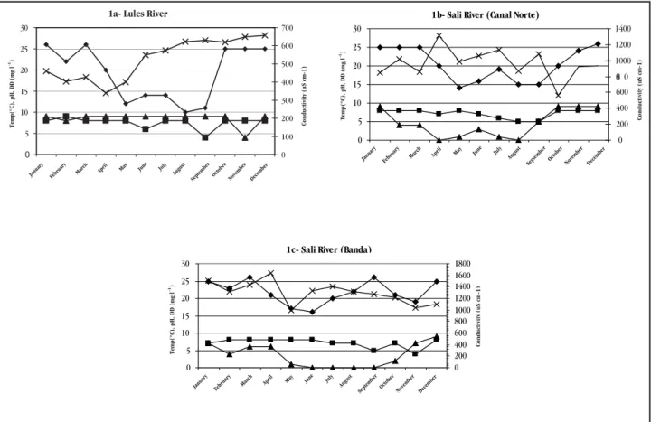
Physical and chemical parameters of the water.
The water
temperature oscillated between 15 and 26°C, pH between 5.5 and 9.0,
DO between 0 and 9mgl
-1and the conductivity between 400 and 1.600µS
cm
-1.
pH values at the Salí River sampling sites (CN and B) oscillated
during the three years between acid and alkaline, whereas fluctuations
at the Lules River sampling site were between 6.59 and 9.06. Regarding
dissolved oxygen, anoxia stages were only observed in the Salí River,
which also showed highest conductivity (Figures 1a, b, c).
Viable culturable Vibrio cholerae.
A total of 613
suspicious colonies (yellow in TCBS) were isolated from the
different sites and 385 were biotypified as
Vibrio cholerae
non-O1, non-O139. The percentage of isolates from each site was: RS
(CN) 33%, RS(B) 41% and RL 26%.
Figure 2 shows
Vibrio cholerae
non-O1, non-O139 isolates
according to their sampling site during the different months and
seasons. The microorganism was mainly isolated during the warm
months, corresponding to spring and summer, with a percentage
of 30 or more.
When analyzing the correlation between isolation of
Vibrio
cholerae
non-O1, non-O139 and physicochemical variables
it was found that the highest number of isolations in the Lules
River in January, March, November and December with a water
temperature over 25°C and pH more than 7.7. DO was between
8.8 and 9mgl
-1and conductivity between 426 and 658µS cm
-1.
1a- Lules River 0 5 10 15 20 25 30 January Te m p (° C ), pH , D D ( m g l -1) 0 100 200 300 400 500 600 700 C onduc tiv it y ( u S cm -1 )
February March April
May June July
August
SeptemberOctoberNovember December
1b- Sali River (Canal Norte)
0 5 10 15 20 25 30 0 200 400 600 80 0 1000 1200 1400
January February March April
May June July
August
SeptemberOctoberNovember December
Te m p (° C ), pH , D D ( m g l -1) C onduc ti vi ty ( u S cm -1 )
1c- Sali River (Banda)
0 5 10 15 20 25 30 0 200 400 600 800 1000 1200 1400 1600 1800
January February March April May June July
August
SeptemberOctoberNovember December
C onduc ti vi ty (uS cm -1) Te m p (° C ), pH , D D ( m g l -1)
Figure 1 - Physicochemical parameters of a) Lules River, b) Salí River (Canal Norte) and c) Salí River (Banda). () Temperature, ŶpH, ŸDissolved Oxygen (DO) and (x) Conductivity.
10 20 30 40 50 60 70
0
Janua
ry
Feb
rua
ry
Mar
ch
Ap
ril
May
Jun
e
Jul
y
Au
gus
t
Sep
tember
Oc
tobe
r
No
vem
ber
Decem
ber
% isolat
es o f V. chol er ae non O1,
non 0139 Spring
3 4 % W inte r 1 5 % Autumn 1 8 % Summer 33%
temperature under 14°C and pH below 6.5. At the Canal Norte
sampling site (Salí River) the microorganism was isolated all year
round with temperatures and pH values that oscillated between
14 and 26°C and 5 and 8.5 respectively. Furthermore, periods
of anoxia were observed and conductivity was generally less than
900µS cm
-1. The highest number of
Vibrio cholerae
non-O1,
non-O139 isolates was recovered at the Banda sampling site (Salí
River) during the period researched. Water temperature varied
from 16 to 26°C and the pH was generally higher than 7. There
were also anoxia periods (June to September) and conductivity
was over 1.000µS cm
-1.
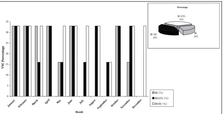
Figure 3 demonstrates that from the 54 water samples from
the different sites analyzed per month 38 were positive for
Vibrio
cholerae
O1 using the direct immunofluorescence (DFA-DVC)
technique.
Vibrio cholerae
O1 (VNC) was detected all year round
in rivers in Tucumán with the highest numbers during January,
February and June. Water temperature in January and February
was over 22°C and pH over 7.5, figures that are different from
those obtained in June with temperatures under 16°C and pH
values below 6.5. Conductivity and DO varied considerably in
January, February and June with values between 400 and 1,550µS
cm
-1and 2.8 and 9mgl
-1respectively. The Pearson correlation
coefficient revealed that there was no relationship between positive
immunofluorescence results and environmental physicochemical
parameters.
Viable but nonculturable Vibrio cholerae.
Even though
no
Vibrio cholerae
O1 strains were obtained by conventional
culture methods, DFA-DVC revealed the presence of
Vibrio
cholerae
O1 (VNC), which appeared as rod-shaped bacteria after
incubation with yeast extract and nalidixic acid (Figure 4).
PCR confirmed presence of genes coding for the somatic antigen
O1 in the 38 positive samples for viable nonculturable
Vibrio
cholerae
using immunofluoresence, but the virulence-associated
ctx
A and/or
tcp
A genes were only confirmed in 24 of them.
0 5 10 15 20 25 30 35
Janu ary
Febru ary
March Ap ril
May June July Augu
st
Septem ber
Octo ber
November Dece mber
Month
VNC Porcentage
RL (%)
RS(CN) (%)
RS(B) (%)
Porcentage
RL 26% RS (B)
40%
RS (CN) 34%
Figure 3 - Detection of Vibrio cholerae O1 VNC using direct immunofluorescence assaying at the different sample sites according to the month. Lules River, Salí River (Canal Norte) and Salí River (Banda).
DISCUSSION
In 1977, Colwell
et al
first hypothesized that coastal waters
were an important reservoir of
Vibrio cholerae
10. Other authors
also detected
Vibrio cholerae
in seawater and other environmental
sources around the world, both in cholera-endemic and in
cholera-free areas
11 18 20 23 24 36 47.
Borroto, Lee
Lee
et al
, Tamplin and Carrillo isolated
Vibrio
from water with temperatures between 25 and 12ºC, which is in
which is in
agreement with our results
3 27 43 44.
Singleton
et al
have concluded that presence of
Vibrio
O1
in aquatic environments is not limited to estuaries, because
its salinity requirements can be met through an adequate
nutrient concentration in fresh water environments. It has been
reported that this microorganism is able to survive in fresh
water for prolonged periods of time
4 13 22 35 37 38 42 45. Feachem
et
al
and Miller
et al
have demonstrated that various biological
and physicochemical factors influence growth, survival, and
distribution of
Vibrio cholerae
in aquatic environments
13 31.
Isolation of the microorganism with classical culture methods may
fail. This depends on the physicochemical properties of the water
or the physiological state of
Vibrio cholerae
O1 itself, either with
actively growing cells or cells in a latent or dormant state
31 37. The
DFA-DVC technique has shown to be useful for detection of viable
but nonculturable
Vibrio cholerae
O1 in water samples
30 44 46. Huq
Huq
et al
isolated
Vibrio cholerae
O1 from fresh water environments
(rivers) using immunofluorescence, but they too were unable to
isolate culturable forms with conventional culture methods in
Bangladesh. Gonçalves
et al
found viable nonculturable forms of
the
Vibrio
organism in two river estuaries in Brazil and Binsztein
et al detected it for the first time in the La Plata River and close to
detected it for the first time in the La Plata River and close to
a marine platform in Argentina
2 15 19.
The rivers in Tucumán are affected by effluents of a variety
of industries (sugar cane, citric fruit processing and paper
among others) that, together with agricultural activities, modify
the aquatic environments, thus generating conditions that allow
survival of
Vibrio cholerae
non-O1, non-O139 and persistence
of the viable nonculturable state of
Vibrio cholerae
O1. The fact
that
Vibrio cholerae
O1 was not detected with classical culture
methods agrees with results obtained by other researchers
21 30.
Kurazono
et al
and Sharma
et al
sustain that the epidemiological
impact of environmental
Vibrio cholerae
strains is not clearly
understood, because most of them do not produce the cholera
toxin and have also lost significant pathogenic factors
26 41. Similarly,
Similarly,
37% of the total number of samples that confirmed somatic
antigen O1 tested negatively for the virulence-associated
ctx
A
and
tcp
A genes.
Our study has for the first time provided evidence of isolation
of
Vibrio cholerae
non-O1, non-O139 and presence of the viable
nonculturable state of
Vibrio cholerae
O1 in rivers in Tucumán all year
round. Consequently, it can be inferred that the Lules and Salí rivers
constitute a reservoir for the microorganism in our province.
The warm temperatures in addition to a high concentration
of organic nutrients from agro-industrial waste, as is the case
in the rivers in Tucumán, create in these developing areas with
poor sanitary conditions an adequate environment so that
Vibrio
cholerae
can persist. Considering that this water is used for human
consumption in rural areas, and that drinking water constitutes
an important transmission vehicle of the pathogen, exhaustive
monitoring studies would be necessary to determine how these
bacteria, present in the rivers, affect public health now and in
the future.
REFERENCES
1. American Public Health Association. Métodos Normatizados para el análisis de aguas potables y residuales. 17° edición Ed Diaz de Santos, SA. Madrid, 1992. 2. Binsztein N, Costagliola M, Pichel M, Jurquiza V, Ramirez F, Akselman R, Vacchino M, Huq
A, Cowell R. Viable but nonculturable Vibrio cholerae O1 in the Aquatic Environment of Argentina. Applied and Environmental Microbiology 70:7481-7486, 2004. 3. Borroto RJ. Supervivencia de Vibrio cholerae en agua dulce superficiales y cólera
endémico: una hipótesis geoecológica. Revista Panamericana de Salud PúblicaRevista Panamericana de Salud Pública 4:371-374, 1998..
4. Bourke A, Cossins Y, Gray B. Investigation of cholera acquired from the riverine environment in Queensland. Medical Journal 144:229-234, 1986.
5. Chakraborty S, Mukhopadhyay AK, Bhadra RK, Ghosh AN, Mitra R, Shimada T, Yamasaki S, Faruque SM, Takeda Y, Cowell RR, Nair GB. Virulence genes in environmental strains of Vibrio cholerae. Applied Environmental Microbiology 66:4022-4028, 2000.
6. Choopun N, Louis V, Huq A, Colwell RR. Simple Procedure for Rapid Identification ofVibrio cholerae from the Aquatic Environment. Applied and Environmental Microbiology 68: 995-998, 2002.
7. Chowdhury MAR, Xu B, Montilla R, Hasan JAK, Huq A, Colwell RR. A simplified immunofluorescence technique for detection of viable cell of Vibrio cholerae
O1 and O139. Journal of Microbiology Methods 24:165-170, 1995. 8. Colwell RR. Global climate and Infectious Disease: the cholera paradigm. Science
274: 2025-2031, 1996.
9. Colwell RR, Brayton PR, Grimes DJ, Roszak DR, Huq SA, Palmer LM. Viable, but non-culturable, Vibrio cholerae and related pathogens in the environment: implications for release of genetically engineered microorganisms. Biotechnology 3:817-820, 1985.
10. Colwell RR, Kaper J, Joseph SW. Vibrio cholerae and Vibrio parahaemolyticus
and other vibrios: occurrence and distribution in Chesapeake Bay. Science 198:394-396, 1977.
11. Colwell RR, Seidler RJ, Kaper J, Joseph SW, Garges S, Lockman H, Maneval D, Bradford H, Roberts N, Remmers E, Huq I, Huq A. Occurrence of Vibrio cholerae serotype O1 in Maryland and Louisiana estuaries. Applied Environmental Microbiology 41:555-558, 1981.
12. Farruque SM, Albert MJ, Makanos JJ. Epidemiology, genetics and ecologyFarruque SM, Albert MJ, Makanos JJ. Epidemiology, genetics and ecologyEpidemiology, genetics and ecology of toxigenic Vibrio cholerae. Microbiology and Molecular Biology Reviews 62:1301-1314, 1998.
13. Feachem R, Miller C, Drasar B. Environmental aspects of cholera epidemiology. II. Occurrence and survival of V. cholerae in the environment. Tropical. Disease Bulletin 78:865-880, 1981.
14. Franco AA, Fix AD, Prada A, Paredes E, Palomino JC, Wright AC, JohnsonFranco AA, Fix AD, Prada A, Paredes E, Palomino JC, Wright AC, Johnson JA, McCarter R, Guerra H, Morris JG. Cholera in Lima, Peru, correlates withCholera in Lima, Peru, correlates with prior isolation of Vibrio cholerae from the environment. American Journal ofAmerican Journal of Epidemiology 146:1067-1075, 1997.
15. Gonçalves EGR, Lopes MJ S, Olivera EG, Hofer E. Associacao de Vibrio cholerae
com o zooplancton de aguas estuarios da Baía de São Luis- MA, Brasil. Revista da Sociedade Brasileira de Medicina Tropical 37:318-323, 2004.
16. Grimes DJ, Atwell RW, Brayton PR, Palmer LM, Rollins DM, Roszak DB, Singleton FL, Tamplin ML, Colwell RR. The fate of enteric pathogenic bacteria in estuarine and marine environments. Microbiological Science 3:324-329, 1986. 17. Hasan J, Bernstein D, Huq A, Loomis L, Tamplin M, Cowell R. Cholera DFA: an
and enumeration of Vibrio cholerae O1. FEMS Microbiology Letters 120: 143-148, 1994.
18. Huq A, Colwell RR. Vibrios in the marine and estuarine environment: tracking ofVibrio cholerae. Journal of Ecosystems and Health 2:198-214, 1996. 19. Huq A, Cowell RR, Rahman R, Ali A, Chowdhury MA, Parveen S, Sack DA,
Russek-Cohen E. Detection of Vibrio cholerae O1 in the aquatic environment by fluorescent-monoclonal antibody and culture methods. Applied Environmental Microbiology 57:2370-2373, 1990.
20. Huq A, Sack R, Colwell RR. Cholera and global ecosystems. In: Aron J, Patz J (eds) Ecosystem change and public health: a global perspective, Johns Hopkins University Press, Baltimore, Maryland, p. 327-347, 2001.
21. Huq A, Small EB, West PA, Huq MI, Rahman R, Colwell RR. Ecological relationship between Vibrio cholerae and planktonic crustacean copepods. Applied and Environmental Microbiology 45:275-283, 1983.
22. Islam MS, Drasar BS, Sack RB. The aquatic flora and fauna as reservoir of Vibrio cholerae: a review. Journal of Diarrhea Disease Research 12:87-96, 1994. 23. Jesudason MV, Balaji V, Mukudan U, Thomas CJ. Ecological study of Vibrio
cholerae in Vellore. Epidemiology Infection 124:201-206, 2000.
24. Kaysner CA, Abeyta Jr C, Wekell MM, DePaola Jr A, Stott RF, Leitch JM. IncidenceC, Wekell MM, DePaola Jr A, Stott RF, Leitch JM. Incidence ofVibrio cholerae from estuaries of the United States West Coast. Applied and Environmental Microbiology 53:1344-1348, 1987.
25. Kogure K, Simidu U, Taga N. A tentative direct microscopic method for counting living marine bacteria. Canadian Journal of Microbiology 25:415-420, 1979. 26. Kurazono H, Pal A, Bag PK, Nair GB, Karasawa T, Mihara T, Takeda Y. DistributionDistribution
of genes encoding cholera toxin, zonula occludens toxin, accessory cholera toxin, and El Tor hemolysin of diverse origins. Microbiology Pathogenic 18:231-235, 1995.
27. Lee J, Bashford D, Donovan T, Furniss A, West P. The incidence of Vibrio cholerae
in water, animals, and birds in Kent, England. Journal of Applied Bacteriology 52:281-291, 1982.
28. Lipp E, Gil AI, Espeland EM, Choopun N, Louis VR, Russek-Cohen E, Huq A,Lipp E, Gil AI, Espeland EM, Choopun N, Louis VR, Russek-Cohen E, Huq A, Colwell RR. Direct Detection ofDirect Detection of Vibrio cholerae and ctxA in Peruvian Coastal Water and Plankton by PCR. Applied and Environmental Microbiology 69:3676-3680, 2003.
29. Louis VR, Russek-Cohen E, Choopun N, Rivera IN, Gangle B, Jiang SC, Rubin A, Huq JA, Colwell RR. Predictability of Vibrio cholerae in Chesapeake Bay. Applied and Environmental Microbiology 69: 2773-2785, 2003.
30. Martins MT, Sanchez PS, Sato MIZ, Brayton PR, Colwell RR. Detection ofDetection of
Vibrio cholerae O1 in the aquatic environment in Brazil employing direct immunofluorescence microscopy. World Journal of Microbiology and Biotechnology 9:390-392, 1993.
31. Miller CJ, Drasar B, Feachem RG. Response of toxogenic Vibrio cholerae O1 to physiological stresses in aquatic environments. Journal of Hygiene 93:475-495, 1984.
32. Nilsson L, Oliver JD, Kjelleberg S. Resuscitation of Vibrio cholerae from the viable but nonculturable state. Journal of Bacteriology 173:5054-5059, 1991.
33. Rivera I, Chun J, Huq A, Sack R, Colwell RR. Genotypes associated with virulence in environmental isolates of Vibrio cholerae. Applied Environmental Microbiology 67: 2421-2429, 2001.
34. Rivera I, Lipp E, Gil A, Choopun N, Huq A, Colwell RR. Method for extraction andMethod for extraction and application of Multiplex PCR to detect Toxigenic V. cholerae O1 and O139 from aquatic ecosystems. Environ Microbiol 5: 599-606, 2003.
35. Rogers R, Cuffe R, Cossins Y, Murphy D, Bourke A. The Queensland cholera incident of 1977. II, The epidemiological investigation. Bulletin World Health Organ 58:665-669, 1980.
36. Rollins DM, Colwell RR. Viable but nonculturable stage of Camylobacter jejuni
and its role in survival in the natural aquatic environment. Applied Environmental Microbiology 52:531-538, 1986.
37. Roszak DB, Colwell RR. Survival strategies of bacteria in the natural environments. American Journal of Public Health 51:365-379, 1987.
38. Roszak DB, Grimes DJ, Colwell RR. Viable but nonrecoverable stage of Salmonella
enteritidis in aquatic systems. Canadian Journal of Microbiology 30:334-338, 1984.
39. Seas C, Miranda J, Gil AI, Leon-Baru R, Patz JA, Huq A, Colwell RR, Sack RB. New insightsSeas C, Miranda J, Gil AI, Leon-Baru R, Patz JA, Huq A, Colwell RR, Sack RB. New insightsNew insights on the emergence of cholera in Latin America during 1991: the Peruvian experience. The American Journal of Tropical Medicine and Hygiene 62:513-517, 2000. 40. Shangkuan Y, Show Y and Wang T. Multiplex polymerase chain reaction to detect
toxigenic V. cholerae and to biotype V. cholerae O1. Journal Applied Bacteriology 79: 264-273,1995.
41. Sharma C, Thungapathra M, Ghosh A, Mukhopadhyay AK, Basu A, Mitra R, Basu I, Bhattacharya SK, Takeda T, Yamasaki S, Takeda Y, Fair GB. Molecular analysis of non -O1, non-O139 Vibrio cholerae associated with an unusual upsurge in the incidence of cholera-like disease in Calcutta, India. Journal of Clinical Microbiology 36:756-763, 1998.
42. Singleton F, Attwel R, Jangi M, Colwel R. Effects of temperature and salinity on Vibrio cholerae growth. Applied and Environmental Microbiology 44:10047-1058, 1982. 43. Tamplin M, Carrillo C, Environmental spread of Vibrio cholerae in Peru. Lancet
338:1216-1217, 1991.
44. Tamplin ML, Gauzens AL, Huq A, Sack DA, Cowell RR. Attachment ofAttachment of Vibrio cholerae serogroup O1 to zooplankton and phytoplankton of Bangladesh waters. Applied and Environmental Microbiology 56:1977-1980, 1990.
45. Venkateswaran KT, Navarro IM, Nakano H, Hashimoto H, Sibling RL. Ecology ofVenkateswaran KT, Navarro IM, Nakano H, Hashimoto H, Sibling RL. Ecology ofEcology of
Vibrio cholerae non O1 and Salmonella spp. and the role of zooplankton in their seasonal distribution in Fukuyama coastal water, Japan. Applied Environmental Microbiology 35:1591-1598, 1989.
46. Xu HS, Roberts NC, Adams LB, West PA, Siebeling RJ, Huq A, Huq MI, Rahman R, Cowell RR. An indirect fluorescent antibody staining procedure for detection ofVibrio cholerae serovar O1 in aquatic environmental samples. Journal of Microbiological Methods 2:221-231, 1984.