Sao Paulo Med J. 2009; 127(3):174-6
174
Case report
Hemosiderotic ibrohistiocytic lipomatous lesion:
case report and review of the literature
Lesão lipomatosa ibrohistiocítica hemossiderótica: relato de caso e revisão da literatura
Antônio Roberto Oliveira Ramalho
1, Marcella Nara Nunes
2, Sheila Jorge Adad
3,
Sebastião Almeida Leitão
4, Adilha Misson Rua Micheletti
5Universidade Federal do Triângulo Mineiro (UFTM), Uberaba, Minas Gerais, Brazil
1MD. Pathology resident, Universidade Federal do Triângulo Mineiro (UFTM), Uberaba, Minas Gerais, Brazil. 2Medical student, Universidade Federal do Triângulo Mineiro (UFTM), Uberaba, Minas Gerais, Brazil.
3PhD. Associate professor, Discipline of Special Pathology, Universidade Federal do Triângulo Mineiro (UFTM), Uberaba, Minas Gerais, Brazil. 4MD. Adjunct professor, Discipline of Orthopedics, Universidade Federal do Triângulo Mineiro (UFTM), Uberaba, Minas Gerais, Brazil. 5PhD.Adjunct professor, Discipline of Special Pathology, Universidade Federal do Triângulo Mineiro (UFTM), Uberaba, Minas Gerais, Brazil.
ABSTRACT
CONTEXT: Lesions of the adipose tissue are the most common type of soft-tissue lesion among adults.
CASE REPORT: We describe the case of a 33-year-old female patient with a soft-tissue lesion in her left knee that was diagnosed as a hemosiderotic ibrohistiocytic lipomatous lesion. This type of lesion, which was described for the irst time in 2000, preferentially affects the ankle region of middle-aged women with a history of previous local trauma. Lesion recurrence is common, caused by incomplete resection, although there have not yet been any
reports of metastases. After a review of the literature, we describe the clinical, radiological, morphological and immunohistochemical characteristics, along with their main differential diagnoses.
RESUMO
CONTEXTO: Lesões de tecido adiposo são as mais comuns de partes moles em adultos.
RELATO DE CASO: Descrevemos o caso de uma paciente do sexo feminino, 33 anos, com lesão em partes moles do joelho esquerdo diagnosticada
como lesão lipomatosa ibrohistiocítica hemossiderótica. Essa lesão foi descrita pela primeira vez em 2000, acometendo preferencialmente a região do tornozelo de mulheres de meia-idade com história de trauma prévio local. Recidiva da lesão é comum devido à ressecção incompleta e não há até o
momento relato de metástase. Após revisão da literatura, descrevemos as características clínicas, radiológicas, morfológicas, imunoistoquímicas assim como seus principais diagnósticos diferenciais.
KEY WORDS:
Hemosiderin. Histiocytes.
Soft tissue neoplasms. Lipoma.
Venous insuficiency.
PALAVRAS-CHAVE: Hemossiderina.
Histiócitos.
Neoplasias de tecidos moles.
Hemosiderotic ibrohistiocytic lipomatous lesion: case report and review of the literature
Sao Paulo Med J. 2009; 127(3):174-6
175
INTRODUCTION
Hemosiderotic ibrohistiocytic lipomatous lesion (HFLL) is a rare benign ibrolipomatous lesion, irst described as an entity in 2000 by Marshall-Taylor and Fanburg-Smith. It accounts for 0.2% of benign li-pomatous lesions.1
HFLL is usually supericial, solitary and circumscribed but not encapsulated. It generally afects the feet and ankles (80 to 92% of the cases), although it may appear in other locations, such as cheeks and hands.1-3 he mean age of the patients is 50.6 years and 80% are women.1-3 In 88% of the cases, there is an association with trauma.1,2 Despite the good prognosis, recurrence occurs in 50% because of in-complete resection.1-5 Venous stasis is implicated in the pathogenesis as an excessive tissue response.2 Mali’s acroangiodermatitis and vascular transformation of the lymph node sinuses, two lesions related to venous stasis, are diferential diagnoses.2
Macroscopically, HFLL is a yellowish lesion, slightly darker than li-pomas, measuring between 1 and 19 cm.1-8 Microscopically, it consists of mature adipose tissue without atypia, associated with fusiform cells that are accompanied by inlammatory iniltrate composed of lympho-cytes, plasmolympho-cytes, histiocytes and mast cells, and abundant hemosid-erin pigmentation.1-4 Cells with slight atypia,1,3 loret-like cells1 or osteo-clast-like cells3 can occasionally be seen. In 20% of the cases, there are small vessels with hyalinization.1
HFLL is positive in most cases (77-100%) for CD34, vimentin (100%) and calponin (100%), focally positive for lysozyme and KP-1 and negative for protein S-100, desmin, smooth-muscle actin, CD68, HMB-45, epithelial membrane antigen, cytokeratins and caldesmon.1,3
Among the diferential diagnoses, various lipomatous lesions should be considered, such as adiponecrosis, ibrolipoma, fusiform cell lipoma and liposarcoma, and also ibrohistiocytic/myoibroblastic lesions such as ibromatosis, nodular fasciitis, pseudo-Kaposi’s sarcoma, ibrohistio-cytoma, dermatoibrosarcoma protuberans and pleomorphic hyaliniz-ing angiectatic tumor. he clinical-morphological-immunohistochem-ical association is suicient for correct diagnosis.1,3,4
CASE REPORT
Our patient was a 33-year-old white woman with pain and tumor on the posterior face of the left knee. hree years earlier, she had twist-ed this knee and, since then, she had been presenting pain and progres-sive swelling. On physical examination, there was a large-volume soft tumor accompanied by varicose veins on the posterolateral face of the left knee. he varicose veins were painful but without signs of inlam-mation. She also presented diiculty in lexing the left knee, with pain on touching and when walking. he patient was HIV-positive without signs of AIDS.
Ultrasound showed an expansive heterogeneous mass, laterally to the left popliteal fossa, of 8 cm in diameter. Tomography revealed a het-erogeneous lesion with lacy highlighting, afecting muscle and subcu-taneous tissues in the distal third of the left thigh. Magnetic resonance characterized it as lipoma.
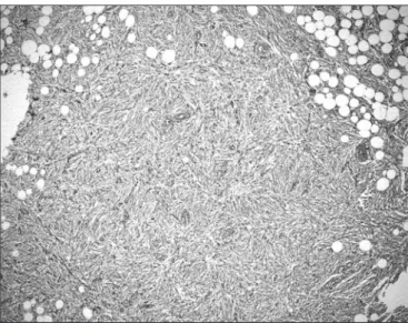
he patient underwent surgery with an incision measuring 1.5 cm on the posterior face of the knee. he lesion was excised with free mar-gins. he material consisted of soft friable brownish-yellowish fragments that together measured 10 x 8 x 6 cm and weighed 80 g. Under the mi-croscope, proliferation of fusiform cells without atypia was observed, with mature adipocytes. he specimen was permeated with abundant hemosiderin pigment, along with small vessels with hyalinized walls (Figures 1 and 2). At the center, there was an old hemorrhage. here was no invasion of vessels and nerves. he margins were diicult to as-sess because the material was fragmented. Immunohistochemistry was positive for vimentin and CD34, and negative for cytokeratin, desmin, smooth-muscle actin, HHF-35, protein S-100 and CD 99.
Fifteen months later, the patient presented with a swelling adjacent to the previous surgical scar measuring 2 x 1.5 x 0.5 cm. On re-exci-sion, the lesion had the same features, except for more ganglionic and multinucleated cells. here were no free margins. Six months after the second excision, the patient continued to show no signs of recurrence or metastasis.
Figure 2. Photomicrograph of the lesion, showing histiocytes, mature adipocytes and small blood vessels with hyalinized walls (hematoxylin-eosin, 20 x).
Sao Paulo Med J. 2009; 127(3):174-6 Ramalho ARO, Nunes MN, Adad SJ, Leitão SA, Micheletti AMR
176
DISCUSSION
We only found 29 cases of HFLL in PubMed1-8 (Table 1). Our case had some unusual characteristics and some interesting associated factors that may have been implicated in the pathogenesis, which is still a mat-ter for discussion.
Among the 29 cases described, 22 (75.86%) were women, of mean age 50 years, and 25 cases (86.2%) were presented in the ankle. Except for one case in a child and a 32-year-old patient described by Browne and Fletcher,3 our patient was younger than the mean age in the litera-ture. Moreover, our lesion is the irst described in the knee. Among the others, 25 cases afected ankles, two afected the hands, one occurred in the cheek and one occurred in the thigh.1-8
One unusual morphological characteristic seen in our case was the hyalinization of the walls of small blood vessels, which has been seen in only 27.5% of the cases (eight cases) in other series.1-4
he idea that this lesion may be associated with venous stasis was irst broached by Marshall-Taylor and Fanburg-Smith in 2000, while discussing the diferential diagnosis with Mali’s acroangiodermatitis. However, the idea was dismissed, given that none of the ten patients in their series presented vascular insuiciency.1 Nevertheless, this likely association was subsequently advocated by Kazakov et al, in relation to two patients who presented this lesion and chronic venous insuicien-cy.2 hus, HFLL would represent an excessive tissue response to venous stasis, since raised pressure in the veins and capillaries, oxygen satura-tion and edema would stimulate the proliferasatura-tion of the elements seen in this lesion.2,9 Kazakov et al. and Michal and Kazakov also showed that transformation of the lymph node sinus was another condition with morphology similar to HFLL and was also associated with venous stasis caused by occlusion of the eferent lymphatic vessels and/or veins.2,9
he patient in our case presented varicose veins in the afected leg and this makes us believe that venous stasis may really play an important role in the pathogenesis of this lesion.
At present, there are two major points for debate regarding HFLL: whether its nature is reactive or neoplastic; and whether it is or is not the precursor of pleomorphic hyalinizing angiectatic tumors, as advocated by Folpe and Weiss.10 Just like Marshall-Taylor and Fanburg-Smith and Kazakov et al., and Michael and Kazakov1,2,9 we believe that HFLL is
reactive, bearing in mind its supericial location, frequent association with previous trauma (11 cases, i.e. 39.3%, including ours), lack of cap-sule, morphology of varied cells and accumulation of hemosiderin. In our case, we also observed a hematoma at the center of the lesion, thus conirming the history of previous trauma and the possibility of a reac-tive lesion.
CONCLUSION
Hemosiderotic ibrohistiocytic lipomatous lesion is a recently de-scribed rare entity that usually afects middle-aged women’s feet and an-kles. Complete resection is necessary in order to avoid local recurrence. here is still much discussion regarding its reactive or neoplastic nature. his has generated controversy, and studies of greater extent are therefore needed to reach a deinitive conclusion.
REFERENCES
1. Marshall-Taylor C, Fanburg-Smith JC. Hemosiderotic ibrohistiocytic lipomatous le-sion: ten cases of a previously undescribed fatty lesion of the foot/ankle. Mod Pathol. 2000;13(11):1192-9.
2. Kazakov DV, Sima R, Michal M. Hemosiderotic ibrohistiocytic lipomatous lesion: clinical correlation with venous stasis. Virchows Arch. 2005;447(1):103-6.
3. Browne TJ, Fletcher CD. Haemosiderotic ibrolipomatous tumour (so-called haemosiderotic ibrohistiocytic lipomatous tumour): analysis of 13 new cases in support of a distinct entity. Histopathology. 2006;48(4):453-61.
4. Prud’homme A, Rousselot C, de Pinieux G, Voche P, Rosset P. Tumeurs ibrolipomateuses hémosidérotiques: une nouvelle entité à ne pas méconnaître [Hemosiderotic ibrohistiocytic lipomatous lesion: a new entity you must remind]. Ann Chir Plast Esthet. 2007;52(6): 616-20.
5. Luzar B, Gasljević G, Juricić V, Bracko M. Hemosiderotic ibrohistiocytic lipomatous lesion: early pleomorphic hyalinizing angiectatic tumor? Pathol Int. 2006;56(5):283-6. 6. Guillou L, Coindre JM. Newly described adipocytic lesions. Semin Diagn Pathol.
2001;18(4):238-49.
7. de Vreeze RS, Koops W, Hass RL, van Coevorden F. An unusual case of hemosiderotic i-brohistiocytic lipomatous lesion: correlation of MRI and pathologic indings. Sarcoma. 2008;2008:893918.
8. West AT, Toms AP, Murphy J, Sultan M. Haemosiderotic ibrohistiocytic lipomatous lesion/ tumor of the foot: MRI and histopathology. Skeletal Radiol. 2008;37(1):71-4.
9. Michal M, Kazakov DV. Relationship between pleomorphic hyalinizing angiectatic tumor and nemosiderotic ibrohistiocytic lipomatous lesion. Am J Surg Pathol. 2005;29(9):1256-7; author reply 1259.
10. Folpe AL, Weiss SW. Pleomorphic hyalinizing angiectatic tumor: analysis of 41 cases su-pporting evolution from a distinctive precursor lesion. Am J Surg Pathol. 2004;28(11): 1417-25.
Place where the paper was presented: Presented in the form of a poster at the 26th Brazilian Congress of Pathology, in Bento Gonçalves, November 2007
Sources of funding: None
Conlict of interest: Not declared
Date of irst submission: January 7, 2008
Last received: June 30, 2009
Accepted: July 1, 2009
Address for correspondence:
Adilha Misson Rua Micheletti
Disciplina de Patologia Especial, Universidade Federal do Triângulo Mineiro Rua Getúlio Guaritá, 130
Bairro Abadia — Uberaba (MG) — Brasil CEP 38025-440
Tel. (+55 34) 3318-5152 Fax. (+55 34) 3318-5846 E-mail: marcella_nara@yahoo.com.br
Database Search strategy Results
PubMed (“Histiocytoma, Malignant Fibrous”[MeSH])
OR (Fibrous Histiocytoma, Malignant) Or (Fibrous Histiocytomas, Malignant) Or (His-tiocytomas, Malignant Fibrous) Or (Malignant Fibrous Histiocytomas) Or (Malignant Fibrous Histiocytoma) Or (Fibrohistiocytic) AND (“Neoplasms, Adipose Tissue”[MeSH]) OR (Adipose Tissue Neoplasms) OR (Adipose Tissue Neoplasm) OR (Neoplasm, Adipose Tis-sue) OR (LIPOMATOSIS) OR (LIPOMATOSES) OR (“Neoplasms, Fibrous Tissue”[MeSH]) OR (Lipomatous Lesion) or (Lipomatous Tumor) AND (haemosiderotic OR hemosiderotic OR hemosideroses OR hemosiderosis)
3 original articles 1 letter to editor 1 updating article 2 case series 3 case reports
