Survival following orbital exenteration at a tertiary brazilian
Survival following orbital exenteration at a tertiary brazilian
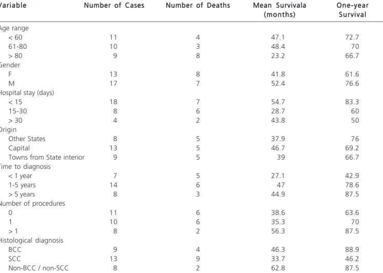
Survival following orbital exenteration at a tertiary brazilian
Survival following orbital exenteration at a tertiary brazilian
Survival following orbital exenteration at a tertiary brazilian
hospital
hospital
hospital
hospital
hospital
Sobrevida pós exenteração de órbita em hospital de referência
Sobrevida pós exenteração de órbita em hospital de referência
Sobrevida pós exenteração de órbita em hospital de referência
Sobrevida pós exenteração de órbita em hospital de referência
Sobrevida pós exenteração de órbita em hospital de referência
J
ULIANAM
IKAK
ATO1, F
ABRICIOL
OPESDAF
ONSECA2, S
UZANAM
ATAYOSHI2A B S T R A C T
A B S T R A C T
A B S T R A C T
A B S T R A C T
A B S T R A C T
Objective: Objective: Objective: Objective:
Objective: to analyze the epidemiology, clinical features and survival rate of patients undergoing orbital exenteration (OE) in a tertiary referral hospital. MethodsMethodsMethodsMethodsMethods: we conducted a retrospective study of all patients undergoing OE at the Hospital das Clínicas, FMUSP between January 2007 and December 2012. We collected data records related to gender, age, origin, length of stay, duration of the disease, other treatments related to the disease, number of procedures outside of the face related to the disease, follow-up and histological diagnosis. ResultsResultsResultsResultsResults: we treated 37 patients in the study period. The average survival in one year was 70%, in two years, 66.1%, and 58.3% in three years. There was no significant difference in the one-year survival related to histological diagnosis (p=0.15), days of hospitalization (p=0.17), gender (p=0.43), origin (p=0.78), disease duration (p=0.27) or the number of operations for the tumor (p=0.31). Mortality was higher in elderly patients (p=0.02). The average years of life lost was 33.9 in patients under 60 years, 14.7 in patients in the 61-80 years range and 11.3 in patients over 80 years. ConclusionConclusionConclusionConclusionConclusion: the present series of cases is significant in terms of prevalence of orbital exenteration; on the other hand, it shows one of the lowest survival rates in the literature. This suggests an urgent need for improved health care conditions to prevent deforming, radical resections.
Key words: Key words: Key words: Key words:
Key words: Orbital Evisceration. Survival Rate. Carcinoma, Squamous Cell. Carcinoma, Basal Cell.
1. Faculdade de Medicina da Universidade de São Paulo (FMUSP), SP, Brasil; 2. Departamento de Oftalmologia, Universidade de São Paulo, SP, Brasil.
INTRODUCTION
INTRODUCTION
INTRODUCTION
INTRODUCTION
INTRODUCTION
O
rbit Exenteration (OE) is one of the most disfiguring
procedures among ophthalmologic operations, and is
characterized by the complete removal of the contents of
the orbital cavity. According to the resection extent, it can
be classified into: 1) total, if there is resection of the eyelids;
2) subtotal, when preserving the eyelids; or 3) extensive,
when including removal of the bone surrounding walls
1 3.
OE is the therapy of choice when other less
radi-cal methods do not result in better prognosis. It is usually
indicated in oncologic resections for local control of
malignant tumors. However, aggressive diseases or benign
tumors that cause uncontrollable pain and structural and/
or extensive lesions also require it. Among the malignant
lesions, Basal cell carcinoma (BCC) is the most common
skin cancer (80-90%), followed by squamous cell
carcino-ma (SCC). Examples of non-carcino-malignant diseases include:
neurofibromatosis, fibrous dysplasia, mucormycosis, sharply
contracted anophthalmic cavity, recurrent meningioma and
orbital myiasis
4,5.
The aesthetic consequences have a strong
psychological impact on the patient and require a
multidisciplinary approach. Many patients are referred to
psychological services after the operation or even refuse to
undergo the surgical procedure. Constant vigilance, good
doctor-patient relationship, early diagnosis and prompt
treatment would provide better prognosis, especially in
emerging countries
6,7.
This retrospective study aims to analyze the
epidemiology, clinical features and survival rate of patients
undergoing orbital exenteration (OE) in a tertiary referral
hospital.
METHODS
METHODS
METHODS
METHODS
METHODS
performed outside the area of the face related to the
disease, follow-up, histologic diagnosis and recurrence of
lesions. To analyze the survival rate, we contacted the
patient’s family members by telephone with the help of
Social Service for identification and active search for the
occurrence of death.
We analyzed the variables by the Kaplan-Meier
method, and compared survival curves using the log-rank
test, with the R software, version 3.1.1. We calculated the
Years of Potential Life Lost (YPLL) by the method proposed
by Romeder
8, adjusted to the life expectancy of Brazilians
in 2013
9. The age of reference used was 78.6 for patients
under 60 years of age, 83.7 for patients between 61 and
81 years, and 96.7 for patients over 80.
RESULTS
RESULTS
RESULTS
RESULTS
RESULTS
We identified 39 patients, of whom two were
excluded due to incorrect coding of the disease.
Demographic and clinical characteristics
Demographic and clinical characteristics
Demographic and clinical characteristics
Demographic and clinical characteristics
Demographic and clinical characteristics
of patients
of patients
of patients
of patients
of patients
The study cohort included 17 men and 20 women,
between 0 and 94 years of age (mean 62.2 years). São
Paulo, capital, was the origin of 15 patients (40.5%), 13
(35.1%) were from towns in the interior of São Paulo and
nine (24.4%) from other Brazilian regions. Thirty-three
patients were white (89.2%), one was black (2.7%) and
three brown (8.1%).
The average time of diagnosis was 43.4 months
(range three months to 12 years), except for congenital
ca-ses. The days of hospitalization ranged from 0 to 62, average
14. Twelve patients (35.3%) were not subjected to any other
surgical procedure related to the current injury, another 12
(35.3%) underwent one operation and 10 (29.4%)
underwent more than one. Seventeen patients had additional
treatment such as radiotherapy (ten patients – 27%),
chemotherapy (two patients – 5.4%) and cryosurgery (three
patients – 8.1%). Most were not submitted to any other
operation outside the face area (81.8%) and eight (21.6%)
were previously treated at least once.
Histopathology
Histopathology
Histopathology
Histopathology
Histopathology
Histopathological findings included 16 cases of
squamous cell carcinoma (43.2%) and ten of basal cell
carcinoma (27.0%). Other diagnoses included adenoid cystic
carcinoma, found in two patients, adenocarcinoma,
sebaceous glands, cystic formation, inflammatory process,
oncocytic schneiderian papilloma, esthesioneuroblastoma,
capillary hemangioma, immature teratoma and malignant
melanoma, each found in one patient (Table 1).
Survival Rate
Survival Rate
Survival Rate
Survival Rate
Survival Rate
We excluded congenital cases from the survival
analysis. Two patients died during hospitalization.
At the time of the study, 15 patients had died,
15 were alive and six could not be contacted. The average
survival rate at one year was 70% and this figure decreased
to 66.1% and 58.3% in two and three years, respectively.
Mean survival was 47.3 months.
The mortality rate was higher in older patients
(p=0.02). There was no significant difference in one-year
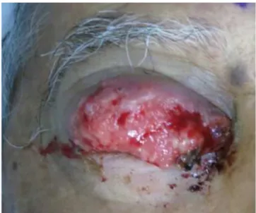
survival as for the histological diagnosis, if SCC (Figure 1),
BCC or non-ECC/non-BCC (p=0.15), days of hospitalization
(p=0.17), gender (p=0.43), origin (p=0.78), time of disease
progression (p=0.27) or number of operations related to
the tumor (p=0.31 – Table 2).
The average age of death in the age group under
60 was 44.7 years; between 61 and 80 years, 69, and in
patients aged over 80 years, 85.4. Considering the life
expectancy of Brazil in 2013, the average years of life lost
were, respectively, 33.9 years, 14.7 years and 11.3 years.
The total YPLL was 191 years (Figure 2).
DISCUSSION
DISCUSSION
DISCUSSION
DISCUSSION
DISCUSSION
Orbital exenteration is not a common procedure
and is usually done in tertiary referral centers. Our case
series presented one of the largest series per year (37
patients in six years). Rahman et al. reported 64 cases in a
period of 13 years
10; Mohr and Esser had 77 in 20 years
11;
Bartley et al. described 102 in 20 years
12; and Maheshwari
et al. published 15 in 10 years
13.
As the hospital where the study was conducted
is a tertiary center, it is expected that 59.9% of patients
originate from other cities as well as from São Paulo. The
geographical distance from the origin to the hospital also
explains the choice for OE, as the imprecise diagnosis of
other health services and lagged time to admission to the
Figure 1 Figure 1 Figure 1 Figure 1
tertiary hospital may have made OE the only possible
procedure for the control of local disease.
Among the patients cohort, three constituted
non-malignant cases. SCC and BCC together accounted for
70.2% of the histological diagnosis, which is consistent with
other studies. BCC is the most common skin cancer in the
periorbital area, but SCC spreads more easily and requires
a quick management to prevent disease
Table 1 -Table 1 -Table 1 Table 1
-Table 1 - Characteristics of patients undergoing orbital exenteration.
P a P aP a P a
P a ttttt i e n ti e n ti e n ti e n t G e n d e ri e n tG e n d e rG e n d e rG e n d e rG e n d e r A g eA g eA g eA g eA g e H o s p i t a lH o s p i t a lH o s p i t a lH o s p i t a lH o s p i t a l O r i g i mO r i g i mO r i g i mO r i g i mO r i g i m Time ofTime ofTime ofTime ofTime of H i s t o p a t h o l o g yH i s t o p a t h o l o g yH i s t o p a t h o l o g yH i s t o p a t h o l o g yH i s t o p a t h o l o g y O t h e rO t h e rO t h e rO t h e rO t h e r Number ofNumber ofNumber ofNumber ofNumber of stay (days)
stay (days) stay (days) stay (days)
stay (days) (State - City)(State - City)(State - City)(State - City)(State - City) disease tilldisease tilldisease tilldisease tilldisease till d i a g n o s i sd i a g n o s i sd i a g n o s i sd i a g n o s i sd i a g n o s i s t r e a t m e n t st r e a t m e n t st r e a t m e n t st r e a t m e n t st r e a t m e n t s p r o c e d u r e sp r o c e d u r e sp r o c e d u r e sp r o c e d u r e sp r o c e d u r e s procedure (years)
procedure (years) procedure (years) procedure (years)
procedure (years) related torelated torelated torelated torelated to current lesion current lesion current lesion current lesion current lesion
1 M 74 49 SP – São Paulo 5 BCC 0 0
2 F 94 8 BA – Caculé 5 BCC 0 1
3 F 72 2 SP – São Paulo 5 CAC RT 0
4 F 83 4 SP – Santos 7 CGS 0 2
5 M 81 22 CE – Cedro 7 SCC 0 1
6 F 65 1 MG 8 SCC Cryosurgery 4
7 F 78 10 CE – Itapipoca 5 BCC 0 0
8 M 52 6 SP – Santos 1,7 SCC QT > 1
9 F 64 62 SP – São Lourenço da Serra 2 SCC 0 0
10 M 71 16 SP – São Paulo 0,4 SCC RT + Cryosurgery 1
11 M 31 44 AM – Boca do Acre 0,3 SCC 0 0
12 F 49 16 SP – São Paulo Unknown SCC Unknown Unknown
13 F 63 2 SP – Mogi Guaçú 0,8 SCC 0 1
14 M 72 39 SP – Presidente Prudente 1 SCC RT 1
15 F 66 2 SP – Uchôa 5 Cystic formation 0 7
16 M 49 9 SP – São Paulo 2 BCC RT indicated 1
17 M 51 30 SP – São Paulo 2 SCC RT 0
18 M 50 10 SP – São Paulo 5 Schneiderian papilloma 0 1
19 F 71 24 SP – Pompéia 0,5 Inflammatory process 0 2
20 M 58 9 SP – São Paulo 12 Esthesioneuroblastoma RT 2
21 F 81 21 SP – São Paulo 2,3 SCC Cryosurgery 2
22 M 82 4 SP – Santo Amaro 0,5 BCC 0 0
23 M 59 7 SP – Guarulhos 0,5 SCC 0 0
24 F 82 9 BA – Jequié 7 BCC 0 1
25 F 9 4 SP – Mogi Mirim 0 Capillary hemangioma 0 > 1
26 F 0 0 SP – São Paulo 0 Immature teratoma QT 0
27 M 49 9 SP – Ibiúna 0,67 SCC 0 0
28 M 42 5 AM – Manaus 1 CAC RT 0
29 M 69 7 SP – São Paulo 0,67 SCC RT 0
30 F 82 7 BA – São Felix 5 BCC 0 1
31 F 42 8 SP – São Paulo 0,67 Adenocarci-noma RT + QT 0
32 M 79 3 SP – Jandira 1 SCC RT 1
33 M 51 7 AM – Manaus 2 SCC 0 4
34 F 82 27 SP – Mairipora 2 Malignant melanoma 0 1
35 M 60 21 SP – São Paulo 11 BCC RT 6
36 M 51 11 SP – São Paulo 8 BCC RT indicated 1
37 M 86 5 SP – São Paulo 6 BCC 0 3
Source: Medical records of the Hospital das Clínicas, Universidade de São Paulo (2007-2012).
BCC: basal cell carcinoma; CAC: cystic adenoid carcinoma; SGC: Sebaceous Glands Carcinoma; SCC: squamous cell carcinoma; RT: radiotherapy; QT: chemotherapy.
progression
2,10,12,14,15. Our findings are similar to current
literature, insofar as BCC represented 27% of the OE
ca-ses, while SCC accounted for 43.2%.
than BCC
16-18. Additional treatments, such as Mohs
micrographic surgery, may have been beneficial in the
management of some SCC cases
19,20.
The average mortality rate after OE also differs
from the literature, since our series showed lower survival.
Rahman et al. reported a survival rate of 93% in one year
10;
Table 2 Table 2 Table 2 Table 2
-Table 2 - Comparison of age, gender, days of hospitalization, origin, time of disease, number of operations and histological diagnosis with survival rate.
V a r i a b l e V a r i a b l e V a r i a b l e V a r i a b l e
V a r i a b l e Number of CasesNumber of CasesNumber of CasesNumber of CasesNumber of Cases Number of DeathsNumber of DeathsNumber of DeathsNumber of DeathsNumber of Deaths Mean SurvivalaMean SurvivalaMean SurvivalaMean SurvivalaMean Survivala O n e - y e a rO n e - y e a rO n e - y e a rO n e - y e a rO n e - y e a r ( m o n t h s )
( m o n t h s ) ( m o n t h s ) ( m o n t h s )
( m o n t h s ) S u r v i v a lS u r v i v a lS u r v i v a lS u r v i v a lS u r v i v a l Age range
< 60 11 4 47.1 72.7
61-80 10 3 48.4 70
> 80 9 8 23.2 66.7
Gender
F 13 8 41.8 61.6
M 17 7 52.4 76.6
Hospital stay (days)
< 15 18 7 54.7 83.3
15-30 8 6 28.7 60
> 30 4 2 43.8 50
Origin
Other States 8 5 37.9 76
Capital 13 5 46.7 69.2
Towns from State interior 9 5 39 66.7
Time to diagnosis
< 1 year 7 5 27.1 42.9
1-5 years 14 6 47 78.6
> 5 years 8 3 44.9 87.5
Number of procedures
0 11 6 38.6 63.6
1 10 6 35.3 70
> 1 8 2 56.3 87.5
Histological diagnosis
BCC 9 4 46.3 88.9
SCC 13 9 33.7 46.2
Non-BCC / non-SCC 8 2 62.8 87.5
Source: Medical records of the Hospital das Clínicas, Universidade de São Paulo (2007-2012). * LogRank Test
BCC: basal cell carcinoma; SCC: squamous cell carcinoma.
Table 3 Table 3 Table 3 Table 3
-Table 3 - Years of life lost according to age group.
Age Group Age Group Age Group Age Group
Age Group NNNNN I n t e r v a lI n t e r v a lI n t e r v a lI n t e r v a lI n t e r v a l Mean ageMean ageMean ageMean ageMean age Average years ofAverage years ofAverage years ofAverage years ofAverage years of YPLLYPLLYPLLYPLLYPLL at death time
at death time at death time at death time
at death time life lostlife lostlife lostlife lostlife lost
< 60 4 31-51 44.7 78.6 33.9
60-80 3 64-71 69 83.7 14.7
> 80 8 81-94 85.4 96.7 11.3
Source: Medical records of the Hospital das Clínicas, Universidade de São Paulo (2007-2012). YPLL: Years of Potential Life Lost.
* According IBGE (Instituto Brasileiro de Geografia e Estatística), 2013
Mohr and Esser had 89%
11and Chih-Hung Kuo, 97%
15.
Karabekmez et al., whose study also come from an
emerging country, showed a low survival rate of 50.5%
7.
Bartley et al. reported a survival rate of 88.6%
12.
Figure 2 -Figure 2 -Figure 2 Figure 2
-Figure 2 - Comparison of age and histological diagnosis with survival rate.
patients lost more than ten years. Not only the aggressiveness
of the disease, but also the lack of information, difficulty in
access to health care and delay in correct diagnosis justify
the current low survival rate
6,21. Studies suggest differences
in post-SCC mortality between developed and developing
countries
22.
Advanced age may act as a confounding variable
because, generally, it is related to comorbidities and other
causes of death unrelated to the tumor. However, the
predominance of advanced malignant disease is already
an indicator of difficulty in access to adequate medical
services for immediate treatment, which could improve
survival even in the older age group.
In conclusion, this case series is significant in terms
of prevalence of Orbit Exenteration; On the other hand, it
displayed one of the lowest survival rates in the literature.
This suggests an urgent need for improved health care
conditions to prevent deforming, radical resections.
R E S U M O
R E S U M O
R E S U M O
R E S U M O
R E S U M O
Objetivo: Objetivo: Objetivo: Objetivo:
Objetivo: analisar o perfil epidemiológico, as características clínicas e a taxa de sobrevida dos pacientes submetidos à exenteração orbitária (EO) em um hospital de referência terciário. Métodos:Métodos:Métodos:Métodos:Métodos: estudo retrospectivo de todos os pacientes submetidos à EO no Hospital das Clínicas da FMUSP entre janeiro de 2007 e dezembro de 2012. Foram coletados em prontuários dados referentes ao sexo, idade, procedência, dias de internação, tempo de evolução da doença, outros tratamentos relacionados à doença, número de procedimentos fora da face relacionados à doença, tempo de seguimento e diagnóstico histológico. Resultados:Resultados:Resultados:Resultados:Resultados: trinta e sete pacientes foram identificados no período de estudo. A sobrevida média em um ano foi 70%, em dois anos, 66,1% e em três anos 58,3%. Não houve diferença significativa na taxa de sobrevida de um ano em relação ao diagnóstico histológico (p=0,15), dias de hospitalização (p=0,17), sexo (p=0,43), procedência (p=0,78), tempo de evolução da doença (p=0,27) ou número de operações referentes ao tumor (p=0,31). A mortalidade foi maior em pacientes idosos (p=0,02). A média de anos de vida perdidos foi 33,9 em pacientes com menos de 60 anos, 14,7 em pacientes de 61-81 anos e 11,3 em pacientes com mais de 80 anos. Conclusão: Conclusão: Conclusão: Conclusão: Conclusão: a presente série de casos é significativa em termos de prevalência de exenteração orbitária; por outro lado, apresenta uma das menores sobrevidas da literatura. Isso sugere uma necessidade urgente de melhora das condições de assistência médica para a prevenção de ressecções radicais deformadoras.
Descritores: Descritores: Descritores: Descritores:
Descritores: Exenteração Orbitária. Taxa de Sobrevida. Carcinoma de Células Escamosas. Carcinoma Basocelular.
REFERENCES
REFERENCES
REFERENCES
REFERENCES
REFERENCES
1. Yeatts RP. The esthetics of orbital exenteration. Am J Ophthalmol. 2005;139(1):152-3.
2. Nassab RS, Thomas SS, Murray D. Orbital exenteration for advanced periorbital skin cancers: 20 years experience. J Plast Reconstr Aesthet Surg. 2007;60(10):1103-9.
4. Roche P, Timon C. Orbital exenteration in periorbital malignancies. Surgeon. 2012;10(4):189-93.
5. Qassemyar A, Aljudaibi N, Wavreille O, Mortier L, Martinot-Duquennoy V, Guerreschi P. Orbital exenteration and periorbital skin cancers. J Oral Maxillofac Surg. 2014;72(4):811-6.
6. Leme VR, Oliveira MVD, Boeira Juìnior N, Cruz AAV. Causas de exenteração. Arq Bras Oftalmol. 1999;62(1):75-7.
7. Karabekmez FE, Selimoglu MN, Duymaz A, Karamese MS, Keskin M, Savaci N. Management of neglected periorbital squamous cell carcinoma requiring orbital exenteration. J Craniofac Surg. 2014;25(3):729-34.
8. Romeder JM, McWhinnie JR. Potential years of life lost between ages 1 and 70: an indicator of premature mortality for health planning. Int J Epidemiol. 1977;6(2):143-51.
9. Instituto Brasileiro de Geografia e Estatística [homepage na Internet]. Tábuas completas de mortalidade [acessado em: jun 14]. Disponível em: http://www.ibge.gov.br/home/estatistica/ populacao/tabuadevida/2013/default.shtm
10. Rahman I, Cook AE, Leatherbarrow B. Orbital exenteration: a 13 year Manchester experience. Br J Ophthalmol. 2005;89(10):1335-40.
11. Mohr C, Esser J. Orbital exenteration: surgical and reconstructive strategies. Graefes Arch Clin Exp Ophthalmol. 1997;235(5):288-95.
12. Bartley GB, Garrity JA, Waller RR, Henderson JW, Ilstrup DM. Orbital exenteration at the Mayo Clinic. 1967-1986. Ophthalmology. 1989;96(4):468-73.
13. Maheshwari R. Review of orbital exenteration from an eye care centre in Western India. Orbit. 2010;29(1):35-8.
14. Sirianni D, Leles CR, Mendonça EF. A 12-year retrospective survey of management of patients with malignant neoplasms in the orbital cavity in a brazilian cancer hospital. Open Dent J. 2013;7:140-5.
15. Kuo CH, Gao K, Clifford A, Shannon K, Clark J. Orbital exenterations: an 18-year experience from a single head and neck unit. ANZ J Surg. 2011;81(5):326-30.
16. Rees JR, Zens MS, Celaya MO, Riddle BL, Karagas MR, Peacock JL. Survival after squamous cell and basal cell carcinoma of the skin: A retrospective cohort analysis. Int J Cancer. 2015;137(4):878-84. 17. Jensen AO, Bautz A, Olesen AB, Karagas MR, Sorensen HT, Friis S. Mortality in Danish patients with nonmelanoma skin cancer, 1978-2001. Br J Dermatol. 2008;159(2):419-25.
18. Johannesdottir SA, Lash TL, Jensen AO, Farkas DK, Olesen AB. Mortality in cancer patients with a history of cutaneous squamous cell carcinoma—a nationwide population-based cohort study. BMC Cancer. 2012;12:126.
19. Harvey DT, Taylor RS, Itani KM, Loewinger RJ. Mohs micrographic surgery of the eyelid: an overview of anatomy, pathophysiology, and reconstruction options. Dermatol Surg. 2013;39(5):673-97. 20. Spencer JM, Nossa R, Tse DT, Sequeira M. Sebaceous carcinoma
of the eyelid treated with Mohs micrographic surgery. J Am Acad Dermatol. 2001;44(6):1004-9.
21. Schayan-Araghi K, Press UP, Hübner H. Orbital exenteration. A preventable course in tumor treatment?! Ophthalmologe. 1994;91(4):536-9.
22. Boyers LN, Karimkhani C, Naghavi M, Sherwood D, Margolis DJ, Hay RJ, et al. Global mortality from conditions with skin manifestations. J Am Acad Dermatol. 2014;71(6):1137-43.e.17.
Received in: 10/10/2015
Accepted for publication: 16/12/2015 Conflict of interest: none.
Source of funding: none.
Mailing address: Mailing address: Mailing address: Mailing address: Mailing address: Juliana Mika Kato