Effects of low-level laser therapy on wound healing
Effects of low-level laser therapy on wound healing
Effects of low-level laser therapy on wound healing
Effects of low-level laser therapy on wound healing
Effects of low-level laser therapy on wound healing
Efeitos da laserterapia de baixa potência na cicatrização de feridas cutâneas
Efeitos da laserterapia de baixa potência na cicatrização de feridas cutâneas
Efeitos da laserterapia de baixa potência na cicatrização de feridas cutâneas
Efeitos da laserterapia de baixa potência na cicatrização de feridas cutâneas
Efeitos da laserterapia de baixa potência na cicatrização de feridas cutâneas
F
ABIANADOS
OCORRODAS
ILVAD
IASA
NDRADE1; R
OSANAM
ARIADEO
LIVEIRAC
LARK2; M
ANOELL
UIZF
ERREIRA2A B S T R A C T
A B S T R A C T
A B S T R A C T
A B S T R A C T
A B S T R A C T
Objective Objective Objective Objective
Objective: To gather and clarify the actual effects of low-level laser therapy on wound healing and its most effective ways of application in human and veterinary medicine. MethodsMethodsMethodsMethodsMethods: We searched original articles published in journals between the years 2000 and 2011, in Spanish, English, French and Portuguese languages, belonging to the following databases: Lilacs, Medline, PubMed and Bireme; Tey should contain the methodological description of the experimental design and parameters used. ResultsResultsResultsResultsResults: doses ranging from 3 to 6 J/cm2 appear to be more effective and doses 10 above
J/cm2 are associated with deleterious effects. The wavelengths ranging from 632.8 to 1000 nm remain as those that
provide more satisfactory results in the wound healing process. ConclusionConclusionConclusionConclusionConclusion: Low-level laser can be safely applied to accelerate the resolution of cutaneous wounds, although this fact is closely related to the election of parameters such as dose, time of exposure and wavelength.
Key words Key words Key words Key words
Key words: Skin. Wound healing. Anti-inflammatory agents. Laser therapy, low-level.
INTRODUCTION
INTRODUCTION
INTRODUCTION
INTRODUCTION
INTRODUCTION
T
he incorporation of laser as a therapeutic tool has been
accompanied in the biomedical field since 1960 by
Theodore Maiman. One of the first published experiments
on the effects of low-level laser dates from 1983, with HeNe
(Helium Neon) laser irradiation of wounds in rats for 14
consecutive days
1.
The effects of low-level laser can be observed in
the behavior of lymphocytes, increasing their proliferation
and activation; on macrophages, increasing phagocytosis;
and on fibroblasts, increasing the secretion of growth factors
and enhancing the uptake of both fibrin as collagen. In
addition, it contributes to increase the motility of epithelial
cells, the amount of granulation tissue and may reduce the
synthesis of inflammatory mediators
2-5. Its action can be
observed on the reduction of the area of skin wounds in
humans and animals, although the adoption of physical
variables involved in the treatments is still not a consensus
among authors
6-9.
Regarding the irradiation protocol, the use of lasers
may differ in the type of activation means, the power and
dose, and also on the manner and time of irradiation and
number of applications
3.
From the above, and with the growing interest
in alternatives to conventional drug therapies, the
objective of this was to gather and clarify the actual
effects of low-level laser therapy on wound healing and
its most effective ways of application in human and
veterinary medicine.
METHODS
METHODS
METHODS
METHODS
METHODS
This was a qualitative study from original articles
published in journals indexed in the following databases:
Lilacs, Medline, PubMed and Bireme. We included all
ori-ginal articles whose publication occurred between the years
1984 and 2011 in Spanish, French, English and Portuguese
languages and provided methodology containing the
parameters used by the applied laser mode. We excluded
the research articles that did not contain the methodology
regarding the description of the parameters used in their
work.
LITERATURE REVIEW
LITERATURE REVIEW
LITERATURE REVIEW
LITERATURE REVIEW
LITERATURE REVIEW
The acronym LASER has its origin in the
English language, abbreviating “light amplification by
stimulated emission of radiation”. The word laser is
e s t a b l i s h e d b y u s a g e a n d d e f i n e s a s o u r c e o f
monochromatic, intense, coherent and collimated light,
whose emission of radiation is done by stimulating the
external field, with varied and growing applications in
industry, engineering, human medicine and more
recently, veterinary medicine
10,11. In the latter, the rat
has been used to study the different aspects involved in
cutaneous healing process, being the elected
experi-mental model due to ease of handling
12.
Lasers are classified into high and low power.
The first is generally applied for the removal, cutting and
coagulating of tissues, while the low-power ones are more
commonly applied in the processes of tissue repair, such as
muscle, joint, nerve, bone and skin injuries
6,13,14.
The photobiological effects of laser radiation can
be conventionally divided into short and long term. The
responses in the short term are those in which the effect
can be observed in a few seconds or minutes after irradiation.
The effects observed in the long term are those that occur
hours or even days after the end of irradiation and usually
involve new cell biosynthesis, especially in the proliferative
phase of inflammation
12,15,16.
A wide variety of lasers that promote wound
healing can be found in the literature, including:.
Helium-Cadmium, Argon, Helium-Neon, Krypton, Gallium Arsenide
and Aluminium and CO
2 6. It is known, however, that the
success of low power therapy and its respective effects is
dependent on wavelength, power, dose and time of
application
4-8,11,16-23(Table 1).
DISCUSSION
DISCUSSION
DISCUSSION
DISCUSSION
DISCUSSION
The repair process is complex and comprises
vascular and cellular alterations, epithelial and fibroblasts
proliferation, synthesis and deposition of collagen, elastin
and proteoglycan production, revascularization and wound
contraction
8. Noteworthy still are the trophic-regenerative,
anti-inflammatory and analgesic effects
2,8,24. It is also
claimed that the low-level laser therapy can lead to
increased mitochondrial activity, with a consequent increase
of adenosine triphosphate (ATP), vasodilation, protein
synthesis, decrease in prostaglandin levels, presence of
cellular mitosis, migration and proliferation of keratinocytes
and neoangiogenesis
18,19,23,25.
In this sense, a study with HeNe laser, applied at
the rate of 4J/cm
2showed better effects in the production
of collagen type III. In another, it was observed that doses
between 7 and 9 J/cm
2caused the opposite effect, reducing
the production of collagen fibers
2,18.
It is understood that the increased collagen
production occurs through photostimulation mechanisms
on which certain frequencies/doses may act, thereby
modulating cellular proliferation and increasing the amount
of fibroblast growth factors. Another possible explanation
for this, according to the authors above, would be the
better absorption of such energy by the mitochondria and
consequently increased production of ATP and nucleic
acid, the result being an increase in collagen production,
accelerated epithelial repair and facilitated growth of
granulation tissue
26.
According to Zanotti et al.
9, excitatory doses (up
to 8J/cm
2) are indicated when the goal of the intervention
includes the enhancement of the sodium/potassium pump;
stimulating production of ATP; restoration of the
membrane potential; increased metabolism and cell
proliferation.
Laser therapy has been administered with the
aim of promoting better resolution of inflammation, reducing
pain, preventing the occurrence of edema and preserving
tissues and nerves adjacent to the site of injury. Such effects
can be achieved via wavelengths between 600 and 1000nm
and power from 1mW to 5 W/cm
2. The authors also
emphasize that very low (2.5W/cm
2) or very high (25 W/
cm
2) power can cause the opposite effect
27.
In a study treating the inflammatory process
present in induced arthritis of the knee joint of rats with
anti-inflammatory and low-level laser therapy, beneficial
effects have been observed both at a dose of 3J/cm
2and
30J/cm
2, although the latter proved more effective in
reducing the painful area over 120 hours after the start of
treatment, when associated with lower power and applied
for ten minutes
19. Bashardoust Tajali et al.
3reported that
the wavelength of 632nm improved resolution of fractures,
thereby demonstrating that there are many results for the
use of this therapy.
Although laser has been successfully applied on
the symptoms of various diseases, investigators showed that
malignant melanoma cells irradiated by
Indium-Gallium-Aluminum-Arsenic-Phosphorus (InGaAlAsP) laser at 660nm
wavelength and dose of 1050J/cm
2revealed worsening
behavior
17. Furthermore, the use of laser is contraindicated
in cases of localized or irradiated malignant tumor; epilepsy;
on the thyroid gland; on pregnant abdomen; high
hypersensitivity; and thrombosis of pelvic or deep leg
veins
28,29.
FINAL CONSIDERATIONS
FINAL CONSIDERATIONS
FINAL CONSIDERATIONS
FINAL CONSIDERATIONS
FINAL CONSIDERATIONS
It is concluded that low-level laser therapy, when
applied to skin wounds, is able to promote major
physiological effects, such as anti-inflammatory resolution,
neoangiogenesis, epithelial and fibroblasts proliferation,
collagen synthesis and deposition, revascularization and
wound contraction. It is also possible to say that doses of
3-6 J/cm
2appear to be more effective and doses above 10 J/
cm
2are associated with deleterious effects. The
Tabela 1 Tabela 1 Tabela 1 Tabela 1
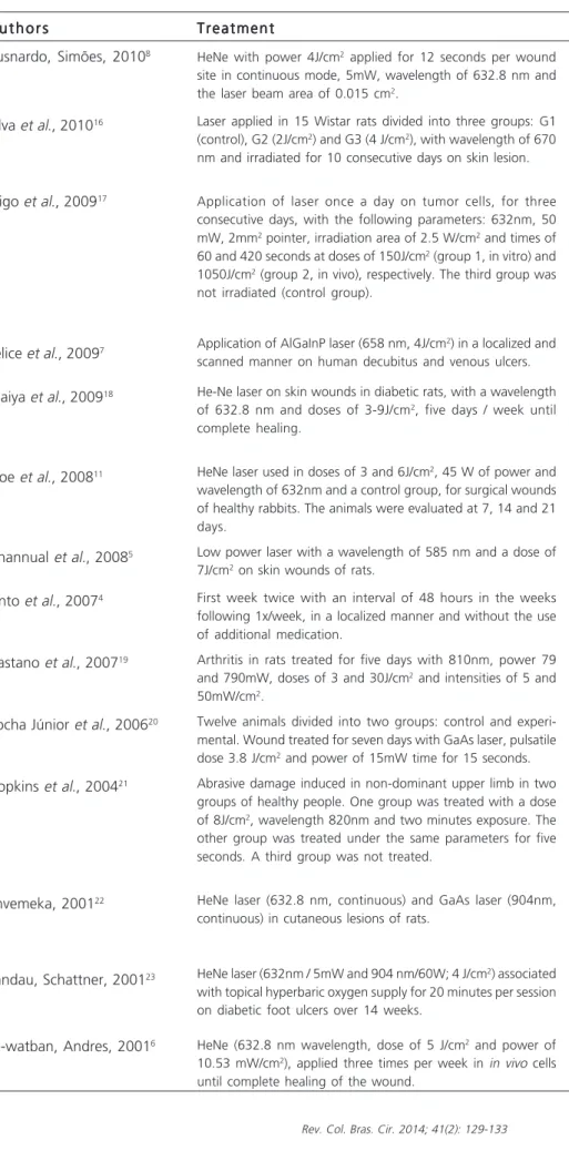
-Tabela 1 - Breve descrição das propostas de tratamento com laserterapia de baixa potência e seus principais resultados.
Treatment TreatmentTreatment Treatment Treatment
HeNe with power 4J/cm2 applied for 12 seconds per wound
site in continuous mode, 5mW, wavelength of 632.8 nm and the laser beam area of 0.015 cm2.
Laser applied in 15 Wistar rats divided into three groups: G1 (control), G2 (2J/cm2) and G3 (4 J/cm2), with wavelength of 670
nm and irradiated for 10 consecutive days on skin lesion.
Application of laser once a day on tumor cells, for three consecutive days, with the following parameters: 632nm, 50 mW, 2mm2 pointer, irradiation area of 2.5 W/cm2 and times of
60 and 420 seconds at doses of 150J/cm2 (group 1, in vitro) and
1050J/cm2 (group 2, in vivo), respectively. The third group was
not irradiated (control group).
Application of AlGaInP laser (658 nm, 4J/cm2) in a localized and
scanned manner on human decubitus and venous ulcers.
He-Ne laser on skin wounds in diabetic rats, with a wavelength of 632.8 nm and doses of 3-9J/cm2, five days / week until
complete healing.
HeNe laser used in doses of 3 and 6J/cm2, 45 W of power and
wavelength of 632nm and a control group, for surgical wounds of healthy rabbits. The animals were evaluated at 7, 14 and 21 days.
Low power laser with a wavelength of 585 nm and a dose of 7J/cm2 on skin wounds of rats.
First week twice with an interval of 48 hours in the weeks following 1x/week, in a localized manner and without the use of additional medication.
Arthritis in rats treated for five days with 810nm, power 79 and 790mW, doses of 3 and 30J/cm2 and intensities of 5 and
50mW/cm2.
Twelve animals divided into two groups: control and experi-mental. Wound treated for seven days with GaAs laser, pulsatile dose 3.8 J/cm2 and power of 15mW time for 15 seconds.
Abrasive damage induced in non-dominant upper limb in two groups of healthy people. One group was treated with a dose of 8J/cm2, wavelength 820nm and two minutes exposure. The
other group was treated under the same parameters for five seconds. A third group was not treated.
HeNe laser (632.8 nm, continuous) and GaAs laser (904nm, continuous) in cutaneous lesions of rats.
HeNe laser (632nm / 5mW and 904 nm/60W; 4 J/cm2) associated
with topical hyperbaric oxygen supply for 20 minutes per session on diabetic foot ulcers over 14 weeks.
HeNe (632.8 nm wavelength, dose of 5 J/cm2 and power of
10.53 mW/cm2), applied three times per week in in vivo cells
until complete healing of the wound.
R e s i u l t s R e s i u l t s R e s i u l t s R e s i u l t s R e s i u l t s
Increase of type III collagen, decreased inflammatory infiltrate and early resolution of wound inflammatory phase.
The dose of 4J/cm2 differed significantly
from the others concerning the re-epithelialization process.
Between the in vitro and the control group, there was no statistically significant difference in the growth of tumor cells. Comparing group 2 and control, there was significant growth of mass and volume to the tumor, as well as a large number of blood vessels in the in vivo group.
Reduction of the wounds area.
Increased production of granulation tissue in the animals that received doses of 4-5J/ cm2, especially on the fifth day of
treatment.
Observed the presence of mature granulation tissue at 14 days and absence of hemorrhage and exudate at day 21.
Permanent vascular proliferation after the fifth day of application.
The results revealed granulation tissue, reducing inflammation and pain relief from the first application.
Increase of adenosine triphosphate (ATP) and improved inflammatory process.
Tissue repair significantly larger and more organized in the experimental group.
The groups treated with low level laser showed a statistically significant reduction in the wound when compared to the control group at the 6th, 8th and 10th days of treatment.
Improvement of wound healing for both wave lengths adopted, though the latter presented more pronounced findings.
Complete healing of ulcers after 25 sessions and only 4% of recurrence.
Increased cell proliferation (fibroblasts and mitochondria), as well as microcirculation, with a consequent increase in cellular metabolism.
A u t h o r s A u t h o r s A u t h o r s A u t h o r s A u t h o r s
Busnardo, Simões, 20108
Silva et al., 201016
Frigo et al., 200917
Felice et al., 20097
Maiya et al., 200918
Inoe et al., 200811
Channual et al., 20085
Pinto et al., 20074
Castano et al., 200719
Rocha Júnior et al., 200620
Hopkins et al., 200421
Envemeka, 200122
Landau, Schattner, 200123
R E S U M O
R E S U M O
R E S U M O
R E S U M O
R E S U M O
Objetivo: Objetivo: Objetivo: Objetivo:
Objetivo: reunir e esclarecer quais os reais efeitos da laserterapia de baixa potência sobre feridas cutâneas e suas formas mais eficazes de aplicação na medicina humana e veterinária. Métodos:Métodos:Métodos:Métodos:Métodos: foram pesquisados artigos originais publicados em periódicos pertencentes às seguintes bases de dados: Lilacs, MedLine, Bireme e PubMed entre os anos de 2000 e 2011, na línguas espanhola, inglesa, francesa e portuguesa, que contivessem a descrição metodológica do modelo experimental e parâmetros utilizados no estudo. Resultados:Resultados:Resultados:Resultados:Resultados: doses compreendidas entre 3-6 J/cm2 parecem ser mais eficazes e que doses acima de 10 J/cm2 estão
associadas a efeitos deletérios. Os comprimentos de onda compreendidos entre 632,8-1000nm seguem como aqueles que apresen-tam resultados mais satisfatórios no processo de cicatrização tecidual. Conclusão:Conclusão:Conclusão:Conclusão:Conclusão: o laser de baixa potência pode ser indicado com segurança para acelerar a resolução de feridas cutâneas, muito embora este fato esteja intimamente ligado à eleição de parâmetros como dose, tempo e comprimento de onda.
Descritores: Descritores: Descritores: Descritores:
Descritores: Pele. Cicatrização de feridas. Anti-inflamatórios. Terapia a laser de baixa intensidade.
REFERENCES
REFERENCES
REFERENCES
REFERENCES
REFERENCES
1. Henriques ACG, Cazal C, Castro JFL. Ação da laserterapia no processo de proliferação celular: revisão de literatura. Rev Col Bras Cir. 2010;37(4):295-302.
2. Bourguignon Filho AM, Feitosa ACF, Beltrão GC, Pagnoncelli GC. Utilização do laser de baixa intensidade no processo de cicatriza-ção tecidual. Revisão de literatura. Rev Port Estomatol Cir Maxilofac. 2005;46(1);37-43.
3. Bashardoust Tajali S, Macdermid JC, Houghton P, Grewal R. Effects of low power laser irradiation on bone healing in animals: a meta-analysis. J Orthop Surg Res. 2010;5:1-13.
4. Pinto NC, Pereira, HC, Stolf NAG, Chavantes MC. Laser de baixa intensidade em deiscência aguda safenectomia: proposta tera-pêutica. Rev Bras Cir Cardiovasc. 2009;24(1);88-91.
5. Channual J, Choi B, Osann K, Pattanachinda D, Lotfi J, Kelly KM. Vascular effects of photodynamic and pulsed dye laser therapy protocols. Lasers Surg Med. 2008;40(9);644-50.
6. Al-watban FAH, Andres BL. Laser photons and pharmacological treatments in wound healing. Laser Therapy. 2001;12:1-9. 7. Felice TD, Pinheiro AR, Menchik EDS, Silva ACD, Souza LS, Caires
CSA, et al. Utilização do laser de baixa potência na cicatrização de feridas. Interbio. 2009;3(2);42-52.
8. Busnardo VL, Biondo-Simões MLP. Os efeitos do laser hélio-neônio de baixa intensidade na cicatrização de lesões cutâneas induzidas em ratos. Rev bras fisioter. 2010;14(1):45-51.
9. Zanotti GB, Oliveira PI, Reis SFS, Silva FS, Araújo AR. Efeitos do laser de baixa potência sobre a regeneração da cartilagem na osteoartrose. Rev fisio bras. 2011;12(2):139-46.
10. Salcido R, Adrian P, Chulhyun A. Animal models in pressure ulcer research. J Spinal Cord Med. 2007;30(2):107-16.
11. Inoe AP, Zafanelli CCG, Rossato RM, Leme MC, Sanches AWD, Araújo CV, et al. Avaliação morfológica do efeito do laser de baixa potência He-Ne em feridas cutâneas de coelhos. Arq ciênc vet zool Unipar. 2008;11(1):27-30.
12. Abergel RP, Lam TS, Dwyer RM, Lesavoy MA, Uitto J. Control of connective tissue metabolism by lasers: recent developments and future prospects. J Am Acad Dermatol. 1984;11(6):142-50. 13. Barreto JG, Salgado CG. Clinic-epidemiological evaluation of ulcers
in patients with leprosy sequelae and the effect of low-level laser therapy on wound healing: a randomized clinical trial. BMC Infect Dis. 2010;10:237-45.
14. Lacerda MS, Nunes TC. Efeitos do cetoprofeno e flunixin meglumine namodulação neuroendócrina à dor pós-operatória em cadelas submetidas a ovário-histerectomia. Biosci J. 2008;24(4):131-7.
15. Dogan SK, Saime AY, Evcki D. The effectiveness of low-level laser therapy in subacromial impingement syndrome: a randomized placebo controlled double-blind prospective study. Clinics. 2010;65(10):1019-22.
16. Silva TS, Mendes F, Alves AMP, Alves EPB, Bertolini GRF. Estudo microscópio da lesão tecidual em pele de ratos Wistar tratados com laser de baixa potência. Rev Bras Bioci. 2010;8(3);264-7. 17. Frigo L, Luppi JSS, Favero GM, Maria DA, Penna SC, Bjordal JM, et
al. The effect of low-level laser irradiation (In-Ga-Al-AsP – 660 nm) on melanoma in vitro and in vivo. BMC Cancer. 2009;9:404-11.
18. Maiya AG, Kumar P, Nayak S. Photo-stimulatory effect of low energy helium-neon laser irradiation on excisional diabetic wound healing dynamics in wistar rats. Indian J Dermatol. 2009;54(4):323-9.
19. Castano AP, Dai T, Yaroslavsky I, Cohen R, Apruzzese WA, Smotrich MH, et al. Low-level laser therapy for zymosan-induced arthritis in rats: importance of illumination time. Lasers Surg Med. 2007;39(6):543-50.
20. Rocha Júnior AM, Oliveira RG, Farias RE, Andrade LCF. Aarestrup FM. Modulação da proliferação fibroblástica e da resposta infla-matória pela terapia a laser de baixa intensidade no processo de reparo tecidual. An Bras Dermatol. 2006;81(2):150-6.
21. Hopkins JT, McLoda TA, Seegmiller JG, Baxter GD. Low-level laser therapy facilitates superficial wound healing in humans: a triple-blind, sham-controlled study. J Athl Train. 2004;39(3):223-9. 22. Envemeka CS. Attenuation and penetration of visible 632.8nm
and invisible infra-red 904nm light in soft tissues. Laser Therapy. 2001;13:95-101.
23. Landau Z, Schattner A. Topical hyperbaric oxygen and low energy laser therapy for chronic diabetic foot ulcers resistant to conventional treatment. Yale J Biol Med. 2001;74(2): 95-100.
24. Verhey JF, Mohammed Y, Ludwig A, Giese K. Implementation of a practical model for light and heat distribution using laser-induced thermotherapy near to a large vessel. Phys Med Biol. 2003;48(21):3595-610.
25. Izikson L, Nelson JS, Anderson RR. Treatment of hypertrophic and resistant port wine stains with a 755nm laser: a case series of 20 patients. Lasers Sur Med. 2009;41(6):427-32.
26. Giuliani A, Lorenzini L, Gallamini M, Massella A, Giardino L, Calzà L. Low infra red laser light irradiation on cultured neural cells: effects on mitochondria and cell viability. BMC Complement Altern Med. 2009;9:8.
27. Huang YY, Chen AC, Carroll JD, Hamblin MR. Biphasic dose response in low level light therapy. Dose Response. 2009;7(4): 358-83.
28. Moshkovska T, Mayberry J. It is time to test low level laser therapy in Great Britain. Postgrad Med J. 2005;81(957):436-41. 29. Bjordal JM, Lopes-Martins RA, Joensen J, Couppe C, Ljunggren
Received on 10/10/2012
Accepted for publication 15/12/2012 Conflict of interest: none.
Source of funding: none.
How to cite this article: How to cite this article: How to cite this article: How to cite this article: How to cite this article:
Andrade FSSD, Clark RMO, Ferreira ML. Effects of low-level laser therapy on wound healing. Rev Col Bras Cir. [periódico na Internet] 2014;41(2). Disponível em URL: http://www.scielo.br/rcbc