INTRODUCTION
Lacrimal gland (LG) secretion of proteins and luid into the tear ilm is essential for maintaining the health of the ocular surface (OS). With regard to the corneal epithelium, its smooth optical properties, its transparency, and its protective efect against pathogenic iniltra-tion are sustained by lacrimal gland funciniltra-tions(1,2). Tears are a complex
mixture of various constituents containing more than a thousand diferent proteins(3). Lysosomal hydrolases, secretory IgA(4), lactoferrin, ABSTRACT
Purpose: In the lacrimal gland (LG) acinar cells, signaling regulates the release of secretory vesicles through specific Rab and SNARE exocytotic proteins. In diabetes mellitus (DM), the LGs are dysfunctional. The aim of this work was to determine if secretory apparatus changes were associated with any effects on the secretory vesicles (SV) in diabetic rats as well as the expression levels of constituent Rab and members of the SNARE family, and if insulin supplementation reversed those changes.
Methods: DM was induced in male Wistar rats with an intravenous dose of strepto-zotocin (60 mg/kg). One of the two diabetic groups was then treated every other day with insulin (1 IU). A third control group was injected with vehicle. After 10 weeks, Western blotting and RT-PCR were used to compared the Rab and SNARE secretory factor levels in the LGs. Transmission electron microscopy evaluated acinar cell SV density and integrity.
Results: In the diabetes mellitus group, there were fewer and enlarged SV. The Rab 27b, Rab 3d, and syntaxin-1 protein expression declined in the rats with diabetes mellitus. Insulin treatment restored the SV density and the Rab 27b and syntaxin expression to their control protein levels, whereas the Vamp 2 mRNA expression increased above the control levels.
Conclusions: Diabetes mellitus LG changes were associated with the declines in protein expression levels that were involved in supporting exocytosis and vesicular formation. They were partially reversed by insulin replacement therapy. These fin-dings may help to improve therapeutic management of dry eye in diabetes mellitus.
Keywords: Diabetes mellitus/chemically induced; Lacrimal apparatus; Exocytosis; secretory vesicles; R-SNARE proteins; Animals; Rats
RESUMO
Objetivo: Células acinares da glândula lacrimal (GL) sinalizam a regulação da libe-ração através de vesículas secretórias específicas Rab proteínas exocitóticas SNARE. No diabetes mellitus (DM), as glândulas lacrimais são disfuncionais. O objetivo deste trabalho foi determinar se em ratos diabéticos, alterações dos aparatos secretórios estão associados a efeitos sobre vesículas secretoras (VS) e sobre os níveis de expressão do constituinte Rab, bem como membros da família SNARE, e se a suplementação de insulina reverte as alterações.
Métodos: DM foi induzido em ratos Wistar machos com uma dose intravenosa de estreptozotocina (60 mg/kg). Um dos dois grupos diabéticos foi então tratado a cada dois dias com insulina (1 UI). Um terceiro grupo controle foi injetado com o veículo. Após 10 semanas, western blot e RT-PCR comparou níveis de fatores secretórios de Rab e SNARE na glândula lacrimal. Microscopia eletrônica de transmissão (MET ) avaliaram a densidade e integridade de VS de célula acinar.
Resultados: No grupo diabetes mellitus , houve poucas e alargadas VS. Rab27b, Rab 3d e Sintaxina-1 diminuiu a expressão da proteína em ratos com Diabetes Mellitus. O tratamento com insulina restaurou a densidade das VS e expressão de Rab 27b e Sintaxina para seus níveis de proteína controle, enquanto a expressão de Vamp 2 RNAm aumentou em relação aos controles.
Conclusões: Alterações na glândula lacrimal de diabetes mellitus estão associadas a reduções nos níveis de expressão de proteínas envolvidas no apoio a exocitose e formação vesicular. Eles são, em parte, revertida por terapia de reposição de insulina. Estes resultados podem ajudar a melhorar a conduta terapêutica do olho seco no diabetes mellitus.
Descritores: Diabetes Mellitus/induzido quimicamente; Aparelho lacrimal; Exocitose; Vesículas secretórias; Proteínas R-SNARE; Animais; Ratos
transferrin(5), and growth factors are secreted through vesicles or
gra-nules in a regulated or constitutive manner(6).
Diabetes mellitus (DM) impairs tear secretion and induces LG and OS changes(7). Although the efects of DM-induced hyperglycemia,
oxidative stress, nerve damage, and impaired insulin signaling have been described in the LG, changes in the secretory mechanism cau-sed by this disease are not clearly understood(8,9). A recent work
re-vealed that high glucose levels reduced the expression of secretory
Insulin replacement restores the vesicular secretory apparatus in the diabetic rat
lacrimal gland
Reposição de insulina restaura o mecanismo secretório da glândula lacrimal de ratos diabéticos
AnA CArolinA DiAs1, ThiAgo MArTins BATisTA2, leTíCiA PrATes roMA2, CArolinA MAriA MóDulo1, leonArDo TAnnus MAlki1, lArA CrisTinA DiAs1, MôniCA Alves1, PeTer sol reinACh1, everArDo MAgAlhães CArneiro2, eDuArDo MelAni roChA1
Submitted for publication: November 18, 2014 Accepted for publication: March 11, 2015
1 Department of Ophthalmology, Otorhinolaryngology and Head and Neck Surgery, Faculdade de Medicina de Ribeirão Preto (FMRP), Universidade de São Paulo (USP), Ribeirão Preto, SP, Brazil. 2 Department of Structural and Functional Biology, Institute of Biology, Universidade Estadual de
Campinas (UNICAMP), Campinas, SP, Brazil.
Funding: This study was supported by grants from the following Brazilian governmental agencies: Fundação de Amparo a Pesquisa do Estado de São Paulo (FAPESP), Conselho Nacional de De sen volvimento Científico e Tecnológico (CNPq), Coordenação de Aperfeiçoamento de Pessoal de Nível Superior (CAPES), Fundação de Apoio ao Ensino, Pesquisa e Assistência do Hospital das Clinicas da Faculdade de Medicina de Ribeirão Preto da Universidade de São Paulo (FAEPA) and Núcleo de Apoio a Pesquisa Fisiopatologia e Terapêutica Ocular (NAP-FTO).
Disclosure of potential conflicts of interest: None of the authors have any potential conflicts of interest to disclose.
granule-associated vesicle-soluble N-ethylmaleimide-sensitive factor attachment protein receptors (SNARE) in the pancreatic beta-cells(10).
Metabolic and neurogenic inputs are needed to sustain LG function. Speciically, they control secretory and anti-oxidant mechanisms(11,12).
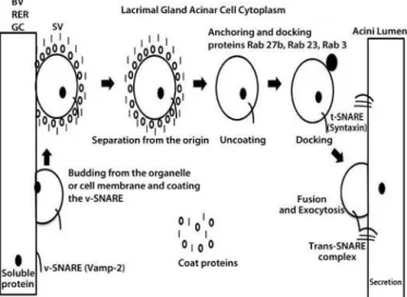
Such support is needed to enable the secretory products in the lacri-mal acini to go through sorting into immature secretory vesicles in the trans-Golgi network and to acquire the external proteins charac-teristic of mature secretory vesicles (SVs) as they move towards the apical membrane prior to their exocytotic release(13).
Local and systemic pathways highly control the LG exocytosis of tear luid components. The exocytosis is modulated by cholinergic and adrenergic stimuli, through changes in protein-kinase A (PKA) activity and intracellular calcium transients(14). This process has several
diverse roles, including the release of neurotransmitters, hormones, enzymes, and cytokines. It is regulated in the intracellular environment by a group of cytosolic proteins that include receptors speciic for anchoring vesicles. SNAREs and some Rab GTPases participate in this process and comprise proteins responsible for mediating secretion from neural, endocrine, and other exocrine tissues(15). They include
vesicle-associated membrane protein 2 (Vamp2), Rab 3d, and syn-taxin 1, which are responsible for the docking of molecules and the driving of the plasma membrane vesicle fusion that leads to the release of their soluble components into tears(16).
In the LG, the most abundant protein in the acinar secretory pathway is Rab3, which is localized to mature SVs. This protein drives vesicular docking and apical membrane fusion and it can also play a role in de termining vesicular size(17) (Figure 1). In previous studies, aging rats
had presented with dry eye disease, an insulin-signaling im pairment, oxidative stress, and declines in the parasympathetic control of LG function; this revealed an interesting similarity with DM dry eye(18,19).
Some SNARE and Rab family members were also altered in aged LG; particularly, Rab 3 expression was reduced at both the mRNA and protein levels(20). In addition, advanced glycation end pro ducts can
accumulate in aged and diabetic rat LGs(7,18).
In the current study, we characterized the impact of DM on rat LG acetylcholine (ACh) content, the expression of Rab and SNARE family proteins, and the structure of secretory vesicles (SV) as well as the protective efects rendered by insulin supplementation on such changes.
METHODS
A
NIMALMODELEight-week-old male Wistar rats were obtained from the Animal Breeding Center of the Faculty of Medicine of Ribeirão Preto (Ribei-rão Preto, São Paulo, Brazil). The animals had free access to standard rodent chow and water. Food was withdrawn 12 h before the experi-ments and diabetes was induced with a single dose of streptozotocin (Sigma, St. Louis, MO, USA), 60 mg/kg body weight, diluted in 1 mL of 0.01 M citrate bufer that was administered through the caudal vein. Controls were injected with citrate bufer alone. Each of the three diferent groups used 10 rats.
Two days later, the diabetic status was veriied with a glucose meter test (Accu-check, Roche Diagnostica Brazil Ltda., São Paulo, SP, Brazil) of blood obtained from the caudal veins of rats that had fasted for 12 h. A fasting hyperglycemia of over 200 mg/dL was thought to indicate the presence of diabetes; on the 4th day, insulin treatment was initiated in part of the diabetic group (subcutaneous injections of 1 IU every other day). This dose was not suicient to maintain ade-quate glycemic control, but was enough to avoid body weight loss and growth retardation(21).
Comparative studies of the three groups, i.e., control (C), diabetic (DM), and diabetic with insulin treatment (IT), were performed 10 weeks later.
The animals were weighed, anesthetized with intraperitoneal in-jection comprising a combination of ketamine (5 mg/100 g b.w.) (União Química Farmacêutica S.A, Embu-Guaçu, SP, Brazil) and xylazine (2 mg/ 100 g b.w.) (Laboratorio Callier S.A., Barcelona, Spain). LGs were ex-tirpated after ensuring that the corneal and caudal relexes were aboli-shed. Thereafter, the rats were euthanized with excess anaesthesia.
T
ISSUECOLLECTIONANDSTORAGEEach LG obtained from the right side of a rat was dissected into two parts and processed according to the protocol for each set of expe-riments. LGs were collected and homogenized in a bufer containing 50 mM Tris at a pH of 7.5, 500 mM NaCl, 0.1% Triton, and the protease inhibitor cocktail set III (Calbiochem, San Diego, CA) with a Polytron (Virsonic, Biopharma, Winchester, UK). RNA from the LGs was extrac-ted with Trizol after homogenization, according to the manufacturer’s protocol. The left LG was used for transmission electron microscopy (EM). This tissue was ixed in 2% glutaraldehyde and 2% paraformal-dehyde (Sciences, Hatield, PA, USA) in 0.1 M of phosphate bufer at a pH of 7.4, for 40 min at room temperature.
A
CETYLCHOLINE(AC
H)
MEASUREMENTSINLG
ACh was measured in LG using an ACh assay kit (Amplex Red; Mo lecular Probes, Eugene, OR, USA) to compare the amounts of this neurotransmitter in LGs of the three groups (n=5/group)(22). In brief,
0.1 mL of medium and tissue aliquots of homogenates containing 200 µg of protein were spotted in duplicate onto 96-well microplates. Standard ACh curves were constructed to evaluate the ACh content in each experiment. A 0.1 mL aliquot of assay bufer (50 mM Tris-HCl at a pH of 7.5) containing 0.2 M of reagent (Amplex Red; Molecular Probes), 2 U/mL of horseradish peroxidase, 0.2 U/mL of choline oxida-se, and 10 U/mL of acetylcholinesterase was added to each well. After incubation, absorbance was determined with a spectrophotometer (Beckman Instruments, Inc., Fullerton, CA) at 530 nm. The ACh levels were expressed on a millimolar (mM) basis.
T
RANSMISSIONELECTRONMICROSCOPYLG tissues from the three groups that were ixed for TEM were rin-sed in a 0.1 M phosphate bufer, dehydrated through a graded ethanol series, rinsed in acetone, and embedded in an Embed 812 (EM Scien-ces). Sections (60-70 nm) were cut with a diamond knife and stained for 25 min each in 2% uranyl acetate and 5 min in Reynolds’ lead citrate. Sections were examined with a TEM (Jeol, Jem 100cx, Tokyo, Japan). Pic-tures were taken and converted to digital iles (Hamamatsu, ORCA-HR Amtv542, Hamamatsu City, Japan). The intra cellular organelles, in-cluding secretory vesicles and nuclei, were eva luated in each group.
RT-PCR
FORR
AB3
D, R
AB23, R
AB27
B,
ANDV
AMP2
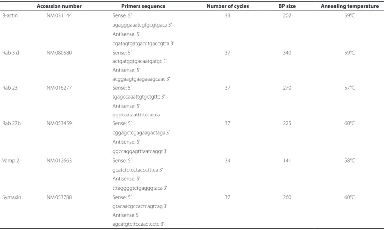
The reverse transcriptase polymerase chain reaction (RT-PCR) com-pared the Rab3d, Rab 23, Rab 27b, and Vamp 2 mRNA levels in the three rat LG groups. In addition, beta-actin mRNA was used for internal nor-malization. The resulting RNA was quantiied by spectrophotometry at 260 nm and the RNA integrity was evaluated on 6.6% formaldehy-de and 1% agarose (Gibco/BRL) gels. Reverse transcriptase, oligo dT priming, and the Advantage RT-for-PCR kits from Clontech Labora-tories Inc. (Palo Alto, CA, USA) were used for the cDNA transcription. PCR ampliication of cDNA was performed with a GeneAmp poly-merase chain reaction (PCR) System 9700 (Applied Biosystems, Foster City, CA, USA) using 1.5 units of Taq DNA polymerase (Gibco/BRL), 0.3 mM each of dATP, dCTP, dGTP, and dTTP (Invitrogen), PCR bufer (Tris -Hcl 60 mM, MgCl2 1.5 mM, NH4 SO4 pH10 15 mM) (Invitrogen), and 10 mM of 5’ and 3’ primers corresponding to rat Rab 3d, Rab 23, Rab 27b, Vamp2, and beta-actin cDNA (Life Technologies, Gaithersburg, MD, USA), after the preliminary assays identiied the parameters to ensure that the products were in the linear range (Table 1). In all PCR proce-dures, the positive and negative control cDNAs were run in parallel. Our attempts to detect syntaxin 1 mRNA were unsuccessful as in a previous study(23).
The PCR program used the following cycle proile: denaturation for 1 min at 94°C, annealing for 1 min at the indicated temperatures, extension for 1.5 min at 72°C, and maximization of strand completion for 7 min at 72°C. Following ampliication, the cDNA fragments were analyzed on 1% agarose gels containing a 100 bp DNA molecular weight ladder (Gibco/BRL) and were post-stained with ethidium bro-mide to conirm the anticipated base pair (bp) sizes for Rab 3d, Rab 27b, Rab 23, and Vamp 2, and beta-actin products.
Positive controls for Rab3d, Rab 23, Rab 27b, and Vamp 2 included cDNA isolated from rat pancreatic islets. Negative controls included samples without reverse transcriptase or samples of cDNA. The re-sults were recorded on the Gel Doc system (Bio-Rad Laboratories, Ri chmond, CA, USA). The membranes were scanned and analyzed by Scion Image Analysis Software (Scion Corp, Frederick, MD, USA).
W
ESTERNBLOTTINGWestern blots evaluated Rab 27b, Rab 3d, Vamp 2, and syntaxin 1 protein expression in cell lysates obtained from the rat LGs from the three diferent groups (n=5/group). LGs were solubilized in 1 mL of homogenization bufer containing the following: 100 mM-2-ami-no-2-hydroxymethyl-propane-1,3-diol (Tris) (pH 7.5), 10 mM-sodium pyrophosphate, 100 mM-sodium luoride, 10 mM-EDTA, 10 mM-so-dium vanadate, 2 mM-phenylmethylsulfonyl luoride, and 1% Triton-X 100. LGs were disrupted using a Polytron PT 1200C homogenizer (Brinkmann Instruments, Westbury, NY, USA). The extracts were then centrifuged at 12,000 rpm at 4°C for 15 min to remove insoluble ma-terials. Protein concentrations in the supernatant fractions were assayed with the Bradford dye method. For SDS gel electrophoresis and Western blotting, the samples were treated with a Laemmli sam-ple bufer containing dithiothreitol. After heating at 95°C for 5 min, the proteins were separated by electrophoresis (70 µg protein/lane, 10%-12% gels). Following electrophoresis, the proteins were
trans-Table 1. RT-PCR primers and parameters for SNARE and Rab mRNA detection in LG. of control, DM, and insulin-treated DM rats
Accession number Primers sequence Number of cycles BP size Annealing temperature
Β-actin NM 031144 Sense: 5’ 33 202 59°C
agagggaaatcgtgcgtgaca 3’
Antisense: 5’
cgatagtgatgacctgaccgtca 3’
Rab 3 d NM 080580 Sense: 5’ 37 340 59°C
actgatggtgacaatgatgc 3’ Antisense: 5’
acggaagtgaagaaagcaac 3’
Rab 23 NM 016277 Sense: 5’ 37 270 57°C
tgagccaaattgtgctgttc 3’
Antisense: 5’ gggcaataatttttccacca
Rab 27b NM 053459 Sense: 5’ 37 225 60°C
cggagctcgagaagactaga 3’ Antisense: 5’
ggccaggagtttaatcaggt 3’
Vamp 2 NM 012663 Sense: 5’ 34 141 58°C
gcatctctcctaccctttca 3’
Antisense: 5’
tttaggggtctgagggtaca 3’
Syntaxin NM 053788 Sense: 5’ 37 260 60°C
gtacaacgccactcagtcag 3’ Antisense 5’
ferred to nitrocellulose membranes. The nitrocellulose ilters were trea ted with a blocking bufer (5% non-fat dried milk, 10 mM-Tris, 150 mM-NaCl, and 0.02% Tween 20) overnight and were subsequen-tly incubated with rabbit polyclonal antibody anti-Rab 3d, Vamp 2, Rab 27b, syntaxin 1, and GAPDH as internal controls (Table 2). Vi-sua lization of speciic protein bands was performed by incubating the membranes for 2 h with a peroxidase-conjugated secondary an tibody (1:10.000; Zymed Laboratories, Inc., San Francisco, CA, USA), followed by detection with enhanced chemiluminescence reagents (Pierce Biotechnology, Rockford, IL, USA) and exposure to X-ray ilm (Kodak, Manaus, AM, Brazil). The band intensities were quantiied by optical densitometry (Scion, Image, Frederick, MD, USA).
S
TATISTICALANALYSISData were reported as mean ± SEM. Comparisons were made using the Kruskal-Wallis test followed by Dunn’s post hoc test (GraphPad 5.0 software; Prism, San Diego, CA). Densitometry values were repor-ted as ratios of beta-actin in RT-PCR and GAPDH in the Western blot assays, respectively. The ratio of densitometric values of one control sample of each blot was deined as 1.0 (100%), and the subsequent values were expressed as a ratio relative to its control value and sub-mitted to statistical analysis with the Kruskal-Wallis test followed by the Dunn’s post hoc test.
RESULTS
Body and LG weight were signiicantly lower in the DM group than in its control group whereas insulin treatment prevented such declines. The ACh levels remained unchanged at 10 weeks after DM induction (Table 3).
In the apical areas of the acinar cells in the DM group, there were fewer and enlarged secretory vesicles (SV) predominantly encapsu-lating the white content and there were fewer dark vesicles than in the controls. On the other hand, the cell size and nuclear appearance were unchanged in the DM group. In the insulin-treated group, the predominant white vesicular coloration in the DM group was less
evident, but there were fewer black SVs than in the apical areas of the acinar cells of the control LGs (Figure 2).
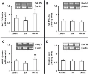
RT-PCR revealed that the Rab 3d, Rab 27b, and Rab 23 mRNA levels were not changed by DM or insulin treatment, but the Vamp 2 level was higher in the insulin-treated group (Figure 3).
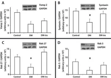
Western blotting revealed that the expression of syntaxin 1, Rab 27b, and Rab 3d were signiicantly lower in the DM group, whereas the decline in Vamp 2 was insigniicant. However, insulin treatment prevented the declines in Rab 27b, syntaxin 1, and Rab 3d expression as they remained at levels similar to those in the control group (Figure 4).
DISCUSSION
The present study indicated that there was a correspondence between changes in the secretory mechanisms in LGs that were
iso-Table 2. Antibodies used for Western blot analysis of SNARE and Rab protein detection in LG of control, DM, and insulin-treated DM rats
Protein Catalog # Type
Molecular
weight Concentration
GAPDH Santa Cruz Rabbit polyclonal 37 KDa 200 μg/mL
SC 367715
Rab 3 d Santa Cruz Goat polyclonal 25 KDa 200 μg/mL SC 26392
Rab 27b Santa Cruz Goat polyclonal 30 KDa 200 μg/mL
SC 22993
Vamp 2 Calbiochem Rabbit polyclonal 12 KDa 1 mg/mL
627724
Syntaxin Santa Cruz Mouse monoclonal 35 KDa 200 μg/mL SC 12736
Table 3. Changes in parameters of the control, DM, and insulin-treated DM rats. Data is expressed as mean ± standard error, *denotes p<0.05
Control DM DM-ins
Body weight (g) 699.8 ± 25.02 205.20 ± 11.37* 504.0 ± 54.67
LG weight (mg) 128.2 ± 04.83 050.10 ± 01.87* 097.0 ± 08.61
Glycemia (mg/dL) 156.5 ± 12.65 542.30 ± 53.53* 295.3 ± 38.15 Acetylcholine (µM) 110.6 ± 18.60 086.70 ± 13.50* 117.6 ± 16.50
Figure 2. Transmission electron microscopy of the acinar cells in the exorbital LG of control (A, D), DM (B, E), and insulin-treated (C, F) DM groups. The images are representative of sections of LGs from 5 animals/group. Magniication ×2000 in the upper lane and ×4000 in the lower lane (scale bar=5 µm and 2.5 µm, respectively).
A
D
B
E
C
F
A
C
B
D
A
C
B
D
Figure 4. Impaired expression of SNARE and Rab proteins was rescued by insulin treat-ment in LG of diabetic rats. Western blotting of (A) Vamp2, (B) syntaxin 1, (C) Rab27b, and (D) Rab3d in LGs of control, DM, and insulin-treated DM rats. Densitometric arbitrary units were normalized to GAPDH expression. Data are expressed are means ± standard error of the mean; * p<0.05. Results are representative of three independent experiments (n = 5/group/experiment).
lated from DM rats and the dry eye syndrome manifestations. Such an insight will provide a better understanding of dry eye pathophy-siology(7,12,21). The major current inding was that lower intracellular
transport protein expression levels in the DM group were associated with declines in delimited SV density and their ultra-structural appea-rance of the acinar apical cell membranes.
Diferent animal models of DM have found that LG secretory product content changes can accompany OS neuronal and struc-tural damage(24). Taken together, the present indings, in particular,
the similar levels of ACh among the diabetic and control groups, suggested that the damage in the signaling machinery of diabetic LG preceded the neurogenic damage to the LG and OS(25). These results
are consistent with a previous study where LG synaptic junctional ultrastructure of the DM rats were preserved after 4 weeks of disease, although the insulin signaling cascade was already impaired just 1 week after DM onset(8,26).
Although animal models in general have some limitations in fully simulating a disease condition (e.g., dry eye syndrome), in streptozo-tocin-diabetic rats, these structural changes were described in the secretory granules of the acinar cells of LGs along with the functional impairment(24). Moreover, in diferent models of diabetic rodents,
me-chanisms other than those described here triggered declines in tear secretion and corneal epithelial damage associated with inlamma-tion, lower proliferative capacity, and oxidative damage(27).
The fact that in LGs of DM rats there is a discrepancy between the declines in Rab 3d and Rab 27 protein expression and the lack of changes in their mRNA expression suggested that DM promoted chan ges at a post-transcriptional level. They may be caused by a re duction in translational events or a decline in the protein half-life. Such decreases could be attributable to hyperglycemia or a secondary vascular disturbance(28).
In an aging rodent, which is another model of the dry eye syndro-me, similar alterations in SV appearance and declines in their density were associated with lower Rab 3d levels(20). Similar to the DM model
used here, this model had high levels of oxidative stress and insulin resistance(19,21). As in that study, we were unable to identify syntaxin 1
at the mRNA levels, but detected it with Western blotting. A possible explanation was that the antibody that we used cross-reacted with another LG acinar cell syntaxin isoform, which we did not probe for at the mRNA level.
A recent publication revealed that SV were highly expressed in human LGs with dry eye induced by prolonged visual display terminal
exposure compared to controls and Sjögren’s syndrome patients. Taken together, those indings indicated that distinct diseases diferentially afected the LG secretory mechanisms, as did evaporative, inlam-matory, and diabetic dry eye. In agreement with those indings in humans subject to video display terminals or Sjogren’s syndrome, the present work revealed that Vamp-2 increased in a DM model of dry eye and the SV were dimorphic; their localization was changed in the acinar cells of LG. This observation may indicate that therapeutic mea sures to prevent such changes could compensate for the abnor-mal secretory changes induced by DM. Insulin induced multiple res ponses through a myriad of interacting intracellular signaling cas-cades(29). It was demonstrated that either systemic insulin or topical
treatment prevented LG damage and reduced ocular surface dama-ge in DM animal models with dry eye(21). In the present work, systemic
insulin was suicient to protect against body and LG weight loss due to increases in catabolism and more severe SV content and ultra-structural changes along with modiied secretory protein proiles(30).
In conclusion, the present work demonstrated that the LG secre-tory impairment secondary to DM was involved in reducing the ex-pression of some Rab and SNARE protein family members that were responsible for intracellular SV traicking that could be prevented by insulin treatment. The consequent reduction in tear secretion and content in the untreated DM rats may help to explain early ocular surface epithelial damage in DM(21). Moreover, the present indings
may help to identify new therapeutic measures in DM for use in the dry eye syndrome, including insulin hormone therapy.
E
THICALSTANDARDSAll experimental procedures adhered to the Principles of Labora tory Animal Care (NIH publication no. 85 to 23) and the ARVO Sta -tement for the Use of Animals in Ophthalmic and Vision Research and were approved by the committee on animal experimentation of the School of Medicine at Ribeirão Preto, University of São Paulo.
REFERENCES
1. Plugfelder SC. Tear dysfunction and the cornea: LXVIII Edward Jackson Memorial Lecture. Am J Ophthalmol. 2011;152(6):900-9 e1.
2. Zhou L, Zhao SZ, Koh SK, Chen L, Vaz C, Tanavde V, et al. In-depth analysis of the human tear proteome. J Proteomics. 2012;75(13):3877-85.
3. Srinivasan S, Thangavelu M, Zhang L, Green KB, Nichols KK. iTRAQ quantitative proteo-mics in the analysis of tears in dry eye patients. Invest Ophthalmol Vis Sci. 2012;53(8): 5052-9.
4. van Haeringen NJ, Glasius E. Lysosomal hydrolases in tears and the lacrimal gland: efect of acetylsalicylic acid on the release from the lacrimal gland. Invest Ophthalmol Vis Sci. 1980;19(7):826-9.
5. Salvatore MF, Pedroza L, Beuerman RW. Denervation of rabbit lacrimal gland increases levels of transferrin and unidentiied tear proteins of 44 and 36 kDa. Curr Eye Res. 1999;18(6):455-66.
6. Hodges RR, Dartt DA. Regulatory pathways in lacrimal gland epithelium. Int Rev Cytol. 2003;231:129-96.
7. Alves M, Calegari VC, Cunha DA, Saad MJ, Velloso LA, Rocha EM. Increased expression of advanced glycation end-products and their receptor, and activation of nuclear factor kappa-B in lacrimal glands of diabetic rats. Diabetologia. 2005;48(12):2675-81. 8. Rocha EM, Lima MH, Carvalho CR, Saad MJ, Velloso LA. Characterization of the
insu-lin-signaling pathway in lacrimal and salivary glands of rats. Curr Eye Res. 2000;21(5): 833-42.
9. Peponis V, Papathanasiou M, Kapranou A, Magkou C, Tyligada A, Melidonis A, et al. Protective role of oral antioxidant supplementation in ocular surface of diabetic patients. Br J Ophthalmol. 2002;86(12):1369-73.
10. Torrejón-Escribano B, Escoriza J, Montanya E, Blasi J. Glucose-dependent changes in SNARE protein levels in pancreatic β-cells. Endocrinology. 2011;152(4):1290-9. 11. Rocha EM, Fernandes ML, Velloso LA. Insulin signaling in the aging nervous system.
Adv Cell Aging Gerontol. 2004;16:107-32.
12. Rocha EM, Alves M, Rios JD, Dartt DA. The aging lacrimal gland: changes in structure and function. Ocul Surf. 2008;6:162-74.
13. Ohnishi H, Ernst SA, Wys N, McNiven M, Williams JA. Rab3D localizes to zymogen granules in rat pancreatic acini and other exocrine glands. Am J Physiol. 1996;271(3 Pt 1):G531-8.
15. An SJ, Almers W. Tracking SNARE complex formation in live endocrine cells. Science. 2004; 306:1042-6.
16. Wu K, Jerdeva GV, da Costa SR, Sou E, Schechter JE, Hamm-Alvarez SF. Molecular me-chanisms of lacrimal acinar secretory vesicle exocytosis. Exp Eye Res. 2006;83(1):84-96. 17. Hamm-Alvarez S, Cadenas E. Mitochondrial medicine and therapeutics, Part II.
Prefa-ce. Adv Drug Deliv Rev. 2009;61(14):1233.
18. Alves M, Cunha DA, Calegari VC, Saad MJ, Boschero AC, velloso LA, et al. Nuclear factor-kappaB and advanced glycation end-products expression in lacrimal glands of aging rats. J Endocrinol. 2005;187(1):159-66.
19. Rocha EM, Carvalho CR, Saad MJ, Velloso LA. The inl uence of ageing on the insulin signalling system in rat lacrimal and salivary glands. Acta Ophthalmol Scand. 2003; 81(6):639-45.
20. Batista TM, Tomiyoshi LM, Dias AC, Roma LP, Módulo CM, Malki LT, et al. Age-de-pen dent changes in rat lacrimal gland anti-oxidant and vesicular related protein ex pression proi les. Mol Vis. 2012;18:194-202.
21. Módulo CM, Jorge AG, Dias AC, Braz AM, Bertazolli-Filho R, Jordão AA Jr, et al. In-l uence of insuIn-lin treatment on the In-lacrimaIn-l gIn-land and ocuIn-lar surface of diabetic rats. Endocrine. 2009;36(1):161-8.
22. Dias AC, Modulo CM, Jorge AG, Braz AM, Jordão AA JR, Filho RB, et al. Inl uence of thyroid hormone on thyroid hormone receptor beta-1 expression and lacrimal gland and ocular surface morphology. Invest Ophthalmol Vis Sci. 2007;48(7):3038-42.
23. Batista TM, Tomiyoshi LM, Dias AC, Roma LP, Módulo CM, Malki LT, et al. Age-de-pendent changes in rat lacrimal gland anti-oxidant and vesicular related protein expression proi les. Mol Vis. 2012;18:194-202.
24. Shetty R, Saeed T, Rashed H, Adeghate E, Singh J. Ef ect of diabetes mellitus on acinar morphology, peroxidase concentration, and release in isolated rat lacrimal glands. Curr Eye Res. 2009;34(10):905-11.
25. Ishida N, Rao GN, del Cerro M, Aquavella JV. Corneal nerve alterations in diabetes mellitus. Arch Ophthalmol. 1984;102(9):1380-4.
26. Cunha DA, de Alves MC, Stoppiglia LF, Jorge AG, Módulo CM, Carneiro EM, et al. Extra-pancreatic insulin production in RAt lachrymal gland after streptozotocin-in-duced islet beta-cells destruction. Biochim Biophys Acta. 2007;1770(8):1128-35. 27. Nguyen CQ, Kim H, Cornelius JG, Peck AB. Development of Sjogren’s syndrome in
nonobese diabetic-derived autoimmune-prone C57BL/6.NOD-Aec1Aec2 mice is de pendent on complement component-3. J Immunol. 2007;179(4):2318-29. 28. Patel NA, Chalfant CE, Yamamoto M, Watson JE, Eichler DC, Cooper DR. Acute
hyper-glycemia regulates transcription and posttranscriptional stability of PKCbetaII mRNA in vascular smooth muscle cells. FASEB J. 1999;13(1):103-13.
29. Myers MG Jr., White MF. Insulin signal transduction and the IRS proteins. Annu Rev Pharmacol Toxicol. 1996;36:615-58.
30. Bonifacino JS, Glick BS. The mechanisms of vesicle budding and fusion. Cell. 2004;116: 153-66.