Mailing Address: Ricardo Tavares de Carvalho • Av. Dr. Enéas de Carvalho Aguiar, 44 - 05403-900 – São Paulo, SP - Brazil E-mail: [email protected] Received on 03/10/05 • Accepted on 12/08/05
Resistive Exercise in the Evaluation of Endothelial
Dysfunction in Heart Failure
Ricardo Tavares de Carvalho, Marcelo Luis Campos Vieira, Ângela Romano, Liliane Kopel, Silvia G. Lage
Instituto do Coração do Hospital das Clínicas – FMUSP - São Paulo, SP - Brazil
O
BJECTIVETo evaluate the use of resistive exercise in the study of endothelial dysfunction in heart failure (HF) comparatively to reactive hyperemia (RH).
M
ETHODSEighteen patients with heart failure and 15 normal volunteers were submitted to intermittent handgrip exercise in a pneumatic bag, at an intensity that corresponds to 75% of the previously assessed maximum load. Patients underwent high-resolution vascular ultrasonography for brachial artery diameter and fl ow evaluation as well as cardiac output determination at rest, RH and after exercise. The systolic fl ow index in the brachial artery and cardiac index were calculated.
R
ESULTSSystolic fl ow index increase in the brachial artery was observed after RH and physical exercise, with the latter presenting the highest increase. There was an increase in the cardiac index after the study conditions in comparison to resting conditions.
C
ONCLUSIONResistive exercise, performed at the assessed load, increases blood fl ow more intensively than RH, constituting a physiological option for the evaluation of endothelial function in HF.
K
EY WORDSHeart failure (HF) is characterized by the impairment of the ventricular function, with progressive physical limitation of affected individuals as the disease advances. However, one of the most remarkable facts regarding this syndrome is that the severity of ventricular dysfunction is not related to the functional capacity assessed by exercise tolerance1-3. Although the central (cardiac) alterations
start and perpetuate the HF, the main limitation to exercise is of peripheral origin, involving alterations in oxygen transportation and consumption4.
The physiopathological basis of this fact consists in the presence of histological and metabolic modifi cations in the skeletal muscle fi bers5,6, as well as alterations in the
capacity of the vessels to distribute fl ow to muscles.
The characteristics of peripheral vessels are important determinants of left ventricular function in HF and are subject to the action of substances released by the endothelium in response to local fl ow7. Among these
substances, nitric oxide (NO) is particularly relevant. Additionally, the endothelial-independent vascular response can also be altered in such patients8-10.
Endothelial dysfunction is a systemic phenomenon and can be assessed by cellular and molecular methods11-12 in
animals. In humans, it is noteworthy the assessment of fl ow-mediated vasodilation in peripheral sites13.
With the description of RH utilization as an assessment method of flow-dependent endothelial dysfunction14, this technique started to be extensively
employed; however, only recently this assessment method has been standardized15.
Physical exercise can also be utilized for endothelial function assessment. The basis of this observation is in the increase of blood fl ow in active muscular sites16,17.
Daily exercise sessions has shown to be benefi cial for the increase of NO expression in normal individuals and for endothelial-dependent vasodilation increase in HF patients18,19. A review of randomized and controlled
studies corroborates the benefi cial effect of physical exercise in HF20. The cardiac output increase that occurs
during the exercise would augment shear stress, and thus, NO expression. It is not a consensus that the relative ischemia produced during localized muscular activity is enough stimulation to cause vasodilation.
Therefore, we tested the hypothesis that resistive exercise can be effective in assessing endothelial dysfunction in HF and it is comparable to reactive hyperemia.
M
ETHODSThirty-three individuals aged between 18 and 65 yrs were studied. They were divided in two groups: 18 patients with congestive heart failure (CHF group) and 15 normal volunteers (NL group) (Table 1).
Table 1 - Characteristics of the groups
CHF GROUP (N=18) NL GROUP (N=15) P
Age (yrs) 50 ± 12 46 ± 11 p= NS
Gender M 14 09 p= NS
Gender F 04 06 p= NS
Weight (Kg) 64.7 ± 11.95 71.5 ± 10.08 p= NS
Height (m) 1.6 ± 0.11 1.7 ± 0.08 p= NS
BMI (Kg/m2) 23.7 ± 2.6 25.9 ± 2.6 p= NS
CHF=congestive heart failure; NL=normal
In the CHF group, left ventricular ejection fraction < 40% at the echocardiogram was the inclusion criterion for the study. Patients with coronary insufficiency, hypertension, permanent pacemaker, cardiac arrhythmias including frequent ventricular and supraventricular extrasystoles, atrial fl utter and fi brillation and II-III degree atrioventricular block were excluded from the study.
The usual medication was withdrawn in all patients before the study for at least twelve hours. Patients were clinically stable with no peripheral edema.
The cause of heart failure was defi ned as dilated cardiomyopathy in 7 cases, Chagas’ cardiomyopathy in 9 cases, alcoholic cardiomyopathy in 1 case and cardiomyopathy secondary to chemotherapy in 1 case. Left ventricular ejection fraction assessed by echocardiogram at rest was 0.31±0.05. Based on the NYHA functional classifi cation, nine patients were class II, fi ve were class III and 4 patients were class IV.
Study Protocol - Each subject was asked out with patients in the supine position and it was started after at least a 20- minute resting period in a quiet, dimly illuminated and temperature-controlled environment (21oC).
Before the study was started, the maximum left hand grip strength was measured using a kilogram (kg) scale dynamometer and afterward a pneumatic bag coupled to a pressure scale in millimeters of mercury (mmHg). Each individual was asked to perform three maximum handgrip movements. The value considered as maximum handgrip strength was the arithmetical mean of the strength obtained at the three gripping movements performed with the pneumatic bag21. This value was used for the
calculation of the exercise load to be applied.
The correlation between the two measurements was carried out (r = 0.90, p < 0.01, n= 33). Only the measurements carried out with the pneumatic bag were utilized in the study, as this was the only way for the individual to control the strength to be applied, according to what had been previously established.
Cardiac rhythm was monitored by electrocardiogram for the echocardiogram to be performed.
Fig. 1- Brachial artery longitudinal section delimitating limits for the vascular diameter measurement. PW: proximal wall; DW: distal wall; D: diameter
recording, for arterial diameter measurement.
In the study protocol, the arterial diameter and flow velocity curve were initially evaluated at rest (B1condition). Afterward, the vascular reactivity was evaluated by reactive hyperemia after a 5-minute cuff occlusion in the left forearm, insuffl ated with a 200 mmHg pressure (HR condition). After a 30-minute rest, brachial artery diameter and flow velocity parameters were reassessed under a new basal condition (B2 condition). After that, each subject received the same pneumatic bag which was utilized to assess maximum handgrip strength capacity and they were asked to performed repeated gripping and relaxation movements with the left hand, with 75% of the maximum load (EX75 condition), in a protocol adaptation developed for this purpose21. After the
fi rst minute of exercise, the aforementioned parameters in brachial territory were assessed.
Cardiac output was evaluated by echocardiogram, using bidimensional mode, during each phase of the study immediately after the analysis of the peripheral parameters. This was performed to verify whether there was a correlation between the increase of local fl ow and the output increase that could take place because of exercise.
Acquisition of arterial diameters - An ultrasonography device (Apogee 800 plus, ATL Inc., Bothell WA, USA) equipped with a high-resolution vascular plane transducer of 5-10 MHz with automatic focus adjustment was utilized to acquire the bidimensional images at longitudinal cuts of the brachial artery, at approximately 5 cm distal to the elbow fold. The transducer was positioned at 90o
to the vessel, so the vessel walls could be visualized. The image acquisition of the vascular diameter under reactive hyperemia was carried out one minute after cuff-occlusion release, and under the exercise conditions, one minute after exercise beggining. The acquisition of arterial images was carried out by the same observer, coupled to the simultaneous recording of the electrocardiographic signal. They were recorded in VHS tapes for posterior determination of the arterial diameter.
The VHS recording was utilized so that, based on the electrocardiographic recording, the images could be selected at the moment of systolic expansion, which corresponds to the fi rst 60 milliseconds of the T wave. A computer software, which was specially developed for this purpose, was utilized to determine the arterial diameter22 (Fig. 1).
Peripheral arterial fl ow - An ultrasonography device (Apogee.-800 plus, ATL Inc, Bothell Wa, USA), equipped with a high-resolution vascular plane transducer of 5-10 MHz was utilized for the evaluation of the brachial artery blood fl ow velocity, using a pulsatile Doppler.
The arterial fl ow velocity images, coupled to the simultaneous electrocardiogram recording, were always
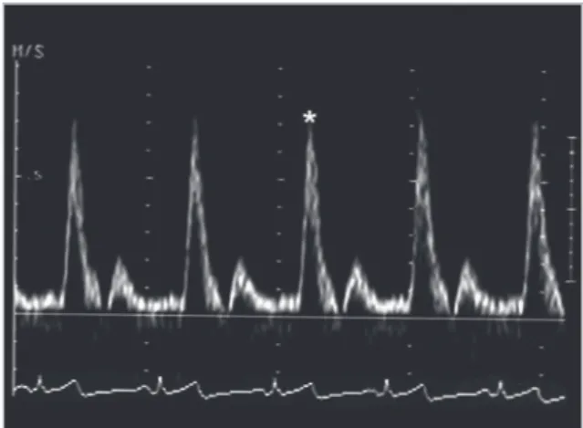
acquired by the same observer and recorded in VHS tapes. Five images were selected and recorded in a computer equipped with video-frame grabber (Willow Publishers VGA, Willow Peripherals, Inc., Bronx, NY, USA), for posterior analysis. Afterward, the curve point that represented the peak systolic velocity (*) was enhanced (Fig. 2). The quantifi cation of the peak systolic velocity of the arterial fl ow was carried out through the use of a software developed for this purpose.
In the reactive hyperemia condition, the fl ow velocity curve of the fi rst heartbeat after cuff-occlusion release was always analyzed (Fig. 3). In the exercise condition, the velocity curves of the fi fth heartbeat of the muscle relaxation phase were analyzed (Fig. 4).
After the fl ow velocity and arterial diameters during systole are measured, the systolic fl ow index (SFI) is calculated using the following formula:
) /m L/min ( BS
0.06 . Vsi . TSA
SFI= 2
where TSA (transverse sectional area of the vessel) = π.radius2, Vsi = peak systolic velocity (m/s), BS=
body surface area (m2) and 0.06 is the correction factor
necessary for the conversion of the units to l/min.
Cardiac assessment - Cardiac assessment was carried out using an ultrasonography device (Apogee-800 plus, ATL Inc, Bothell Wa., USA), with a setorial 3 MHz mechanical transducer. The M-mode echocardiogram and the bidimensional echocardiogram were always performed by the same observer, as well as the study of intracardiac fl ow using pulsatile Doppler device. The Doppler echocardiogram was recorded in VHS tapes. All quantifi cations were carried out based on the images recorded in 3 to fi ve heartbeats, and the mean value of the measurements was considered for the analysis.
The bidimensional echocardiogram images were acquired according to the guidelines of the American Society of Echocardiography23.
PW
Fig. 2 - Flow velocity variation curve in the brachial artery showing the reference point for analysis of the systolic peak velocity (Vsy); * Systolic peak
Fig. 3 - Curve of fl ow velocity variation in the brachial artery for reactive hyperemia analysis. The arrow indicates the velocity curve of the fi rst heartbeat after cuff-occlusion release
Fig. 4 - Curve of brachial fl ow velocity variation in the relaxation phase of the exercise condition with 75% of maximum load (EX75). The arrow indicates the fi fth heartbeat, used for the analysis
The bidimensional left parasternal longitudinal cut was utilized for the measurement of left ventricular outfl ow diameter, necessary for the calculation of the systolic volume. The systolic fl ow of the left ventricle outfl ow tract was obtained from the apical cut, with the integral of the fl ow velocity being determined through the use of an adequate software for the ultrasound equipment. The
systolic volume (SV) was then calculated based on the following formula:
SV = FVI .TSA
where: FVI=fl ow velocity integral and TSA = transverse sectional area of the aortic ring.
Heart rate was obtained based on the RR interval of the electrocardiogram. Cardiac output was calculated as the product of the systolic volume and heart rate. The cardiac index was obtained dividing the cardiac output by the body surface area.
Statistical analysis - To analyze the behavior of the groups considering the studied conditions, repeated measures analysis of variance was utilized (TIMM, 1975). The use of this technique presumes normal distribution of data. Signifi cance level was set at 5%. The calculations were carried out with the SAS system (Statistical Analysis System) (SAS, 1989).
R
ESULTSFor the normal group, the systolic fl ow index mean values in brachial artery were 0.25 ± 0.08 l/min/m2
for reactive hyperemia; 0.20 ± 0.06 l/min/m2; 0.48 ±
0.17 l/min/m2 and 0.60 ± 0.24 l/mim/m2, for basal
conditions (B1 and B2), for post-reactive hyperemia (RH) and exercise with 75% of the maximum load (EX75), respectively.
For the CHF group, the systolic fl ow index mean values in brachial territory were 0.19 ± 0.08 l/min/m2 and
0.21 ± 0.09 l/min/m2 for basal conditions (B1 and B2),
respectively and 0.43 ± 0.14 l/min/m2 and 0.45 ± 0.18
l/min/m2 for post-reactive hyperemia (RH) and exercise
with 75% of the maximum load (EX75), respectively. There was a positive and significant correlation between the systolic fl ow indexes in RH and EX75 conditions (r=0.66, p=0.005).
The groups did not present a statistically signifi cant difference regarding behavior during the evaluations performed (p=NS). The groups did not present a signifi cant difference regarding their mean values at each evaluation moment (p=NS). There was an alteration of this parameter during the evaluations (p<0.001) for both groups.
When analyzing the means of the systolic fl ow index under the stimulation conditions (RH and EX75), no signifi cant difference was observed between them.
The comparison between basal conditions and those that came subsequently (B1XRH and B2XEX75) showed a signifi cant difference in all evaluations performed for both groups (p<0.001) (Fig.5).
Fig. 5 - Systolic fl ow index in the brachial artery. Comparison between basal conditions (B1 and B2) and the subsequent stimulations. RH: reactive hyperemia; EX75: exercise with 75% of maximum load
0 0,2 0,4 0,6 0,8
B1 HR B2 EX75
*p<0,001
IFS (l/mln/m
2 )
*
*
Systolic Flow Index – brachial artery
0 100 200 300 400
Delta 1 Delta 2
*p<0,001
*
∆%
Systolic Flow Index – brachial artery
∆
%Fig. 6 - Percentage variation between the systolic fl ow indexes in the brachial artery between B1 and RH (∆ 1) and B2 and EX75 (∆ 2)
The analysis of percentage variations was carried out by assessing the value at a post-stimulation condition, and relatively taking into account its respective basal value. Thus, we call delta 1 (∆1) that value observed between the B1 and RH conditions, and delta 2 (∆2) the one observed between B2 and EX75 conditions.
The percentage variations of the brachial artery systolic fl ow index means (∆1 and ∆2) for the CHF group were, respectively: 134.1 ± 48.5% and 208.3 ± 96.5%. For the normal group, the same parameters were, respectively: 94.5 ± 56.8% and 193.3 ± 90.1%. The groups did not present differences regarding either behavior (p=NS) or difference between the means in ∆1 as well as in ∆2
(p=NS). There was a difference between the conditions 1 (B1 and RH) and 2 (B2 and EX75) for both groups (p< 0.001). ∆1 showed a signifi cant difference compared to
∆2 (p<0.001) (Fig. 6).
D
ISCUSSIONThis study proposes a new methodology in the assessment of the endothelium-dependent vascular reactivity. Physical exercises have been increasingly considered in HF24,25. Despite that, few studies have
associated this practice to the peripheral hemodynamic response in HF patients18,26.
We studied clinically compensated patients, especially regarding the presence of peripheral edema. This factor can interfere in the vasodilating response due to the increase of vascular stiffness27, related to the increase
amount of sodium levels in the vessel wall. In addition, the edema can lead to an increase in tissue pressure with mechanical compression of the capillaries28.
In EX75 condition, the choice of the fi fth heartbeat in the relaxation phase was carried out in order to facilitate the standardization of the analysis of fl ow velocity curves, as there is a rapid and progressive decrease until their normalization, with time (Fig. 4).
The choice of manual stress involving a small muscle mass, instead of exercise utilizing the lower limbs, seemed a more interesting physiologic. The evaluation of the vascular response to a submaximum stress would be more compatible with the reality of patients who present limitations to physical activity and who perform household tasks that demand more muscular resistance than aerobic conditioning.
It was not possible to observe a difference in local blood fl ow in our study, even under elevated load, between reactive hyperemia and localized exercise. However, in terms of percentage, the variation of post-exercise fl ow was more intense than post-reactive hyperemia, especially when considering that the post-exercise analysis was carried out at the fi fth heartbeat after exercise interruption, and not in the fi rst heartbeat as under the reactive hyperemia condition.
At the evaluation of fl ow in calf and forearm muscles,
after fi ve minutes of local ischemia in patients with compensated HF and no edema, and in normal age-matched individuals, a decrease in blood flow was observed, compared to normal volunteers, only in the calf29. In this study, there was also no difference regarding
the blood fl ow in the forearm between the two groups. Therefore, it is suggested that vascular abnormalities in CHF are regionally specifi c not only for the fl ow differences observed in distinct territories but also for the fl ow distribution inside the muscle itself, favoring oxidative fi bers30.
Additionally, it is possible that, as HF advances, the alterations that were initially evident in lower limbs also start to occur in the upper limbs. Thus, it seems that the disparity among the results that assess vascular reactivity in the presence of exercise in heart failure is due to differentiation regarding disease severity, to the state of clinical compensation of the studied population and the type of stimulation employed (aerobic exercise, resistive exercise, distinct loads and muscular site territory evaluated).
It is noteworthy that, from a methodological point of view, the non-invasive studies utilizing arterial ultrasound are more appropriate and accurate for the analysis in the upper limbs than in the lower limbs.
We conclude that the study of a smaller muscular site that imitates daily activities regarding the type of work as well as the load, demonstrated that the exercise with 75% of the maximum load increased the peripheral
SFI
0.8
0.6
0.4
0.2
0
*p<0.001
R
EFERENCES1. Franciosa JA, Leddy CL, Wilen M. Relation between hemodynamic and ventilatory responses in determining exercise capacity in severe congestive heart failure. Am J Cardiol. 1984; 53: 127-34.
2. Baker BJ, Wilen MM, Boyd CM, Dinh HA, Franciosa JA. Relation of right ventricular ejection fraction to exercise capacity in chronic left ventricular failure. Am J Cardiol. 1984; 54: 596-9.
3. Cohn J, Johnson G, Shabetai R. Ejection fraction, peak exercise oxygen consumption, cardiothoracic ratio, ventricular arrhythmia and plasma norepinephrine as determinants of prognosis in heart failure. Circulation. 1993; Suppl 6: VI5 – VI16.
4. Drexler H, Reide U, Munzel T, Konig H, Funke E, Just H. Alterations of skeletal muscle in chronic heart failure. Circulation. 1992; 85: 1751-59.
5. Kemp GS, Thompson CH, Straton JR. Abnormalities in exercising skeletal muscle in congestive heart failure can be explained in terms of decreased mitochondrial ATP synthesis, reduced metabolic effi ciency and increased glycogenolysis. Heart. 1996; 76: 35-41.
6. Andrews R, Walsh JT, Evans A, et al. Abnormalities of skeletal muscle metabolism in patients with chronic heart failure: evidence that they are present at rest. Heart. 1997; 77: 159-63.
7. Kopel L, Carvalho RT, Lage SG. Endotélio na insufi ciência cardíaca. In: Endotélio & Doenças Cardiovasculares. São Paulo: Ed. Atheneu; 2003: 221-9.
8. Hambrecht R, Fiehn E, Weigl C, et al. Regular physical exercise corrects endothelial dysfunction and improves exercise capacity in patients with chronic heart failure. Circulation. 1998; 98: 2709-15.
9. Imaizumi T, Takeshi A, Ashihara T, Nakamura M. The effects of sublingually administered nitroglycerine on forearm vascular resistance in patients with heart failure and normal subjects. Circulation. 1985; 72: 747-52.
10. Katz SD, Biasucci L, Sabba C, et al. Impaired endothelium-mediated vasodilation in the peripheral vasculature of patients with congestive heart failure J Am Coll Cardiol. 1992; 19: 918-25.
11. Morgan DML. Isolation and culture of human umbelical vein endothelial cells..In Jones FE. Methods in molecular medicine. Human Cell Culture Protocols 1998; 101:109.
12. Takano M, Meneshan A, Sheikh E, et al. Rapid upregulation of endothelial P-selectin expression via reactive oxygen species generation. Am J Physiol Heart Circ Physiol. 2002; 283: H2054 – H61.
13. Anderson TJ, Uehata A, Gerhard MD, et al. Close relation of endothelial function in the human coronary and peripheral circulations. J Am Coll Cardiol. 1995; 26: 1235-41.
14. Celermajer DS, Sorensen KE, Gooch VM, et al. Non-invasive detection of endothelial dysfunction in children and adults at risk of atherosclerosis. Lancet. 1992; 340: 1111-5.
15. Corretti MC, Anderson TJ, Benjamin EJ, et al. Guidelines for the ultrassound assessment of endothelial-dependent fl ow-mediated vasodilation of the brachial artery J Am Coll Cardiol. 2002; 39: 254-65.
16. Sanders JS, Mark AL, Ferguson DW. Evidence for cholinergically mediated vasodilation at the beginning of isometric exercise in humans. Circulation. 1989; 79: 815-24.
17. Green DJ, O´Driscoll G, Blankbly BA, et al. Control of skeletal muscle blood fl ow during dynamic exercise – contribution of endothelium-derived nitric oxide. Sports Med. 1996; 21: 119-46.
18. Katz SD, Yuen J, Bijou R, LeJemtel TH. Training improves endothelium-dependent vasodilation in resistance vessels of patients with heart failure. J Appl Physiol. 1997; 82: 1488-92
19. Coats AJS, Adamopoulos S, Meyer TE, et al. Effects of physical training in chronic heart failure. Lancet. 1990; 335: 63-6.
20. Piepoli MF, Falther M, Coats AJS & on behalf of the European Heart Failure Training Group Experience from controlled trials of physical training in chronic heart failure: protocol and patient factors in effectiveness in the improvement in exercise tolerance. Eur Heart J. 1998; 19: 466-75.
21. Zelis R, Longhurst J, Capone RJ, Mason DT. A comparison of regional blood fl ow and oxygen utilization during dynamic forearm exercise in normal subjects and in patients with congestive heart failure. Circulation. 1974; 50: 137-43.
22. Guttierrez MA, Pilon PE, Lage SG, Kopel L, Carvalho RT. Assesment of carotid diameter and wall thickness in ultrasound images using active contours improved by multiresolution technique. Proc SPIE. 2002; 4683: 248-55.
23. Henry WL, DeMaria A, Gramiak R, et al. Report of American Society of Echocardiography Committee on nomeclature and standarts in two-dimensional echocardiography. Circulation. 1980; 62: 211-7.
24. Belardinelli R, Georgiou D, Cianci G, Purcaro A. Randomized, controlled trial of long-term moderate exercise training in chronic heart failure: effects on functional capacity, quality of life, and clinical outcome. Circulation. 1999; 99: 1173-82.
25. Piepoli M, Capucci AJS. Exercise training in heart failure: effect on morbidity and mortality. Int J Cardiol. 2000; 73: 3-6.
26. Hambrecht R, Fiehne E, Neig LC, et al. Regular physical exercise corrects endothelial dysfunction and improves exercise capacity in patients with chronic heart failure. Circulation. 1998; 98: 2709-15.
27. Zelis R, Flaim SF. Alterations in vasomotor tone in congestive heart failure. Prog Cardiovasc Dis. 1982; 24: 437-59.
28. Beer G. Role of tissue fl uid in blood fl ow regulation. Circ Res. 1971; Suppl 1.
29. Jondeau G, Katz S, Toussaint J. Regional specifi city of peak hyperhemic response in patients with chronic heart failure: correlation with peak aerobic capacity. J Am Coll Cardiol. 1993; 22: 1399-402.
30. Drexler H, Banhardt U, Meinertz T, Wollschläger H, Lehmann M, Just H. Contrasting peripheral short-term and long-term effects of converting enzyme inhibition in patients with congestive heart failure. A double-blind, placebo controlled trial. Circulation. 1989; 79: 491-502.
fl ow more intensively than the reactive hyperemia, in percentage terms.
Thus, it is possible that the assessment of vascular reactivity, using resistive exercise, is a physiological, more comfortable and easy to perform option, and that it is comparable to reactive hyperemia in the study of endothelium-mediated vasodilation.
Potencial Confl ict of Interest